TABLE 1.1 Macrophage Characteristics
Histochemical Surface Antigens
Receptors Functions
5´-Nucleotidase OKM1 Fc Phagocytosis
Esterase Class II antigens
Alkaline phosphodiesterase
Aminopeptidase
Insulin
Immunoglobulin M (IgM) Pinocytosis
Lymphokine Immune activation
Lactoferrin Secretory
Microbicidal
Cb3 Tumoricidal
Fibrinogen
Lipoprotein
Fig. 1.1 Macrophage differentiation.
and F4/80). Other cell-surface markers are also present, such as class II antigens, Fc receptors (for antibody), and receptors (for complement). These enzymes and cell markers help identify this class of cells and their state of activation. The presence of esterase is a useful marker to distinguish macrophages from granulocytes and lymphocytes. Monocytes will leave the bloodstream because of either a predetermined maturational process or induced migration into an area as a result of chemotactic substances, often produced during inflammatory events. After taking up residence in various tissues, they become macrophages, which are frequently known by other names (Fig. 1.1). Dendritic cells, such as Langerhans’ cells, are found in the skin and the cornea and play an important role in presenting antigens to lymphocytes.
Macrophages play at least three major roles within the immune system. The first is to directly destroy foreign pathogens and clear dying or diseased tissue. Killing of invading microbes is, in part, mediated by a burst of hydrogen peroxide (H2O2) activity by the activated macrophage. An example with ocular importance is the engulfment of the toxoplasmosis organism, with the macrophage often being a repository for this parasite if killing is inadequate. The second is to activate the immune system. Macrophages, dendritic cells, or other cells with similar characteristics are mandatory for antigen-specific activation of T lymphocytes. Internalizing and processing of the antigen by the macrophage are thought to be integral parts of this mechanism, and the macrophage or the dendritic cell is named antigen-presenting cell (APC). Other cells, such as B cells, can also serve this function. The macrophage and the lymphocyte usually need to be in close contact with each other for this transfer to occur. Another requirement is for the cells to have in common a significant portion of their major histocompatibility complex (MHC), genes that express various cell-surface membranes essential for cellular communication and function. Thus this MHC stimulation leads to the initiation of an immune response, ultimately with both T and B cells potentially
participating. Other cell-surface markers are needed for activation. This “two signal” theory has centered on other cell-surface antigens, such as the B7–CD28 complex. The engagement of B7 (on the macrophage side) with CD28 enhances the transcription of cytokine genes. Third, the macrophage is a potent secretory cell. Proteases can be released in abundance, and this can degrade vessel surfaces and perivascular areas. Degradation products that result from these reactions are chemotactic and further enhance an immune response. Interleukin (IL)-1, a monokine with a molecular weight of 15 kilodaltons (kDa), is produced by the macrophage (and other cells) after interaction with exogenous pathogens or internal stimuli, such as immune complexes or T cells. IL-1 release directly affects T-cell growth and aids this cell in releasing its own secretory products. IL-1 is noted to act directly on the central nervous system (CNS), with a by-product being induction of fever. Still other macrophage products stimulate fibroblast migration and division, all of which have potentially important consequences in the eye.
Macrophages also produce IL-12, IL-18, IL-10, and transforming growth factor (TGF)-β. In a feedback mechanism, interferon (IFN)-γ can activate macrophages, and the production of IL-12 by the macrophage plays an important role in T-cell activation. The role of macrophages in the eye remains to be fully explored.
Dendritic Cells
Although macrophages play an important role, it is conjectured that dendritic cells are important macrophage-like cells in tissues. They are a subset of cells, perhaps of different lineage from macrophages, and they can be distinguished by lack of persistent adhesion and by the bearing of an antigen, 33D1, on their surface, features that macrophages do not possess. The major role of dendritic cells is to serve as APCs for both CD4+ and CD8+ cells. Like macrophages, dendritic cells produce IL-12, an important activator of T-cell responsiveness. They are rich in MHC II intracellular compartments, an important factor in antigen presentation. The MHC class II compartments move to the surface of the cell when the dendritic cell matures, stimulated by IFN-γ and the CD40 ligand. Dendritic cells are special in that they inhabit tissues where foreign antigens may enter. Experiments with painting of the skin brought seminal observations. Antigens painted on the skin are “brought” to the draining lymph nodes by the dendritic cells of the skin (Langerhans’ cells) where T-cell activation can occur. What is interesting is the migratory nature of these cells: They constantly carry important information to the peripheral centers of the immune response. Whether dendritic APCs can activate T cells efficiently in the tissues themselves is an open question, and the answer is important to our understanding of immune responses in the eye. Dendritic cells are thought to be the APCs (or one of the major players) in corneal graft rejection. Thus the concept of removing dendritic cells from a graft has been proposed and used in experimental models. However, there is an opposing concept that peripheral immune tolerance, induced by antigens that foster programmed cell death (apoptosis), may depend on presentation of antigen by dendritic cells in the tissue.
T cells
T-cell responses to antigens provide the basis for a large part of the inflammatory process. They are generated in bone marrow and mature in the thymus, the first lymphoid organ to develop. The thymus consists of two compartments, the cortex and the medulla. In the cortex, immature thymocytes develop through a complex process; their T-cell receptors (TCRs; see later) then interact with thymic epithelium, a process that determines their becoming CD4 (“helping”) or CD8 (“cytotoxic”) T cells. Thymocytes that fail this process die by apoptosis (“positive selection”), whereas thymocytes that succeed in this selection migrate to the thymic medulla, where epithelial and dendritic cells express all the body’s major autoantigens. Thymocytes
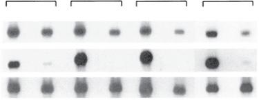
Fig. 1.2 Transcription of S-antigen (S-Ag) and interphotoreceptor retinoid-binding protein (IRBP) genes (uveitogenic antigens) in eyes and thymuses of mouse strains. S-Ag and IRBP are abundant in the eyes of all animals and S-Ag is found in the thymuses of all four strains tested. However, IRBP was seen only in thymuses of two strains –BALB/c and AKR/J – and not in those of B10.A or B10.RIII. The last two animals are susceptible to induction of uveitis with IRBP. (From Egwuagu CE, Charukamnoetkanok P, Gery I. Thymic expression of autoantigens correlates with resistance to autoimmune disease. J Immunol. 1997;159:3109–3112. Copyright 1997, The American Association of Immunologists, Inc.)
expressing TCRs with strong affinity to autoantigens are deleted (“negative selection”), and the remaining T cells enter the lymphoid system. Importantly, the negative selection is incomplete and T cells specific to autoantigens do escape the negative selection (see later).
A major component of the negative selection system is the autoimmune regulator (AIRE), a protein that is produced by medullary cells and that controls the expression of organ-specific antigens. Loss of the AIRE gene leads to autoimmunity,1 which is known to occur in humans who develop autoimmune polyglandular syndrome (APS) type I, an autoimmune disease that is inherited in an autosomal recessive fashion. In addition to adrenal insufficiency, mucocutaneous infections, and hypoparathyroidism, these patients can have diabetes, Sjögren syndrome, vitiligo, and uveitis.2
The expression of ocular self-antigens in the thymus was investigated in both mice and humans. Egwuagu et al.3 have shown in different mouse strains an inverted relationship between thymic expression of ocular-specific retinal antigens and the susceptibility to induction of experimental autoimmune uveitis (EAU): Thymic expression of retinal antigens causes lack of responsiveness to these antigens. An example of this phenomenon is shown in Fig. 1.2. Four inbred strains of mice were evaluated for the expression in their thymus of two uveitogenic antigens, interphotoreceptor retinoid-binding protein (IRBP) and S-antigen (S-Ag). All four strains were resistant to the induction of uveitis when S-Ag was used as the immunizing antigen, and all four expressed S-Ag in their thymus. However, two of the four strains, B10.A and B10.RIII, did not express IRBP in their thymuses and were susceptible to uveitis induction when IRBP was used as the immunizing antigen. These observations were extended to include other rodents and primates.4 In the Lewis rat, which is susceptible to both antigens, neither message was found in the thymus. These observations may provide an insight into the propensity for the disease in humans. Takase et al.5 evaluated 18 human thymus samples taken from patients undergoing surgery for congenital heart disease. They found that there was expression of the four antigens that can induce experimental uveitis (S-Ag, recoverin, RPE65, and IRBP) in the thymuses of the tested patients. However, the expression of the various antigens was very variable, with some thymus samples showing strong expression and others not. The implication of the findings from these studies is that expression of these antigens in the thymus is very variable in humans, similar to what is seen in the differences among various rodent strains. T cells with specificity to ocular self-antigens that escape the negative selection are found in the circulation, but do not induce uveitis. This observation is explained by two mechanisms: (1) the inhibitory effect of T-regulatory (Treg) cells, which are normally present in the body; and
(2) the retina being isolated from circulating cells by the blood–retina barrier. The normal presence of T cells is indicated by the finding of T cells specific to retinal antigens in healthy individuals with no eye disease.
One important quality possessed by T cells is their immunologic recall or anamnestic capacity after re-exposure to their specific target antigen. The exposure to the antigen increases the number of specific cells and changes them into a “memory” phenotype. A memory T cell to a particular antigen can retain this immunologic memory (see later) essentially for its lifetime. With a repeat encounter, this memory response leads to an immune response that is more rapid and more pronounced than the first. An example is the positive skin response seen after purified protein derivative (PPD) testing.
The central role of the T cell in the immune system cannot be overemphasized. T cells function as pivotal modulators of the immune response by helping production of antibody by B cells and augmenting cellmediated reactions through the release of molecules, named cytokines, which activate immune-related and other cells. T cells also may downregulate or prevent immune reactions through active suppression. (i.e., Treg cells). The cytotoxic (CD8) T-cell subset plays a major role in transplantation rejection crises. Accumulated evidence supports the importance of T cells in many aspects of the intraocular inflammatory process – from the propagation of disease to its subsequent downregulation.
A state of suspended animation can be induced in T cells; this is termed anergy. For T cells to be activated, several signals need to be given: one through the TCR and the other through costimulatory receptors, such as CD28; the third is the costimulant B7 linking to CD28 (which is on the T cell). If the TCR is activated but the costimulant is not, a growth arrest can be seen in these cells: They simply stop functioning but do not die. A second way this can occur is when a weakly adherent peptide is linked to the TCR, even if costimulation occurs. It would seem to be a mechanism to prevent unwanted or nuisance immune responses. The full response takes place only if all the appropriate interactions have occurred.
T-cell Receptor
Much interest has focused on the TCR. T cells need to produce the TCR on their cell surface to recognize the target immunogenic peptide on the MHCs of APCs. This complex interaction involves either CD4 or CD8 and their TCRs. The TCR is similar in structure to an immunoglobulin, having both α and β chains. The more distal ends of these chains are variable, and the hypervariable regions are termed V (variable) and J (joining) on the α chain and V and D (diversity) regions on the β chain. Compared with the number of immunoglobulin genes, there are fewer V genes and more J genes in the TCR repertoire. It is logically assumed that the target peptide, which has a special shape and therefore fits specifically in a lock-and-key fashion into the groove between the MHC and the TCR, would be the “cement” of this union. It has been suggested that of all the possible combinations of gene arrangements that could possibly produce the variable region believed to cradle the peptide, certain genes within a family seem to be noted more frequently in autoimmune disease. One such group is the Vα family, with Vβ8.2 receiving much attention. A small number of cells have a TCR made up not of α and β chains but, rather, of γ and δ chains (detailed later). In addition to these physiologic mechanisms, T cells may also be activated by “superantigens,” which are bacterial products, such as enterotoxins, or plant products, such as phytohemagglutinin. In addition, T cells may be activated by antibodies to certain surface antigens, mostly CD3 and CD28.
Major Populations of T Cells
The functions that have been briefly described are carried out by several subsets of CD4 T cells, identified by their products and functions. It was observed early on that T cells (and other cells) manifest myriad different molecules on their surface membranes, some of which are
TABLE 1.2 Selected human leukocyte Differentiation antigens (Incomplete list)
Cluster Designation Main Cellular Distribution Associated Functions
CD3 T cells, thymocytes Signal transduction
CD4 Helper T (Th) cells MHC class II coreceptor
CD8 Suppressor T cells, cytotoxic T cells MHC class I receptor
CD11a Leukocytes LFA-1, adhesion molecule
CD11b Granulocytes, MΦ Mac-1, adhesion molecule
CD11c Granulocytes, MΦ T cells, B cells α integrin, adhesion molecule
CD19 B cells B-cell activation
CD20 B cells B-cell activation
CD22 B cells B-cell regulatory
CD25 T cells, B cells α chain of IL-2 receptor (Tac) activation
CD28 T cells Costimulatory T-cell marker
CD45 Leukocytes Maturation
CD54 Endothelial, dendritic, and epithelial cells; activated T and B cells
ICAM-1, adhesion molecule; ligand of LFA-1 and Mac-1
CD56 NK cells N-CAM, adhesion molecule
CD68 Macrophages
CD69 NK cells, lymphocytes Signal transmission receptor
CX3CR1 Monocytes Chemoattractant
CXCR3 T cells Cell maturation
CCR7 T cells Migration to inflammation
CCR5 T cells Chemokine receptor
ICAM, intercellular adhesion molecule; IL, interleukin; LFA, lymphocyte function-associated molecule; MHC, major histocompatibility complex; N-CAM, neural cell adhesion molecule; NK, natural killer.
expressed uniquely at certain periods of cell activation or function. It was noted that certain monoclonal antibodies directed against these unique proteins bind to specific subsets of cells, thereby permitting a way to identify them (Table 1.2). The antibodies to the CD3 antigen in humans (e.g., OKT3) are directed against an antigen found on all mature human T cells in the circulation; approximately 70% to 80% of lymphocytes in the systemic circulation bear this marker. Antibodies to the CD4 antigen (e.g., OKT4) define the helper subgroup of human T cells (Th cells; about 60%–80% of the total T cells). These CD4+ cells respond to antigens complexed to MHCs of the class II type. The CD4 cells are particularly susceptible to human immunodeficiency virus (HIV) that causes acquired immunodeficiency syndrome (AIDS), with the percentage of this subset decreasing dramatically as this disease progresses. Furthermore, these helper cells are necessary components of the autoimmune response seen in the experimental models of ocular inflammatory disease induced by retinal antigens (see discussion of autoimmunity later in this chapter). Antibodies to the CD8 antigen (i.e., OKT8) distinguish a population that includes cytotoxic T cells, making up about 20% to 30% of the total number of T cells. Antibodies directed against the CD8 antigen block class I histocompatibility-associated reactions.
The two major populations of T cells (CD4 and CD8) are further divided into subpopulations that are detailed later. These subpopulations are generated by combinations of cytokines, which are products of other cells, and affect the immune system by the specific cytokines they produce.
T-cell Subsets
The population of Th cells has been further subdivided on the basis of their functional characteristics into several subsets. The major subsets are named Th1, Th2, and Th17. In normal conditions, Th1 cells defend against intracellular pathogens, Th2 cells defend against extracellular parasites and mediate antibody production, and Th17 cells defend against extracellular pathogens. Pathologically, Th1 and Th17 cells are responsible for initiation of “cell-mediated” immune responses, such as foreign tissue rejection and pathogenic autoimmune processes, whereas Th2 cells are involved in allergic responses and in immunoregulation. Th1 cells (Fig. 1.3) produce mostly IFN-γ, IL-2, and tumor necrosis factor-α
Fig. 1.3 Helper T-cell subsets now recognized. (From: Chen, Z, O’Shea JJ. Th17 cells: a new fate for differentiating helper T cells. Immunol Res. 2008;41:87, with permission.)
(TNF-α). The cytokine profile of Th2 cells consists of IL-4, IL-5, IL-10, IL13, and perhaps TGF-β , and the major cytokines produced by Th17 cells are IL-17, IL-21, and IL-22. In many animal models of human diseases, Th1 and Th17 cells are associated with initiation of disease, whereas Th2 cells are associated with disease downregulation and allergy initiation. Importantly, under experimental conditions, Th17 cells may switch to a Th1 phenotype, but Th1 cells maintain their phenotype and do not change.6
IL-22 is part of the IL-17 group of cytokines produced during an inflammatory response.7 Albeit made by lymphocytes, its receptors are present on epithelial cells. Thus it has been suggested that one of its major roles is to be the cross-talk lymphokine between resident tissue cells and infiltrating inflammatory cells, particularly T cells. This proinflammatory cytokine is found in the synovia of patients with rheumatoid arthritis and is upregulated in both Crohn disease and ulcerative colitis8,9 and in both the serum and intraocular fluids of patients with uveitis.10,11
Gamma Delta (γδ) T Cells
γδ Τ cells, which constitute a small fraction of peripheral T cells, play important roles in inflammatory processes, such as EAU.12 Of particular importance is the involvement of γδ Τ cells in mucosal tissue inflammation in the conjunctiva, as shown by St Leger et al.,13 where an inflammatory response to a commensal bacterium is mediated to a large extent by IL-17 produced by γδ cells.
T-regulatory Cells
It is now clear that just as the immune system needs cells to initiate a response, it needs cells to suppress or modify immune responses. One of the ways this need is met is with Treg cells.14 These cells derive from a naive T-cell population under the influence of cytokines that are different from those involved in Th1, Th17, or Th2 cell development (see Fig. 1.3). Treg cells can be found in the thymus or in the peripheral circulation, where a large portion is “induced” (iTregs). An interesting report by Kemper et al.15 described stimulating CD4+ cells with CD3 and CD46 (a complement regulator) and inducing Treg cells, that is, producing large amounts of IL-10, moderate amounts of TGF-β, and little IL-2. The literature is replete with information about different types of Treg cells, and these cells have been reported in several organs, such as the gut, where peripheral immune tolerance needs to be induced.16 Certain Treg cells are characterized by their ability to produce IL-10 and TGF-β. They are capable of downregulating both CD4- and CD8mediated inflammatory responses and apparently require cell-to-cell contact. The majority of Treg cells bear CD25 (the IL-2 receptor) on their cell surface and express the transcription factor forkhead/winged helix (FoxP3),17 which is a reliable marker for the development and function of naturally occurring Treg cells.18 When we evaluated the T cells of patients with ocular inflammatory disease, we found that the FoxP3 marker varied considerably among patients and was not a very good indicator of poor Treg cell function.19 An interesting observation is the noted increase in a subset of NK cells (so-called CD56 “bright”) after daclizumab therapy; this subset makes large amounts of IL-10, indicating the regulatory nature of these cells. The increase is seen when the patient’s disease is well controlled, and it has also been seen in patients with multiple sclerosis receiving daclizumab therapy.
Lymphocytes of Innate Immune System
In addition to adaptive immunity cells and molecules, mentioned earlier, the protection against invasion of pathogenic agents is carried out by components of “innate” immunity that lack antigenic specificity but are capable of providing immunity. The cell populations of the innate immunity are discussed in the following sections.
Innate Lymphoid Cells
The recently discovered ILCs are lymphocytes that lack antigen specificity and are involved in the immune response by releasing cytokines or carry cytotoxic capacity (NK cells; see later). ILCs are mainly tissue resident and play major roles in keeping the homeostasis in these tissues.20 Unlike T or B cells, ILCs react promptly to stimulations, such as pathogen invasion. ILCs are separated into three major groups (ILC1, ILC2, and ILC3) that selectively collaborate with Th1, Th2, and Th17 lymphocytes in the defense against intracellular pathogens and tumors, large extracellular pathogens and allergens, and extracellular pathogens, respectively.20
Natural Killer and Invariant Nature Killer T Cells
NK cells and invariant natural killer T (iNKT) cells are major components of the innate immune response, whose main function is to carry out the rejection by cytolytic activity of both tumors and virally infected cells. The main difference between NK cells and iNKT cells is their morphology, with NK cells being large granular lymphocytes, whereas iNKT cells express highly conserved TCRs. Both populations release large amounts of cytokines upon activation. NK cells seem to be involved in the pathogenesis of EAU because the disease was found to be diminished in mice with no NK cells.21 Grajewski et al.22 showed that similar to NK cells, iNKT cells ameliorate the EAU process. However, iNKT cells were also found to have a dual effect in the pathogenic process of EAU: The disease was enhanced in iNKT-deficient mice, but activation of these cells also exacerbated the pathologic process.23
Cytokines
Intercellular communication, which is crucial for active immune response, is mediated by cytokines, chemokines, and adhesion molecules. Cytokines are produced by lymphocytes, macrophages, and other cells. They are hormone-like proteins capable of amplifying an immune response and of suppressing it. With activation of a T lymphocyte, the production and release of various lymphokines will occur. One of the most important cytokines is IL-2, with a molecular weight of 15 kDa in humans. The release of this lymphokine stimulates lymphocyte growth and augments specific immune responses, including stimulation of Treg cells. Of particular interest are cytokines involved in the inflammatory process. They include proinflammatory cytokines, IFN-γ, IL-1, IL-17, and TNF-α and the anti-inflammatory IL-4, IL-5, IL-10, and TGF-β. Of interest, IFN-γ and TGF-β are active in both activation and suppression of immune responses. The number of lymphokines that have been purified and for which effects have been described continues to grow rapidly. An incomplete list is shown in Table 1.3 and a more recent list has been provided by Akdis et al.24
Chemokines
This family of chemoattractant cytokines is characterized by its ability to induce directional migration of movable cells. They direct cell adhesion, homing, and angiogenesis. There are four major subfamilies of chemokines: CXC (nine of which are found on chromosome 4); CC (11 of which are found on chromosome 17); C (only one well-defined member, lymphotactin, is found on chromosome 11); and CX3C (fractalkine is found on chromosome 16). The nomenclature is based on the cysteine molecules. The CC chemokines have two adjacent cysteines at their amino terminus; the CXC chemokines have their N-terminal cysteines separated by one amino acid; the C chemokines have only two cysteines, one at the terminal end and one downstream; the CX3C chemokines have three amino acids between their two N-terminal cysteines. Each chemokine family has special functions that affect different types of cells. An example of this fine specificity is seen with the CXC family. These chemokines, with a Glu–Leu–Arg sequence near the end
TABLE 1.3 Cytokines: An incomplete list
Type Source
IFN-γ T cells
TGF-β T cells, resident ocular cells
Target and Effect
Antiviral effects; promotes expression of MHC II
Antigens on cell surfaces; increases MΦ tumor killing; inhibits some T-cell proliferation
Suppresses generation of certain T cells; involved in ACAID and oral tolerance Interleukin
IL-1 Many nucleated cells, high levels in MΦ, keratinocyte, endothelial cells, some T and B cells
IL-2 Activated T cells
IL-3 T cells
IL-4 T cells
IL-5 T cells, eosinophils
T- and B-cell proliferation; fibroblasts – proliferation, prostaglandin production; CNS – fever; bone and cartilage resorption; adhesion-molecule expression on endothelium
Activates T cells, B cells, MΦ, NK cells
Affects hemopoietic lineage that is nonlymphoid eosinophil regulator; similar function to IL-5 GM-CSF
Regulates many aspects of B-cell development; affects T cells, mast cells, and MΦ
Affects hemopoietic lineage that is nonlymphoid, eosinophil regulator: similar function to IL-3 GMCSF; induces B-cell differentiation into IgG- and IgM-secreting plasma cells
IL-6 MΦ T cells fibroblasts; endothelial cells, RPE B cells – cofactor for Ig production; T cells – comitogen; proinflammatory in eye
IL-7 Stromal cells in bone marrow and thymus
IL-8 NK cells, T cells
IL-9 T cells
IL-10 T cells, B cells, stimulated MΦ
IL-11 Bone marrow stromal cells (fibroblasts)
IL-12 B cells, T cells
IL-13 T cells
IL-14 T cells
IL-15 Variety of cells
IFN-α Variety of cells
IFN-β Variety of cells
IFN-γ T and NK cells
TNF-α MΦ
TNF-β T cells
Stimulates early B-cell progenitors; affects immature T cells
Chemoattractant of neutrophils, basophils, and some T cells; aids in neutrophils adhering to endothelium; induced by IL-1, TNF-α, and endotoxin
Supports growth of helper T cells; may be enhancing factor for hematopoiesis in presence of other cytokines
Inhibits production of lymphokines by T helper 1 (Th1) cells
Stimulates cells of myeloid, lymphoid, erythroid, and megakaryocytic lines; induces osteoclast formation; enhances erythrocytopoiesis, antigen-specific antibodies, acute-phase proteins, fever
Induces IFN-γ synthesis; augments T-cell cytotoxic activity with IL-2; is chemotactic for NK cells and stimulates interaction with vascular endothelium; promotes lytic activity of NK cells; antitumor effects regulate proliferation of Th1 cells but not Th2 or Th0 cells
Anti-inflammatory activity as IL-4 and IL-10; downregulates IL-12 and IFN-α production and thus favors Th2 T-cell responses; inhibits proliferation of normal ανδ leukemic human B-cell precursors; monocyte chemoattractant
Induces B-cell proliferation, malignant B cells; inhibits immunoglobulin secretion
Stimulates proliferation of T cells; shares bioactivity of IL-2 and uses components of IL-2 receptor
Antiviral
Antiviral
Inflammation, activates MΦ
Inflammation, tumor killing
Inflammation, tumor killing, enhanced phagocytosis
ACAID, anterior chamber acquired immune deviation; CNS, central nervous system; GM-CSF, granulocyte macrophage–colony-stimulating factor; IFN, interferon; Ig, immunoglobulin; MΦ, macrophage; MHC, major histocompatibility complex; NK, natural killer; RPE, retinal pigment epithelium; TGF, transforming growth factor; TNF, tumor necrosis factor.
of the N terminus, bind well to the CXCR2 on neutrophils. CXC chemokines not possessing that sequence are chemotactic for monocytes and lymphocytes. IL-8 can bind with either CXCR1 or CXCR2 chemokine receptors. Organisms have adapted to these chemokines as well. HIV gp120 binds to CCR5 and CCR3, aiding its entry into the lymphocyte. This area of knowledge is still evolving. Clearly, cell homing has importance in ocular inflammatory disease but probably in other conditions as well, such as diabetes and age-related macular degeneration (AMD), in which the immune components of the disease are just being explored but which may be important areas for therapeutic interventions.
Cell-Adhesion Molecules and Their Role in Lymphocyte Homing and in Disease
Cell-adhesion molecules (CAMs) are cell-surface glycoproteins important for the interaction between cells and for the interaction of cells with the extracellular matrix. CAMs play an integral role in the
development of the inflammatory response. These adhesion molecules are especially important for directing leukocytes to areas of inflammation. The upregulation of CAM expression on the vascular endothelium and surrounding area allows inflammatory cells to home to inflamed tissues.25 CAMs are also involved in the interaction of lymphocytes and APCs, important for lymphocyte stimulation.
CAMs are divided into three structural groups: selectins, integrins, and the immunoglobulin gene superfamily. The selectins are a group of CAMs that appear to mediate the initial adhesion of inflammatory cells to the vascular endothelium, leading to a rolling of the cells along the vascular wall.26 The integrins and members of the immunoglobulin supergene family then interact to form a more firm adherence between the leukocytes and the vascular endothelium, leading to transendothelial migration of the cells into the inflamed tissue.26
E-selectin, also known as endothelial leukocyte adhesion molecule-1 (ELAM-1, CD62E), mediates the attachment of polymorphonuclear
leukocytes to endothelial cells in vitro and appears to be important in the recruitment of neutrophils in a local endotoxin response in the skin.27 We investigated the expression of E-selectin in eyes with endotoxin-induced uveitis (EIU), a useful animal model for the study of acute ocular inflammation,28 which is characterized by iris hyperemia, miosis, increased aqueous humor protein, and inflammatory cell infiltration into the anterior uvea and anterior chamber.29,30 Inflammatory cells first enter the eye 6 hours after endotoxin injection, and the resultant uveitis peaks within 24 hours. EIU is thought to result from mediators released by activated cells, including macrophages, but the exact mechanism causing infiltration into the eye is not clearly defined. Recent data suggest that CAMs play an important role in the pathogenesis of this animal model of disease and that CAM expression is important for the recruitment of leukocytes into eyes with EIU.
ICAM-1 binds not only to Mac-1, but also to lymphocyte functionassociated molecule-1 (LFA-1, CD11a/CD18), a second β2-integrin expressed on all leukocytes predominantly involved in lymphocyte trafficking. A number of groups have studied how ICAM-1 and LFA-1 affect the development of EIU. In eyes with EIU in C3H/HeN mice, ICAM-1 is first expressed on the ciliary body epithelium 6 hours after endotoxin injection and, later, on the vascular endothelium of the ciliary body and iris and on the corneal endothelium.31 Elner et al.32 demonstrated the expression of ICAM-1 (CD54) on the corneal endothelium, and the expression of this cell adhesion molecule also appears to be important to the development of keratic precipitates. In experiments on Lewis rats, we have seen that EIU can be prevented by treating the animals with anti-ICAM-1 or anti-LFA-1 antibody at the time of endotoxin injection,33 even when administered 6 hours after endotoxin injection when the eyes are already clinically inflamed. Rosenbaum and Boney34 also showed that antibody to LFA-1 significantly reduced the cellular infiltrate associated with rabbit models of uveitis but that vascular permeability was less affected. An ICAM-neutralizing antibody can inhibit viral infection of the RPE by HTVL-1.35
The secretion of cytokines, particularly by infiltrating T lymphocytes, appears to regulate adhesion molecule expression. IFN-γ, IL-1, and TNF induce strong ICAM-1 expression at a transcriptional level, although the response to cytokines varies among cell types.36-38 In vitro studies have shown that ICAM-1 expression on the cornea and the RPE is upregulated by cytokines, such as IL-1.39,40 It is clear that one of the major effects of cytokines in the pathogenesis of EIU involves the upregulation of adhesion molecule expression.
CAMs have also been shown to play a critical role in the pathogenesis of EAU. We studied the expression of ICAM-1 and LFA-1 in B10.A mice with EAU.41 ICAM-1 was first expressed on the vascular endothelium of the retina and ciliary body by 7 days after immunization, whereas infiltrating leukocytes expressing LFA-1 were not observed until 9 days after immunization, and clear histologic evidence of ocular inflammation did not occur until 11 days after immunization.
Treatment with monoclonal antibodies against ICAM-1 and LFA-1 inhibited the development of EAU, suggesting that antiadhesion molecule antibodies could inhibit EAU by interfering with immunization and antigen sensitization and/or by blocking leukocyte homing and migration into the eye. These data indicate that antibodies against ICAM-1 and LFA-1 inhibit EAU by interfering with both the induction and the effector phases of the disease. Adhesion molecules are also involved in the pathogenesis of lens-induced uveitis. Till et al.42 showed that antibodies against adhesion molecules reduced ocular inflammation in lens-induced uveitis.
Recent studies in humans have shown that cell-adhesion molecules are important in the development of ocular inflammation. We have shown that ICAM-1 is expressed in the retina and choroid of human eyes with posterior uveitis.43 In addition, we demonstrated increased expression of ICAM-1 in corneas with allograft rejection.44
As indicated by animal data, clinical trials in 18 patients who received cadaver donor renal allografts showed that immunosuppression with anti-ICAM-1 antibody resulted in significantly less rejection.45 These data showed not only that CAMs are involved in the pathogenesis of inflammation but also that treatment with drugs to block these adhesion molecules should provide effective therapy for inflammatory disease. We used efalizumab (Raptiva), a CD11a antibody that inhibits binding of LFA-1 to ICAM-1, in the treatment of patients with uveitis in a small pilot study, with positive therapeutic effects .46
B Cells
B cells make up the second broad arm of the lymphocyte immune response. Originating in mammals from the same pluripotent stem cells in bone marrow as T cells, the maturational process and role of B cells are quite different. The term B cell originates from the finding that in chickens, antibody-producing cells mature in the bursa of Fabricius, a uniquely avian structure. The mammal equivalent appears to be bone marrow. The role of the B cell is to function as the effector cell in humoral immunity. The unique characteristic of these cells is the presence of surface immunoglobulin on their cell membranes. There are two major subgroups of B cells. Innate-like B1 cells originate in the fetal liver, are long lived, and self-renew. They produce natural antibodies, mostly immunoglobulin M (IgM), in the absence of antigen stimulation. In contrast, B2 cells are crucial for adaptive immunity. They derive from bone marrow and produce high-affinity antibodies in response to exogenous stimuli. They produce immunoglobulins other than IgM (see next section). B2 cells also mediate the anamnestic, rapid, high-affinity antibody response to previously sensitizing antigens. When activated for antibody production B cells undergo morphologic change and are named “plasma cells” that are typical by having a round, eccentric nucleus with coarse clumps of heterochromatin and euchromatin.
The maturation process of B cells is complex and not fully understood. What is clear is that various gene regions that control the B cell’s main product, that is, immunoglobulins, are not physically next to each other. Through a process of translocation, these genes align themselves next to each other, excising intervening genes. IL-7 is an important factor in this maturation process. B cells can be activated through their interaction with CD4+ T cells, which express class II MHC antigens and CD40 ligand on their surface. This process is promoted by T-cell cytokines, including IL-2, IL-4, IL-5, IL-6, and IL-17.
B cells initially express surface IgM and IgD simultaneously, with differentiation occurring only after appropriate activation. Five major classes of immunoglobulins are identified on the basis of the structure of their heavy chains: α, γ, μ, δ, and ε, corresponding to IgA, IgG, IgM, IgD, and IgE (Table 1.4). The structure of the immunoglobulin demonstrates symmetry, with two heavy chains and two light chains uniformly seen in all classes except IgM and IgA (Fig. 1.4). The production of immunoglobulins usually requires T-cell participation. Many “relevant” antigens are T cell dependent; that is, the addition of antigen to a culture of pure B cells will not induce immunoglobulin production. However, polyclonal B-cell activators, such as lipopolysaccharide, pokeweed mitogen, dextran, and certain viruses, such as Epstein-Barr virus, have the capacity to directly induce B-cell proliferation and immunoglobulin production. For a primary immune response, B cells will produce IgM, which binds complement. With time – and if they encounter these antigens again – B cells will switch immunoglobulin production to IgG, usually during the primary response. This immunoglobulin class switching, which requires a gene rearrangement, is inherent in the B cell and is partly controlled by lymphokines. IL-4 has been associated with a switch to express IgG (in mouse IgG1, in human IgG4) and IgE, whereas IFN-γ controls a switch to IgG2a and TGF-β to IgA.
TABLE
1.4 Characteristics of human immunoglobulins
Crosses placenta +
Serum
Complement
IN EYE
Conjunctiva Rich Rich Varies Varies Varies
Cornea Moderate Moderate 0 ? 0
Aqueous Low Low Low ? 0
Iris Low Low Low Varies Varies
Choroid Rich Rich Rich Varies Varies
Retina Low Low Low 0 0
Vitreous – – – – –
From Allansmith M. Unpublished data 1987. Used with permission.
Surprisingly, recent studies have revealed that B cells also function as immunoregulatory cells. Wang et al.47 showed that the cytokine IL-35 induces B cells with immunoregulatory capacity (“Breg” cells), which release the immunosuppressive cytokines IL-10 and IL-35. Furthermore, Bregs were found to inhibit the development of EAU by inhibiting pathogenic Th1 and Th17 and promoting the expansion on Treg cells. Adoptive transfer of Breg cells was found to inhibit the development of EAU.48
Classes of Immunoglobulins
More IgA is made than any other immunoglobulin, most of it in the gut. IgG is the major circulating immunoglobulin class found in humans; it is synthesized at a very high rate and makes up about 75% of the total serum immunoglobulins. Plasma cells that produce IgG are found mainly in the spleen and the lymph nodes. Four subclasses of IgG have been identified in humans (G1–G4). G1 and G3 fix complement readily and can be transmitted to the fetus. The production of these subclasses is not random but reflects the antigen to which the antibody is being made. When doing tests in the serum or the chambers of the eye (aqueous or vitreous), we usually look at IgG production.
IgM is a pentamer made up of the typical antibody structure linked by disulfide bonds and J chains (Fig. 1.5). In conventional responses of B2 cells, IgM is produced in minute amounts. Because of its size, IgM generally stays within the systemic circulation and, unlike IgG, will not cross the blood–brain barrier or the placenta. This antibody is expressed early on the surface of B cells. Therefore initial antibody responses to exogenous pathogens, such as Toxoplasma gondii, are of this class. The observation of an IgM-specific antibody response helps confirm a newly acquired infection. IgM has a complement-binding site and can mediate phagocytosis by fixing C3b, a component of the complement system.
One major role of both IgG and IgM is to interact with both effector cells and the complement system to limit the invasion of
exogenous organisms. These immunoglobulins help effector cells through opsonization, which occurs by the antibody coating an invading organism and assisting the phagocytic process. The Fc portion of the antibody molecule then can readily interact with effector cells, such as macrophages, thereby helping effectively resolve the infection. Persons with deficiencies in IgG and IgM are particularly prone to infections by pyogenic organisms, such as Streptococcus and Neisseria species. In addition, both these antibodies will activate the complement pathway, inducing cell lysis by that mechanism as well.
IgA is the major extravascular immunoglobulin, although it comprises only about 10% to 15% of the intravascular total. Two isotypes of IgA are
Fig. 1.4 Structure of human immunoglobulin G (IgG) molecule.
Fig. 1.5 Immunoglobulin M (IgM) pentamer with J chain.
noted: IgA1 is more commonly seen intravascularly, whereas IgA2 is somewhat more prevalent in the extravascular space. The IgA-secreting plasma cells are found in the subepithelial spaces of the gut, respiratory tract, tonsils, and salivary and lacrimal glands. IgA is an important component to the defense mechanism of the ocular surface, being found in a dimer linked by a J chain, a polypeptide needed for polymerization. In addition, a secretory component, a unique protein with parts of its molecule having no homology to other proteins, is needed for the IgA to appear in the gut and outside vessels. The secretory component is produced locally by epithelial cells, which then form a complex with the IgA dimer/J chain (Fig. 1.6). This new complex is internalized by mucosal cells and then released on the apical surface of the cell through a proteolytic process. The amount of IgA within the eye is quite small. IgA can fix complement through the alternate pathway and can serve as an opsonin for phagocytosis. IgA appears to exert its major role by preventing entry of pathogens into the internal environment of the organism by binding with the infectious agent. It may also impede the absorption of potential toxins and allergens into the body. Furthermore, it can induce eosinophil degranulation.
IgE is slightly heavier than IgG because its heavy chain has an additional constant domain. Mast cells and basophils have Fc receptors for IgE, and IgE is thought to be one of the major mediators of the allergic or anaphylactoid reaction. It appears to be an important defense mechanism against parasites: one way IgE accomplishes this is to prime basophils and mast cells. Although its role in ocular surface disease has been well recognized, this has not been the case for intraocular inflammation.
IgD is found in minute quantities in the serum (0.5% of serum immunoglobulin). It is found simultaneously with IgM on B cells before specific stimulation. Little else is known about this antibody other than that it is a major B-cell membrane receptor for antigen.
Antibodies directed toward specific antigens, particularly cellsurface antigens of the immune system, have provided clinical and basic investigators with a powerful tool with which to identify various components of the immune system, as was described in the section on the T cell. The development of monoclonal antibodies by using hybridoma technology has allowed for the production of these immune probes in
almost unlimited quantities. Immortalized myeloma cells can be fused with a B cell committed to the production of an antibody directed toward a relevant antigen. This is usually accomplished with the use of polyethylene glycol, which promotes cell membrane fusion. By careful screening, clones of these fused cells (i.e., hybrid cells or hybridomas) can be identified as producing the antibody needed. These can be isolated and grown, yielding essentially an unlimited source of the antibody derived from one clone of cells and directed against one specific determinant. Monoclonal antibodies have been raised against cell markers of virtually all cellular components of the immune system. Antibodies can now be “humanized” so that only small parts of the variable end remain of mouse origin. The advantage of this is the reduced probability of an immune response against the foreign protein.
Other Cells
Mast Cells
This large (15–20 μm) cell is intimately involved in type I hypersensitivity reactions (see next section). Its most characteristic feature is the presence of large granules in the cytoplasm. It is clear that there are subtypes of mast cells. In humans, mast cells are characterized by the presence or absence of the granule-associated protease chymase. It has been suggested that tryptase-positive, chymase-negative human mast cells are suggestive of mucosal mast cells found in the mouse. Mast cells contain a large number of biologically active agents, including histamine, serotonin, prostaglandins, leukotrienes, chemotactic factors of anaphylaxis, and cytokines and chemokines. Histamine is stored within the mast-cell granules. Once released into the environment, histamine can cause smooth muscle to contract and can increase small vessel permeability, giving the typical “wheal and flare” response noted in skin tests. Serotonin, in humans, appears to have a major effect on vasoconstriction and blood pressure, whereas in rodents, it may also affect vascular permeability. Prostaglandins, a family of lipids, are capable of stimulating a variety of biologic activities, including vasoconstriction and vasodilation. Leukotrienes are compounds produced de novo with antigen stimulation. Leukotriene B4 is a potent chemotactic factor for both neutrophils and eosinophils, whereas leukotrienes C4 and D4, for example, enhance vascular permeability. At least two chemotactic factors of anaphylaxis attract eosinophils to a site of mast-cell degranulation, whereas other factors attract and immobilize neutrophils.
Mast-cell involvement in several external ocular conditions has been established. However, it is not yet clear what role this cell may play in intraocular inflammatory disorders. Mast cells are present in abundance in the choroid and appear to be related to the susceptibility of at least one experimental model for uveitis (see discussion on autoimmunity). Findings from human studies have supported the hypothesis that many cytokine-dependent processes are implicated in IgE-associated disorders. Many different cytokines and chemokines have been seen in mast cells. These include IL-4, IL-6, IL-8, TNF-α, vascular endothelial growth factor (VEGF), and macrophage inflammatory protein (MIP)-1α
All of these findings link the mast cell to a whole variety of immune processes. It can be speculated that when a mast cell degranulates in the choroid, it also releases chemokines and lymphokines, which may be the initiating factors of what we describe as a T-cell–mediated disorder.
The role of mast cells in the pathogenesis of EAU has been noted in early studies. Mochizuki et al.49 have noted that rat strain susceptibility to EAU induced with S-Ag was dramatically associated with the number of mast cells in the choroid, and de Kozak et al.50 have shown that mast cells in the choroid degranulate just before the influx of T cells into the eye, suggesting that these cells “open the door” into the eye for the T cells. This concept is especially provocative because Askenase et al.51 showed that mast-cell degranulation can be induced not only by IgE antibodies but also by T cells.
Fig. 1.6 Immunoglobulin A (IgA) dimer with J chain and secretory piece.
Eosinophils
These bilobed nucleated cells are about 10 to 15 μm in size and are thought to be terminally differentiated granulocytes. Their most morphologically unique characteristic is the approximately 200 granules that are highly acidophilic (taking up eosin in standard staining procedures) and which are found in the cytoplasm. The granules are almost entirely made up of major basic protein (molecular weight 9 kDa), but other toxic cationic granules include eosinophil-derived neurotoxia, eosinophil cationic protein, and eosinophil peroxidase. A minor percentage of these cells (5%–25%) have IgG receptors, and about half may have complement receptors on their surface membranes, although it is not clear whether receptors for IgE are present. Eosinophils contain an abundant number of enzymes, which are quite similar in nature to those contained in neutrophils. Both cells contain a peroxidase and catalase, both of which can be antimicrobial, but eosinophils lack lysozymes and neutrophils lack the major basic protein. Eosinophils also contain several anti-inflammatory enzymes, such as kininase, arylsulfatase, and histaminase. In addition, eosinophils produce growth factors, such as IL-3 and IL-5; chemokines, such as RANTES and MIP-1; and cytokines, such as TGF-α and TGF-β, VEGF, TNF-α, IL-1α, IL-6, and IL-8.
The eosinophil arises in bone marrow from a myeloid progenitor, perhaps from a separate stem cell than neutrophils. The time spent in the systemic circulation is probably quite short, and the number seen on a routine blood smear is usually very low (≤1% of nucleated cells). These cells can be attracted to an area in the body through the release of mastcell products and, once localized to an inflammatory site, are capable of performing several functions. The eosinophil may play an immunomodulatory role in the presence of mast-cell and basophil activation.
As mentioned, the eosinophil contains the anti-inflammatory agents histaminase and arylsulfatase, which are capable of neutralizing the effect of histamine release and slow-reacting substance, both products of mast cells. Furthermore, basophil function may be inhibited by prostaglandins E1 and E2, both produced by eosinophils. An additional immunomodulatory mechanism is the capacity of the eosinophil to ingest immunoreactive granules released by mast cells. An extremely important role played by these cells is in the response of the immune system to parasitic organisms. Eosinophils are seen in high numbers at the site of a parasitic infiltration and are known to bind tightly to the organism through receptors. Furthermore, the release of the major basic protein granules or an eosinophil-produced peroxidase complexed with H2O2 and deposited on the parasite’s surface membrane will lead to the death of the invading organism. Major basic protein may play a role in corneal ulceration in severe cases of allergy.
Neutrophils
Neutrophils are the most abundant type of white blood cells (WBCs), and it is clear that they play an important role in acute inflammation. They do not live as long as monocytes or lymphocytes and are attracted to inflammatory sites by IL-8, IFN-γ, and C5a. One of their main functions is phagocytosis, in particular killing microbes by using reactive oxygen species (ROS) and hydrolytic enzymes. Although their role in innate immunity seemed clear, some thought-provoking findings have suggested a relationship with IL-17. IL-17 is made by not only by T cells and macrophages but also by neutrophils. Furthermore, IL-17 appears to mobilize lung neutrophils after a bacterial challenge.52 This would therefore suggest that neutrophils are responding to immune responses from both the innate and the acquired sides of the immune process.
Resident Ocular Cells
The interaction of the resident ocular cells with those of the immune system is an interesting concept. It is clear that several cells of the eye, including the RPE and Müller cells, either have functions similar to
cells within the immune system or can be induced to bear markers that potentially permit them to participate in immune-mediated events; for example, Müller cells were found to inhibit lymphocyte stimulation.53 There are microglia in the retina that are of hematopoietic origin. One can speculate that the initial priming of the immune system may occur through this interchange or that the continued recruitment of immune cells may be mediated through these mechanisms. Interestingly, microglia were recently found to be critical for the initiation of EAU by T cells.54 The effects of immune cells and their products may also be important for certain ocular conditions, inasmuch as macrophages and T-cell products have a profound effect on fibrocyte growth and division, and the RPE and Müller cells may respond in like fashion. The RPE, when activated, can act as efficient APCs. Numerous lymphokines are found in the eye, many of which are produced by ocular resident cells. As mentioned earlier, it is not clear whether there can be antigen presentation in the eye, but in experimental models, these cells do modulate this process. We also know that resident ocular cells modulate the ocular environment by eliciting molecules that alter the immune process (anterior chamber associated immune deviation [ACAID]; see section on ACAID).
Complement System
The complement system is a cascade of soluble proteins that “complement” the function of antibodies in the immune system. Each complement protein is a proteolytic enzyme that acts as a substrate for the enzymes that precede it in the cascade, and which then acts as a part of a proteolytic complex for the next protein in the cascade. The classic complement pathway begins when C1q, C1r, and C1s (parts of the first component of complement) interact with membrane-bound antigen–antibody complexes to form an enzyme that cleaves C4 into C4a and C4b. C4b binds to the cell membrane, followed by C2, which is then split by C1s to yield a complex called C4b,2a. This complex splits C3 into C3a and C3b, which then joins the complex to make C4b,2a,3b. This complex cleaves C5 into C5a and C5b. C5b then binds to the cell membrane, and C6, C7, and C8 bind to it. The resulting C5b,6,7,8 complex then leads to C9 polymerization into the membrane of the target cell or a pathogen.
The alternate pathway of complement does not require antibody but can be activated directly by bacterial cell walls and is therefore a nonspecific defense mechanism. In this pathway, a small amount of pre-existing C3b cleaves factor B into Ba and Bb. The bacterial cell wall or other membranes assist in this step. The resulting C3b,Bb complex then cleaves more C3, forming a C3b,Bb,3b complex, which can then cleave C5, and the pathway proceeds as already described.
The result is the generation of chemotactic protein fragments (C5a), protein fragments that cause smooth muscle contraction (C3a and C5a), protein fragments that cause mast-cell degranulation (C5a), molecules that assist in neutrophil phagocytosis (C3b), and molecules that are capable of promoting cell lysis (C5b,6,7,8,9). The complement system is therefore involved in many of the effectors of the inflammatory response.
Complement has become an area of special focus because of its possible role in the pathogenesis of AMD. Complement factors have been found in the drusen of AMD eyes, suggesting that an immune response may have occurred after the activation of the complement cascade.55 Several reports have appeared showing an association between a complement factor H variant and AMD.56,57 These observations are thought provoking and remain functionally undefined. However, we felt that it may be part of a larger series of mechanisms that we collectively called the downregulatory immune environment of the eye.58 Indeed, this concept is now supported by the report that the complement regulatory gene factor H (CFH) variant is associated with some forms of uveitis, hence an alteration not unique to AMD.59
Cellular Interactions: Hypersensitivity Reactions
Fig. 1.7 is a simplified version of the myriad interactions that have been identified in the immune system’s repertoire in the eye. Although many exceptions and alternative (sometimes contradictory) mechanisms have been proposed or partially demonstrated, certain useful basic concepts can be of help to the observer. The initiation of a response leading to immune memory requires antigen to be presented to T cells. Classically, this is performed by dendritic cells and perhaps macrophages bearing the same class II (human leukocyte antigen [HLA]-DR) antigens as the T cells. Other cells, however, may also be equally competent in performing this task. Potential candidates in the eye include the vascular endothelium, RPE, and Müller cells. Macrophages release factors, such as IL-1, which are essential for the activation of the T cell. IL-1 also may be necessary as a cell-membrane component for antigen presentation to occur.
The subsets of T cells, discussed earlier, cover a wide range of functions, from aiding B cells to produce antibody, to cell-mediated killing, to modulation of the immune response. A point worth bearing in mind is that T-cell recruitment is very much dependent on the release of factors (cytokines) that help recruit and activate other initially uncommitted T cells. This seems to be a basic underlying mechanism for T-cell function.
Other cells also have a major impact on this T cell–B cell–macrophage axis. Mast-cell degranulation may assist the egress of immune cells into an organ, and the eosinophils and neutrophils will aid in killing and/or preparing pathogens for disposal by other parts of the immune system. T cells have a direct effect on mast-cell maturation in bone marrow by the release of IL-3, whereas the T cell and other immune components have similar effects on other cells of the nonlymphoid series by the release of colony-stimulating factors.
Classic Immune Hypersensitivity Reactions
Although it is not rare for any inflammatory response to involve several arms of the immune repertoire, it frequently appears that one arm of the system predominates. Inflammatory reactions were originally classified into four types or “hypersensitivity reactions” by British immunologists Philip Gell and Robin Coombs, with some recent additions.
Type I. This inflammatory reaction is mediated by antibodies, especially IgE. The binding of this antibody to mast cells or basophils results in the degranulation of these cells and the release of pharmacologically active products, as already mentioned. An ocular example of this reaction is hay fever. Typically, a large amount of edema without structural damage is noted. The role for this immune mechanism in intraocular inflammatory disease remains unclear. It is not inconceivable that mast cells could play an ancillary role in some cases, but hard evidence is still lacking.
Type II. This type of reaction is mediated by cytotoxic antibodies and is thought to mediate hemolytic disorders, such as blood mismatch reactions and the scarring seen in ocular pemphigoid. It is clear that in ocular pemphigoid, antibodies directed to the basement membrane of mucosal surfaces are present and may, indeed, be cytotoxic. One might consider the antibody effect of carcinoma- or melanoma-associated retinopathy to be a type II reaction. Intravitreal injections of human mixed antiglobulin reaction (MAR) IgG has been shown to alter retinal signaling.60 Another ocular example may be the rare disorder acute annular outer retinopathy.61 However, T cells can be noted to be infiltrating into the lesion in this disease. Some have suggested including reactions termed antibody-dependent cell-mediated cytotoxicity in this category, thereby making this category one that has a mixed mechanism.
Type III. This reaction is frequently referred to as an immune complex–mediated inflammatory response. The binding of antibody to
an antigen – either fixed in tissue or free floating, which then deposits as a complex – can initiate the complement cascade, which, in turn, attracts cells capable of causing tissue damage. An example is the Arthus reaction, seen approximately 4 hours after the injection of antigen into the skin of a sensitized person or animal having substantial levels of circulating antibody directed to the antigen being injected locally. The role of immune complex–mediated tissue damage in the eye still needs to be defined. Immune complexes have been demonstrated in the aqueous humor of patients with uveitis.61,62 Circulating immune complexes have been reported in patients with Behçet disease (see Chapter 27) and HLA-B27+ uveitis (see Chapter 20).63,64 It is now believed, however, that this hypersensitivity reaction has a minor role in the pathogenesis of intraocular inflammatory disease, such as Behçet disease, but may play a major role in phacoanaphylaxis, a disorder that follows after damage to the lens and release of lens proteins and appears to be immune complex driven, at least partially.
Type IV. This category of immune response comprises those mediated solely by T cells. It is therefore termed a cell-mediated immune mechanism, rather than a humoral mechanism, as was the case for the other three types of hypersensitivity reactions. The positive skin test reaction noted 48 hours after a PPD skin test is an example of a type IV hypersensitivity reaction. Granulomatous responses, as seen in sarcoid and sympathetic ophthalmia, are mediated by this mechanism. In all of these cases, the humoral arm of the immune system is not thought to play a significant role in the inflammatory reaction. To date, evidence suggests that the pathogenic process is mediated by T cells sensitized against an ocular antigen. This notion is supported by similar pathologic changes in the eye seen in animals immunized against ocular-specific antigens. The animal disease EAU is discussed in detail later in the chapter.
Type V. This reaction has been added to the original four. In this reaction, an antibody can act as a stimulant to a target cell or organ. An example is long-acting thyroid stimulator (LATS) antibody, a feature of Graves’ disease. The LATS antibody is directed toward a portion of the thyroid-stimulating hormone (TSH) receptor in the thyroid and mimics the function of TSH.
CONCEPTS OF DISEASE PATHOGENESIS
The potential mechanisms by which tissue damage is mediated by the immune system pose a question that has been hotly debated for some time. The debates are particularly vociferous because most arguments are difficult to support. However, recently these potential mechanisms have opened some of their secrets to observers, and the arguments of a previous generation are no longer acceptable. With our increased understanding of immune mechanisms comes the realization of the network’s complexity: that the system has many alternative choices and that there is an extraordinary intertwining of events that appears to be necessary for the immune system to respond appropriately and inappropriately. It still is conceptually valid to simplify these potential mechanisms, and in the following sections we attempt to do that – to provide the reader with concepts rather than numerous specific details. The understanding of these mechanisms is certainly an intellectually stimulating undertaking. However, it has a practical aspect as well. Therapeutic interventions will be increasingly specific, tailored to the problem at hand. Therefore, in the not-too-distant future, an understanding of the mechanisms of ocular inflammatory disease will be invaluable in choosing the appropriate therapy for the patient.
Immune Characteristics of the Eye
It seems reasonable to begin a section on immune mechanisms that may be responsible for intraocular inflammatory disease by reviewing
the characteristics of the eye that might influence these responses. For years, the eye was considered a “privileged” immune site. The implication of this was that the immune system somehow ignored or was tolerant of the antigens in the eye. We think it appropriate to consider the eye as being, indeed, immune privileged, but in a different way from that implied by the original notion. Although the characteristics to be reviewed are not always unique to the eye, the combination of all these factors does elevate this organ to a special relationship with the immune system.
Absence of Lymphatic Drainage
Like the brain, placenta, and testes, the eye has no direct lymphatic drainage, although in mice, submandibular nodes do collect antigen from the eye.65 The environment in which antigen presentation occurs plays an important role in the type of immune response the organism may mount. Experimentally, for example, antigen placed in an area with good lymphatic drainage will elicit an excellent immune response, with a measurable antibody response and cell-mediated immune response. However, the same antigen given intravenously may elicit a very different immune response, the ultimate response being immune tolerance (or anergy). Therefore this anatomic phenomenon may have a profound effect on the types of immune response elicited in the eye.
Intraocular Microenvironment
It has been suggested that the eye has at least four ways to protect itself against unwanted or nuisance inflammatory processes. The first is having a barrier, such as the blood–ocular barrier. The second is the presence of soluble or membrane-bound inhibitors that block the function of invading organisms. The third strategy is to kill an invading organism or cell that may be inducing an unwanted inflammation (by perhaps speeding up apoptosis or programmed cell death), and the fourth is to devise a method by which a state of tolerance is induced.66 All of these barriers appear to exist in the eye.
Anterior Chamber Associated Immune Deviation
This phenomenon could be seen as an example of the fourth strategy mentioned above. The immune response elicited by antigen placement into the anterior chamber has interested immunologists for some time.67,68 Allogeneic tissue implants (i.e., tissue from the same species but not an identical twin) in the anterior chamber were noted to survive longer than those placed in other orthotopic sites.69 The placement of alloantigens into the anterior chamber of the eye has been noted to elicit a transient depression of cell-mediated immunity but an intact humoral response.70,71 The model has been further extended to include soluble antigen alone.72 The capacity of the immune system to enhance or suppress tumor growth can be successfully manipulated by using this phenomenon. Good antibody responses and cytotoxic T cells directed against the intraocularly placed tumor (or antigen) develop. However, although cells that mediate delayed hypersensitivity reactions do not form, antigen-specific suppressor cells do. ACAID can be also induced in primates.73
Of prime importance in ACAID is the presence of an ocular–splenic axis. The induction of suppressor T cells is enhanced when antigen processing bypasses the lymphatic drainage system normally present. There appears to be a unique processing of antigen in the dendritic cells of the eye. Cells then carry the ACAID signal to the spleen for the activation of regulatory T cells.74 It has been reported that this signal in blood was associated with F4/80+ macrophages, which populate the anterior uvea. Also of interest is the fact that in vitro exposure of APCs to aqueous humor – or TGF-β – confers ACAID-like properties on these cells. Indeed, TGF-β appears to play one of the important roles in ACAID.75
Physiologic Mechanisms for Inhibition of Ocular Inflammation: A Summary
The sight system is composed of numerous cell populations, many of which cannot be replaced, and the inflammatory process can cause damage to these cells and to vision. To protect against such damage, the eye is endowed with several anti-inflammatory preventive mechanisms. In addition, the potential development of damaging autoimmune processes is suppressed by two mechanisms: (1) the great majority of lymphocytes specific to ocular antigens are eliminated by the negative selection in the thymus; and (2) cells that escaped this process are inhibited by the population of Treg cells. On the latter immunosuppressive mechanisms see a recent review.76
Fas-Fas Ligand Interactions and Programmed Cell Death (Apoptosis)
Fas ligand (FasL) is a type II membrane protein that belongs to the TNF superfamily. It is found in the eye and can induce apoptotic cell death in cells that express Fas. Fas is part of the TNF receptor family and is found on lymphocytes. It is believed that apoptosis is one method of immune privilege in the eye.77 Organs that appear to be able to limit immune responses, such as the eye, testes, and brain, express FasL. Other organs, such as the liver and the intestine, express this antigen only during severe inflammatory processes. Gene therapy experiments performed on other organs where FasL is transferred can confer immune privilege. It is clear that the Fas-FasL interaction works in concert with several factors. One cofactor appears to be TNF. Activated lymphocytes producing TNF will be more at risk to become apoptotic. Other mechanisms induce apoptosis through IL-2 activation of lymphocytes. These highly activated cells will ultimately die a programmed death. This raises the interesting question whether blockage of part of either the TNF system or the IL-2 circuitry, despite being beneficial on the one hand, could, on the other, prevent apoptosis of these cells, thereby leaving them at a site of inflammation longer or circulating longer.
Resident Ocular Cells and Immune System
Although communication between resident organ cells and the immune system is not unique to the eye, the number of cells potentially capable of fulfilling this role in the eye is, indeed, remarkable. The list begins at the cornea with Langerhans’ cells and includes cells in the ciliary body that can express Ia antigens on their surfaces, the Müller cells, which are capable of profound effects on the immune response, and the RPE, with characteristics similar to those of macrophages. Finally, the vascular endothelium of the eye, as in other organs, may be of great importance in regulating immune system activity.
Müller cells have been shown to have a profound effect on T cells.53 Isolated pure cultures of rat Müller cells downregulate the proliferative capabilities of S-Ag–specific T cells capable of inducing experimental uveitis. Cell-to-cell contact is needed to see this phenomenon. It is interesting to note that when Müller cells are killed with a specific poison, the disease induced by S-Ag immunization in rats appears to be worse than in rats with “intact” retinal Müller cells in the retina.78 Such experiments would suggest that Müller cells play a role similar to that of ACAID – that is, as part of the protective mechanisms that downregulate “nuisance” inflammatory responses in the eye.
As mentioned, RPE cells have many of the characteristics of macrophages. These cells have the capacity to migrate and engulf particles and have characteristics that strongly suggest a capacity to participate in the local immune response. The RPE has been shown to produce cytokines, perhaps one of the most notable, to date, being IL-6,79 a lymphokine capable of inducing intraocular inflammatory disease when injected into the eye. RPE cells, which express MHC class I antigens constitutively on their surface, can express class II antigens when
BOX 1.1 Cytokines
Proinflammatory Cytokines
IL-1 TNF
IL-6 IL-2
IL-3 IL-8
IFN-γ IL-4
IL-12 IL-17
Anti-inflammatory Cytokines
TGF-β IL-4 (systemic)
IFN-γ IL-10
IFN, interferon; IL, interleukin; TGF, transforming growth factor; TNF, tumor necrosis factor.
activated80 (see later discussion). Furthermore, RPE cells in culture can act as APCs for S-Ag–specific T cells. Here, then, it would appear that we have an example of an ocular resident cell capable of augmenting (or initiating?) an immune response in the eye, but there is no clinical proof to support this concept. However, we do have further experimental evidence that it could, indeed, happen. We have shown that the glucocorticoid-induced TNF-related receptor ligand (GITRL) is expressed constitutively at low levels on the RPE (and other ocular cells). When GITRL expression is upregulated on RPE cells, the suppressive effects of the RPE on T-cell proliferation is abrogated and so is the production of TGF-β, an important contributor to the downregulatory environment. GITRL upregulation also induced proinflammatory cytokines in T cells.81 Interestingly, GITR serves as a negative regulator for NK cell activation.82 Indeed, one may argue that there are so many APCs, such as macrophages and dendritic cells, in the eye that it really does not seem reasonable to think that these ocular resident cells would initiate an immune response.
Cytokines and Chemokines and the Eye
A large number of cytokines, some produced locally by ocular resident cells and others by cells of the immune system, have been implicated in the ocular immune response. In addition to cytokines, numerous neuropeptides and other factors have been cited as being involved in the ocular immune response (see Fig. 1.7, which shows the complex nature of this response). Cytokines can be termed “proinflammatory” or “immunosuppressive” in the intraocular milieu (Box 1.1). Some cytokines have been noted to both stimulate and suppress the immune response, depending on the environment in which the cytokine is found. Instead of considering it contradictory, this phenomenon should be viewed as evidence of the complex immune response we are studying. IL-6 (produced locally), IL-2, and IFN-γ are perhaps the most important cytokines to be considered when an intraocular inflammatory response occurs. Foxman et al.83 evaluated the simultaneous expression of several cytokines, chemokines, and chemokine receptors in the eye during an inflammatory episode. Of interest were the relatively high levels of chemokine activity in noninflamed eyes. For experimental autoimmune uveitis, IL-1α, IL-1β, IL-1 receptor antagonist, IL-6, and TNF-α were highly expressed (Fig. 1.8). IFN-β is found in the serum of a large number of retinal vasculitis patients (including those with Behçet disease).84 Importantly, in more recent studies, IL-17 was found to play a major role in most inflammatory eye conditions including Behçet disease.85
The ocular downregulatory immune environment (DIE) appears to be rich with many factors, as already noted: in addition to TGF-β, 86 which has been localized to trabecular cells,87 α-melanocyte-stimulating hormone,88 calcitonin gene-related peptide,89 and vasoactive intestinal peptide are also found in the eye.90 Other factors, such as hormones, may significantly affect the microenvironment. Sternberg et al.91 have shown that rats not capable of mounting a major intrinsic cortisol response to trauma (or immunization with protein) are more prone to the development of autoimmune disorders. This observation is of
Fig. 1.7 Schematic representation of (1) numerous interactions in the eye of cells of the immune system, and (2) cells resident in the eye. (Courtesy Rachel Caspi, PhD.)
Fig. 1.8 Upregulation of cytokines, chemokines, and chemokine receptor messenger ribonucleic acid (mRNA) transcripts in eyes with experimental autoimmune uveitis (EAU). Animals were immunized with interphotoreceptor retinoid-binding protein (IRBP) to induce disease. (From Foxman EF, Zhang M, Hurst SD, et al. Inflammatory mediators in uveitis: differential induction of cytokines and chemokines in Th1- versus Th2-mediated ocular inflammation. J Immunol. 2002;168:2483–2492. Copyright 2002, American Association of Immunologists.)
further interest because the aqueous is deficient in cortisol-binding globulin; therefore this hormone could play a most important role in downregulating an immune response in the eye.92
Oral Tolerance
Oral tolerance has long been recognized as inducing systemic tolerance. It was first described in 1911 by Wells,93 who prevented anaphylaxis in guinea pigs by feeding them egg protein. Information about positive mechanisms has been gained over the past few years.94 Three possible immune mechanisms can be hypothesized: clonal deletion of autoaggressive cells, clonal anergy, and active suppression. Most information would suggest that active suppression is perhaps a predominant mechanism, but it is also clear that clonal anergy can be demonstrated under certain circumstances.95,96 TGF-β appears to be the basic mediator of the active suppression seen after feeding. In studies using myelin basic protein, oral tolerance has been shown to markedly alter the expression of S-Ag–induced EAU.97 Feeding S-Ag to Lewis rats before immunization with this antigen suppressed the expression of EAU. Feeding of S-Ag even after immunization with S-Ag still was capable of suppressing EAU. Furthermore, regulatory cells could be isolated from the spleen of fed animals. These are Th2 cells, cells that are capable of downregulating,
as opposed to the pro–immune-augmenting Th1 cells. An intact spleen appears to be important in the development of this phenomenon.98 It is of interest to note that nasal administration of retinal antigens can also suppress E.99
Because of these initial data and information being gathered from our collaborators working in the realm of other animal models and with patients having multiple sclerosis, we embarked on a pilot study, in which we fed S-Ag to two patients who had uveitis and were receiving immunosuppressive therapy for their disease. Feeding S-Ag could replace the suppressive therapy in reducing the inflammatory process. A double-masked study resulted from these initial findings (see Chapter 8). Feeding either the antigen itself, or an HLA peptide that cross-reacts with S-Ag, has shown promise.100,101
Choroidal Circulation and Anatomy
The choroid has a blood flow comparable only with that of the kidney. Therefore systemic influences can be assumed to rapidly affect this portion of the eye. Indeed, the relatively large blood flow and its anatomy would act as a sort of trap for many bloodborne problems, most notably fungal disorders. Therefore most fungal lesions begin as a choroiditis.102 The choroid has the capacity to function as a repository