Vibrational Spectroscopy Applications in Biomedical, Pharmaceutical
and Food Sciences
Andrei A. Bunaciu
Hassan Y. Aboul-Enein
Vu Dang Hoang
Elsevier
Radarweg 29, PO Box 211, 1000 AE Amsterdam, Netherlands
The Boulevard, Langford Lane, Kidlington, Oxford OX5 1GB, United Kingdom 50 Hampshire Street, 5th Floor, Cambridge, MA 02139, United States
© 2020 Elsevier Inc. All rights reserved.
No part of this publication may be reproduced or transmitted in any form or by any means, electronic or mechanical, including photocopying, recording, or any information storage and retrieval system, without permission in writing from the publisher. Details on how to seek permission, further information about the Publisher’s permissions policies and our arrangements with organizations such as the Copyright Clearance Center and the Copyright Licensing Agency, can be found at our website: www.elsevier.com/permissions
This book and the individual contributions contained in it are protected under copyright by the Publisher (other than as may be noted herein).
Notices
Knowledge and best practice in this field are constantly changing. As new research and experience broaden our understanding, changes in research methods, professional practices, or medical treatment may become necessary.
Practitioners and researchers must always rely on their own experience and knowledge in evaluating and using any information, methods, compounds, or experiments described herein. In using such information or methods they should be mindful of their own safety and the safety of others, including parties for whom they have a professional responsibility.
To the fullest extent of the law, neither the Publisher nor the authors, contributors, or editors, assume any liability for any injury and/or damage to persons or property as a matter of products liability, negligence or otherwise, or from any use or operation of any methods, products, instructions, or ideas contained in the material herein.
Library of Congress Cataloging-in-Publication Data
A catalog record for this book is available from the Library of Congress
British Library Cataloguing-in-Publication Data
A catalogue record for this book is available from the British Library
ISBN: 978-0-12-818827-9
For information on all Elsevier publications visit our website at https://www.elsevier.com/books-and-journals
Publisher: Susan Deans
Acquisitions Editor: Kathryn Eryilmaz
Editorial Project Manager: Lena Sparks
Production Project Manager: Debasish Ghosh
Designer: Greg Harris
Typeset by SPi Global, India
Preface
Vibrational spectroscopy, comprising infrared absorption and Raman scattering spectroscopy, is being currently and widely used in different branches of natural science such as chemistry, physics, astronomy, biology, medicine, geology, and mineralogy.
These spectroscopic techniques have been unceasingly matured since the historical discovery of infrared radiation by Sir Frederick William Herschel (1738–1822) and Raman scattering by Sir Venkata Raman (1888–1970).
Typically, they are used in connection with each other so as to get a more complete picture of molecular structure that is extensively useful for characterizing and identifying compounds.
The application of vibrational spectroscopy is ever expanding, due to its nondestructive and versatile nature. Historically speaking, it started with pioneer works in the field of infrared spectroscopy by Coblentz in 1913 and Raman spectroscopy by Garfinkel and Edsall in 1958. Since then, its development has been undeniably evidenced by an enormous number of review and research papers published every year, especially in biomedical, pharmaceutical, and food analysis.
Bearing this in mind, this book specifically aims at providing readers with up-to-date applications of vibrational spectroscopy in biomedical, pharmaceutical, and food analysis. It is suitable for both graduate students and experienced researchers in academia and industry. It contains 10 chapters, being organized into four main sections. The first section deals with the theoretical aspects of vibrational spectroscopy, sampling methods, and instrumentation used by infrared and Raman spectroscopy. The last three sections focus on describing studies selectively and illustratively related to biomedical, pharmaceutical, and food analysis. An appendix is also provided to highlight the importance of the chemometric tools used in vibrational spectroscopy data analysis.
Andrei A. Bunaciu
Hassan Y. Aboul-Enein
Vu Dang Hoang
Chapter 1 Introduction
Vibrational spectroscopy is one of the classical instrumental methods of chemical analysis that can shed light on molecular chemical composition and architecture of molecules. Since the discovery, at the end of the 19th century, this branch of molecular spectroscopy could be used as an approach to understand the bond lengths/bond angles/bond distortion relationship, measured with picometer precision. It is also suitable for studying redox state, interactions with the environment—like hydrogen bonding and electric fields—as well as conformational degrees of freedom.
Nowadays, it has become one of the most commonly used techniques in the field of biomedical, pharmaceutical, and food sciences for identification, structural elucidation, characterization, reaction monitoring, quality control, and quality assurance. This standing is due to the fact that any kind of substances (i.e., liquids, solutions, powders, pastes, films, fibers, gaseous, and different surfaces) can be investigated by using vibrational spectroscopy with a thoughtful choice of sampling techniques. With modernized machines and informatics information development, more sensitive analytical procedures have been increasingly developed in order to examine samples previously intractable.
As a collective term, vibrational spectroscopy encompasses several techniques, i.e., infrared (IR) and Raman spectroscopy. It involves the study of changes in molecular vibrational state caused by photon energy transfer in the interaction of electromagnetic radiation with the molecule. While IR bands arise from an electric dipole-mediated transition between vibrational energy levels by cause of absorbing mid-IR radiation (a resonance condition), Raman bands arise from a change in polarizability of the molecule (an off-resonance condition).
In principle, mid-IR and Raman spectroscopy yield characteristic fundamental vibrations, which is useful for the interpretation of molecular structure. On the other hand, near-IR spectra are suitable for rapid and accurate quantitation because they are generated by two processes: broad overtone and combination bands of some fundamental vibrations transitions (only the higher frequency modes). To fully assess molecular vibrational modes of a molecule, Raman and mid-IR spectroscopy are commonly requested for symmetric vibrations of nonpolar groups and asymmetric vibrations of polar groups, respectively.
Vibrational
To really appreciate the importance of vibrational spectroscopy in analytical sciences, its brief history is introduced at first in this chapter with the most pioneering works in biomedical, pharmaceutical, and food application.
For more details on the theoretical knowledge that are beyond the scope of this book, readers may refer to other textbooks edited by Chalmers and Griffiths [1–3] on vibrational spectroscopy.
Brief history of vibrational spectroscopy
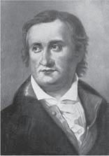
Although Sir Isaac Newton was the first to understand the visible spectrum of light by using a prism, in 1666, when refracting white light into various colors [4] , vibrational spectra experiments started with the first studies of the astronomer Sir Frederich William Herschel in 1800 [5, 6] , in the IR region. In a systematic study of the heating power of colored rays, he created the spectrum—a rainbow, by directing the sunlight through a glass prism placed in front of a thin slit made in a window shutter. The temperature of each color was measured by using three mercury-in-glass thermometers with blackened bulbs (i.e., placing one bulb in a visible color and the other two beyond the spectrum as control). His findings were published in the first article [5] speculating on the fact that the maximum heat effect lies beyond the red edge of the visible spectrum, now named as IR radiation.
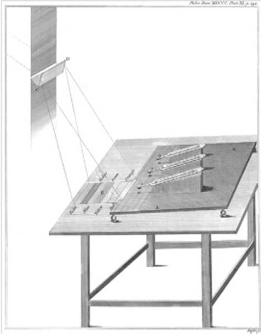
In the second article [6], he stated the detection of IR radiation with the apparatus presented in Fig. 1.1: “the four last experiments prove that the maximum of the heating power is vested among the invisible rays.”
It must be noted that when IR radiation was discovered with Herschel’s glass prism, most scientists did not approve the wave theory of light proposed by Christiaan Huygens [7] stating that wavelength is what determines the color of light. This did not change for more than a decade, even though shortly afterwards an English physicist, Thomas Young, determined the wavelengths of the colors of visible light by using narrowly separated slits to isolate the interference fringes [8, 9].
Thomas J. Seebeck (1770–1831)
Sir Frederich W. Herschel (1738–1822)
FIG. 1.1 Experimental setup for the discovery of IR radiation in 1800. A prism dispersed sunlight; the spectrum fell on a table and a movable stand with mounted thermometers. Thermometers 1 and 2 were exposed to the radiation, whereas thermometer 3 served as a control. (Reproduced from N. Sheppard, The historical development of experimental techniques in vibrational spectroscopy, in: J.M. Chalmers, P.R. Griffits (Eds.), Handbook of Vibrational Spectroscopy, John Wiley & Sons, Ltd., 2002, pp. 1–32 with permission.)
At the same time, further advances on IR spectroscopy depended on the possibility to replace the mercury-in-glass thermometer by more sensitive temperature-measurement methods to find out better IR optical materials and sources of heat radiation for laboratory work other than sunlight. These objectives were accomplished by the discovery [10] of the thermoelectric effect by Thomas Johann Seebeck and the discovery of the thermocoupling effect by Jean Claude Athanase Peltier and Thomas Balt-Johann Seebeck in 1822. Nobili, in 1825, developed the astatic galvanometer; and together with his younger colleague Melloni, in 1833, initially used a thermopile (a series-connected
array of thermocouples) and a galvanometer to increase the sensitivity for measuring temperature and IR radiation.
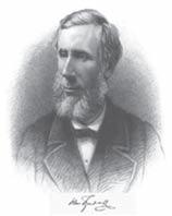
Using thermopile technology, in the 1850s, John Tyndall, at the Royal Institution of Great Britain in London, was the first who measured correctly the relative infrared absorptive powers of a wide variety of liquids and gases [11]. He was a pioneer in attributing IR absorption bands to vibrational degrees of freedom of the molecules concerned, and demonstrating that visually transparent gaseous elements (e.g., O2, N2, and H2) were IR emitters.
Tyndall (1820–1893)
In 1881, Sir William de Wiveleslie Abney and Edward Robert Festing employed photographic means to record the first NIR spectra for about 48 organic substances [12] up to 1.3 μm showing that some molecules (e.g., CCl4 and CS2) did not absorb in this spectral region. They postulated the correlation of observed absorption bands to different types of bonds involving the light hydrogen atoms (CH, NH, OH, etc.) available in the molecules under study. The association of individual NIR absorption bands with smaller functional groups was also done for complex organic molecules. His work was further developed by Julius, who extended the range of measurements to 10 μm for 20 organic liquids and assigned the maxima at 3.45 and 6–7 μm to the existence of methyl groups ( CH3) in the molecule.
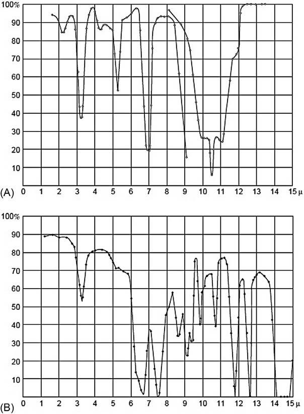
In this direction, William Weber Coblentz studied a very broad range of compounds mostly in IR absorption but also in IR reflection or emission spectroscopy, under the guidance of professor Edward Leamington Nichols at Cornell University. His collected data were later published by the Smithsonian Institution of Washington, DC [13], and some examples are displayed in Fig. 1.2 Coblentz listed 15 group-characteristic bands for aromatic rings, most polar groupings (NO2, CN, SCN, and NCS), and types of XH groups (CH3, CH2, NH2, OH, etc.). Surprisingly, the specificity of the strong CO bond-stretching absorptions was not recognized, possibly due to their (still systematic) variations in position in aldehydes, ketones, carboxylic acids, esters, etc.
John
Sir William de Wiveleslie Abney (1843–1920)
William W. Coblentz (1873–1962)
FIG. 1.2 Coblentz’s IR spectra of (A) ethylene (ethene) and (B) nitrobenzene. (Reproduced N. Sheppard, The historical development of experimental techniques in vibrational spectroscopy, in: J.M. Chalmers, P.R. Griffits (Eds.), Handbook of Vibrational Spectroscopy, John Wiley & Sons, Ltd., 2002, pp. 1–32 with permission.)
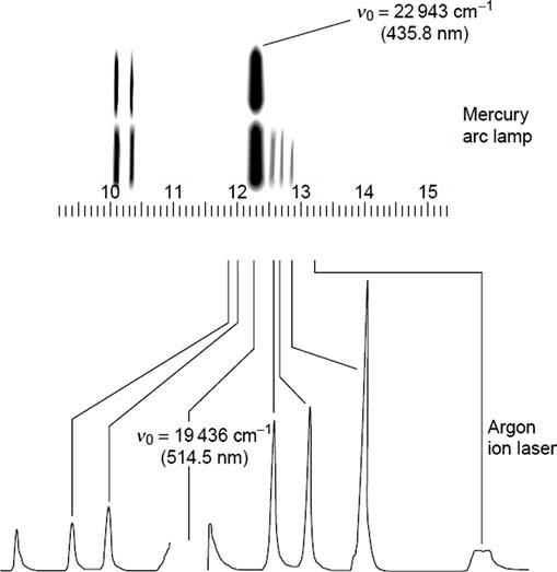
Raman scattering or the Raman effect is the inelastic scattering of photons of light upon the interaction with matter. The effect was discovered by Sir Chandrasekhara Venkata Raman and his student Kariamanickam Srinivas Krishnan [14], in Calcutta in 1928 while trying to use a green filter to intercept the scattered light emerged at right angles to the original beam (i.e., the violet portion of the sunlight) after it had passed through a liquid. In fact, this effect had
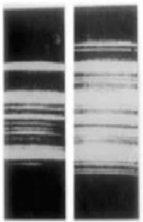
been predicted by Smekal [15] in 1925. Some of the first spectra, obtained by Raman and Krishnan [14], are presented in Fig. 1.3 (the left photograph shows the incident light from a mercury arc lamp after passing through a blue filter, while the right photograph shows the same spectrum after passing through liquid benzene).
The interest in Raman discovery blossomed into some 70 papers by the end of that year because this spectroscopic technique could provide a second method for studying the frequency ranges linked with molecular vibrations and rotations.
As early as in 1931, Karl Wilhelm Friedrich Kohlrausch summarized the measurements of many Raman spectra of organic liquids in several monographs [16, 17]. Raman spectra could be used in addition to IR data to enable at least nearly complete and accurate vibrational assignments of fundamental normal modes, i.e., chemical grouping that shows weak or missing bands in the IR region, but often gives strong Raman features.
FIG. 1.3 The first Raman spectra obtained by photography. (Reproduced from The Raman Effect—75 years, Curr. Sci. 84(5) (2003) 627, https://www.jstor.org/stable/24108480 (Accessed 10 January 2020) with permission.)
Sir C.V. Raman (1888–1970)
Sir K.S. Krishnan (1898–1961)
Because the experimental setup could be much more easily established in the visible light than in the IR region, more than 1800 papers were published on the Raman effect by 1939.
By the late 1930s, Raman spectroscopy was principally chosen for nondestructive chemical analysis for both organic and inorganic compounds, identification was made by referring to the unique spectrum of Raman scattered light of any particular substance served as a “fingerprint” for its identification, whereas the intensity of the spectral lines was related to the amount of the substance.
In 1942, a remarkable progress happened in Raman measurement as the first photoelectric Raman spectrograph (using a cooled cascade-type RCA IP21 photomultiplier detector) was introduced by Rank and Wiegand for quantitative hydrocarbon analysis. This Raman instrument improved the limited photometric accuracy as compared to the use of photographic plates as detector [18]. Photographic and photoelectric spectra of CCl4 are presented for comparison in Fig. 1.4
During the World War II, IR measurements quickly became routine operations due to the availability of sensitive detectors and advances in electronics. At that time, however, the requirement of skilled operators and darkroom facilities
FIG. 1.4 The Raman spectrum of carbon tetrachloride, CCl4, taken by photographic recording (Hg 435.8 nm excitation) and by photoelectric recording (Ar 514.5 nm excitation). (Reproduced from N. Sheppard, The historical development of experimental techniques in vibrational spectroscopy, in: J.M. Chalmers, P.R. Griffits (Eds.), Handbook of Vibrational Spectroscopy, John Wiley & Sons, Ltd., 2002, pp. 1–32 with permission.)
C.W. Townes (1915–2015)
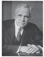
made Raman spectroscopy less competitive than IR spectroscopy. Moreover, Raman scattering was a relatively weak process, so it needed more intense light sources for effect amplification. This problem was solved by Charles Hard Townes, who developed the laser [19] (Light Amplification Stimulated Emission of Radiation), a much more powerful light source to serve as a probe exploring properties to generate dramatically new effects. Basically, a laser is a maser (i.e., Microwave Amplification by Stimulated Emission of Radiation ) that works with higher-frequency photons in the UV-Vis spectrum. The first papers about the maser were coauthored by C.H. Townes, J.P. Gordon, and H.J. Zeiger, who created the first ammonia-beam maser at Columbia University to produce amplification of microwaves at a frequency of about 24.0 GHz.
The late 1980s experienced a resurgence in the use of the original Raman effect by virtue of commercially available Fourier Transform (FT) Raman spectrophotometers. It was also realized that the use of a NIR laser in place of a visible laser as the excitation source could circumvent fluorescence and photodecomposition, but reduce sensitivity in Raman experiments. Fortunately, a successful approach to overcome the latter has engaged with the adoption of interferometry and FT techniques for signal processing.
In practice, Fourier transform infrared (FTIR) is the preferred technique of IR spectroscopy. An FTIR spectrum arises from the mathematical method of Fourier-transformation of an interferogram being yielded by the interference of radiation between two beams. An FTIR instrument is better than a dispersive IR one with reference to shorter analysis time (no energy separation into individual frequency), less reflection loss (no individual frequency limit and fewer mirror surfaces), and spectral comparison with confidence (the laser is available as a source of wavelength calibration within the instrument).
Jean-Baptiste
Joseph Fourier (1768–1830)
J. von Neumann (1903–1957)
Vibrational spectroscopy experienced a final impetus and advance through its digital measuring devices, fathered by the computer scientist J. von Neumann (real name Neumann János Lajos) [20].
Table 1.1 briefly summarizes some of the differences between the techniques of vibrational spectroscopy.
IR spectroscopy was probably first exploited in the field of biomedical analysis by Coblentz [21], in 1911 for radiometric investigation of water of crystallization, light filters, and standard absorption bands, Stair and Coblentz [22], in 1936, for measuring IR absorption spectra of plant, animal tissue, and various other substances.
The pioneers of Raman spectroscopy utilization in biomedical analysis were probably Garfinkel and Edsall [23] in 1958. These authors used a high-pressure mercury lamp for scattering excitation and photographic plates for detection to record the first Raman spectrum of a protein, lysozyme.
The first paper that used MIR spectroscopy in order to characterize fats and oils dates back to 1905, when Coblentz published the first compilation of IR spectra of several vegetable oils and fatty acids [13].
In conclusion, a short family tree of vibrational spectroscopy can be presented in Fig. 1.5.
TABLE 1.1 Comparison of Raman, mid-IR, and near-IR spectroscopy.
Ease
Liquids
Powders
Polymers
Gases
Fingerprinting
Group
Quantitative
Low-frequency
Reproduced from P. Larkin, Infrared and Raman Spectroscopy; Principles and Spectral Interpretation, Elsevier Science, Oxford, 2011 with permission.
Albert A. Michelson
Joseph Fourier
William W. Coblenz
C.V. Raman
C.H. Townes
J. Von Neumann
Vibrational spectroscopy
FIG. 1.5 Chronology of major contribution to vibrational spectroscopy. (Reproduced from J.E. Katon, G.E. Pacey, J.F. O’Keefe, Vibrational molecular microspectroscopy, Anal. Chem. 58(3) 1986, 465A–478A with permission.)
References
[1] J.M. Chalmers, P.R. Griffiths (Eds.), Handbook of Vibrational Spectroscopy. Volume 1: Theory and Instrumentation, John Wiley & Sons, Ltd., 2002.
[2] J.M. Chalmers, P.R. Griffiths (Eds.), Handbook of Vibrational Spectroscopy. Volume 2: Sampling Techniques for Vibrational Spectroscopy, Wiley& Sons, Ltd., 2002.
[3] J.M. Chalmers, P.R. Griffiths (Eds.), Handbook of Vibrational Spectroscopy. Volume 3: Sample Characterization and Spectral Data Processing, John Wiley & Sons, Ltd., 2002.
[4] W.W. Rouse Ball, A Short Account of the History of Mathematics, Dover, New York, 1908, p.325.
[5] F.W. Herschel, Experiments on the solar, and on the terrestrial rays that occasion heat; with a comparative view of the laws to which light and heat, or rather the rays which occasion them, are subject, in order to determine, whether they are the same, or different. Part I, Philos. Trans. R. Soc. Lond. 90 (1800) 255–283.
[6] F.W. Herschel, Experiments on the solar, and on the terrestrial rays that occasion heat; with a comparative view of the laws to which light and heat, or rather the rays which occasion them, are subject, in order to determine, whether they are the same, or different. Part II, Philos. Trans. R. Soc. Lond. 90 (1800) 284–292.
[7] C. Huygens, Treatise on light containing the explanation of reflection and of refraction and especially of the remarkable refraction which occurs in Iceland Spar, in: J.S. Ames (Ed.), Scientific Memoirs—X. The Wave-Theory of Light, American Book Company, 1900, pp. 1–42.
[8] E.S. Barr, Historical survey of the early development of the infrared spectral region, Am. J. Phys. 28 (1960) 42–54.
[9] E.S. Barr, Men and milestones in optics. II: Thomas Young, Phys. Teach. 5 (1967) 53–60. (reprint of Appl. Opt. 2, 639, (1963)).
[10] T.J. Seebeck, Magnetic polarization of metals and minerals by temperature differences, Treatises R. Acad. Sci. Berl. (1825) 265–373.
[11] J. Tyndall, Heat, a Mode of Motion, sixth ed., Longmans, Green and Co., London, 1880.
[12] W. Abney, R.E. Festing, On the influence of atomic grouping in the molecules of organic bodies on their absorption in the infra-red region of the Spectrum, Philos. Trans. R. Soc. Lond. 172 (1881) 887–918.
[13] W.W. Coblentz, Investigations of Infrared Spectra, Parts I to V, Carnegie Institution, 1905–1908. Publication No. 35, 65 and 97. (Republished by Coblentz Society and the PerkinElmer Corp., 1962, Norwalk, CT).
[14] C.V. Raman, K.S. Krishnan, A new type of secondary radiation, Nature 121 (1928) 501–502.
[15] A. Smekal, Zur quantentheorie der dispersion, Naturwissenschaften 11 (43) (1923) S873–S875.
[16] K.W.F. Kohlrausch, Der Smekal-Raman-Effekt, in: Struktur und Eigenschaften der Materie, vols. XII and XIX, Springer Verlag, Berlin, 1931/1938.
[17] K.W.F. Kohlrausch, Ramanspektren, in: Hand- und Jahrbuch der Chemischen Physik, Vol. 9, Part VI, Akad. Verlagsgesellschaft, Leipzig, 1943.
[18] D.H. Rank, R.V. Wiegand, A photoelectric Raman spectrograph for quantitative analysis, J. Opt. Soc. Am. 36 (6) (1946) 325–334.
[19] J. Gordon, H. Zeiger, C.H. Townes, The maser—new type of microwave amplifier, frequency standard, and spectrometer, Phys. Rev. 99 (4) (1955) 1264–1274.
[20] D. Knuth, Von Neumann’s first computer program, in: W. Aspray, A. Burks (Eds.), Papers of John von Neumann on Computing and Computer Theory, MIT Press, Cambridge, ISBN: 978-0-262-22030-9, 1987, pp. 89–95.
[21] W.W. Coblentz, Radiometric investigation of water of crystallization, light filters and standard absorption bands, Bull. Natl. Bur. Stand. (U.S.) 7 (1911) 619–663.
[22] R. Stair, W.W. Coblentz, Infrared absorption spectra of plant and animal tissue and of various other substances, J. Res. Natl. Bur. Stand. 15 (1935) 295–316.
[23] D. Garfinkel, J.T. Edsall, Raman spectra of amino acids and related compounds. X. The Raman spectra of certain peptides and of lysozyme1–3, J. Am. Chem. Soc. 80 (15) (1958) 3818–3823.
Further reading
J.E. Katon, G.E. Pacey, J.F. O’Keefe, Vibrational molecular microspectroscopy, Anal. Chem. 58 (3) (1986) 465A–478A.
P. Larkin, Infrared and Raman Spectroscopy; Principles and Spectral Interpretation, Elsevier Science, Oxford, 2011.
N. Sheppard, The historical development of experimental techniques in vibrational spectroscopy, in: J.M. Chalmers, P.R. Griffits (Eds.), Handbook of Vibrational Spectroscopy, John Wiley & Sons, Ltd., 2002, pp. 1–32.
The Raman Effect—75 years, Curr. Sci. 84 (5) (2003) 627. https://www.jstor.org/stable/24108480 (Accessed 10 January 2020).
Chapter 2 Basic theory, sampling techniques, and instrumentation
Basic theory
Spectroscopy is defined to be a branch of natural sciences concerned with the study of the absorption and emission of light and other electromagnetic radiation by matter as well as the interactions between particles (e.g., electrons, protons, and ions) as a function of their collision energy [1].
In molecular spectroscopy, more specifically speaking, molecules can undergo three types of quantized transitions (electronic, rotational, and vibrational) when being excited by ultraviolet (UV), visible (Vis), and infrared (IR) radiation [2]. For electronic transition, an electron residing in a low-energy orbital is pushed forward to a higher-energy orbital as the energy hν of the photon in the UV-Vis region exactly matches the energy gap between the two orbitals. Unlike UV-Vis rays, IR radiation (from 1 to 15 kcal/mol) is not energetic enough to induce electronic transitions, but it can generate transitions in the rotational and vibrational states involved in the ground-state electronic energy of a molecule given its being in resonance with a vibrating bond. In other words, absorbing IR radiation is characteristic of molecular species having a small energy discrepancy between the rotational and vibrational states.
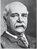
Provided that n is the number of atoms in a molecule and each atom has three degrees of freedom of motion (corresponding to its position in threedimensional space described by the Cartesian coordinate system), the internal degrees of freedom (that describes the vibrational motion of a molecule, i.e., change in the distance between atoms (stretching) or the angle between bonds (bending)) will be 3n 6 and 3n 5 for nonlinear and linear molecules, respectively. The two main modes of vibration (i.e., stretching and bending) may be further given descriptive names as shown in Fig. 2.1 [3]. It is mentioned that some general trends are applicable to vibrational modes as follows: (i) the frequencies of stretching are higher than those of bending because it is much easier to bend than to stretch or compress a bond; (ii) the frequencies of stretching are higher for bonds to hydrogen than those to heavier atoms; (iii) the decreasing order for the frequencies of stretching is: triple bonds > double bonds > single bonds (except for bonds to hydrogen).
Conventionally, the IR region covers the electromagnetic spectrum range from 13,000 to 100 cm 1, which can be subdivided into three regions (near-IR, mid-IR, and far-IR) as follows [4]:
● The mid-IR region (4000–400 cm 1) is often generalized as the X-H stretching region [4000–400 cm 1: O H stretching observed as a broad band in the range 3700–3600 cm 1; N H stretching (usually much sharper than O H stretching) seen between 3400 and 3300 cm 1; C H stretching occurred in the range 3800–2850 cm 1 (from aliphatic compounds) or between 3100 and 3000 cm 1 (if adjacent to a double bond or aromatic ring)], the triple-bond region [2500–2000 cm 1: CC (normally very weak intensity) and CN (medium intensity) stretching occurred in the ranges 2300–2050 and 2300–2200 cm 1, respectively], the double-bond region [2000–1500 cm 1: CN, CC and CO (usually the most intense band) stretching], and the fingerprint region [1500–600 cm 1: uniquely found for most bending and skeletal vibrations making it difficult to assign all the absorption bands].
● The near-IR region (13,000–4000 cm 1) includes weak and overlapping absorption bands because they arise from overtones (i.e., a vibrational mode is excited from the ground state to a higher state and the quantum number v ≥ 2) and combinations (i.e., two molecular vibrations are simultaneously excited) of C H, N H, or O H stretching bands. This region is less useful than mid-IR region for qualitative analysis, but it can be often exploited for quantitative analysis because of important differences existed between different functional groups.
● The far-IR region (400–100 cm 1) is rarely used for structural elucidation, but does provide information on the intramolecular stretching modes involving heavy atoms, skeleton bending modes involving an entire molecule containing heavier atoms, torsional modes (i.e., certain small groups bonded to a large group undergo a motion with regard to the heavier “anchor” group), and crystal lattice vibrations (i.e., the movement of the whole molecular chains with regard to each other in crystalline solids).
FIG. 2.1 Possible vibrational modes of a molecule.
When one of the electrons of a molecule is excited to a higher energy level, the molecule almost instantaneously relaxes to the lowest level in the excited electronic state without emitting radiation by collision with other molecules (i.e., internal conversion). It is followed by emitting fluorescence light in the deexcitation process when the excited molecule goes back to one of the vibrational levels of the ground state.
Conversely, if a molecule is shined with the light (being more energetic to excite any vibrational or rotational states, but less energetic to bring it out of the ground state), it is excited to a virtual state (i.e., a very short-lived, unobservable quantum state) and decays back down to lower energy states. In this case, Raman or Rayleigh scattering may occur [5]. For Rayleigh scattering, the scattered photon has its energy preserved because the molecule decays back to the initial state (i.e., elastically scattered radiation); whereas for Raman scattering (i.e., inelastically scattered radiation), the shifted photons can be of either higher (anti-Stokes radiation) or lower (Stokes radiation) energy as compared to Rayleigh radiation, depending upon the vibrational state of the molecule under study (Fig. 2.2). The Stokes line is much more intense than the anti-Stokes line since only molecules vibrationally excited prior to irradiation may give rise to the anti-Stokes line. Hence, in Raman spectroscopy only the more intense Stokes line is normally measured and the Raman effect is relatively weak with an observed intensity of ca. 10 6 times that of the incident light for strong Raman scattering. However, the intensity of Raman-active vibrations (associated with

FIG. 2.2 Energy diagram of IR and Raman processes: IR absorption (A), Rayleigh scattering (B), Stokes Raman scattering (C), anti-Stokes Raman scattering (D), resonance Raman scattering (E), and fluorescence (F). The numbers represent different vibrational levels within each electronic state. (Modified from H. Baranska, An introduction to Raman scattering, in: H. Baranska, A. Labudzinska, J. Terpinski, (Eds.), Laser Raman Spectrometry: Analytical Applications, Ellis Horwood, Chichester, 1987, pp. 9–31.)
the absorbing chromophore) could be enhanced by a factor of 102–104 (resonance Raman effect) if the incident laser line in a Raman experiment is tuned near, and finally through, the electronic transition of a molecule [6]. Moreover, the Raman scattering from a molecule (or ion) absorbed on or even within a few Angstroms of the surface of suitably nanostructured metallic substrates can be strongly amplified (i.e., 103–106 times greater than in solution). This surfaceenhanced Raman scattering (aka. SERS) is related to both the electromagnetic and chemical effects as illustrated in Fig. 2.3 [7]. Alternatively stated, SERS may arise from two mechanisms: (i) an enhanced electromagnetic field produced at the metal surface (conduction electrons in the metal surface are excited to a state called a surface plasmon resonance when the wavelength of the incident light
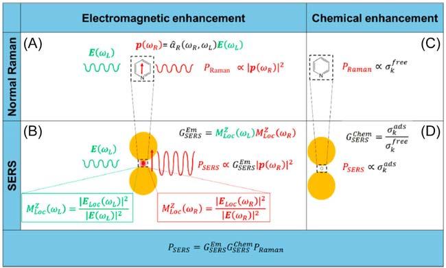
FIG. 2.3 Electromagnetic enhancement. (A) Normal Raman. A laser radiation, with electric field E(ωL) oscillating at (angular) frequency ωL impinges on a molecule, characterized by a Raman polarizability tensor ˆ αR (ωR, ωL). The laser induces a dipole oscillating at the Raman frequency (vertical red arrow (ωR)); the Raman power radiated by this dipole is proportional to the square modulus of the dipole itself. (B) Surface-enhanced Raman scattering (SERS) electromagnetic enhancement. When the molecule is placed near a plasmonic substrate, the electric field experienced by the molecule is ELoc (ωL), normally much stronger than the input laser E(ωL); this local field enhancement is quantified by M Z Loc (ωL). Moreover, the presence of the plasmonic substrate also enhances the efficiency with which the dipole emits Raman radiation; this reradiation enhancement is quantified by M Z Loc (ωR). The total electromagnetic enhancement factor, within the | E |4 approximation, is defined as: G Em SERS = M Z Loc (ωL) M Z Loc (ωR). Chemical enhancement. (C) Normal Raman. The vibrational modes of a molecule in free space are characterized by the cross-section(s) σk free; (D) SERS chemical enhancement. The interaction with the plasmonic substrate modifies the structure of the molecule and consequently also the cross-section(s) of its modes (σk ads). The chemical enhancement is quantified as G Em SERS = σ σ k ads k free .(Reproduced with permission from R. Pilot, R. Signorini, C. Durante, L. Orian, M. Bhamidipati, Laura Fabris, A review on surface-enhanced Raman scattering, Biosensors 9 (2019) 57.)
is close to the plasma wavelength of the metal. It makes molecules absorbed or in close proximity experience an exceptionally large electromagnetic field, most strongly enhancing vibrational modes normal to the surface); (ii) the formation of a charge-transfer complex between the surface and analyte molecule (the resonance enhancement happens as a result of the electronic transition of many charge-transfer complexes in the Vis region. The strongest SERS effect is observed for molecules with lone pair electrons or pi clouds).
Theoretically, the symmetry of a molecule, or the lack of it, will determine what vibrations are IR and Raman active. In general, Raman spectra could be most easily recorded for symmetric or in-phase vibrations and nonpolar groups; on the other hand, IR spectra could be most conveniently ascribed for asymmetric or out-of-phase vibrations and polar groups. It was suggested that the mathematical theory of group could be applied for predicting the number of vibrational bands, their shape and polarization, and the qualitative description of their associated normal modes [8]. It is based on the fact that a molecule may have at least one symmetry element, allowing it to be classified by a point group (i.e., a set of compactible symmetry operations). For small molecules, the IR and Raman activities may be often defined by simply inspecting vibrational forms, i.e., according to the rule of mutual exclusion, no vibration can be active in both the IR and Raman spectra of molecules having a center of symmetry. For instance, vibrations (retaining the center of symmetry) are IR inactive and may be Raman active, and vice versa for vibrations (not retaining the center of symmetry). Conversely, some vibrations can be active in both the IR and Raman spectra (for molecules without any center of symmetry) or in only either one of the IR and Raman spectra (for molecules having other suitable symmetry elements other than a center of symmetry).
For spectral interpretation, IR and Raman frequencies of common functional groups are displayed in Tables 2.1 and 2.2 [4].
Sampling techniques and instrumentation
Basically, IR absorption and Raman scattering differ from each other with respect to the underlying principle by which molecular vibrations occur, i.e., the molecule must be subjected to a change in polarizability in Raman spectroscopy, while there is a change in the net molecular dipole in IR spectroscopy. As a consequence, each technique requires very different instrumentation for spectral registration, i.e., an IR spectrum is obtained by projecting the image of the IR source through a sample onto a detector, by contrast a Raman measurement is performed by imaging the focused laser beam in a sample [9].
In practice, there have been so far two basic types of vibrational instrumentation: (i) dispersive instruments and (ii) Fourier transform instruments. For the former (sometimes called grating or scanning spectrometers, emerged in the 1940s), a diffraction grating is used to sort polychromatic radiation spatially into monochromatic components and direct the dispersed radiation through
TABLE 2.1 IR and Raman frequencies of common functional organic groups.
Functional group
OH for water
H bonded OH
OH group
R CH3
3700–3300 s w
3550–3230 str br
3670–3680 str ms
2975–2950 asym str vs vs
R CH3 2885–2860 sym str vs vs
R CH3 1470–1440 asym bend ms ms
R CH3 1380–1370 sym bend m vw
R CH(CH3)2 1385–1380 Bend-bend m vw
R CH(CH3)2 1373–1365 Bend-open m vw
Aryl CH3
2935–2915 Sym str + bend overtone ms ms 2875–2855 m m
R(CH3)3 1395–1385 Bend-bend m vw
R(CH3)3 1373–1365 Bend-open ms ms
Aliphatic CH2
Aliphatic CH2
Aliphatic CH2
2936–2915 asym str vs vs
2895–2833 sym str vs vs
2920–2890 Fermi resonance w m
Aliphatic CH2 1475–1445 Bend ms ms
(CH2)> 3 1305–1295 In-phase twist – m
(CH2)> 3 736–720 In-phase rock m –
R3CH 1360–1320 CH bend m m
C CC H 3340–3267 CH str s w C CC H 2140–2100 CC str w vs C CC H 710–578 CH wag sbr w
>CC< trans, tri, tetra 1600–1665 CC str w-0 s
>CCH R mono, cis, trans 3020–2995 CH str m m
CC mono, cis 1,1 1660–1630 CC str m s
>CCH2 mono 1,1 3090–3075 CH2 asym str m m
>CCH2 mono 1,1 3000–2980 CH2 sym str m s
TABLE 2.1 IR and Raman frequencies of common functional organic groups—cont’d
Functional group
>CCH2 mono 1,1 1420–1400 CH2 bend w m
R CHCH2 995–985 trans CH2 in-phase wag s w
R CHCH2 910–905 >CH2 wag s w
Aryl CH 3100–3000 CH str mw s
Aromatic ring 1620–1585 Quadrant str var m
Aromatic ring 1590–1565 Quadrant str var m
Aromatic ring 1525–1470 Semicircle str var vw
Aromatic ring 1465–1400 Semicircle str m vw
Mono, meta, (1,3,5), (2,4,6) 1010–990 In-phase str vw vs
Meta, (1,2,4), (1,3,5) 9365–810 Lone H wag m –
Para, (1,2,4) 880–795 2 adj. H wag s –
Meta, (1,2,3) 825–750 3 adj. H wag s –
Ortho, meta 800–725 4 and 5 adj. H wag s –
Mono, meta, (1,3,5) 710–665 Ring out-of-plane bend s –
Para 650–630 Ring in-plane bend – m
Mono 630–605 Ring in-plane bend w m R
s
TABLE 2.1 IR and Raman frequencies of common functional organic groups—cont’d
R COO
OC NH About 3300 NH str s w
OC NH Near 3100 (overtone of 1550) NH str w w
TABLE 2.1 IR and Raman frequencies of common functional organic groups—cont’d
Functional group
CNHX 3
CNHX 3
CNHX 22
CNHX 22
C3NH+…X
1625–1560 NH3 out-of-phase str mw vw
1550–1505 NH3 in-phase str w vw
1620–1560 NH2 bend mw w 3000–2700 NH2 str sbr w
2700–2300 NH2 str s w
CH2 NO2 1600–1530 NO2 out-of-phase str s mw
CH2 NO2 1380–1310 NO2 in-phase str s vs
Ar NO2 1555–1485 NO2 out-of-phase str s –
Ar NO2 1357–1318 NO2 in-phase str s vs
CH2 Cl 830–560 C Cl str s s
CH2 Br 700–515 C Br str s vs
CH2 F 1100–1000 C F str s w
Pyridine 3100–3300 Aryl CH str m m
Pyridine 1615–1570 Quadrant str s m
Pyridine 1400–1440 Semicircle str s mw
Pyridine 1035–1025 2,4,6 carbon radial str m vs
Pyridine 995–985 Ring breath/str m s
Pyridine 660–600 Quadrant in-plane bend – m
Pyrrole 3500–3000 NH str s m-w
Pyrrole 3135–3103 CH str m s
Pyrrole 1530 Quadrant str + CH rock s –
Pyrrole 1468 Quadrant str + CH rock m s
Continued