Morphology-controlled synthesis of 3D flower-like TiO2 and the superior performance for selective catalytic reduction of NOx with NH3 Luyao Zong & Guodong Zhang & Jiuhu Zhao & Fang Dong & Jiyi Zhang & Zhicheng Tang
Electrocardiography of Arrhythmias-A Comprehensive Review-A Companion to Cardiac Electrophysiology, 2e (Mar 8, 2021)_(032368050X)_(Elsevier).pdf Mithilesh Kumar Das
Brown University, Emerita, and University of Bristol
Chapter 26 “Primate Evolution and the Emergence of Humans” by Sergi López-Torres, University of Toronto Scarborough and Roman Kozlowski Institute of Paleobiology, Polish Academy of Sciences
Oxford University Press is a department of the University of Oxford. It furthers the University’s objective of excellence in research, scholarship, and education by publishing worldwide. Oxford is a registered trade mark of Oxford University Press in the UK and certain other countries.
Published in the United States of America by Oxford University Press 198 Madison Avenue, New York, NY 10016, United States of America
Sinauer Associates is an imprint of Oxford University Press.
For titles covered by Section 112 of the US Higher Education Opportunity Act, please visit www.oup.com/us/he for the latest information about pricing and alternate formats.
All rights reserved. No part of this publication may be reproduced, stored in a retrieval system, or transmitted, in any form or by any means, without the prior permission in writing of Oxford University Press, or as expressly permitted by law, by license, or under terms agreed with the appropriate reproduction rights organization. Inquiries concerning reproduction outside the scope of the above should be sent to the Rights Department, Oxford University Press, at the address above.
You must not circulate this work in any other form and you must impose this same condition on any acquirer.
Address editorial correspondence to: Sinauer Associates 23 Plumtree Road Sunderland, MA 01375 U.S.A. publish@sinauer.com
Address orders, sales, license, permissions, and translation inquiries to: Oxford University Press U.S.A. 2001 Evans Road Cary, NC 27513 U.S.A. Orders: 1-800-445-9714
Library of Congress Cataloging-in-Publication Data
Names: Pough, F. Harvey, author. | Janis, Christine M. (Christine Marie), 1950- author.
Title: Vertebrate life / F. Harvey Pough, Rochester Institute of Technology, Emeritus, Christine M. Janis, Brown University, Emerita, University of Bristol ; chapter 26 “Primate Evolution and the Emergence of Humans” by Sergi López-Torres, University of Toronto Scarborough, Roman Kozlowski Institute of Paleobiology, Polish Academy of Sciences
Description: Tenth edition. | New York : Oxford University Press, [2019] | Revised edition of: Vertebrate life / F. Harvey Pough, Christine M. Janis, John B. Heiser. 9th ed. Boston : Pearson, c2013. | Includes bibliographical references and index.
Identifiers: LCCN 2018005330 (print) | LCCN 2018005884 (ebook) | ISBN 9781605357218 (ebook) | ISBN 9781605356075 (casebound)
LC record available at https://lccn.loc.gov/2018005330
9 8 7 6 5 4 3 2 1
Printed in the United States of America
Brief Contents
CHAPTER 1 Evolution, Diversity, and Classification of Vertebrates 1
CHAPTER 2 What Is a Vertebrate? 19
CHAPTER 3 Jawless Vertebrates and the Origin of Jawed Vertebrates 41
CHAPTER 4 Living in Water 65
CHAPTER 5 Geography and Ecology of the Paleozoic Era 83
CHAPTER 6 Radiation and Diversification of Chondrichthyes 95
CHAPTER 7 Extant Chondrichthyans 103
CHAPTER 8 Radiation and Diversity of Osteichthyes 121
CHAPTER 9 Extant Bony Fishes 133
CHAPTER 10 Origin and Radiation of Tetrapods 161
CHAPTER 11 Extant Amphibians 181
CHAPTER 12 Living on Land 211
CHAPTER 13 Geography and Ecology of the Mesozoic Era 233
CHAPTER 14 Synapsids and Sauropsids 241
CHAPTER 15 Ectothermy: A Low-Energy Approach to Life 269
CHAPTER 16 Turtles 283
CHAPTER 17 Lepidosaurs 301
CHAPTER 18 Crocodylians 329
CHAPTER 19 Mesozoic Diapsids: Dinosaurs and Others 343
CHAPTER 20 Endothermy: A High-Energy Approach to Life 371
CHAPTER 21 The Origin and Radiation of Birds 387
CHAPTER 22 Extant Birds 399
CHAPTER 23 Geography and Ecology of the Cenozoic Era 435
CHAPTER 24 Synapsida and the Evolution of Mammals 451
CHAPTER 25 Extant Mammals 481
CHAPTER 26 Primate Evolution and the Emergence of Humans 519
Preface xv
Chapter 1
Evolution, Diversity, and Classification of Vertebrates 1
1.1 The Vertebrate Story 1
Major extant groups of vertebrates 2
Non- amniotes 2
Amniotes 4
1.2 Classification of Vertebrates 5
Binominal nomenclature 5
Phylogenetic systematics 5
Applying phylogenetic criteria 6
Morphology-based and molecular-based phylogenies 7
Using phylogenetic trees 8
1.3 Crown and Stem Groups 9
1.4 Genetic Mechanisms of Evolutionary Change 9
Phenotypes and fitness 10
Developmental regulatory genes 11
Intragenerational versus transgenerational phenotypic modification 14
1.5 Ear th History and Vertebrate Evolution 14
Chapter 2
What Is a Vertebrate? 19
2.1 Vertebrates in Relation to Other Animals 19
2.2 Characteristics of Chordates 20
Chordate origin and evolution 21
Extant nonvertebrate chordates 21
2.3 What Distinguishes a Vertebrate? 23
2.4 Vertebrate Embryonic Development 24
Development of the body 24
Development of the pharyngeal region 25
Development of the brain 27
Unique developmental features of vertebrates 27
2.5 Basic Vertebrate Structures 29
Adult tissue types 30
Mineralized tissues 30
The skeletomuscular system 30
2.6 Basic Vertebrate Systems 32
The alimentary system 32
The cardiovascular system 33
The excretory and reproductive systems 35
The sense organs 35
Chapter 3
Jawless
Vertebrates and the Origin of Jawed Vertebrates 41
3.1 The Earliest Evidence of Vertebrates 41
The origin of bone and other mineralized tissues 42
The mysterious conodonts 43
The environment of early vertebrate evolution 43
3.2 Cyclostomes: The Extant Jawless Fishes 46
Characters of cyclostomes 46
Fossil cyclostomes 46
Extant hagfishes: Myxiniformes 46
Lampreys: Petromyzontiformes 48
Cyclostomes and humans 49
3.3 Ostracoderms: Extinct Jawless Fishes 50
Characters of ostracoderms 50
Ostracoderm evolutionary patterns 50
3.4 The Basic Gnathostome Body Plan 51
Gnathostome biology 51
What about soft tissues? 52
3.5 The Origin of Jaws 52
Early hypotheses of jaw origins 52
The importance of the nose 55
Developmental studies of extant vertebrates 55
Transitional anatomy in fossils 56
The selective value of jaws 56
3.6 The Origin of Paired Appendages 56
The advantages of fins 56
Fin development and the lateral somitic frontier 56
Origin of the neck region 57
3.7 Extinct Paleozoic Jawed Fishes 58
Placoderms: Armored fishes 58
Acanthodians 60
The surviving gnathostome groups 61
Chapter 4
Living in Water 65
4.1 The Aquatic Environment 65
Obtaining oxygen from water: Gills 65
Obtaining oxygen from air: Lungs and other respiratory structures 66
Adjusting buoyancy 67
4.2 Water and the Sensory World of Aquatic
Vertebrates 69
Vision 69
Hearing 69
Chemosensation: Taste and smell 70
Detecting water displacement 70
Electrical discharge 72
Electroreception by sharks and rays 72
4.3 The Internal Environment of Vertebrates 74
4.4 Exchange of Water and Ions 75
Nitrogen excretion 75
The vertebrate kidney 75
Regulation of ions and body fluids 76
4.5 Vertebrates in Different Environments 76
Marine vertebrates 76
Freshwater vertebrates: Teleosts and amphibians 78
Euryhaline vertebrates 79
Terrestrial vertebrates 80
Chapter 5
Geography and Ecology of the Paleozoic Era 83
5.1 Shifting Continents and Changing Climates 83
5.2 Continental Geography of the Paleozoic 84
5.3 Paleozoic Climates 85
5.4 Paleozoic Ecosystems 86
Aquatic life 86
Terrestrial floral ecosystems 88
Terrestrial faunal ecosystems 89
5.5 Paleozoic Extinctions 90
Chapter 6
Radiation and Diversification of Chondrichthyes 95
6.1 Chondrichthyes: The Cartilaginous Fishes 95
Distinctive characters of chondrichthyans 95
6.2 Evolutionary Diversification of Chondrichthyes 98
Paleozoic chondrichthyan radiations 98
The Mesozoic chondrichthyan radiation 100
Paleozoic and Mesozoic chondrichthyan paleobiology 101
Chapter 7
Extant Chondrichthyans 103
7.1
Morphology of Extant Chondrichthyans 103
Skeleton 103
Jaws 103
Skin 104
7.2 Sharks (Selachii) 106
Sensory systems and prey detection 106
Ecology of sharks 108
Heterothermy 109
Feeding 109
Reproduction 110
7.3 Skates and Rays (Batoidea) 112
Morphology 112
Ecology 112
Courtship and reproduction 113
7.4 Chimaeras (Holocephali) 113
7.5 Declining Shark Populations: An Ecological Crisis 114
Chapter 8
Radiation and Diversity of Osteichthyes 121
8.1 The Origin of Bony Fishes 121
Earliest osteichthyans and the major groups of bony fishes 123
8.2 Evolution of Actinopterygii 125
Basal actinopterygians 125
Neopterygii 125
Evolution of jaw protrusion 126
Pharyngeal jaws 127
Specializations of fins 128
8.3 Evolution of Sarcopterygii 128
Actinistia 129
Dipnoi 129
Tetrapodomorpha 130
Chapter 9
Extant Bony Fishes 133
9.1 Actinopterygians: Ray-Finned Fishes 133
Non-teleosts 133
Teleosts 134
9.2 Swimming 139
Minimizing drag 141
Steering, stopping, and staying in place 141
9.3 Actinopterygian Reproduction 142
Oviparity 142
Viviparity 143
9.4 The Sex Lives of Teleosts 144
Protandry 144
Protogyny 145
Hermaphroditism 146
All-female species 147
9.5 Teleosts in Different Environments 147
Deep-sea fishes 147
Coral reef fishes 152
9.6 Heterothermal Fishes 152
Warm muscles 152
Hot eyes 153
9.7 Sarcopterygians:Lobe-Finned Fishes 154
Actinistians: Coelacanths 154
Dipnoans: Lungfishes 155
9.8 Pollution, Overfishing, and Fish Farming 155
Freshwater fishes 155
Marine fishes 156
Chapter 10
Origin and Radiation of Tetrapods 161
10.1 Tetrapod Origins 161
Tetrapodomorph fishes 161
Earliest tetrapods of the Late Devonian 163
10.2 Moving onto Land 165
Terrestrial and walking fishes today 165
How are fins made into limbs? 166
Body support and locomotion 167
Lung ventilation and dermal bone 168
10.3 Radiation and Diversity of Non-Amniote Tetrapods 169
10.4 Amniotes 172
Derived features of amniotes 173
The amniotic egg 174
Patterns of amniote temporal fenestration 175
Chapter 11
Extant Amphibians 181
11.1 Diversity of Lissamphibians 181
Salamanders 181
Anurans 184
Caecilians 188
11.2 Life Histories of Amphibians 188
Salamanders 189
Anurans 191
The ecology of tadpoles 196
Caecilians 197
11.3 Amphibian Metamorphosis 198
11.4 Exchange of Water and Gases 198
Cutaneous respiration 198
Blood flow in larvae and adults 199
Cutaneous permeability to water 199
Behavioral control of evaporative water loss 201
Uptake and storage of water 202
11.5 Toxins, Venoms, and Other Defense Mechanisms 202
Skin glands 202
Toxicity and diet 203
Venomous amphibians 204
11.6 Why Are Amphibians Vanishing? 205
Disease 205
Synergisms 206
Chapter 12
Living on Land 211
12.1 Support and Locomotion on Land 211
The skeleton 211
The cranial skeleton 212
The axial skeleton: Vertebrae and ribs 212
Axial muscles 213
The appendicular skeleton: limbs and limb girdles 215
Size and scaling 217
Locomotion 217
12.2 Eating on Land 219
12.3 Breathing Air 220
12.4 Pumping Blood Uphill 220
12.5 Sensory Systems in Air 222
Vision 222
Hearing 224
Olfaction 225
Proprioception 226
12.6 Conserving Water in a Dry Environment 226
12.7 Controlling Body Temperature in a Changing Environment 227
Ectothermy 227
Endothermy 228
Ectothermy, endothermy, and heterothermy 228
Chapter 13
Geography and Ecology of the Mesozoic Era 233
13.1 Continental Geography of the Mesozoic 233
13.2 Mesozoic Climates 233
13.3 Mesozoic Aquatic Life 234
13.4 Mesozoic Terrestrial Ecosystems 235
The Triassic 235
The Jurassic 236
The Cretaceous 236
13.5 Mesozoic Extinctions 238
Chapter 14
Synapsids and Sauropsids 241
14.1 The Conflict between Locomotion and Respiration 241
Locomotion and lung ventilation of synapsids 242
Locomotion and lung ventilation of sauropsids 243
14.2 Limb-Powered Locomotion 243
The basal amniote ankle joint 245
The sauropsid ankle joint 245
The synapsid ankle joint 245
14.3 Increasing Gas Exchange 247
Synapsid lungs 248
Sauropsid lungs 248
The respiratory system of birds 248
Why are synapsid and sauropsid lungs so different? 248
14.4 Transporting Oxygen to the Muscles: The Heart 250
14.5 The Evolution of Endothermy 253
How did endothermy evolve? 253
Evaluating the models 254
When did endothermy evolve? 254
14.6 Getting Rid of Wastes: The Kidneys 255
Nitrogen excretion by synapsids: The mammalian kidney 256
Nitrogen excretion by sauropsids: Renal and extrarenal routes 260
14.7 Sensing and Making Sense of the World 263
Vision 263
Chemosensation: Gustation and olfaction 263
Hearing 264
Brains 264
Chapter 15
Ectothermy: A Low-Energy Approach to Life 269
15.1 Vertebrates and Their Environments 269
15.2 Dealing with Dryness: Ectotherms in Deserts 269
Desert tortoises 270
The chuckwalla 271
Desert amphibians 274
15.3 Coping with Cold: Ectotherms in Subzero Conditions 275
Frigid fishes 275
Frozen frogs 276
15.4 Energetics of Ectotherms and Endotherms 277
Body size 277
Body shape 278
Conversion efficiency 279
15.5 The Role of Ectotherms in Terrestrial Ecosystems 279
Chapter 16
Turtles 283
16.1 Everyone Recognizes a Turtle 283
Shell and skeleton 283
Families of extant turtles 285
16.2 Turtle Structure and Function 287
Lung ventilation 287
The heart 288
Patterns of circulation and respiration 289
Body size and temperature regulation 289
16.3 Reproductive Biology of Turtles 289
Moisture and egg development 289
Temperature-dependent sex determination 290
Parental care 291
Hatching and the behavior of baby turtles 291
16.4 Social Behavior, Communication, and Courtship 293
16.5 Navigation and Migrations 293
Navigation by adult turtles 294
Navigation by hatchling and juvenile sea turtles 295
16.6 The Fateful Life-History Characteristics of Turtles 295
Chapter 17
Lepidosaurs 301
17.1 Rhynchocephalians and the Biology of Tuatara 301
17.2 Radiation of Squamates 303
Lizards 303
Snakes 306
17.3 Foraging Modes 308
Correlates of foraging mode 309
17.4 Skull Kinesis 312
17.5 Feeding Specializations of Snakes 312
Venom and fangs 314
Hear ts and stomachs 315
17.6 Predator Avoidance and Defense 315
Crypsis 315
“Eavesdropping” 316
Deterrence 316
Autotomy 317
Venomous and poisonous snakes 317
17.7 Social Behavior 318
Territoriality 318
Sociality 320
17.8 Reproductive Modes 320
Sex determination 320
O viparity and viviparity 320
Parthenogenesis 321
Parental care 321
17.9 Thermal Ecology 322
Organismal performance and temperature 322
17.10 Lepidosaurs and Climate Change 323
Chapter 18
Crocodylians 329
18.1 Diversity of Extant Crocodylians 329
18.2 The Crocodylomorph Lineage 331
Notosuchia 331
Neosuchia 332
18.3 Predatory Behavior and Diet of Extant Crocodylians 333
18.4 Communication and Social Behavior 335
18.5 Reproduction and Parental Care 336
Temperature-dependent sex determination 336
Parental care 336
18.6 The Skin Trade 337
Chapter 19
Mesozoic Diapsids: Dinosaurs and Others 343
19.1 Characteristics of Diapsids 343
19.2 Diversity of Mesozoic Diapsids 343
19.3 Lepidosauromorphs: Marine Diapsids 346
Terrestrial lepidosauromorphs 346
Marine lepidosauromorphs 346
19.4 Metriorhynchid Crocodylomorphs 349
19.5 Pterosaurs: The First Flying Vertebrates 349
The structure of pterosaurs 349
Reproduction, eggs, and parental care 352
Did the evolution of birds doom the pterosaurs? 352
19.6 Triassic Faunal Turnover 352
19.7 The Structure and Function of Dinosaurs 353
Hips and legs 353
Dinosaur lineages 354
19.8 Ornithischian Dinosaurs 355
Thyreophora 355
Marginocephalia 357
Ornithopoda 358
Social behavior of ornithischian dinosaurs 358
Nesting and parental care by ornithischians 360
19.9 Herbivorous Saurischians 360
The structure of sauropods 360
Social behavior of sauropods 362
Nesting and parental care by sauropods 362
19.10 Carnivorous Saurischians 363
Tyrannosauroides 363
Ornithomimisauria 363
Maniraptora 364
Social behavior of theropods 365
Nesting and parental care by theropods 365
19.11 Gigantothermy and the Body Temperature of Dinosaurs 366
Chapter 20
Endothermy: A High-Energy Approach to Life 371
20.1 Balancing Heat Production with Heat Loss 371
Whole-body metabolism 372
Shivering and non-shivering thermogenesis 372
Insulation 372
Evaporative cooling 373
20.2 Endotherms in the Cold 373
Avoiding cold and sharing heat 374
20.3 Facultative Hypothermia 375
Seasonal hypothermia 375
Rest-phase hypothermia 375
Hibernation 376
20.4 Endotherms in the Heat 378
Temperature stress and scarcity of water 379
Strategies for desert survival 379
Avoidance 380
Relaxation of homeostasis by hyperthermia 381
Hypothermia in the desert 383
Chapter 21
The Origin and Radiation of Birds 387
21.1 Avian Characters in Nonavian Theropods 387
Skeletal characters 387
Feathers 388
Reproduction and parental care 390
Body size 391
21.2 The Mosaic Evolution of Birds 392
How—and why—birds got off the ground 392
The appearance of powered avian flight 393
21.3 Early Birds 394
21.4 The Mesozoic Radiations of Birds 394
Chapter 22
Extant Birds 399
22.1 The Structure of Birds: Specialization for Flight 399
Feathers 401
Streamlining and weight reduction 403
Skeleton 403
Muscles 404
22.2 Wings and Flight 404
Wing muscles 406
Wing shape and flight characteristics 407
22.3 Feet 408
Hopping, walking, and running 408
Climbing 409
Swimming 409
22.4 Feeding and Digestion 410
Beaks, skulls, and tongues 410
The digestive system 413
22.5 Sensory Systems 414
Vision 415
Hearing 416
Olfaction 416
22.6 Social Behavior 417
Plumage colors and patterns 417
Vocalization, sonation, and visual displays 419
22.7 Oviparity 421
Egg biology 421
Sex determination 422
Maternal control of sex of offspring 422
22.8 Monogamy: Social and Genetic 423
22.9 Nests and Parental Care 423
Incubation 423
Parental care 425
Brood parasitism 425
22.10 Orientation and Navigation 426
22.11 Migration 427
Migratory movements 427
Costs and benefits of migration 427
22.12 Birds and Urbanization 428
Success in the city 429
Noise pollution 429
Not so sex y in the city 429
Chapter 23
Geography and Ecology of the Cenozoic Era 435
23.1 Continental Geography of the Cenozoic 435
23.2 Cenozoic Climates 436
Paleogene and Neogene climates 436
The Pleistocene ice ages 438
23.3 Cenozoic Terrestrial Ecosystems 439
23.4 Biogeography of Cenozoic Mammals 441
The isolation of Australian mammals 441
The isolation of mammals on other continents 444
23.5 Cenozoic Extinctions 446
Chapter 24
Synapsida and the Evolution of Mammals 451
24.1 The Origin of Synapsids 451
24.2 The Diversity of Non-Mammalian Synapsids 451
Pelycosaurs: Basal non-mammalian synapsids 452
Therapsids: More derived non-mammalian synapsids 456
Therapsid diversity 459
24.3 Evolutionary Trends in Synapsids 460
Evolution of the diaphragm 462
Evolution of a double occipital condyle 462
Evolution of jaws and ears 462
24.4 The First Mammals 465
Metabolic and growth rates 466
Skeletomuscular system 466
Feeding and mastication 467
Brain, senses, and behavior 468
The integument: Epidermis and glands 469
Lactation and suckling 471
Food processing and swallowing 472
Facial musculature 472
Internal anatomy 473
24.5 Mesozoic Mammals 474
Dual radiations of Mesozoic mammals 474
Chapter 25
Extant Mammals 481
25.1 Major Lineages of Mammals 481
Multituberculates 482
Monotremes 482
Marsupials 485
Placentals 487
25.2 Differences between Therians and NonTherians 491
Craniodental features 491
Postcranial skeletal features 492
Gait and locomotion 493
Hearing 494
Vision 494
Lactation 495
Information from the genes 495
Sex determination and sex chromosomes 496
25.3 Differences between Marsupials and Placentals 496
25.4 Mammalian Reproduction 497
Mammalian urogenital tracts 498
Genitalia 498
Reproductive mode of monotremes: Matrotrophic oviparity 499
Reproductive mode of therians: Matrotrophic viviparity 500
The earliest therian condition, and the discredited notion of placental superiority 502
25.5 Specializations for Feeding: Teeth and Jaws 503
Mammalian teeth 503
Differences between carnivorous and herbivorous mammals 503
Rodents: Specialized feeders 505
25.6 Specializations for Locomotion 506
Cursorial limb morphology 506
Fossorial limb morphology 507
25.7 The Evolution of Aquatic Mammals 508
Morphological adaptations for life in water 508
The evolution of cetaceans 509
25.8 Trophy Hunting 513
Endangering the endangered: The effect of perceived rarity 514
The extinction vortex 515
Chapter 26
Primate Evolution and the Emergence of Humans 519
26.1 Primate Origins and Diversification 519
Evolutionary trends and diversity in primates 519
26.2 Origin and Evolution of Hominoidea 526
Diversity and social behavior of extant apes 527
Relationships within Hominoidea 531
Diversity of fossil hominoids 531
26.3 Origin and Evolution of Humans 533
Early hominins 533
Ecological and biogeographic aspects of early hominin evolution 537
26.4 Derived Hominins: The Genus Homo 537
Homo erectus and Homo ergaster 537
The Dmanisi hominins 539
Homo floresiensis 539
Homo naledi 540
Precursors of Homo sapiens 540
The Neandertals 541
The Denisovans 542
Origins of modern humans 542
What happened to the humans who were already there? 543
26.5 Evolution of Human Characteristics 543
Bipedalism 543
Large brains 544
Speech and language 545
Loss of body hair and development of skin pigmentation 545
Human technology and culture 546
26.6 Why Is Homo sapiens the Only Surviving Hominin Species? 546
Hybridization among species of Homo 547
26.7 Humans and Other Vertebrates 547
Humans as superpredators and environmental disruptors 547
Megafaunal extinctions 548
Is this the Anthropocene? 549
Appendix A-1
Glossary G-1
Illustration Credits IC-1
Index I-1
Preface
The sustainability of populations, and even the continued existence of some species of vertebrates, is becoming ever more problematic. In addition to overarching events that affect all living organisms—such as global climate change and acidification of the seas—each lineage of vertebrates faces threats that are intimately entwined with the biological characteristics of that lineage.
Thus, as we have prepared this tenth edition of Vertebrate Life , the study of vertebrates has taken on new urgency. Many areas of vertebrate biology have seen enormous advances since the previous edition:
• Phylogenies based on molecular data increasingly supplement, reinforce, and in some cases contradict phylogenies based on morphology. We have incorporated this information, including some of the cases where the two approaches have generated different hypotheses about the timing or sequence of evolutionary change.
• O ur understanding of the genetic and epigenetic control of development has advanced greatly, and this evo-devo perspective provides a mechanistic understanding of evolutionary changes in phenotype.
• Newly discovered fossil sites, fossils, and even fossils that preserve soft tissue structures have added enormous detail (and often greater complexity) to our understanding of evolutionary lineages, especially early tetrapods, feathered nonavian dinosaurs, and humans.
This edition reflects these changes. What has not changed is the authors’ view of vertebrates as complex and fascinating organisms that can best be understood by considering the interactions at multiple levels of biological organization that shape the biology of a species. This book presents vertebrates in a way that integrates all facets of their biology— from anatomy and physiology to ecology and behavior—in an evolutionary context
Authors
Two changes of authorship have occurred in this edition. Ill health has compelled John B. Heiser to withdraw from active participation. John has been an author since the first edition of Vertebrate Life, enriching the chapters on fishes
with the experience gained from his field work in nearly every ocean. We thank him for his contributions, and wish him well.
We are delighted to have gained the assistance of Sergi López-Torres who authored the chapter on primates and the evolution of humans. Primate evolution is an extraordinarily active field of research, and Sergi brings the depth and breadth of knowledge of human evolution needed for an overview of this increasingly complex field.
Organization
The scope of vertebrate biology and of evolutionary time is vast, and encapsulating this multiplicity of themes and mountains of data into a book or a semester course is a continuing challenge. Following suggestions from users, the text has been extensively reorganized to improve the flow of information. Topics have been split or merged, and some have been deleted, resulting in presentation of information in more manageable segments. Each chapter includes a list of sources that will be useful to students, and all of the sources that we consulted in preparing this edition, as well as many sources from earlier editions, are available on the book’s web page, oup.com/us/vertebratelife10e.
Sources
We have relied on these sources for the numbers of extant species and their common and scientific names.
• FishBase: http://www.fishbase.se/search.php
• A mphibiaWeb: https://amphibiaweb.org/
• T he Reptile Database: http://www.reptile-database.org/
• IOC World Birds list: http://www.worldbirdnames.org/
• M ammal Species of the World: https://www.departments.bucknell.edu/biology/resources/msw3/
• A SM Mammal Diversity Database: https://mammaldiversity.org/
• I UCN Red List of threatened species: http://www. iucnredlist.org/
Acknowledgments
Authors are only the visible tip of the iceberg that is a textbook. Expert librarians are essential to any scholarly undertaking, and we are fortunate to have had the outstanding assistance of Adwoa Boateng and Morna Hilderbrand (Wallace Library, Rochester Institute of Technology) and Sue O’Dell (Hatch Science Library, Bowdoin College).
The figure legends cite the many artists and photographers who kindly allowed us to use their work, often provided additional information, and in some cases took new photos at our request.
We are grateful to the many colleagues who answered questions and suggested sources for data and photographs:
Albert Bennett
Lucille Betti-Nash
Daniel Blackburn
Caleb M. Brown
Grover Brown
Larry Buckley
Mark Chappell
Charles J. Cole
Alison Cree
Chris Crippen
Mark Dimmitt
Vladimir Dinets
Col leen Farmer
Vincent Fernandez
Luciano Fischer
Linda Ford
Margaret Fusari
Harry W. Greene
Katrina Halliday
James Hanken
Karsten E. Hartel
Gene Helfman
Peter E. Hillman
Andrew Holmes
Osamu Kishida
Harvey B. Lillywhite
Jessie Maisano
Patrick Moldowan
Andrew Moore
Kenneth Nagy
Darren Naish
Gavin Naylor
Stewart Nicol
Martin Nyffler
Kouki Okagawa
Todd Pierson
Theodore Pietsch
Christopher Raxworthy
Shawn M. Robinson
Caroline S. Rogers
Robert Rothman
Timothy B. Rowe
Michael J. Ryan
Sue Sabrowski
Allen Salzberg
Colin Sanders
Jay Savage
Kurt Schwenk
Wade C. Sherbrooke
Rick Shine
Matthew Simon
Megan Southern
Zachary Stahlschmidt
Hyla Sweet
Ryan Taylor
Frank Tiegler
Hugh Tyndale-Biscoe
Wayne van Devender
John K. VanDyk
David B. Wake
Cliff White
Mark Witton
Stephen Zozaya
We leaned especially heavily on many colleagues, and thank them for their generosity:
Robin M. Andrews, Virginia Tech
James Aparicio, Museo Nacional de Historia Natural de Bolivia
Paul M. Barratt, Natural History Museum, London
Robin M. D. Beck, University of Salford
David R. Begun, University of Toronto
William E. Bemis, Cornell University
Michael J. Benton, University of Bristol
Christopher Brochu, University of Iowa
Edmund D. Brodie, Jr., Utah State University
William S. Brown, Skidmore College
Emily A. Buchholtz, Wellesley College
Ann C. Burke, Wesleyan University
Kenneth Catania, Vanderbilt University
Jennifer A. Clack, University of Cambridge
René C. Clark, Tucson
Chris Crippen, Virginia Living Museum
Michael I. Coates, University of Chicago
A. W. “Fuzz” Crompton, Harvard University
Martha L. Crump, Utah State University
David Cundall, Lehigh University
John D. Damuth, University of California, Santa Barbara
Christine Dahlin, University of Pittsburgh at Johnstown
Dominique Didier, Millersville University
Kenneth Dodd, University of Florida
Phillip C. J. Donoghue, University of Bristol
William N. Eschmeyer, California Academy of Sciences
Robert E. Espinoza, California State University, Northridge
David E. Fastovsky, University of Rhode Island
Sharon Gilman, Coastal Carolina University
Pamela G. Gill, University of Bristol
Frank M. Greco, New York Aquarium
Gordon Grigg, The University of Queensland
David F. Gruber, City University of New York
Célio F. B. Haddad, Universidade Estadual Paulista “Júlio de Mesquita Filho”
Lindsay Hazely, The Southland Museum and Art Gallery
Axel Hernandez, Università di Corsica Pasquale Paoli
Thomas R. Holtz, University of Maryland
Carlos Jared, Instituto Butantan
Bruce C. Jayne, University of Cincinnati
Jeffrey Lang, University of North Dakota
Manuel S. Leal, University of Missouri
Agustín Martinelli, Museo Argentino de Ciencias Naturales “Bernardino Rivadavia”
Phillip Motta, University of South Florida
Sterling J. Nesbit, Virginia Tech
Dina L. Newman, Rochester Institute of Technology
Daniel Pinchiera-Donoso, University of Lincoln
P. David Polly, Indiana University
Malcolm S. Ramsay, University of Toronto
Mario Sacramento, Biologist & Photographer, Passos, Brazil
Mary Beth Saffo, National Museum of Natural History
Steven Salisbury, The University of Queensland
Colin Sanders, University of Alberta
Alan H. Savitzky, Utah State University
Nalani Schnell, Muséum National d’Histoire Naturelle, Paris
Mary T. Silcox, University of Toronto
Matthew G. Simon, Skidmore College
Janet M. Storey, Carleton University
Glenn Tattersall, Brock University
Christopher R. Tracy, University of California, Riverside
C. Richard Tracy, University of Nevada, Las Vegas
Jaime Troncoso-Palacios, Universidad de Chile
Peter Uetz, Virginia Commonwealth University
Laura Verrastro, Universidade Federal do Rio Grande do Sul
T. Bence Viola, University of Toronto
Richard Vogt, Instituto Nacional de Pesquisas da Amazônia
Christopher Walmsley, Monash University
Nicholas C. Wegner, NOAA Fisheries
Kentwood D. Wells, University of Connecticut
Lisa Whitenack, Allegheny College
L. Kate Wright, Rochester Institute of Technology
We are especially grateful to Sharon Gilman (Coastal Carolina University) who assembled and edited the active learning exercises that are available online. And finally, we recognize that no textbook of this magnitude comes into being without the help of dedicated publishing professionals. Beth Wilbur of Pearson Higher Education was endlessly helpful as the editor of previous editions and wonderfully generous during the transition to Sinauer Associates. Words cannot express the gratitude and admiration we feel for the people we have worked with at Sinauer and Oxford University Press: Carol Wigg and Laura Green, whose skill, experience, and extraordinary patience accomplished the Herculean task of transferring the book to a new publisher in midstream; Elizabeth Pierson, whose editing identified inconsistencies and vastly improved style and diction; Michele Beckta, whose patience and knowledge of copyright law has kept us safe; Joan Gemme, whose design and layout artistry turned the many complicated pieces into an elegant and beautiful whole; Mark Siddall, who located obscure photographs and, when all else failed, photographed the needed material himself; Elizabeth Morales, who transformed our scribbled instructions into glorious and colorful finished art; Jason Dirks and Nathaniel Nolet, who organized the extensive instructors’ resources; and of course, Andy Sinauer, whose energy, spirit, and commitment to excellence has always guided Sinauer Associates.
Media and Supplements
to accompany Vertebrate Life, TENTH EDITION
eBook
(ISBN 978-1-60535-721-8)
Vertebrate Life, Tenth Edition is available as an eBook, in several different formats, including RedShelf, VitalSource, and Chegg. All major mobile devices are supported.
For the Student
Companion Website (oup.com/us/vertebratelife10e)
The following resources are available to students free of charge:
• Active Learning Exercises: Activities that engage students with topics discussed in the textbook.
• C hapter References: All of the sources that the authors consulted in preparing the Tenth Edition of Vertebrate Life, as well as many sources from earlier editions.
• New References: Important new papers published since the publication of the textbook, updated on a regular basis.
• Video Links: Links to interesting videos illustrating some of the organisms and processes related to each chapter, updated on a regular basis.
• News Links: Links to news items related to vertebrate biology, updated on a regular basis.
For the Instructor
Ancillary Resource Center (oup-arc.com)
Includes a variety of resources to aid instructors in developing their courses and delivering their lectures:
• Textbook Figures and Tables: All the figures and tables from the textbook are provided in JPEG format, reformatted for optimal readability, with complex figures provided in both whole and split formats.
• PowerPoint Resources: A PowerPoint presentation for each chapter includes all of the chapter’s figures and tables, with titles and captions.
• A nswers to Discussion Questions: Sample answers are provided for all of the in-text discussion questions.
Evolution, Diversity, and Classification of Vertebrates
Evolution is central to vertebrate biology because it provides a principle that organizes the diversity we see among living vertebrates and helps fit extinct forms into the context of extant (currently living) species. In this chapter, we present an overview of the environments, the participants, and the events that have shaped the evolution and biology of vertebrates.
1.1 The Vertebrate Story
Say the word “animal” and most people picture a vertebrate. Vertebrates are abundant and conspicuous parts of people’s experience of the natural world. They are also remarkably diverse—the more than 67,000 extant species of vertebrates range in size from fishes weighing as little as 0.1 gram to whales weighing over 100,000 kilograms. Vertebrates live in virtually all of Earth’s habitats. Bizarre fishes, some with mouths so large they can swallow prey bigger than their own bodies, live in the depths of the sea, sometimes luring prey with glowing lights. Some 15 kilometers above the fishes, migrating birds fly over the peaks of the Himalayas.
The behaviors of vertebrates are as diverse and complex as their body forms and habitats. Life as a vertebrate is energetically expensive, and vertebrates obtain the energy they need from the food they eat. Carnivores eat the flesh of other animals and show a wide range of methods of capturing prey. Some predators actively search the environment to find prey, whereas others remain stationary and wait for prey to come to them. Some carnivores pursue their prey at high speeds, and others pull prey into their mouths by suction. Many vertebrates swallow their prey intact, sometimes while it is alive and struggling, and other vertebrates have specific methods of dispatching prey. Venomous snakes, for example, inject complex mixtures of toxins, and cats (of all sizes, from house cats to tigers) kill their prey with a distinctive bite on the neck. Herbivores eat plants. Plants cannot run away when an animal approaches, so they are easy to catch, but they are hard to chew and digest and frequently contain toxic compounds. Herbivorous vertebrates show an array
of specializations to deal with the difficulties of eating plants. Elaborately sculptured teeth tear apart tough leaves and expose the surfaces of cells, but the cell walls of plants contain cellulose, which no vertebrate can digest directly. Herbivorous vertebrates rely on microorganisms living in their digestive tracts to digest cellulose and to detoxify the chemical substances that plants use to protect themselves.
Reproduction is a critical factor in the evolutionary success of an organism, and vertebrates show an astonishing range of behaviors associated with mating and reproduction. In general, males court females and females care for the young, but many species of vertebrates reverse those roles. At the time of birth or hatching, some vertebrates are entirely self-sufficient and never see their parents, whereas other vertebrates (including humans) have extended periods of obligatory parental care. Extensive parental care is found in seemingly unlikely groups of vertebrates—fishes that incubate eggs in their mouths, frogs that carry their tadpoles to water and then return to feed them, and birds that feed their nestlings a fluid called crop milk that is similar in composition to mammalian milk.
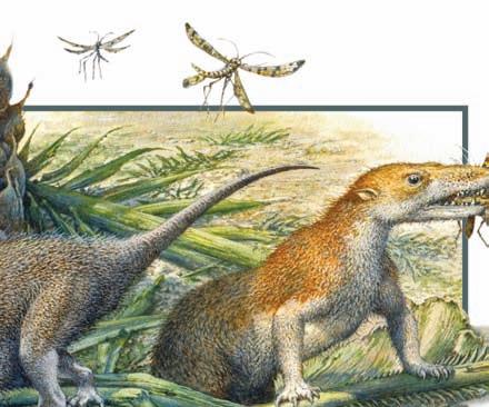
The diversity of living vertebrates is enormous, but the extant species are only a small proportion of the species of vertebrates that have existed. For each extant species, there may be hundreds of extinct species, and some of these have no counterparts among extant forms. For example, the dinosaurs that dominated Earth for 180 million years are so entirely different from extant animals that it is hard to reconstruct the lives they led. Even mammals were once more diverse than they are now. The Pleistocene saw giants of many kinds, such as ground sloths as big as modern rhinoceroses and raccoons as large as bears. The number of species of terrestrial vertebrates probably reached its maximum in the middle Miocene, between 14 and 12 million years ago, and has been declining since then.
Where and when the vertebrates originated, how they evolved, what they do, and how they work provide endless intriguing details. In preparing to tell this story, we first introduce some basic information, including what the different kinds of vertebrates are called, how they are classified, and what the world was like as their story unfolded.
and Petromyzontiformes
Major extant groups of vertebrates
Figure 1.1 Diversity of extant vertebrates. Areas in the pie chart correspond to the approximate numbers of extant species in each group as of 2017; the numbers change as new species are described or existing species become extinct. Common names are in the center circle, with formal names for the groups shown in the outer circle. The two major lineages of extant vertebrates are Actinopterygii (ray-finned fishes; lavender) and Sarcopterygii (all other colors except gray), each of which includes more than 33,000 extant species. In its formal sense, Sarcopterygii includes the lineages Actinistia, Dipnoi, Urodela, Anura, Gymnophiona, Testudines, Lepidosauria, Crocodylia, Aves, and Mammalia (i.e, the lobe-finned fishes and all their descendants, including all amphibians, reptiles, birds, and mammals.)
temperature regulation. Figure 1.1 shows the major groups of vertebrates and the approximate number of extant species in each. Next we briefly describe these vertebrate groups.
Non-amniotes
The embryos of non-amniotes are enclosed and protected by membranes that are produced by the reproductive tract of the female. This is the condition seen among the Pough Vertebrate Life 10E Sinauer Associates Morales Studio VL10E_01.01.ai 11-15
Two major groups of vertebrates are distinguished on the basis of an innovation in embryonic development: the appearance of three membranes formed by tissues that are generated by the embryo itself. The innermost of these membranes, the amnion, surrounds and cushions the embryo, and animals with this structure (the reptiles, birds, and mammals) are called amniotes. The split between non-amniotes and amniotes corresponds roughly to the division between aquatic and terrestrial vertebrates, although many amphibians and a few fishes lay non-amniotic eggs in nests on land. Among the amniotes, we can distinguish two major evolutionary lineages: the sauropsids (reptiles, including birds) and the synapsids (mammals). These lineages separated from each other in the mid-Carboniferous, before vertebrates had developed many of the characters we see in extant species. As a result, synapsids and sauropsids represent parallel but independent origins of basic characters such as lung ventilation, kidney function, insulation, and
invertebrate relatives of vertebrates, and it is retained in the non-amniotes (fishes and amphibians).
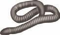
Hagfishes and lampreys: Myxiniformes and Petromyzontiformes Lampreys and hagfishes are elongate, limbless, scaleless, and slimy and have no internal bony tissues. They are scavengers and parasites and are specialized for those roles. Hagfishes are marine, living on the seabed at depths of 100 meters or more. In contrast, many species of lampreys are migratory, living in oceans and spawning in rivers. Hagfishes and lampreys are unique among extant vertebrates because they lack jaws.
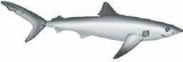
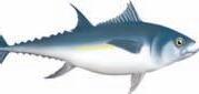
Sharks, rays, and ratfishes: Chondrichthyes The name Chondrichthyes (Greek chondros, “gristle”; ichthys, “fish”) refers to the cartilaginous skeletons of these fishes. Extant sharks and rays form a group called Neoselachii (Greek neos, “new”; selachos, “shark”), but the two kinds of fishes differ in body form and habits. Sharks have a reputation for ferocity that most species would have difficulty living up to. Some sharks are small (25 cm or less), while the largest species, the whale shark (Rhincodon typus), grows to 17 m long and is a filter feeder that subsists on plankton it strains from the water. Rays are mostly bottom feeders; they are dorsoventrally flattened and swim with undulations of their extremely broad pectoral fins.
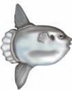
The second group of chondrichthyans, the ratfishes or chimaeras, gets its name, Holocephali (Greek holos , “whole”; kephale , “head”), from the single gill cover that extends over all four gill openings. These are bizarre marine animals with long, slender tails. Some species have a bucktoothed face that looks rather like a rabbit. They forage on the seafloor and feed on hard-shelled prey, such as crustaceans and mollusks.
Bony fishes: Osteichthyes Osteichthyes (Greek osteon, “bone”) are so diverse that any attempt to characterize them briefly is doomed to failure. Two broad categories can be recognized: the ray-finned fishes (Actinopterygii; Greek aktis, “ray”; pteron, “wing” or “fin”) and the lobe-finned or fleshy-finned fishes (Sarcopterygii; Greek sarco, “flesh”).
Actinopterygians have radiated extensively in both fresh and salt water, and more than 32,000 species have been named. Nearly 400 species have been described annually since 1997, and several thousand additional species may await discovery. A single project, the Census of Marine Life, is describing 150–200 previously unknown species of rayfinned fishes annually.
Actinopterygians can be divided into three groups: (1) the monophyletic and derived Neopterygii (almost all extant rayfinned fishes); (2) the more basal Chondrostei (sturgeons and paddlefish); and (3) Cladistia (polypterids, including bichirs and reedfish, which are swamp- and river-dwellers from Africa). Sturgeons are large fishes with protrusible, toothless mouths that suck food items from the bottom. Sturgeon eggs are the source of caviar, and many species have been driven close to extinction by overharvesting of females for
their eggs. Paddlefishes (two species, one in the Mississippi system of North America and another nearly extinct species in China’s Yangtze River) have a paddlelike snout with organs that locate prey by sensing electrical fields.
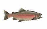
Neopterygii, the modern radiation of ray-finned fishes, can be divided into two lineages. One these—Holostei, the gars and bowfins—is a relict of an earlier radiation. These fishes have cylindrical bodies, thick scales, and jaws armed with sharp teeth. They seize prey in their mouths with a sudden gulp, and lack the specializations of the jaw apparatus that allow the bony fishes to use more complex feeding modes. The second neopterygian lineage, Teleostei, includes 95% of actinopterygians, embracing every imaginable combination of body form, ecology, and behavior. Most of the fishes that people are familiar with are teleosts, from sport fishes like trout and swordfishes, to food staples like tuna and salmon, to the “goldfish” and exotic tropical fishes in home aquaria. Modifications of the body and jaw apparatus have allowed many teleost species to be highly specialized in their swimming and feeding habits.
The third neopterygian lineage, Teleostei, includes 95% of actinopterygians, embracing every imaginable combination of body form, ecology, and behavior. Most of the fishes that people are familiar with are teleosts—the trout, bass, and panfishes that anglers seek; the sole (a kind of flounder) and swordfishes featured by seafood restaurants; and the salmon and tuna that find their way into both sushi and cat food. Modifications of the body and jaw apparatus have allowed many teleosts to be highly specialized in their swimming and feeding habits.
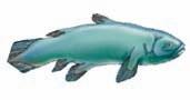
In one sense, only eight species of sarcopterygian fishes survive: the six species of lungfishes (Dipnoi) found in South America, Africa, and Australia; and two species of coelacanths (Actinistia), one from deep waters off the east coast of Africa and a second species discovered near Indonesia. These are the extant fishes most closely related to terrestrial vertebrates, however, and from an evolutionary standpoint the diversity of sarcopterygians includes all of their their terrestrial descendants—amphibians, turtles, lepidosaurs (the tuatara [Sphenodon punctatus], lizards, and snakes), crocodylians, birds, and mammals. From this perspective, bony fishes include two major evolutionary radiations—one in the water and the other on land—each containing more than 33,000 species.
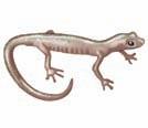
Salamanders, frogs, and caecilians: Urodela, Anura, and Gymnophiona These three groups of vertebrates are popularly known as amphibians (Greek amphis, “double”; bios, “life”) in recognition of their complex life histories, which often include an aquatic larval form (the larva of a salamander or caecilian and the tadpole of a frog) and a terrestrial adult. All amphibians have bare skins (i.e., lacking scales, hair, or feathers) that are important in the exchange of water, ions, and gases with their environment. Salamanders are elongate animals, mostly terrestrial, and usually with four legs; anurans (frogs) are short-bodied, with large heads
and large hindlegs used for walking, jumping, and climbing; and caecilians are legless aquatic or burrowing animals.
Amniotes
A novel set of membranes associated with the embryo appeared during the evolution of vertebrates. These are called fetal membranes because they are derived from the embryo itself rather than from the reproductive tract of the mother. As mentioned at the start of this chapter, the amnion is the innermost of these membranes, and vertebrates with an amnion are called amniotes. In general, amniotes are more terrestrial than non-amniotes; but there are also secondarily aquatic species of amniotes (such as sea turtles and whales) as well as many species of salamanders and frogs that spend their entire lives on land despite being non-amniotes. However, many features distinguish non-amniotes (fishes and amphibians) from amniotes (mammals and reptiles, including birds), and we will use the terms to identify which of the two groups is being discussed.
By the Permian, amniotes were well established on land. They ranged in size from lizardlike animals a few centimeters long through cat- and dog-size species to the cow-size pareiasaurs. Some were herbivores; others were carnivores. In terms of their physiology, we can infer that they retained ancestral characters. They had scale-covered skins without an insulating layer of hair or feathers, a simple kidney that could not produce highly concentrated urine, simple lungs, and a heart in which the ventricle was not divided by a septum. Early in their evolutionary history, terrestrial vertebrates split into two lineages—synapsids (today represented by mammals) and sauropsids (modern reptiles, including birds).
Terrestrial life requires lungs to extract oxygen from air, a heart that can separate oxygen-rich arterial blood from oxygen-poor venous blood, kidneys that can eliminate waste products while retaining water, and insulation and behaviors to keep body temperature stable as the external temperature changes. These features evolved in both lineages, but because synapsids and sauropsids evolved terrestrial specializations independently, their lungs, hearts, kidneys, and body coverings are different.
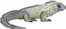
Sauropsid amniotes Extant sauropsids are the animals we call reptiles: turtles, the scaly reptiles (tuatara, lizards, and snakes), crocodylians, and birds. Extinct sauropsids include the forms that dominated the world during the Mesozoic—dinosaurs and pterosaurs (flying reptiles) on land and a variety of marine forms, including ichthyosaurs and plesiosaurs, in the oceans.
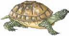
• Turtles: Testudines Turtles (Latin testudo, “turtle”) are probably the most immediately recognizable of all vertebrates. The shell that encloses a turtle has no exact duplicate among other vertebrates, and the morphological modifications associated with the shell make turtles extremely peculiar animals. They are, for example, the only vertebrates with the shoulders (pectoral girdle) and hips (pelvic girdle) inside the ribs.
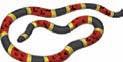
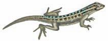
• Tuatara, lizards, and snakes: Lepidosauria T hese three kinds of vertebrates can be recognized by their scale-covered skin (Greek lepisma, “scale”; sauros, “lizard”) as well as by characters of the skull. The tuatara, a stocky-bodied animal found only on some offshore islands of New Zealand, is the sole living remnant of an evolutionary lineage of animals called Rhynchocephalia, which was more diverse in the Mesozoic. In contrast, lizards and snakes (which are highly specialized lizards) are now at the peak of their diversity.
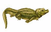
• A lligators and crocodiles: Crocodylia T hese impressive animals, which draw their name from the ancient Latin word for them (crocodilus) are in the same evolutionary lineage (Archosauria) as dinosaurs and birds. The extant crocodylians are semiaquatic predators with long snouts armed with numerous teeth. They range in size from the saltwater crocodile (Crocodylus porosus), which can grow to 6 m, to dwarf crocodiles and caimans that are 1.5 m long. Crocodylian skin contains many bones (osteoderms; Greek osteon, “bone”; derma, “skin”) that lie beneath their scales and provide a kind of armor plating. Crocodylians are noted for the parental care they provide for their eggs and young.
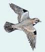
• Birds: Aves Birds (Latin avis, “bird”) are a lineage of dinosaurs that evolved flight in the Mesozoic. Feathers are characteristic of extant birds, and feathered wings are the structures that power a bird’s flight. Discoveries of dinosaur fossils with feathers show that feathers evolved long before flight. This disparity between the time feathers are first seen and the time flight appeared illustrates an important principle: The function of a trait in an extant species is not necessarily the same as that trait’s function when it first appeared. In other words, current utility is not the same as evolutionary origin. The original feathers were almost certainly structures used in courtship displays, and their modifications as airfoils, for streamlining, and as insulation in birds were secondary events.
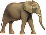
Synapsid amniotes: Mammalia The synapsid lineage contains the three kinds of extant mammals: the monotremes (prototheria; platypus and echidnas), marsupials (metatherians), and placentals (eutherians). Extinct synapsids include forms that diversified in the Paleozoic—pelycosaurs and therapsids—and the rodentlike multituberculates of the late Mesozoic.
The mammals (Latin mamma, “teat”) originated in the Late Triassic. Extant mammals include about 5,400 species in three groups: placentals (by far the largest group), marsupials, and monotremes. The name “placentals” is misleading, because both placentals and marsupials have a placenta, a structure that transfers nutrients from the mother to the embryo and removes the waste products of the embryo’s metabolism. Placentals have a long gestation period and have largely completed development at birth, whereas marsupials have a short gestation period and give
birth to very immature young that continue their development attached to a nipple, often within a pouch on the mother’s abdomen. Marsupials dominate the mammalian fauna only in Australia, although some 100 species are found in South and Central America, and one species, the Virginia opossum (Didelphis virginiana), has migrated from South America to North America. Kangaroos, koalas, and wombats are familiar Australian marsupials. Finally, the monotremes—platypus and echidnas—are unusual mammals whose young hatch from eggs. All mammals, including monotremes, feed their young with milk.
1.2 Classification of Vertebrates
The diversity of vertebrates (more than 67,000 extant species and perhaps 100 times that number of extinct species) makes organizing them into a coherent system of classification extraordinarily difficult. Yet classification has long been at the heart of evolutionary biology. Initially, classification of species was seen as a way of managing the diversity of organisms, much as an office filing system manages the paperwork of the office. Each species could be placed in a pigeonhole marked with its name; when all species were in their pigeonholes, the diversity of vertebrates would have been encompassed. This approach to classification was satisfactory as long as species were regarded as static and immutable: once a species was placed in the filing system, it was there to stay. Acceptance of the fact that species evolve has made that kind of classification inadequate. Now biologists must express evolutionary relationships among species by incorporating evolutionary information in the system of classification. Ideally, a classification system should not only attach a label to each species but also encode the evolutionary relationships between that species and other species. Modern techniques of systematics (the evolutionary classification of organisms) have become methods for generating testable hypotheses about evolution.
Binominal nomenclature
Our system of naming species is pre-Darwinian. It traces back to methods established by the naturalists of the seventeenth and eighteenth centuries, especially those of Carl von Linné, the Swedish naturalist better known by his Latin pen name, Carolus Linnaeus. The Linnaean system identifies a species using two names—a genus name and a species name; hence, this convention is called binomial nomenclature. Species are arranged in hierarchical categories (family, order, and so on). These categories are called taxa (singular taxon), and the discipline of naming organisms is called taxonomy (Greek taxo, “to arrange”; nomos, “order”). The scientific naming of species became standardized when Linnaeus’s monumental work Systema Naturae (The System of Nature) was published in sections between 1735 and 1758. Linnaeus attempted to give an identifying name to every known species of plant and animal. Familiar
examples include Homo sapiens for human beings (Latin homo, “human” and sapiens, “wise”), Passer domesticus for the house sparrow (Latin passer, “sparrow” and domesticus, “belonging to the house”), and Canis familiaris for the domestic dog (Latin canis, “dog” and familiaris, “domestic”). Why use Latin? Latin was the early universal language of European scholars and scientists. It provided a uniform usage that scientists, regardless of their native language, continue to recognize worldwide. The same species may have different colloquial names, even in the same language. For example, Felis concolor (Latin, “uniformly colored cat”) is known in various parts of North America as the cougar, puma, mountain lion, American panther, painter, and catamount. In Central and South America it is called león Colorado, león de montaña, pantera, onça vermelha, onça parda, yagua pytá, and suçuarana. But biologists of all nationalities recognize the name Felis concolor as referring to this specific kind of cat.
Phylogenetic systematics
All methods of classifying organisms, even Linnaean systems, are based on similarities shared by the included species, but some similarities are more significant than others. For example, nearly all terrestrial vertebrates have paired limbs, but only a few kinds of vertebrates have mammary glands. Consequently, knowing that two species have mammary glands tells you more about the closeness of their relationship than does knowing they have paired limbs. A way to assess the relative importance of different characteristics was developed in the mid-20th century by Willi Hennig, who introduced a method of determining evolutionary relationships called phylogenetic systematics (Greek phylon, “tribe”; genesis, “origin”).
The groups of organisms recognized by phylogenetic systematics are called natural groups, and the members of these groups are linked by a nested series of characters that trace the evolutionary history of the lineage. Hennig’s contribution was to insist that these groups can be identified only on the basis of homologous derived characters —that is, characters that have the same evolutionary origin (i.e., are homologous) and that differ from the ancestral condition (are derived). A derived character is called an apomorphy (Greek apo, “away from” and morphe, “form,” which is interpreted as “away from the ancestral condition”).
For example, the feet of terrestrial vertebrates have distinctive bones—the carpals, tarsals, and digits. This arrangement of foot bones is different from the ancestral pattern seen in lobe-finned fishes, and all lineages of terrestrial vertebrates either have that derived pattern of foot bones or had it at some stage in their evolution. Many groups of terrestrial vertebrates—horses, for example—have subsequently modified the foot, and some, such as snakes, have lost the limbs entirely. The significant point is that those evolutionary lineages include ancestral species that had the derived terrestrial pattern. Thus, the terrestrial pattern of foot bones is a shared derived character of terrestrial vertebrates. Shared derived characters