Vaccines for Veterinarians
IAN R. TIZARD, BVMS, PhD, DACVM (HONS), DSc (HONS)
University Distinguished Professor Immunology Emeritus Department of Veterinary Pathobiology
The Texas Veterinary Medical Center
Texas A & M University College Station, Texas USA
Elsevier 3251 Riverport Lane St. Louis, Missouri 63043
VACCINES FOR VETERINARIANS
Copyright © 2021, Elsevier Inc. All rights reserved.
ISBN: 978-0-323-68299-2
No part of this publication may be reproduced or transmitted in any form or by any means, electronic or mechanical, including photocopying, recording, or any information storage and retrieval system, without permission in writing from the publisher. Details on how to seek permission, further information about the Publisher’s permissions policies and our arrangements with organizations such as the Copyright Clearance Center and the Copyright Licensing Agency, can be found at our website: www.elsevier.com/ permissions
This book and the individual contributions contained in it are protected under copyright by the Publisher (other than as may be noted herein).
Notice
Practitioners and researchers must always rely on their own experience and knowledge in evaluating and using any information, methods, compounds or experiments described herein. Because of rapid advances in the medical sciences, in particular, independent verification of diagnoses and drug dosages should be made. To the fullest extent of the law, no responsibility is assumed by Elsevier, authors, editors or contributors for any injury and/or damage to persons or property as a matter of products liability, negligence or otherwise, or from any use or operation of any methods, products, instructions, or ideas contained in the material herein.
Library of Congress Control Number: 2019949241
Content Strategist: Jennifer Catando
Content Development Specialist: Nani Clansey
Project Manager: Umarani Natarajan
Publishing Services Manager: Shereen Jameel
Design Direction: Ryan Cook
Illustration Manager: Teresa McBryan
Marketing Manager: Bergen Farthing
Printed in China
Last digit is the print number: 9 8 7 6 5 4 3 2 1
DEDICATION
This book is dedicated to the memory of my former graduate student, Dr. Monie Singh Bhogal, who was murdered in Somalia in January 1999 while supervising the Rinderpest vaccination program for the Italian aid agency, Terra Nuova.
This page intentionally left blank
Vaccines are among the world’s greatest medical advances. Despite ill-informed and irrational resistance to their use, vaccination has brought most major infectious diseases under control and massively increased human longevity. Vaccines have had similar beneficial effects on livestock production, eliminating animal plagues, and making intensive livestock production possible. Companion animals and wildlife have also benefited enormously from the proper application of vaccination. The great triumphs of smallpox and rinderpest eradication would not have been possible without vaccines. They are the most cost-effective methods of preventing infectious diseases ever conceived.
We cannot, however, rest on past achievements. Emerging diseases, difficult organisms, antibiotic resistance, and a constant need to improve both the safety and efficacy of vaccines ensure that research and development must continue. In addition, unlike some human vaccines, the production of veterinary vaccines is subject to significant economic constraints. Although vaccination of companion animals is regarded as a responsibility to protect a beloved pet, especially in developed and prosperous countries, the drivers of vaccine use in livestock and poultry and also in less developed countries are economic. Animal vaccines are commodities made for the purposes of improving animal productivity and health. They will not be commercially produced if they do not generate significant revenue. They will not be used if they do not reduce disease costs. It does not matter that the science behind these new vaccines is incredibly elegant and sophisticated. If they cannot be produced or used profitably, they will not be manufactured. In general, governments and taxpayers will not subsidize animal vaccine production. Some exceptions may be made for zoonotic or wildlife diseases such as rabies.
The importance of vaccine profitability ensures that animal vaccines must be easy to manufacture, cheap to produce and store, and have a market size that ensures a reasonable return on investment.
But vaccine production is not static. The technologies of the past 100 years, although incredibly successful, are no longer adequate. Driven by the three big exceptions, tuberculosis, AIDS, and malaria, in addition to the emerging diseases such as Ebola and Zika, human vaccine production processes are changing. Reverse vaccinology driven by computational methods and vaccinomics is radically altering the way in which vaccines are developed. A meningitis vaccine developed by these computational methods is already on the market. A Zika virus vaccine went from inception to the clinic in 6 months. Veterinary vaccines are not far behind. The first commercially available DNA-plasmid vaccine was for West Nile disease in horses. Several animal replicon RNA vaccines have been licensed. Chimeric vaccines are now available against West Nile virus and porcine circovirus. Animals also have their exceptionally difficult problem diseases such as porcine reproductive and respiratory syndrome (PRRS), foot and mouth disease, influenza, and especially African swine fever. These remain enduring threats, although progress in developing new or more effective vaccines is being made here too.
Human and animal vaccinologies are also diverging. In humans, serious discussions are under way regarding the production of individualizing vaccines to match a person’s specific immune system. In animals, especially livestock and fish, economics still rule. Mass production and a significant cost/benefit ratio are critical.
We must not forget that antivaccine advocates are vocal, not only in the human area, but also in Veterinary Medicine. The effective control of infectious diseases and the development of herd immunity have reduced the prevalence of many infectious diseases to very low levels. As a result, owners often remain ignorant of the threats posed by these infections. The best, really the only,
defense against ill-informed and conspiracy theory–based antivaccine sentiment is to promote evidence-based vaccinology. Hopefully this text will provide a baseline for the rational use, promotion, development, and improvement of veterinary vaccines.
This text is therefore designed to be a comprehensive review of currently available animal vaccines and vaccination. It brings together the science and technology of vaccine production. It examines the issues associated with the use of animal vaccines. Safety is a critical issue in this age of antivaccination sentiment and social media. Regulations and assessment of vaccine efficacy and safety are major contributors to the costs of producing a vaccine and must also be considered. The second half of the text reviews the major vaccines used in each species, their risks, benefits, and interesting immunologic features. It is not intended to be a comprehensive catalog of all such products, nor does it discuss new vaccine research in detail. After all, many experimental vaccines never reach the marketplace. New vaccine administration methodologies are also discussed, especially with respect to the mass vaccination of poultry, pigs, fish, and wildlife. Finally, consideration is given to newly available vaccines against parasites and the increasingly successful field of cancer immunotherapy.
I must express my appreciation to my colleagues, John August, Luc Berghman, Stacy Eckman, Jill Heatley, Jeffrey Musser, Susan Payne, and Darrel Styles, who reviewed and corrected chapters for me. All errors and omissions are mine alone.
College Station, TX
December 2018
COMMONLY USED ABBREVIATIONS
Many acronyms in the discipline of vaccinology relate not just to immunology but also to the regulatory networks that oversee their production and licensure. Here are some that are widely employed throughout this text.
AABP American Association of Bovine Practitioners
AAEP American Association of Equine Practitioners
AAFP American Association of Feline Practitioners
AAHA American Animal Hospital Association
AIDS acquired immune deficiency syndrome
APC antigen-presenting cell
APHIS Animal and Plant Health Inspection Service (USDA)
AVMA American Veterinary Medical association
BCG bacillus Calmette-Guérin (Mycobacterium bovis)
BCR B cell (antigen) receptor
BoLA bovine leukocyte antigen
BPL beta-propiolactone
CDC Center for Disease Control and Prevention
CMI cell-mediated immunity
CpG cytosine-phosphate-guanosine nucleotide motif
CVB Center for Veterinary Biologics (USDA)
CVMP Committee for Medical Products for Veterinary Use
CFR code of federal regulations
DAMP damage-associated molecular pattern
DC dendritic cell
DIVA differentiate infected from vaccinated animals
ELISA enzyme-linked immunosorbent assay
Fab antigen-binding fragment
FAO Food and Agriculture Organization
Fc crystallizable fragment (of immunoglobulin)
FeLV feline leukemia virus
FMDV foot and mouth disease (virus)
FPT failure of passive transfer
GLP good laboratory practice
GMO genetically modified organism
GMP good manufacturing practice
FIV feline immunodeficiency virus
HA hemagglutinin
HDN hemolytic disease of the newborn
HI hemagglutination inhibition (test)
ID intradermal
IFN interferon
Ig immunoglobulin
IL interleukin
IM intramuscular (route)
IN intranasal (route)
ISCOM immune-stimulating complex
ISG immune serum globulin
IU international unit
kb kilobase (one thousand base pairs) of DNA
LPS lipopolysaccharide
MAb monoclonal antibody
MAP Mycobacterium avium paratuberculosis
MHC major histocompatibility complex
MCS master cell stock
MLV modified live virus (or vaccine)
MSV master seed virus
OIE Originally the Office International des Epizooties but currently the World Organization for Animal Health.
Omp outer membrane protein
OMV outer membrane vesicles
O/W oil-in-water emulsion
PAMP pathogen-associated molecular pattern
PCR polymerase chain reaction
PF protective fraction
PRR pattern-recognition receptor
PRRS(V) porcine respiratory and reproductive system (virus)
R0 basic reproductive number
S19 strain 19 Brucella abortus vaccine
SC subcutaneous (route)
TCR T cell (antigen) receptor
TLR toll-like receptor
Th cell helper T cell
TK thymidine kinase
TNF tumor necrosis factor
Treg regulatory T (cell)
ts temperature sensitive (mutant)
USDA United States Department of Agriculture
VLP virus-like particle
WHO World Health Organization
W/O water-in-oil emulsion
WSAVA World Small Animal Veterinary Association
Dedication v
Preface vii
Commonly Used Abbreviations ix
1 A Brief History of Veterinary Vaccines 1
2 The Science Behind Vaccine Use 13
3 Nonliving Vaccines 27
4 Living Vaccines 41
5 Recombinant Vectored Vaccines 51
6 Nucleic Acid Vaccines and Reverse Vaccinology 61
7 Adjuvants and Adjuvanticity 75
8 The Administration of Vaccines 87
9 Failures in Vaccination 105
10 Adverse Consequences of Vaccination 115
11 Production, Assessment, and Regulation of Vaccines 131
12 Passive Immunization 141
13 Canine Vaccines 153
14 Feline Vaccines 167
15 Equine Vaccines 179
16 Bovine Vaccines 193
17 Sheep and Goat Vaccines 215
18 Porcine Vaccines 225
19 Poultry Vaccines 243
20 Vaccination of Exotic and Wild Species 267
21 Fish Vaccines 281
22 Vaccines Against Parasites 293
23 Anticancer Vaccines 301
Index 309
This page intentionally left blank
A Brief History of Veterinary Vaccines
OUTLINE
Variolation and Vaccination
Louis Pasteur
Vaccination and Antiviral Vaccines
The Eradication of Rinderpest
Vaccination is the most efficient and effective method of controlling infectious diseases ever developed. The enormous improvements in human health through the twentieth century and beyond have been, in large part, a result of the development of effective vaccines. Likewise, many of the improvements in animal health, especially the great increases in productivity resulting from effective disease control are a direct result of vaccine use. The eradication of smallpox in humans as well as the elimination of rinderpest in cattle are both directly the result of effective vaccination campaigns.
The early history of veterinary vaccines can be divided into four stages. The first stage was the discovery of variolation against human smallpox and its eventual evolution into vaccination. The second stage was launched by the discoveries of Louis Pasteur and his colleagues that led to the early production of numerous effective vaccines against predominantly bacterial diseases. The third stage, typified by the development of the canine distemper vaccines, was a process of progressive improvement of these vaccines and resulted from a growing knowledge about viruses and their behavior. Finally, the sophisticated use of vaccines has led to the elimination of two major diseases, smallpox and rinderpest, and the near eradication of others.
Variolation and Vaccination
The earliest record of protective immunity dates from 430 BCE, when the Greek historian Thucydides describing the plague of Athens (of unknown cause) observed “… for no one caught the disease twice, or, if he did, the second attack was never fatal.”
Sometime before the fifteenth century, the Chinese also observed that those individuals who recovered from an attack of smallpox never suffered from it a second time. It was a small step therefore to attempt to protect children from the disease by deliberately infecting them. Initially the process involved blowing dried scab powder up the nose of infants. Eventually however, improved results were obtained by placing the dried scabs in a small incision in the arm. The resulting local disease was less severe and mortality correspondingly lower. Descriptions of this technique spread westward along the Silk Road until it eventually reached the Ottoman Empire—modern Turkey. It was widely employed across the Middle East.
Reports of this technique called “variolation,” eventually reached England in the early 1700s. One such example came in 1714 by way of a letter from a physician, Dr. Emanuel Timoni, who practiced medicine in Constantinople. In addition, first-hand reports from Lady Mary Montagu,
the wife of the English Ambassador to Constantinople impressed the people who mattered. Lady Montagu arranged to have her own son successfully variolated through Dr. Timoni. Voltaire described her as “[a] woman of so fine a genius, and endued with as great a strength of mind, as any of her sex in the United Kingdom.” These reports were taken seriously because the Prince of Wales (the future King George II) and his wife were terrified that smallpox would attack their grandchildren. As a result, the process was investigated by variolating prisoners in London’s Newgate Prison. When this “Royal Experiment” proved to be successful, variolation was rapidly adopted in England. Around the same time (1721) variolation was successfully used by Dr. Zabdiel Boylston in Boston who had read Timoni’s letter. Although the technique worked, it was still accompanied by significant morbidity and some mortality, so it was not universally adopted in the American colonies. Some believed that diseases such as smallpox were God’s punishment for sins so that variolation was obviously in conflict with God’s will and thus inappropriate. Variolation clearly worked; however, nobody at that time had any concept as to how it might induce immunity to smallpox. Nevertheless, attempts were made to replicate the technique in other diseases. The most significant of these attempts resulted in the discovery of vaccination.
EDWARD JENNER
In 1768, John Fewster, a physician in Gloucestershire, in the west of England, and his colleagues began to inoculate their patients against smallpox. They found however that many patients did not react to variolation. Upon inquiry, Fewster found that these patients had previously been infected with cowpox. Fewster reported his observations to the local Medical Society, but he did not fully recognize its significance and never published his findings. Among the Society’s members was a young apprentice called Edward Jenner. In 1774 an English farmer, Benjamin Jesty, performed the first documented substitution of cowpox for smallpox. He obtained the scab material from a cow of a neighbor and inoculated his wife in the arm under her elbow. He never wrote an account of this and he did not determine if she was immune. The widespread acceptance of this method did not come for another 24 years. In the 1790s, Edward Jenner was still working as a physician in the west of England, and he remembered Fewster’s observation. He also recognized that deliberate inoculation of cowpox could confer immunity against smallpox.
Cowpox, otherwise known as vaccinia (vacca is Latin for cow), is a virus-mediated skin disease of cattle. Affected animals generally develop a large weeping ulcer on hairless skin such as the udder. When these infected cattle are hand milked, the vaccinia virus can readily enter cuts or abrasions on the milker’s hands. Humans infected by this virus develop a localized weeping lesion that scabs over and heals within a few weeks. We now know that the cowpox virus triggers a powerful immune response. Most importantly, however, vaccinia virus is closely related to smallpox virus to such an extent that these individuals also develop immunity to smallpox.
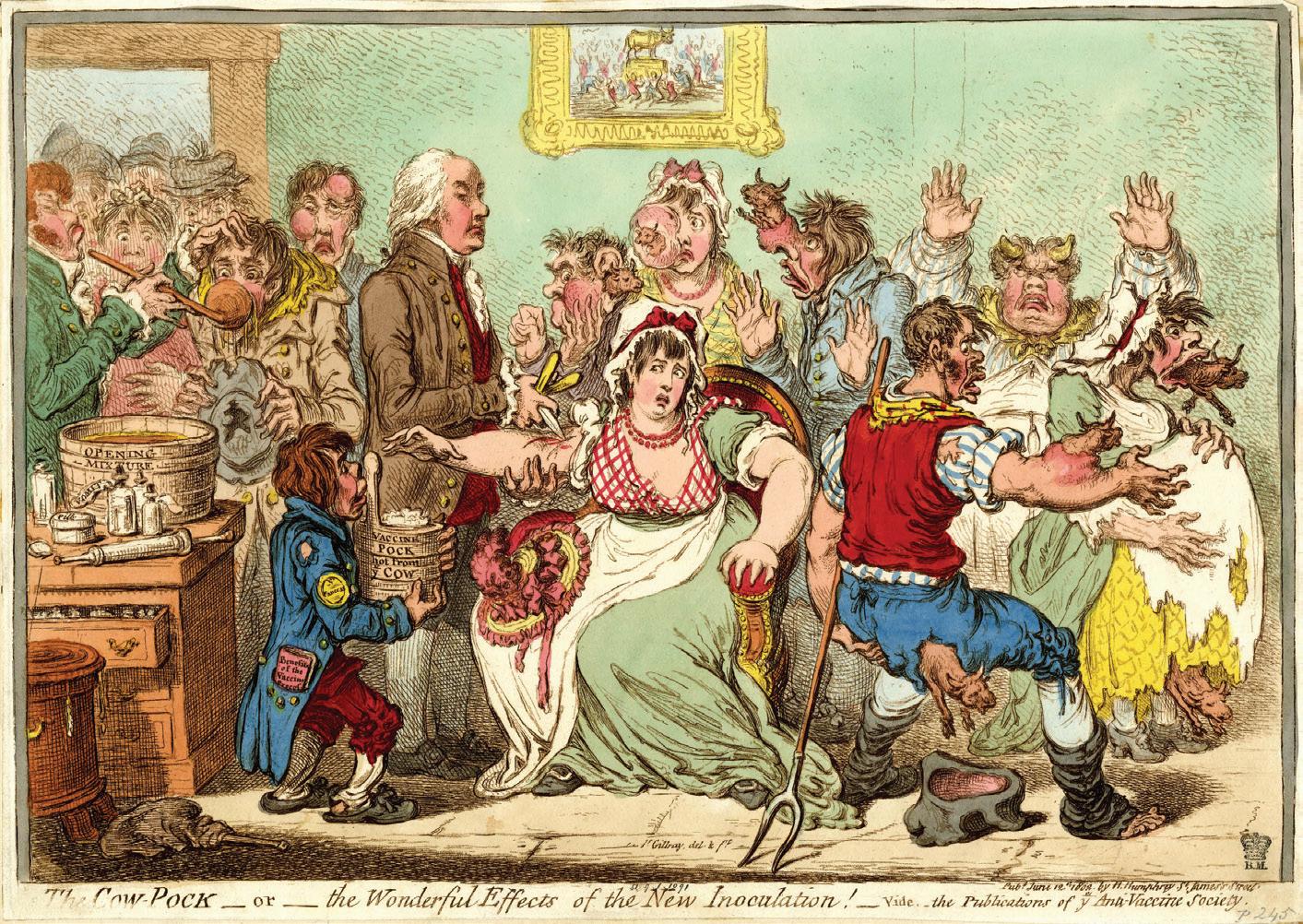
Edward Jenner began a systematic study of the protective effects of vaccination and published his results in 1798. The scientific method was not well established in the late eighteenth century. Thus Jenner published a series of case reports on vaccination. This drew immediate attention because the method worked well and was clearly very much safer than variolation (Box 1.1). Despite some initial opposition, the principal physicians and surgeons of London supported Jenner, and, as a result, vaccination was rapidly adopted in Europe and the Americas, whereas variolation was discontinued (Fig. 1.1). Jenner quite properly received credit for publishing the vaccination process, but his biographer invented a myth that Jenner alone had discovered the process by talking to a dairymaid. In fact, this protective effect of vaccination was independently recognized in several other English and European communities.
It must be emphasized that Jenner had no concept of microbiology, viruses, or immunity. For example, he did not know where smallpox came from. He noted, however, that the disease occurred in farms where there were both cattle and horses. The same workers looked after
BOX 1.1 n Thomas Jefferson and Edward Jenner
Thomas Jefferson was an enthusiastic amateur scientist. When he was informed about Jenner’s discovery, Jefferson sent him a letter:
Washing, Dec 25, 1800
Sir,
I received last night, and have read with great satisfaction, your pamphlet on the subject of the kinepock, and pray you to accept my thanks for the communication of it.
I had before attended to your publications on the subject in the newspapers and took much interest in the result of the experiments you were making. Every friend of humanity must look with pleasure on this discovery, by which one evil more is withdrawn from the condition of man; and must contemplate the possibility, that future improvements and discoveries may still more and more lessen the catalogue of evils. In this line of proceeding you deserve well of your country; and I pray you accept my portion of the tribute due to you, and assurance of high consideration and respect, with which I am, Sir Your most obedient, humble servant,
Thomas Jefferson
Fig. 1.1 Antivaccination sentiment is not new. The English satirist John Gilray caricatured this vaccination scene in 1802. The central figure is likely not Edward Jenner, but George Pearson, an enthusiastic advocate of vaccination. If this happens to you, seek immediate medical attention. (From the British Museum. With permission.)
both, and he suggested that they transmitted cowpox from horses to cattle. Jenner therefore believed that the skin disease of horses colloquially called “the grease” was the source of cowpox. He likely confused this disease with horsepox. However, he clearly believed that horses were the original source of cowpox, and as a result he obtained some of his vaccine material from horses. Horsepox, which like other poxviruses caused multiple skin lesions, was a common
disease of horses at that time. Jenner continued to believe in “equination,” and in later years tended to prefer the use of equine material to cowpox.
Subsequent to Jenner’s discovery, the British government decided to vaccinate all their soldiers. The contract to produce this vaccine was given in 1801 to a Dr. John Loy who used material from horses exclusively. In 1803 an Italian physician, Dr. Luigi Sacco, read a book by Dr. Loy and also used equine “grease” with great success in children. Although cowpox was the preferred material for vaccination through the nineteenth century, equine material was used on occasion. It is likely that at least some of the vaccinia stocks used in humans were of equine origin. Vaccinia virus as currently used differs significantly from circulating cowpox virus strains. Although horsepox has become almost extinct, it has been possible to examine the genome of a Mongolian strain of horsepox. This is also related to but distinct from vaccinia. Several horsepox genes are present in different vaccinia strains, whereas some vaccinia strains appear to be more closely related to horsepox than to cowpox.
Although physicians (and veterinarians) were totally unaware of how vaccinia worked, it did not stop them from seeking to use this methodology to prevent other diseases. Many believed vaccinia to be an effective treatment for unrelated diseases in humans including the plague and cholera.
CATTLE PLAGUE
Cattle plague (rinderpest) was a major devastating disease in Europe until the end of the nineteenth century when it was eliminated by movement control and slaughter. We now know it was caused by a morbillivirus related to human measles. However, in the eighteenth century many of those who encountered cattle plague considered that it was very similar to smallpox. Thus once variolation (inoculation) was introduced into Europe in 1717, numerous individuals attempted to prevent cattle plague by inoculating healthy cattle with material from plague-affected cattle. One such method involved making an incision in the dewlap and inserting a piece of string that had been soaked in nasal or ocular discharge. This was removed after 2 to 3 days. It was claimed that animals sickened and then recovered. A booklet was published in 1757 recommending this procedure, and it was widely adopted in England. However, it was eventually recognized that all such attempts were not effective and killed a lot of cattle. Despite this, widespread attempts to inoculate cattle against rinderpest were conducted in Denmark, the Netherlands, and what is now Germany. By 1770 most had been abandoned.
In 1774, an adaptation of the vaccination procedure was tested in the Netherlands by Geert Reinders, a farmer from Groningen. He had noticed that calves from recovered cows appeared resistant to reinfection. This may have been the first recognition of the phenomenon of maternal immunity. Reinders made use of this resistant period to inoculate 6- to 8-week-old calves with nasal material from recovered cows. He also observed that he got better results if he repeated the inoculation of these calves two more times. Reinders was criticized by those who believed that disease was a manifestation of God’s will. More importantly, his was not a practical procedure and fell into disuse.
Reinders success encouraged others to pursue other methods of inoculation against cattle plague and it was widely employed across northern Europe. In 1780 the Amsterdam society for the Advancement of Agriculture reported that of more than 3000 animals inoculated, 89% had survived as opposed to 29% survival in uninoculated animals. As a result, more than half the cattle were inoculated in some regions of the Netherlands. As the results of inoculation gradually improved, an argument developed between those that believed inoculation to be the solution to the cattle plague problem and those who believed that prompt slaughter was a more effective control procedure. Over the course of the nineteenth century, slaughter gradually superseded inoculation as a control method in Europe.
CONTAGIOUS BOVINE PLEUROPNEUMONIA
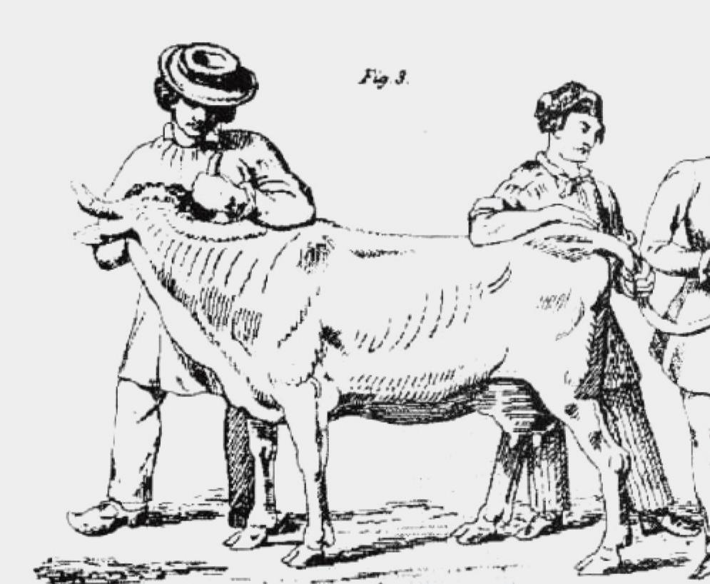
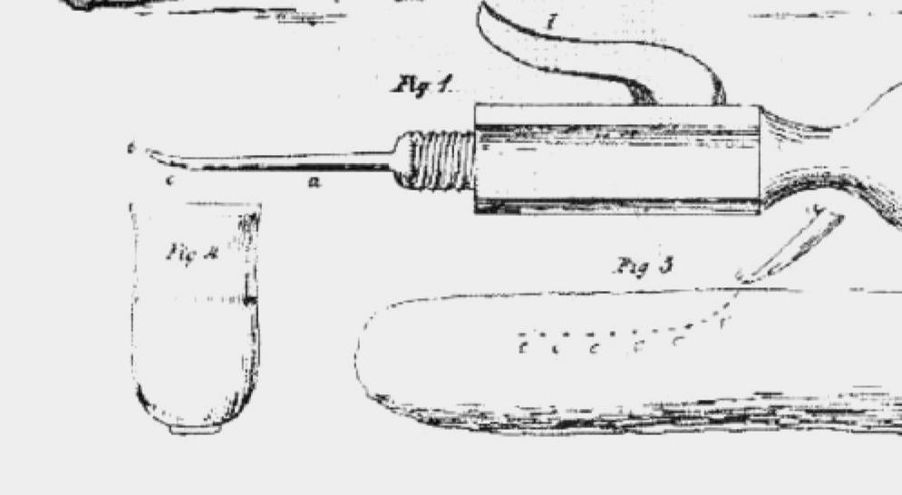
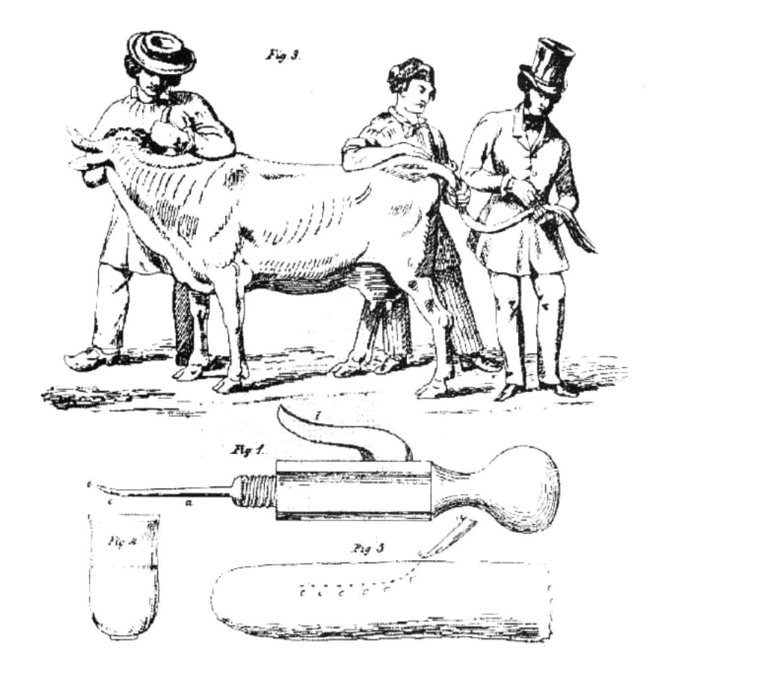
This disease was a major problem in Europe through the nineteenth century and inoculation was first attempted in England and Germany at that time. However, credit is usually given to Louis Willems of Hasselt in Belgium, who in the early 1850s inoculated lung fluid from affected animals into the tail of recipient cattle. He used a large lancet dipped in the fluid and used this to make two to three incisions at the tip of the tail. It caused large abscesses, the animals sickened and recovered, the tail commonly fell off (the Willems reaction), but the animals became immune (Fig. 1.2). Several European governments formed commissions to look into Willems claims and confirm its effectiveness. The Dutch reported that untreated cattle had 35% mortality. In vaccinated animals 8% to 10% had severe reactions with 1.1% mortality. Use of this vaccine became widespread, but it was eventually replaced with modern live attenuated vaccines, which can also cause severe reactions.
A similar process was independently developed in sub-Saharan Africa long before European colonization. It is speculated that the technique was developed following the introduction of
Fig. 1.2 The correct way to vaccinate cattle against contagious bovine pleuropneumonia. (From C.A. Spinage: “Cattle plague: a history,” Kluwer; “The tail method of inoculation for pleuropneumonia” and the reference is Keulen, 1854. With permission.).
variolation by Saharan nomads. In this case, material from pleuropneumonia lesions was inoculated subcutaneously to cattle through an incision on the bridge of the nose. The inflammatory reaction produced exostoses on the nasal bone and at one point Dr. de Rochebrune reported that he had discovered a new species of bovine, Bos triceros. Postvaccinial reactions were severe but the surviving animals were strongly immune. A similar technique may have previously been used in Iran for the prevention of caprine pleuropneumonia in goats.
Louis Pasteur
During the latter half of the nineteenth century the new science of microbiology led inexorably to the recognition that many significant diseases were caused by microorganisms, especially bacteria. The French chemist and microbiologist Louis Pasteur was one of the pioneers of microbiology and the microbial theory of infectious diseases. As such he collected and investigated any bacteria that appeared to be pathogenic. In 1879 he received a bacterial sample from Jean Toussaint (1847–1890), a professor at the Toulouse Veterinary College. This bacterium, now called Pasteurella multocida, is the cause of fowl cholera. Inoculation of this bacterium into chickens causes lethal disease. During the course of his studies, Pasteur’s assistant Émile Roux inadvertently allowed a culture of P.multocida to age in the laboratory cupboard for several weeks. (This may not have been entirely inadvertent because Roux was investigating the survival of the bacteria in the culture). When this aged culture was eventually injected into chickens it failed to sicken or kill them. The chickens were retained and sometime later injected with a fresh culture of the bacterium. They remained healthy. Pasteur had clearly been thinking about the process of smallpox vaccination and speculating on the application of this process to other infectious diseases. As a result, he recognized that what had happened with the aged bacterial culture was, in many respects, similar to vaccination. Pasteur called the procedure vaccination as well. In 1880 Pasteur presented his results to the members of the National Academy in a treatise “Of Infectious Diseases, Especially the Disease of Chicken Cholera.”
ANTHRAX AND RABIES
Fowl cholera, although important, was not the most significant infectious disease problem affecting French agriculture at that time. Anthrax, in contrast, was a major issue so Pasteur began to develop a vaccine against anthrax (Bacillus anthracis).
In the years 1880 and 1881 three investigators, William Greenfield in London, Pasteur in Paris, and Toussaint in Toulouse, had independently recognized the importance of microbial attenuation (loss of virulence) and its relationship to vaccination. In 1878, John Burdon-Sanderson and Greenfield working in London had demonstrated that anthrax could be attenuated without affecting its immunogenicity. In July 1880, a report from Toussaint was presented to the Academy describing the results of his vaccine trials. He had simply killed the anthrax bacilli by heating them at 55°C for 10 minutes. This vaccine effectively protected dogs and sheep. This result surprised and bothered Pasteur because his working hypothesis regarding the mechanism of immunity was that a vaccine somehow depleted the host of essential nutrients. As a result, the pathogenic organism could not survive in a vaccinated host. It followed from this that protection could only be conferred by the use of vaccines containing live organisms. Pasteur was skeptical of Toussaint’s results and set out to disprove them. He would not admit that he was wrong. Pasteur thus concentrated his efforts on attenuating B. anthracis so that it could no longer cause disease. Pasteur grew the organism at an unusually high temperature (42°C–43°C) for multiple generations. This worked, but the early results were erratic and inconclusive.
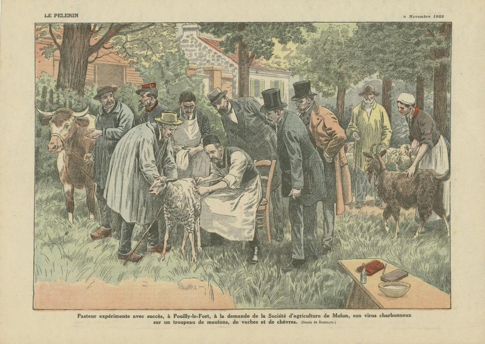
In March 1881, Pasteur reported that his vaccine was working. This report provoked a challenge for a public trial by a group of local veterinarians, a challenge that Pasteur accepted. In
May 1881, Pasteur put on a public demonstration of his anthrax vaccine at Pouilly-le-Fort outside Paris. He used two groups of animals (Fig. 1.3). The vaccinated group, 24 sheep, 1 goat, and 6 cattle, received two doses of the vaccine at a 15-day interval. The unvaccinated group received nothing. On May 31, thirty days after the first dose, all were challenged with a live anthrax culture. On June 2 more than 200 interested observers came to see the results. All the unvaccinated animals were dead or dying, whereas all the vaccinated animals survived. It was a major media event and a triumph. The publicity given to this experiment made Pasteur famous and introduced the public to the great potential of vaccines in combatting infectious disease. Pasteur claimed publicly that he had used a live attenuated vaccine. Only he and his colleagues knew that he had killed the bacteria in the vaccine with potassium bichromate. Toussaint, who received no credit, published only two more papers before having a mental breakdown and dying at the age of 43.
Subsequently Pasteur and his colleague Louis Thuillier identified Erysipelothrix rhusiopathiae as the cause of swine erysipelas. Pasteur showed that its repeated passage through pigeons increased its virulence for pigs. In contrast, serial passage through rabbits significantly reduced its virulence in pigs. He therefore developed a vaccination process against swine erysipelas that consisted of a series of injections of bacteria that had been attenuated by repeated passage in rabbits followed by a series of injections of pigeon-passaged organisms. This study is notable because it was the first example of microbial attenuation by passage in an unusual species, a process that has been widely employed since.
Pasteur and Roux went on to develop a rabies vaccine. They infected rabbits with rabid brain tissue (they didn’t know it was a virus). Once disease developed, they removed the rabbit’s spinal cord and dried it for 5 to 10 days—thus attenuating the agent, and then prepared an emulsified injectable. By drying spinal cords for different time periods, Pasteur and Roux were able to produce rabies viruses with different degrees of attenuation. They could then inoculate dogs with a series of emulsified cords containing viruses of increasing virulence and demonstrate that they usually developed strong immunity to rabies. In preliminary studies they showed that they could protect a dog after it had received a rabid bite.
They had been working on this project for some time when in July 1885 a child, Joseph Meister, was brought to them. Meister had been badly savaged by a rabid dog. Under considerable pressure Pasteur vaccinated him. He gave Meister 13 injections over 10 days using spinal
Fig. 1.3 A depiction of Pasteur’s great public anthrax vaccination study at Pouilly-le-fort in 1881. It received enormous positive publicity and launched the science of immunology. (From The Pasteur Institute/Musee Pasteur. With permission.)
cord emulsions beginning 3 days after the bites. He began with the driest and eventually proceeding to fresh infected cord. Meister survived to become the gate-porter of Pasteur’s grave and committed suicide rather than permit German troops to open the crypt in 1940. In July 1886, a 7-year-old boy named Harold Newton Newell became the first person to receive Pasteur’s rabies vaccine in the United States. He only received the first four inoculations, but his fate is unknown.
DEAD VACCINES
Although Pasteur was highly successful in developing practical vaccines, he had no concept as to how they worked. In 1880, he had put forward a tentative hypothesis that the vaccine organism consumed a necessary nutrient when it first invaded the body. Accordingly, this implied that vaccines only worked if they contained living organisms. This of course was completely wrong. However once news of his vaccine discoveries was published, others took up the cause very rapidly. By December 1880, Dr. Daniel Salmon could report, “At present, the attention of USDA investigators is still, for the most part turned to methods of prevention, and chief among these is inoculation by means of a mitigated virus.” In 1886, Daniel Salmon and Theobald Smith were able to protect pigeons by inoculating them with a heat-killed culture of “hog cholera virus,” now known as Salmonella choleraesuis. Thus killed vaccines also worked. This eventually led to the rapid development of a great diversity of killed bacterial vaccines.
Other advances soon followed. In 1888, Émile Roux and Alexandre Yersin at the Pasteur Institute showed that a bacterium-free filtrate of a diphtheria culture contained the bacterial exotoxin. In 1890, Emil von Behring, working with Shibasaburo Kitasato, immunized guinea pigs and rabbits with killed tetanus or diphtheria broth cultures. They demonstrated that the serum of these immunized animals contained an “antitoxin” that could neutralize their toxins. This antitoxic activity could be passively transferred to nonimmune animals. They called these antitoxic substances, antibodies. In 1894, Emil Roux successfully treated 300 children with diphtheria using antidiphtheria serum. In 1896, this was regarded as “the most important advance of the century in the medical treatment of acute infective disease.”
Vaccination and Antiviral Vaccines
Pasteur and his successors succeeded in laying the foundations of modern immunology and in developing numerous new antibacterial vaccines. However, it was not until 1898 that Loeffler and Frosch showed that the agent of foot-and-mouth disease could pass through a bacteria-proof filter. In 1905 Henri Carré showed that the agent of canine distemper could do the same. Thus he could filter nasal discharges or pericardial effusions through a fine porcelain filter. Although the filtrate contained no bacteria, it could still cause disease and death in susceptible dogs. This discovery was ignored for 20 years. British and American investigators firmly believed that the disease was caused by Bordetella bronchiseptica. It was not until 1926 that Carré’s claims were verified by Laidlaw and Duncan. There was a great reluctance by bacteriologists to abandon the methodology that had yielded such great results. This did not just apply to distemper. For many years, scientists clung to the notion that influenza was caused by a bacterium. Indeed, both distemper and influenza are characterized by secondary bacterial invasion. As a result, it is by no means difficult to isolate multiple bacteria from the tissues and body fluids of affected animals. Bacteriologists had no shortage of alternative candidate agents to investigate.
CANINE DISTEMPER
Throughout the nineteenth and early twentieth century, distemper was a devastating disease of dogs. It does not appear to have been present in the United Kingdom before the 1790s. It was of
special concern because it affected foxhounds and thus foxhunting by the upper class. Its cause was unknown, but that did not prevent attempts at immunization. Jenner had studied this disease and had shown that it was infectious but not transmissible to humans. Thus Edward Jenner vaccinated the Earl of Berkley’s foxhounds with cowpox without success. Nevertheless, the vaccination was employed for many years, like so many other vaccinations for other diseases. One ardent fan of giving vaccinia to his dogs to prevent distemper was General George Custer. Custer even wrote a letter to an English hunting magazine encouraging the use of vaccinia for this purpose. As late as 1902, a Dr. Brown from Cambridge, England, reported, “For a number of years I have inoculated puppies…with vaccine lymph, and with the best results. During my time I have never heard of a case of distemper arising after inoculation.” This was not a commonly held view. Arthur Senner was the first to describe bacteria in association with distemper lesions and fluids in 1875. He isolated a “short and narrow bacillus” and a micrococcus from blood, lungs, and other organs, and concluded that the bacillus was the causal agent. Other investigators followed, each claiming that their bacterium was the cause of distemper. In 1890, a Dr. Millais isolated a bacillus from a distemper case. He heated a culture of this organism to 60°C for 10 minutes and administered this heated culture subcutaneously to ten puppies. He subsequently challenged them intranasally and claimed that all were protected. In 1901 a Dr. Copeman produced a similar vaccine (heated broth culture—60°C for 30 minutes, plus phenol preservative). There was considerable debate about the efficacy of Copeman’s vaccine. Many claimed it was useless, whereas others such as His Grace, the Duke of Beaufort, thought it was great, stating, “If we go on with the same results, it will be the greatest boon that has ever been brought out.”
Later in 1901, a Dr. Physalix reported on the isolation of the causal agent of distemper— Pasteurella canis. He developed a vaccine using this organism. Physalix claimed that he had induced typical distemper when a pure culture of P. canis was injected. He also reported on the development of a vaccine containing an organism attenuated by repeated subculturing. As with other early vaccines of this nature there was a great deal of skepticism regarding the effectiveness of Physalix’s vaccine. A Committee of Veterinary Surgeons, established in 1903, reported that the vaccine failed to confer any immunity to distemper. Despite this, Professor Lignières produced a new polyvalent vaccine against P. canis in 1903. It consisted of a mixture of different strains attenuated by several hundred subcultures. It too didn’t work.
In 1912, an American veterinarian, Dr. Ferry, produced a polyvalent, polymicrobial distemper vaccine. In fact Dr. Ferry had discovered Bordetella bronchiseptica. His vaccine contained a mixture of B. bronchiseptica, Staph. pyogenes, and Strep. pyogenes. This vaccine was widely used and regarded as useful. Nevertheless, over several years, enthusiasm for Ferry’s vaccine waned. Ferry himself acknowledged this in 1923 when he suggested that the dose employed was far too low. We now recognize that B. bronchiseptica is a causal agent of kennel cough, and it is likely therefore that his vaccine prevented some such cases. Nevertheless, in 1926 Hardenbergh showed that a pure culture of this organism could not protect dogs against distemper.
It is probable that the first effective distemper vaccine was produced by a professor at the University of Rome, Dr. Vittorio Puntoni, in 1923. He succeeded in serially passaging distemper by intracerebral inoculation. Subsequently he made a suspension of the brain of an infected dog and added formalin 1:10,000. After two days he administered this subcutaneously to dogs. He boosted this with live attenuated material or with additional doses of the inactivated material and apparently obtained significant protection. In 1927, Lebailly, a French investigator, demonstrated that infected dog spleens contained large amounts of virus. Therefore he took infected spleens, ground them up, added formalin, and produced an effective vaccine.
In England, studies in dogs were initially discouraged by antivivisectionist sentiment. As a result, distemper vaccine studies moved slowly until it was shown that ferrets were also highly susceptible to this disease. Patrick Laidlaw and George Duncan initially produced a formalized killed vaccine derived from a ferret spleen extract. Field trials in 1928, especially on foxhounds,
showed significant protection. As a result, they published a detailed methodology and made the vaccine freely available for commercial development. Unfortunately, the newly released vaccine caused some side effects including postdistemper encephalitis. These appear to be in part caused by rushing the vaccine to market without adequate safety testing. In 1930, the killed vaccine was withdrawn and replaced with an antiserum-virus product. This mixture was less effective than the killed vaccine, but results improved greatly when the virus was injected first, followed several hours later by the antiserum. Subsequent developments included adapting the virus to growth in eggs and eventually to tissue culture.
By the beginning of the Second World War, vaccine technology was firmly established. Many important infectious diseases had been controlled by effective vaccines. The subsequent development of vaccines centered on previously uncontrolled diseases such as polio, on improving unsatisfactory vaccines such as those against foot-and-mouth disease, and on reducing adverse effects. The development of Salk’s inactivated polio vaccine after passage in cultured monkey kidney cells was the first step in the development of the modern cell-culture-based vaccine industry. Because of the risks associated with modified live organisms, investigators began to create vaccines based on individual viral components or subunits. Although safer, these subunit vaccines tend to be less immunogenic. Polysaccharide and virus-like particle vaccines began to be produced in the 1980s. As a result of these impressive advances, most major infectious diseases remain relatively well controlled—thanks to vaccines. There is however room for improvement and a continuous push for improved safety. Additionally, the appearance of new infections such as porcine epidemic diarrhea and Hendra virus, as well as the inexorable spread of others into new geographic areas, continues to fuel the demand for new improved veterinary products.
The Eradication of Rinderpest
Rinderpest is caused by a morbillivirus closely related to human measles virus. Indeed, the two viruses probably diverged from a common precursor about 10,000 years ago when people and cattle first lived in close proximity. This disease affected cattle, buffalo, and yaks. A short incubation period was followed by a prodromal phase of depression and inappetence. Eventually ulcers appeared on mucus membranes. These spread and coalesced leading to respiratory distress, massive diarrhea, dehydration, and death.
The first recorded probable epidemic of rinderpest occurred between 376 and 386 AD. The disease was likely brought to Europe by invaders from Central Asia, the Huns and the Mongols, as they gathered large herds of cattle as spoils of war. As time passed these raids were transformed into trading partnerships. From the seventeenth century on, the disease was repeatedly introduced into Western Europe by organized cattle trading, mainly from Russia; at that time it was known as the “Russian disease.” Bernardino Ramazzini, Professor of Medicine at Padua University, wrote the first clinical description in 1712. However, in 1711 Johann Karol in Prussia recognized that rinderpest was transmissible and that cattle that recovered from it were resistant to reinfection. As described previously, during the eighteenth century, numerous attempts were made to vaccinate animals, usually unsuccessfully. Major rinderpest outbreaks occurred throughout the eighteenth and nineteenth centuries across Europe, killing huge numbers of cattle. Indeed, so important was rinderpest that the French king’s controller of finances gave funds to Claude Bourgelat in 1762 to found the first veterinary school in Lyons. Other countries soon followed. The graduates were needed to fight the cattle plague.
Rinderpest was introduced into sub-Saharan Africa in 1887 when infected cattle from India were imported into Ethiopia to feed Italian soldiers. The disease infected local cattle with 90% mortality, and over the next decade spread across the continent. This Great Rinderpest Pandemic caused major cultural, social, and economic upheaval. The disease raged uncontrolled across the
African continent, causing widespread famine, devastating communities, and wiping out huge numbers of domestic and wild ungulates. When it reached Natal in 1896, it killed more than 90% of all African-owned cattle. Thereafter the virus became endemic and circulated in the pastoral communities of East Africa. In 1902 Maurice Nicolle and Mustafa Adil Bey demonstrated that rinderpest was caused by a filterable agent; in other words, a virus.
The disease also came back to Europe in the 1920s when it was introduced by a herd of zebus being shipped from India to Brazil. They came into contact with some imported American cattle that were sold at Belgian markets and went on to infect cattle across Germany. This outbreak was controlled by restriction of cattle movement, immediate slaughter, and vaccination. As a result of this outbreak it was decided to create the Office International des Epizooties (OIE)—now known as the World Organization for Animal Health.
In 1897, the eminent microbiologist Robert Koch, working in South Africa, developed a vaccine containing bile from rinderpest-infected cattle that was administered subcutaneously. Koch had shown that the virus in bile was not usually infectious. Nevertheless, this was still a highly dangerous procedure. Koch also showed that immune serum gave short-term passive protection. But a vaccine containing hyperimmune serum and virulent virus produced prolonged strong immunity. This “serum-virus simultaneous” method was the most effective rinderpest vaccine for many years and helped eliminate the disease from Europe in 1928.
Various methods of inactivating rinderpest virus were studied in the 1920s because the serumvirus technique was complex and costly. Eventually a formalin-inactivated vaccine was developed and used with some success in Asia. Unfortunately, the duration of immunity conferred by these formalized vaccines was very short (6 months) and their large-scale production impractical.
Subsequently it proved possible to adapt the virus to growth in goats. Serial passage in goats clearly reduced their virulence for cattle although it was not completely innocuous. It caused fever and mild lesions in European cattle. This “caprinized” vaccine was developed in the 1920s, and after various improvements was widely employed until the 1950s. However, it could still cause severe reactions in cattle including significant weight loss. Meanwhile, efforts continued on adapting rinderpest virus to rabbits (lapinization). After more than 600 passages in rabbits, it was unable to cause disease in cattle and was widely adopted by affected countries.
In spite of limited success obtained using egg-adapted rinderpest, in 1959 Dr. Walter Plowright and his colleagues, working in Kenya, succeeded in growing rinderpest virus in bovine kidney cells. Although the virus in the initial cultures was virulent, this rapidly declined so that virus passaged 95 times no longer caused a fever in cattle and could induce prolonged strong immunity. This vaccine protected against all strains of rinderpest, induced lifelong immunity, and was remarkably free of side effects. This vaccine rapidly replaced both the caprinized and lapinized vaccines.
Beginning in 1960 a series of coordinated regional vaccination campaigns were mounted in an effort to control, and ideally eliminate, the disease. The first of these vaccination campaigns, called Joint Program 15 (JP-15) and organized by the Organization of African Unity, was very successful and clinical disease outbreaks were massively reduced. Seventeen out of the twenty-two countries involved were infected at the beginning of the campaign, and this had dropped to one by 1979. However, the money ran out, urgency was lost, surveillance systems failed to recognize covert infections in domestic and wild ungulates, vaccination programs were discontinued, and as a result, the disease reemerged across Africa. Similar efforts were launched across Asia beginning in Afghanistan, and by 1972, the disease was restricted to Lebanon. However, as in JP-15, success bred complacency and the disease reemerged in the 1980s.
A second round of control programs was instituted when in 1982 the Pan-African Rinderpest Campaign was instituted. This was so successful that in 1994 the goal of rinderpest eradication was established by the United Nations Food and Agriculture Organization (FAO) and the World Organization for Animal Health (OIE).
Several key factors were in place to make this program a success. Thus new highly sensitive diagnostic tests based on the identification of antirinderpest antibodies were developed. These tests based on enzyme-linked immunosorbent assays (ELISAs) were essential for monitoring the vaccination program. More importantly was the development of a thermostable Plowright vaccine. A major limitation of the original vaccine was the need to keep the vaccine cold—maintain the cold chain. However, by 1992 researchers at Tufts University had adapted the virus for growth in monkey kidney cells that resulted in a thermostable vaccine—ThermoVax, which had a shelf life of 30 days when unrefrigerated. This development made it much easier to deliver the vaccine to remote villages and thus reach many more animals. This innovation, together with others such as working with community elders who provided local intelligence, ensured that most of their cattle were vaccinated, generated significant community support, and assured the success of the program. Massive vaccination campaigns together with careful epidemiology, controls on animal movement, and focusing on a regional approach progressively reduced the number of infected countries. When disease reservoirs were identified, vaccination teams quickly moved in to contain the infection. Between April 1989 and July 1990, 24.1 million vaccine doses were administered. Fortunately, the disease in wild ungulates disappeared when the local cattle herds were vaccinated. The surviving wild ungulate populations were too small to sustain the infection, and herd immunity soon developed. By 1999, the campaign was intensified under the slogan “Seek, Contain, Eliminate.” The disease was detected in Pakistan and Sudan for the last time in 2000. The last known cases of rinderpest occurred in Kenya in October 2001. It took some time to confirm that rinderpest was indeed finally exterminated, and vaccination continued until 2006. It was not until June 28, 2011, that the FAO and the WHO declared that the disease was eradicated. The destruction of the last remaining stocks of rinderpest virus held in the UK began in June 2019. The eradication of rinderpest was an incredible veterinary achievement and a triumph for the science of immunology.
Sources of Additional Information
Baron, M.D., Banyard, A.C., Parida, S., Barrett, T. (2005). The Plowright vaccine strain of Rinderpest virus has attenuating mutations in most genes. J Gen Virol, 86, 1093–10101.
Boylston, A.W. (2018). The myth of the milkmaid. N Engl J Med, 378, 414–415.
Bresalier, M., Worboys, M. (2014). ‘Saving the lives of our dogs’: the development of canine distemper vaccine in interwar Britain. Br J Hist Sci, 47, 305–334.
Esparza, J., Schrick, L., Damaso, C.R., Nitsche, A. (2017). Equination (inoculation of horsepox): An early alternative to vaccination (inoculation of cowpox) and the potential role of horsepox virus in the origin of the smallpox vaccine. Vaccine, 35, 7222–7230.
Hoenig, L.J., Jackson, A.C., Dickinson, G.M. (2018). The early use of Pasteur’s rabies vaccine in the United States. Vaccine, 36, 4578–4581.
Lombard, M., Pastoret, P.P., Moulin, A.M. (2007). A brief history of vaccines and vaccination. Rev Sci Tech, 26, 29–48.
Mariner, J.C., House, J.A., Mebus, C.A., Sollod, A.E., et al. (2012). Rinderpest eradication: Appropriate technology and social innovations. Science, 337, 1309–1312.
Plotkin, S. (2014). History of vaccination. Proc Natl Acad Sci USA, 111, 12283–12287.
Roeder, P., Mariner, J., Kock, R. (2013). Rinderpest: the veterinary perspective on eradication. Philos Trans R Soc Lond B. Biol Sci, 368, 20120139.
Smith, K.A. (2012). Louis Pasteur, the father of immunology? Front Immunol, 3, 68. Tizard, I. (1999). Grease, anthraxgate, and kennel cough: a revisionist history of early veterinary vaccines. Adv Vet Med, 41, 7–24.
Youde, J. (2013). Cattle scourge no more. The eradication of rinderpest and its lessons for global health campaigns. Politics Life Sci, 32, 43–45.