USMLE STEP 2
Fifth Edition
1600 John F. Kennedy Blvd. Ste 1800 Philadelphia, PA 19103-2899
USMLE STEP 2 SECRETS, 5th edition
Copyright © 2018 by Elsevier, Inc. All rights reserved. Previous editions copyrighted 2014, 2010, 2004
ISBN: 978-0-323-49616-2
No part of this publication may be reproduced or transmitted in any form or by any means, electronic or mechanical, including photocopying, recording, or any information storage and retrieval system, without permission in writing from the publisher. Details on how to seek permission, further information about the Publisher’s permissions policies and our arrangements with organizations such as the Copyright Clearance Center and the Copyright Licensing Agency, can be found at our website: www.elsevier.com/permissions.
This book and the individual contributions contained in it are protected under copyright by the Publisher (other than as may be noted herein).
Notices
Knowledge and best practice in this field are constantly changing. As new research and experience broaden our understanding, changes in research methods, professional practices, or medical treatment may become necessary.
Practitioners and researchers must always rely on their own experience and knowledge in evaluating and using any information, methods, compounds, or experiments described herein. In using such information or methods they should be mindful of their own safety and the safety of others, including parties for whom they have a professional responsibility.
With respect to any drug or pharmaceutical products identified, readers are advised to check the most current information provided (i) on procedures featured or (ii) by the manufacturer of each product to be administered, to verify the recommended dose or formula, the method and duration of administration, and contraindications. It is the responsibility of practitioners, relying on their own experience and knowledge of their patients, to make diagnoses, to determine dosages and the best treatment for each individual patient, and to take all appropriate safety precautions.
To the fullest extent of the law, neither the Publisher nor the authors, contributors, or editors, assume any liability for any injury and/or damage to persons or property as a matter of products liability, negligence or otherwise, or from any use or operation of any methods, products, instructions, or ideas contained in the material herein.
Library of Congress Cataloging-in-Publication Data
Names: O’Connell, Theodore X., author.
Title: USMLE step 2 secrets / Theodore X. O’Connell.
Other titles: USMLE step two secrets | United States Medical Licensing Examination step 2 secrets | Secrets series.
Description: Fifth edition. | Philadelphia, PA : Elsevier, [2018] | Series: Secrets series | Includes bibliographical references and index.
Identifiers: LCCN 2017007823 | ISBN 9780323496162 (pbk. : alk. paper)
Subjects: | MESH: Clinical Medicine | Examination Questions
Classification: LCC RC58 | NLM WB 18.2 | DDC 616.0076--dc23 LC record available at https://lccn.loc.gov/2017007823
Content Strategist: Jim Merritt
Content Development Specialist: Angie Breckon
Publishing Services Manager: Patricia Tannian
Project Manager: Ted Rodgers
Design Direction: Bridget Hoette
CHAPTER 29 PEDIATRICS 249
CHAPTER 30 PHARMACOLOGY 256
CHAPTER 31 PREVENTIVE MEDICINE 261
CHAPTER 32 PSYCHIATRY 264
CHAPTER 33 PULMONOLOGY 275
CHAPTER 34 RADIOLOGY 281
CHAPTER 35 RHEUMATOLOGY 284
CHAPTER 36 SHOCK 291
CHAPTER 37 SMOKING 294
CHAPTER 38 UROLOGY 295
CHAPTER 39 VASCULAR SURGERY 300
CHAPTER 40 VITAMINS AND MINERALS 304
INDEX 307
11. Arterial blood gas analysis: in general, pH tells you the primary event (acidosis vs. alkalosis), whereas carbon dioxide and bicarbonate values give you the cause (same direction as pH) and suggest any compensation present (opposite of pH).
12. Exogenous causes of hyponatremia to keep in mind: oxytocin, surgery, narcotics, inappropriate intravenous (IV) fluid administration, diuretics, and antiepileptic medications.
13. EKG findings in electrolyte disturbances: tall, tented T waves in hyperkalemia; loss of T waves/T-wave flattening and U waves in hypokalemia; QT prolongation in hypocalcemia; QT shortening in hypercalcemia.
14. Shock: first give the patient oxygen, start an IV line, and set up monitoring (pulse oximetry, EKG, frequent vital signs). Then give a fluid bolus (30 cc/kg of normal saline or lactated Ringer solution) if no signs of congestive heart failure (e.g., bibasilar rales) are present while you try to figure out the cause if it is not known. If the patient is not responsive to fluids, start cardiac pressors.
15. Virchow triad of deep venous thrombosis: endothelial damage (e.g., surgery, trauma), venous stasis (e.g., immobilization, surgery, severe heart failure), and hypercoagulable state (e.g., malignancy, birth control pills, pregnancy, lupus anticoagulant, inherited deficiencies).
16. Therapy for congestive heart failure: diuretics (e.g., furosemide), angiotensinconverting enzyme inhibitors, and beta-blockers (for stable patients) are the mainstays of pharmacologic treatment. Be sure to screen for and address underlying atherosclerosis risk factors (e.g., smoking, hyperlipidemia). Most patients should be on antiplatelet therapy (e.g., aspirin).
17. Cor pulmonale: right-sided heart enlargement, hypertrophy, or failure due to primary lung disease (usually chronic obstructive pulmonary disease). The most common cause of right-heart failure, however, is left-heart failure (not cor pulmonale).
18. In patients with atrial fibrillation, assess for an underlying cause with thyroid-stimulating hormone, electrolytes, urine drug screen, and echocardiogram. The main management issues are ventricular rate control (if needed, slow the rate with medications) and atrial clot formation/embolic disease (consider anticoagulation with warfarin or a novel oral anigoagulant).
19. Ventricular fibrillation and pulseless ventricular tachycardia are treated with immediate defibrillation followed by epinephrine, vasopressin, amiodarone, and lidocaine. If ventricular tachycardia with a pulse is present, treat with amiodarone and synchronized cardioversion.
20. Obstructive vs. restrictive lung disease: the FEV1/FVC ratio is the most important parameter on pulmonary function testing to distinguish the two (FEV1 may be the same). In obstructive lung disease, the FEV1/FVC ratio is less than normal (<0.7). In restrictive disease, the FEV1/FEV ratio is often normal.
21. The most common type of esophageal cancer in the United States is adenocarcinoma occurring as a result of longstanding reflux disease and the development of Barrett esophagus. Smoking and alcohol abuse contribute to the development of squamous cell carcinoma, the second most common histologic type of esophageal cancer.
22. All gastric ulcers must be biopsied or followed to resolution to exclude malignancy.
23. Testing a nasogastric tube aspirate for blood is the best initial test to distinguish an upper from a lower gastrointestinal (GI) bleed, although bright red blood via mouth or anus is a fairly reliable sign of a nearby bleeding source.
24. Irritable bowel syndrome is one of the most common causes of GI complaints. Physical exam and diagnostic studies are by definition negative; this is a diagnosis of exclusion. The classic patient is a young adult female with a chronic history of alternating constipation and diarrhea.
25. Crohn disease vs. ulcerative colitis
CROHN DISEASE ULCERATIVE COLITIS
Place of origin
Thickness of pathology
Progression
Distal ileum, proximal colon Rectum
Transmural
Irregular (skip lesions)
Location From mouth to anus
Bowel habit changes
Classic lesions
Colon cancer risk
Obstruction, abdominal pain
Fistulas/abscesses, cobblestoning, string sign on barium x-ray
Slightly increased
Surgery No (may make worse)
Mucosa/submucosa only
Proximal, continuous from rectum; no skipped areas
Involves only colon, rarely extends to ileum
Bloody diarrhea
Pseudopolyps, lead-pipe colon on barium x-ray, toxic megacolon
Markedly increased
Yes (proctocolectomy with ileoanal anastomosis)
26. All forms of viral hepatitis can present similarly in the acute stage; serology testing and history are needed to distinguish them. Hepatitis B, C, and D are transmitted parenterally and can lead to chronic infection, cirrhosis, and hepatocellular carcinoma.
27. Hereditary hemochromatosis is currently the most common known genetic disease in white people. The initial symptoms (fatigue, impotence) are nonspecific, but patients often have hepatomegaly, skin pigmentation changes (“bronze diabetes”), and diabetes. Initial tests include with transferrin saturation (serum iron/total iron binding capacity) and ferritin level. Treat with phlebotomy after confirming the diagnosis with genetic testing and liver biopsy.
28. Sequelae of liver failure: coagulopathy (that cannot be fixed with vitamin K), thrombocytopenia, jaundice/hyperbilirubinemia, hypoalbuminemia, ascites, portal hypertension, hyperammonemia/encephalopathy, hypoglycemia, disseminated intravascular coagulation, and renal failure.
29. Pancreatitis is usually due to alcohol or gallstones. Patients present with abdominal pain, nausea/vomiting, and elevated amylase and lipase. Treat supportively and provide pain control. Complications include pseudocyst formation, infection/abscess, and adult respiratory distress syndrome.
30. Jaundice/hyperbilirubinemia in neonates is usually physiologic (only monitoring, follow-up lab tests, and possibly phototherapy are needed), but jaundice present at birth is always pathologic.
31. Primary vs. secondary endocrine disturbances. In primary disorders (e.g., Graves, Hashimoto, or Addison disease), the gland malfunctions, but the pituitary or another gland and the central nervous system respond appropriately (e.g., thyroid-stimulating hormone, thyrotropin-releasing hormone, or adrenocorticotropic hormone elevates or depresses as expected in the setting of a malfunctioning gland). In secondary disorders (e.g., adrenocorticotropic hormone-secreting lung carcinoma, heart failure–induced hyperreninemia, renal failure–induced hyperparathyroidism), the gland itself is doing what it is told to do by other controlling forces (e.g., pituitary gland, hypothalamus, tumor, disease); they are the problem, not the gland itself.
32. Corticosteroid side effects (aka Cushing syndrome): weight gain, easy bruising, acne, hirsutism, emotional lability, depression, psychosis, menstrual changes, sexual dysfunction, insomnia, memory loss, buffalo hump, truncal and central obesity with wasting of extremities, round plethoric facies, purplish skin striae, weakness (especially of the proximal muscles), hypertension, peripheral edema, poor wound healing, glucose intolerance or diabetes, osteoporosis, and hypokalemic metabolic alkalosis (due to mineralocorticoid effects of certain corticosteroids). Growth can also be stunted in children.
33. Osteoarthritis is by far the most common cause of arthritis (≥75% of cases) and usually does not have hot, swollen joints or significant findings if arthrocentesis is performed.
34. Cancer incidence and mortality in the United States.
Prostate
Lung
Colon
Breast
Lung
Colon
Colon
35. Sequelae of lung cancer: hemoptysis, Horner syndrome, superior vena cava syndrome, phrenic nerve involvement/diaphragmatic paralysis, hoarseness from recurrent laryngeal nerve involvement, and paraneoplastic syndromes (Cushing syndrome, syndrome of inappropriate antidiuretic hormone secretion, hypercalcemia, Eaton-Lambert syndrome).
36. Bitemporal hemianopsia (loss of peripheral vision in both eyes) is due to a space-occupying lesion pushing on the optic chiasm (classically a pituitary tumor) until proven otherwise. Order a computed tomography (CT) or magnetic resonance imaging of the brain.
37. Potential risks and side effects of estrogen therapy (e.g., contraception, postmenopausal hormone replacement): endometrial cancer (with unopposed estrogen, therefore must be given with progesterone in patients with a uterus), hepatic adenomas, glucose intolerance/diabetes, deep venous thrombosis, pulmonary embolism, stroke, cholelithiasis, hypertension, endometrial bleeding, depression, weight gain, nausea/vomiting, headache, drug-drug interactions, teratogenesis, and aggravation of preexisting uterine leiomyomas (fibroids), breast fibroadenomas, migraines, and epilepsy. The risks of coronary artery disease and breast cancer may be increased with combined estrogen and progesterone therapy.
38. ABCDE characteristics of a mole that should make you suspicious of malignant transformation: Asymmetry, Borders (irregular), Color (change in color or multiple colors), Diameter (the bigger the lesion, the more likely that it is malignant), and Evolution over time. Do an excisional biopsy of such moles and/or if a mole starts to itch or bleed.
39. Bronchiolitis vs. croup vs. epiglottitis
BRONCHIOLITIS
Child’s age 0–18 months
Common Yes
Common cause(s)
Respiratory syncytial virus (≥75%), parainfluenza, influenza
Symptoms/ signs Initial viral URI symptoms followed by tachypnea and expiratory wheezing
X-ray findings
Hyperinflation
Treatment Generally supportive care with humidified oxygen and suctioning
CROUP (ACUTE LARYNGOTRACHEITIS) EPIGLOTTITIS
1–2 yr
Yes
Parainfluenza virus (50%–75% of cases), influenza
Initial viral URI symptoms followed by “barking” cough, hoarseness, and inspiratory stridor
Subglottic tracheal narrowing on frontal x-ray (steeple sign)
Dexamethasone, nebulized racemic epinephrine, humidified oxygen
2–5 yr
No
Haemophilus influenzae type b, Staphylococcus spp., Streptococcus spp.
Rapid progression to high fever, toxicity, drooling, and respiratory distress
Swollen epiglottis on lateral neck x-ray (thumb sign)
Prepare to establish an airway, antibiotics (e.g., third-generation cephalosporin and an antistaphylococcal agent active against MRSA such as vancomycin or clindamycin).
40. Sequelae of group A streptococcal infection: rheumatic fever, scarlet fever, and poststreptococcal glomerulonephritis. Only the first two can be prevented by treatment with antibiotics.
41. Multiple sclerosis should be suspected in any young adult with recurrent, varied neurologic symptoms/signs when no other causes are evident. Best diagnostic tests: magnetic resonance imaging (most sensitive), lumbar puncture (elevated immunoglobulin G oligoclonal bands and myelin basic protein levels, mild elevation in lymphocytes and protein), and evoked potentials (slowed conduction through areas with damaged myelin).
42. For the unconscious or delirious (encephalopathic) patient in the emergency department with no history or signs of trauma: consider empiric treatment for hypoglycemia (glucose), opioid overdose (naloxone), and thiamine deficiency (thiamine should be given before glucose in a suspected alcoholic). Other commonly tested causes are alcohol, illicit or prescription drugs, diabetic ketoacidosis, stroke, epilepsy or postictal state, subarachnoid hemorrhage (e.g., aneurysm rupture), sepsis, electrolyte imbalance/metabolic causes, uremia, hepatic encephalopathy, and hypoxia.
43. Delirium vs. dementia
DELIRIUM
Onset Acute and dramatic
DEMENTIA
Chronic and insidious
Common causes Illness, toxin, withdrawal Alzheimer disease, multiinfarct dementia, HIV/AIDS
Reversible Usually Usually not
Attention Poor
Usually unaffected
Arousal level Fluctuates Normal
44. Always consider the possibility of pregnancy (and order a pregnancy test to rule it out, unless pregnancy is an impossibility) in reproductive-age women before advising potentially teratogenic therapies or tests (e.g., antiepileptic drugs, x-ray, CT scan). Pregnancy is in the differential diagnosis of both primary and secondary amenorrhea.
45. Anaphylaxis is commonly caused by bee stings, food allergy (especially peanuts and shellfish), medications (especially penicillins and sulfa drugs), or rubber glove allergy. Patients become agitated and flushed and shortly after exposure develop itching (urticaria), facial swelling (angioedema), and difficulty in breathing. Symptoms develop rapidly and dramatically in true anaphylaxis. Treat immediately by securing the airway (laryngeal edema may prevent intubation, in which case do a cricothyroidotomy, if needed), and give subcutaneous or IV epinephrine. Antihistamines and corticosteroids are not useful for immediate, severe reactions that involve the airway.
46. Cancer screening in asymptomatic adults
CANCER
PROCEDURE AGE FREQUENCY
Colorectal Colonoscopy or >50 yr for all studies
Every 10 yr
Flexible sigmoidoscopy or Every 5 yr
Double contrast barium enema or Every 5 yr
CT colonography or Every 5 yr
Fecal occult blood test or Annually
Fecal immunochemical test or
Stool DNA test
Annually
Every 3 yr
CANCER PROCEDURE AGE FREQUENCY
Colon, prostate Digital rectal exam >40 yr Annually, though benefits are uncertain and guidelines vary
Prostate Prostate specific antigen test >50 yr* Have a risk/benefit discussion with appropriate patients. Routine screening no longer recommended.
Cervical Pap smear
Pap and HPV co-test
Beginning at age 21 yr regardless of sexual activity
If conventional Pap test is used, test every 3 yr from ages 21 to 65. Pap and HPV co-testing should not be used for women <30 yr old. Test every 5 yr in women 30–65 yr of age if both HPV and cytology results are negative. Screening may be stopped after age 65 yr with adequate negative prior screening and no history of CIN2 or higher.
Endometrial Endometrial biopsy Menopause No recommendation for routine screening in the absence of symptoms
Breast Mammography >50 yr
Lung Low-dose CT scan
Every 1–2 yr, depending on guidelines. Controversial if patients 40–49 should be screened.
Annual CT scan is controversial but may be indicated for smokers and former smokers ages 55–74 who have smoked at least 30 pack-years.
This table is for the screening of asymptomatic, healthy patients. Other guidelines exist, but you’ll be fine if you follow this table for the USMLE. Controversial areas usually are not tested on the USMLE. *Start at age 45 in African Americans and at age 40 for patients with a first-degree relative diagnosed at an early age.
47. Biostatistics calculations using a 2 × 2 table
Test or Exposure
(+) (–)
Sensitivity
Specificity
PPV
NPV
Odds ratio
Relative risk
A/(A + C)
D/(B + D)
A/(A + B)
D/(C + D)
(A × D)/(B × C)
[A/(A + B)] – [C/(C + D)] (+) (–) AB CD
Attributable risk
[A/(A + B)] / [C/(C + D)]
48. The P-value reflects the likelihood of making a type I error, or claiming an effect or difference where none existed (i.e., results were obtained by chance). When we reject the null hypothesis (i.e., the hypothesis of no difference) in a trial testing a new treatment, we are saying that the new treatment works. We use the P-value to express our confidence in the data.
49. Side effects of antipsychotics: acute dystonia (treat with antihistamines or anticholinergics), akathisia (beta-blocker may help), tardive dyskinesia (switching to newer agent may have benefit), parkinsonism (treat with antihistamines or anticholinergics), neuroleptic malignant syndrome, hyperprolactinemia (may cause breast discharge, menstrual dysfunction, and/or sexual dysfunction), and autonomic nervous system–related effects (e.g., anticholinergic, antihistamine, and alpha1-receptor blockade).
50. Asking about depression and suicidal thoughts/intent is important in the right setting and does not cause people to commit suicide. Hospitalize psychiatric patients against their will if they are a danger to self or others or gravely disabled (unable to care for self).
51. Drugs of abuse: potentially fatal in withdrawal include alcohol, barbiturates, and benzodiazepines. Alcohol, cocaine, opiates, barbiturates, benzodiazepines, phencyclidine (PCP), and inhalants are potentially fatal in overdose.
52. Pelvic inflammatory disease is the most common preventable cause of infertility in the United States and the most likely cause of infertility in younger, normally menstruating women.
53. Polycystic ovarian syndrome is classically associated with women who are “heavy, hirsute, and [h]amenorrheic.” It is the most common cause of dysfunctional uterine bleeding. Remember the increased risk of endometrial cancer due to unopposed estrogen.
54. Fetal/neonatal macrosomia is due to maternal diabetes until proven otherwise. Treat gestational/maternal diabetes by aiming for tight glucose control through diet, oral agents, or insulin.
55. Low maternal serum alpha-fetoprotein causes: Down syndrome, inaccurate dates (most common), and fetal demise. High maternal serum alpha-fetoprotein associated with: neural tube defects, ventral wall defects (e.g., omphalocele, gastroschisis), inaccurate dates (most common), and multiple gestation. Measurement is generally obtained between 16 and 20 weeks’ gestation.
56. Hypertension plus proteinuria in pregnancy equals preeclampsia until proven otherwise.
57. Positive pregnancy test (i.e., not a clinically apparent pregnancy) plus vaginal bleeding and abdominal pain equals ectopic pregnancy until proven otherwise. Order a pelvic ultrasound if the patient is stable.
58. Decelerations during maternofetal monitoring: early decelerations are normal and due to head compression. Variable decelerations are common and usually due to cord compression (turn the mother on her side, give oxygen and fluids, stop oxytocin, and consider amnioinfusion). Late decelerations are due to uteroplacental insufficiency and are the most worrisome pattern (turn the mother on her side, give oxygen and IV fluids, stop oxytocin, and measure fetal oxygen saturation or scalp pH). Prepare for prompt delivery.
59. Always perform an ultrasound before a pelvic exam in the setting of third-trimester bleeding (in case placenta previa is present).
60. Uterine atony is the most common cause of postpartum bleeding and is typically due to uterine overdistention (e.g., twins, polyhydramnios), prolonged labor, fibroids, and/or oxytocin usage. The risk is increased in multiparous women.
61. Acute abdomen pathology localization by physical exam
AREA
Right upper quadrant
Left upper quadrant
Right lower quadrant
Left lower quadrant
Epigastric area
ORGAN (CONDITIONS)
Gallbladder/biliary (cholecystitis, cholangitis) or liver (abscess)
Spleen (rupture with blunt trauma)
Appendix (appendicitis), pelvic inflammatory disease
Sigmoid colon (diverticulitis), pelvic inflammatory disease
Stomach (peptic ulcer) or pancreas (pancreatitis)
62. The “6 Ws” of postoperative fever: water, wind, walk, wound, “wawa,” and weird drugs. Water stands for urinary tract infection, wind for atelectasis or pneumonia, walk for deep venous thrombosis, wound for surgical wound infection, “wawa” for breast (usually relevant only in the postpartum state), and weird drugs for drug fever. In patients with daily fever spikes that do not respond to antibiotics, think about a postsurgical abscess. Order a CT scan to locate, then drain the abscess if one is present.
63. ABCDEs of trauma (follow in order if you are asked to choose): airway, breathing, circulation, disability, and exposure.
64. Six rapidly fatal thoracic injuries that must be recognized and treated immediately:
1. Airway obstruction (establish airway)
2. Open pneumothorax (intubate, place chest tube at different site, and close defect on three sides)
3. Tension pneumothorax (perform needle thoracentesis followed by chest tube)
4. Cardiac tamponade (perform pericardiocentesis)
5. Massive hemothorax (place chest tube to drain; thoracotomy if bleeding does not stop)
6. Flail chest (consider intubation and positive pressure ventilation if oxygenation inadequate)
65. Neonatal conjunctivitis may be caused by chemical reaction (in the first 12–24 hours of giving drops for prophylaxis), gonorrhea (2–5 days after birth; usually prevented by prophylactic drops), and chlamydial infection (5–14 days after birth; often not prevented by prophylactic drops).
66. Glaucoma is usually (90%) due to the open-angle form, which is painless (no “attacks”) and asymptomatic until irreversible vision loss (that starts in the periphery) occurs. Thus, screening is important. Open-angle glaucoma is the most common cause of blindness in African Americans.
67. Uveitis is often a marker for systemic conditions: juvenile rheumatoid arthritis, sarcoidosis, inflammatory bowel disease, ankylosing spondylitis, reactive arthritis, multiple sclerosis, psoriasis, or lupus. Photophobia, blurry vision, and eye pain are common complaints.
68. Bilateral (though often asymmetric) painless gradual loss of vision in older adults is usually due to cataracts, macular degeneration, or glaucoma, which can be distinguished on physical exam. Presbyopia is a normal part of aging and affects only near vision (i.e., accommodation).
69. Compartment syndrome, usually in the lower extremity after trauma or surgery, causes the “6 Ps”:
1. Pain (present on passive movement and often out of proportion to the injury)
2. Paresthesias (numbness, tingling, decreased sensation)
3. Pallor (or cyanosis)
4. Pressure (firm feeling muscle compartment, elevated pressure reading)
5. Paralysis (late, ominous sign)
6. Pulselessness (very late, ominous sign)
Treat with fasciotomy to relieve compartment pressure and prevent permanent neurologic damage.
70. Peripheral nerve evaluation
NERVE MOTOR FUNCTION
Radial Wrist extension (watch for wrist drop)
Ulnar Finger abduction (watch for “claw hand”)
Median Forearm pronation, thumb opposition
Axillary Shoulder abduction, lateral rotation
Peroneal Foot dorsiflexion, eversion (watch for foot drop)
SENSORY FUNCTION CLINICAL SCENARIO
Back of forearm, back of hand (first 3 digits)
Front and back of last 2 digits
Palmar surface of hand (first 3 digits)
Lateral shoulder
Dorsal foot and lateral leg
Humeral fracture
Elbow dislocation
Carpal tunnel syndrome, humeral fracture
Upper humeral dislocation or fracture
Knee dislocation
or hypocalcemia). In this setting, pH correction is needed (rather than direct treatment of the calcium or potassium levels). Magnesium depletion can also make hypocalcemia and hypokalemia unresponsive to replacement therapy (until magnesium is corrected).
88. Adult patients of sound mind are allowed to refuse any form of treatment. Watch for depression as a cause of “incompetence.” Treat depression before wishes for death are respected.
89. If a patient is incompetent (including younger minors who lack adequate decisionmaking capacity) and an emergency treatment is needed, seek a family member or court-appointed guardian to make health care decisions. If no one available, treat as you see fit in an emergency, or contact the courts in a nonemergency setting.
90. Respect patient wishes and living wills (assuming that they are appropriate) even in the face of dissenting family members, but take time to listen to family members’ concerns.
91. Always be a patient advocate and treat patients with respect and dignity, even if they refuse your proposed treatment or are noncompliant. If patients’ actions puzzle you, do not be afraid to ask them why they are doing or saying what they are.
92. Break doctor-patient confidentiality only in the following situations:
• The patient asks you to do so.
• Child abuse is suspected.
• The courts mandate you to do so.
• You must fulfill the duty to warn or protect (if a patient says that he is going to kill someone or himself, you have to tell the someone, the authorities, or both).
• The patient has a reportable disease.
• The patient is a danger to others (e.g., if a patient is blind or has seizures, let the proper authorities know so that they can revoke the patient’s license to drive; if the patient is an airplane pilot and is a paranoid, hallucinating schizophrenic, then authorities need to know).
93. Causes of “false” lab disturbances: hemolysis (hyperkalemia), pregnancy (elevated sedimentation rate and alkaline phosphatase), hypoalbuminemia (hypocalcemia), and hyperglycemia (hyponatremia).
94. EKG findings of myocardial infarction: flipped or flattened T waves, ST-segment elevation (depression means ischemia; elevation means injury), and/or Q waves in a segmental distribution (e.g., leads II, III, and AVF for an inferior infarct). ST depression may also be seen in “reciprocal”/opposite leads.
95. Drugs that may be useful in the setting of acute coronary syndrome: aspirin, morphine, nitroglycerin, beta-blocker, angiotensin-converting enzyme inhibitor, clopidogrel, HMG-CoA reductase inhibitor, glycoprotein IIb/IIIa receptor inhibitors, heparin (unfractionated or low-molecular-weight heparin), and tissue-plasminogen activator (t-PA; strict criteria for use).
96. Cholesterol management guidelines
The following information is from the 2013 American College of Cardiology/American Heart Association (ACC/AHA) Guidelines on the Treatment of Blood Cholesterol to Reduce Atherosclerotic Cardiovascular Risk in Adults. This new guideline differs from the previous recommendations in that it moves away from specific LDL targets. Instead, overall LDL reduction is recommended.
GROUP
Anyone with an LDL level at or above 190 mg/dL
Anyone with diabetes between 40 and 75 yr old and LDL above 70 mg/dL
Reduce LDL by >50%
Reduce LDL by 30%–50%
High-intensity statin
Moderate-intensity statin
Anyone with current or past cardiovascular disease (angina, MI, PAD) with LDL above 70 mg/dL
Anyone with a 7.5% or greater chance of developing atherosclerotic cardiovascular disease in the next 10 yr, using a specific calculator*
Reduce LDL by >50% High-intensity statin
Reduce LDL by 30%–50% Moderate-intensity statin
*The Pooled Cohort Equations (available online) MI, Myocardial infarction; PAD, peripheral arterial disease
High-dose statin means a statin at a sufficient dose to reduce LDL by at least 50%. This includes atorvastatin 40–80 mg or rosuvastatin 20–40 mg. Moderate-dose statin means a statin at a sufficient dose to reduce LDL by 30%–50%. This includes atorvastatin 10–20 mg, simvastatin 20–40 mg, rosuvastatin 5–10 mg, pravastatin 40–80 mg, and lovastatin 40 mg.
97. Type 1 vs. type 2 diabetes
TYPE 1 (10% OF CASES)
TYPE 2 (90% OF CASES)
Age at onset Most commonly <30 yr Most commonly >30 yr
Associated body habitus Thin Obese
Development of ketoacidosis Yes Less likely
Development of hyperosmolar state No Yes
Level of endogenous insulin Low to none Normal to high (insulin resistance)
Twin concurrence <50% >50%
HLA association Yes No
Response to oral hypoglycemics No Yes
Antibodies to insulin Yes (at diagnosis) No
Risk for diabetic complications Yes Yes
Islet-cell pathology Insulitis (loss of most B cells) Normal number, but with amyloid deposits
Remember, however, that these findings may overlap. HLA, human leukocyte antigen.
98. Hypertension classification
≥160
*Classification is based on the worst number (e.g., 168/60 mm Hg is considered stage II hypertension even though diastolic pressure is normal).
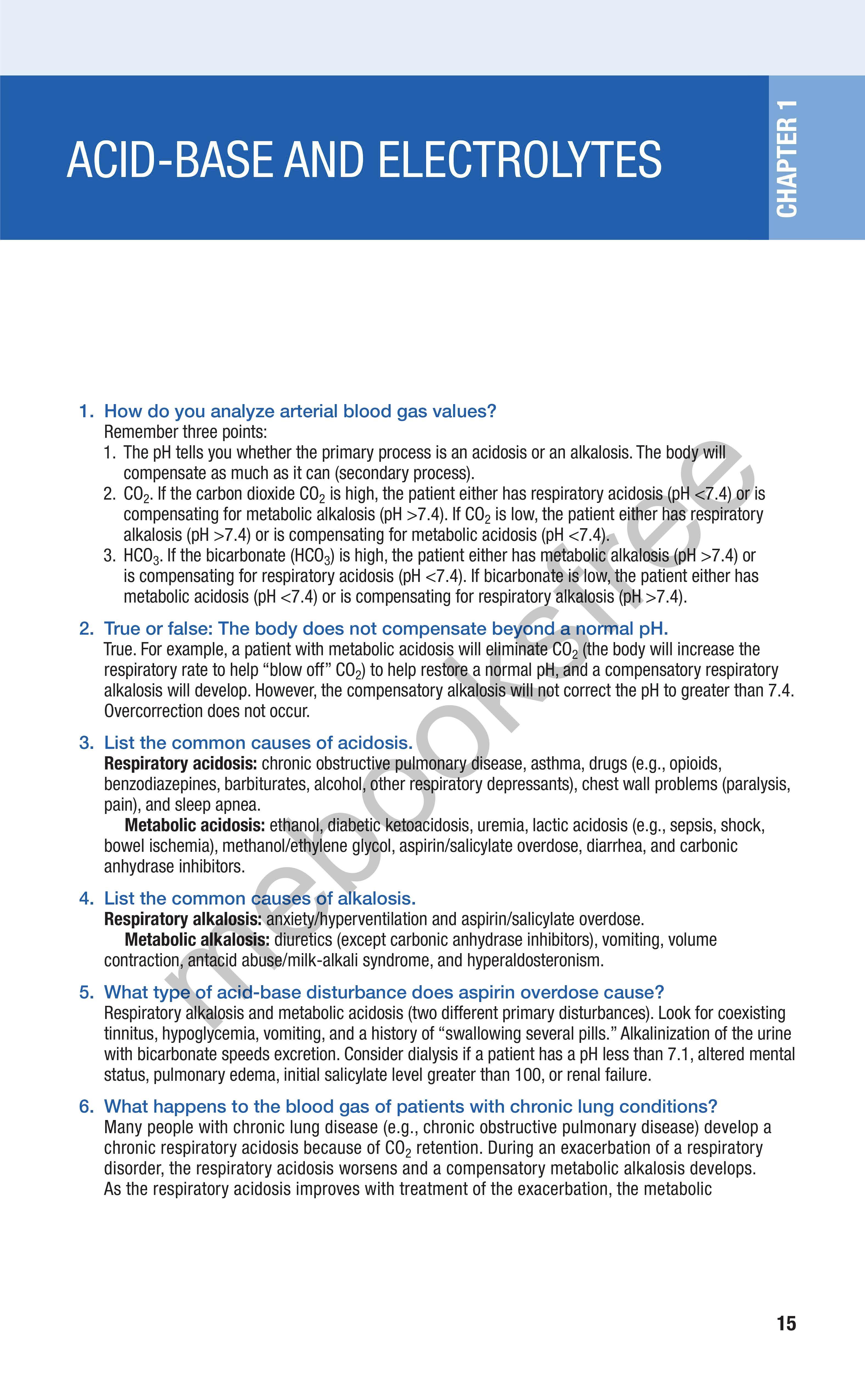
1. How do you analyze arterial blood gas values?
Remember three points:
1. The pH tells you whether the primary process is an acidosis or an alkalosis. The body will compensate as much as it can (secondary process).
2. CO 2 If the carbon dioxide CO 2 is high, the patient either has respiratory acidosis (pH <7.4) or is compensating for metabolic alkalosis (pH >7.4). If CO 2 is low, the patient either has respiratory alkalosis (pH >7.4) or is compensating for metabolic acidosis (pH <7.4).
3. HC0 3 If the bicarbonate (HC0 3) is high, the patient either has metabolic alkalosis (pH >7.4) or is compensating for respiratory acidosis (pH <7.4). If bicarbonate is low, the patient either has metabolic acidosis (pH <7.4) or is compensating for respiratory alkalosis (pH >7.4).
2. True or false: The body does not compensate beyond a normal pH. True. For example, a patient with metabolic acidosis will eliminate CO 2 (the body will increase the respiratory rate to help "blow off" CO 2) to help restore a normal pH, and a compensatory respiratory alkalosis will develop. However, the compensatory alkalosis will not correct the pH to greater than 7.4. Overcorrection does not occur.
3. List the common causes of acidosis.
Respiratory acidosis: chronic obstructive pulmonary disease, asthma, drugs (e.g., opioids, benzodiazepines, barbiturates, alcohol, other respiratory depressants), chest wall problems (paralysis, pain), and sleep apnea.
Metabolic acidosis: ethanol, diabetic ketoacidosis, uremia, lactic acidosis (e.g., sepsis, shock, bowel ischemia), methanol/ethylene glycol, aspirin/salicylate overdose, diarrhea, and carbonic anhydrase inhibitors.
4. List the common causes of alkalosis.
Respiratory alkalosis: anxiety/hyperventilation and aspirin/salicylate overdose.
Metabolic alkalosis: diuretics (except carbonic anhydrase inhibitors), vomiting, volume contraction, antacid abuse/milk-alkali syndrome, and hyperaldosteronism.
5. What type of acid-base disturbance does aspirin overdose cause?
Respiratory alkalosis and metabolic acidosis (two different primary disturbances). Look for coexisting tinnitus, hypoglycemia, vomiting, and a history of "swallowing several pills." Alkalinization of the urine with bicarbonate speeds excretion. Consider dialysis if a patient has a pH less than 7.1, altered mental status, pulmonary edema, initial salicylate level greater than 100, or renal failure.
6. What happens to the blood gas of patients with chronic lung conditions?
Many people with chronic lung disease (e.g., chronic obstructive pulmonary disease) develop a chronic respiratory acidosis because of CO 2 retention. During an exacerbation of a respiratory disorder, the respiratory acidosis worsens and a compensatory metabolic alkalosis develops. As the respiratory acidosis improves with treatment of the exacerbation, the metabolic
alkalosis is no longer a compensatory mechanism and becomes a primary disturbance. However, in certain people with chronic lung conditions (especially those with sleep apnea), pH may be alkaline during the day because breathing improves when awake. As a side note, remember that sleep apnea, like other chronic lung diseases, can cause right-sided heart failure (cor pulmonale).
7. Should you give bicarbonate to a patient with acidosis?
For purposes of Step 2, almost never. First, give intravenous (IV) fluids, and treat the underlying disorder. If all other measures fail and the pH remains less than 7.0, bicarbonate may be given.
8. The blood gas of a patient with asthma has changed from alkalotic to normal, and the patient seems to be sleeping. Is the patient ready to go home?
For Step 2, this scenario means that the patient is probably crashing. Asthmatic patients are supposed to be slightly alkalotic during an asthma attack. Remember that pH is initially high in patients with an asthma exacerbation because they are breathing rapidly, eliminating CO2, and developing a metabolic alkalosis. If the patient becomes tired and breathing slows, CO2 will begin to rise and pH will begin to normalize. Eventually the patient becomes acidotic and requires emergency intubation if appropriate measures are not taken. If this scenario is mentioned on boards, the appropriate response is to prepare for possible elective intubation and to continue aggressive medical treatment with beta2-agonists, steroids, and oxygen. Fatigue secondary to work of breathing is an indication for intubation.
9. List the signs and symptoms of hyponatremia.
• Lethargy
• Seizures
• Mental status changes or confusion
• Cramps
• Anorexia
• Coma
10. How do you determine the cause of hyponatremia?
The first step in determining the cause is to look at the volume status:
HYPOVOLEMIC
Think of Dehydration, diuretics, diabetes, Addison disease/hypoaldosteronism (high potassium)
EUVOLEMIC HYPERVOLEMIC
SIADH, psychogenic polydipsia, oxytocin use
SIADH, Syndrome of inappropriate antidiuretic hormone secretion
11. How is hyponatremia treated?
Heart failure, nephrotic syndrome, cirrhosis, toxemia, renal failure
For hypovolemic hyponatremia, the Step 2 treatment is normal saline. Euvolemic and hypervolemic hyponatremia are treated with water/fluid restriction; diuretics may be needed for hypervolemic hyponatremia.
12. What medication is used to treat SIADH if water restriction fails? Demeclocycline, which induces nephrogenic diabetes insipidus.
13. What happens if hyponatremia is corrected too quickly?
You may cause osmotic demyelination syndrome (ODS, formerly called central pontine myelinolysis) which can result in irreversible or only partially reversible symptoms such as dysarthria, paresis, behavioral disturbances, lethargy, confusion, and coma. Hypertonic saline is used only when a patient has seizures from severe hyponatremia—and even then, only briefly and cautiously. Normal saline is a better choice 99% of the time for board purposes. In chronic severe symptomatic hyponatremia, the rate of correction should not exceed 0.5–1 mEq/L/hr.
14. What causes spurious (false) hyponatremia?
• Hyperglycemia (once glucose is >200 mg/dL, sodium decreases by 1.6 mEq/L for each rise of 100 mg/dL in glucose). Make sure you know how to make this correction.
• Hyperproteinemia


