Solving Urban Infrastructure Problems Using Smart City Technologies: Handbook on Planning, Design, Development, and Regulation 1st Edition John R. Vacca
No part of this publication may be reproduced or transmitted in any form or by any means, electronic or mechanical, including photocopying, recording, or any information storage and retrieval system, without permission in writing from the Publisher. Details on how to seek permission, further information about the Publisher’s permissions policies and our arrangements with organizations such as the Copyright Clearance Center and the Copyright Licensing Agency, can be found at our website: www.elsevier.com/permissions
This book and the individual contributions contained in it are protected under copyright by the Publisher (other than as may be noted herein).
Notices
Knowledge and best practice in this field are constantly changing. As new research and experience broaden our understanding, changes in research methods, professional practices, or medical treatment may become necessary.
Practitioners and researchers must always rely on their own experience and knowledge in evaluating and using any information, methods, compounds, or experiments described herein. In using such information or methods they should be mindful of their own safety and the safety of others, including parties for whom they have a professional responsibility.
With respect to any drug or pharmaceutical products identified, readers are advised to check the most current information provided (i) on procedures featured or (ii) by the manufacturer of each product to be administered, to verify the recommended dose or formula, the method and duration of administration, and contraindications. It is the responsibility of practitioners, relying on their own experience and knowledge of their patients, to make diagnoses, to determine dosages and the best treatment for each individual patient, and to take all appropriate safety precautions.
To the fullest extent of the law, neither the Publisher nor the authors, contributors, or editors, assume any liability for any injury and/or damage to persons or property as a matter of products liability, negligence or otherwise, or from any use or operation of any methods, products, instructions, or ideas contained in the material herein.
Library of Congress Cataloging-in-Publication Data
Names: Fitzpatrick, James E., 1948- author. | High, Whitney A., author. | Kyle, W. Lamar, author.
Title: Urgent care dermatology : symptom-based diagnosis / James E. Fitzpatrick, Whitney A. High, W. Lamar Kyle.
Description: Philadelphia, PA : Elsevier, [2018] | Includes bibliographical references and index.
Identifiers: LCCN 2017019068 | ISBN 9780323485531 (pbk. : alk. paper)
Classification: LCC RL74 | NLM WR 141 | DDC 616.5—dc23 LC record available at https://lccn.loc.gov/2017019068
Senior Content Strategist: Charlotta Kryhl
Director, Content Development: Rebecca Gruliow
Publishing Services Manager: Patricia Tannian
Senior Project Manager: Sharon Corell
Book Designer: Bryan Salisbury
Printed in Canada. Last digit is the print number: 9 8 7 6 5 4 3 2 1
This book is dedicated to my sons, Jeff and Josh -JEF and Misha, Madison, and Morgan – “my three M’s” -WAH
Preface
“Everything should be made as simple as possible, but not simpler.”
ALBERT EINSTEIN
Urgent Care Dermatology: Symptom-Based Diagnosis had its genesis on a bass fishing trip on the Arkansas River in 2000. Lamar Kyle, a private practice physician, who had worked in an emergency room and urgent care clinic environment for decades, complained to me, an academic dermatologist, that dermatology textbooks were not organized in a useful fashion. He opined that dermatology books for a primary care provider needed to be organized by physical findings and not grouped by pathogenesis, needed copious photographs, and needed written text to be short and to the point. From this pivotal conversation, this textbook was born.
We caught very few fish, but when I returned home, I started outlining the book and experimenting with formats. For years, the book was something that I worked on early in the morning before the family woke up and on planes when traveling. After 7 years, the book had begun to take shape and was about two-thirds finished when a simultaneous computer disaster on two different computers containing the original and backup versions of the book brought the project to a halt. An older version of the manuscript on a third computer was found, but more than 100 completed pages of the manuscript were absent in this
dated version. Disheartened, my interest in returning to work on the manuscript lagged, and the venture lay in limbo. The project was brought back to life by my energetic colleague, Whitney High, who worked with me in the Department of Dermatology at the University of Colorado. He offered to help write portions of the book and bring the book into a publishable state.
The concept of the book was simple. First, the dermatologic diseases needed to be organized by physical findings and not by pathogenesis. Whereas this would on the surface appear to be easy to do, it was difficult because we were using a morphologic model that is rarely utilized in dermatology textbooks. Second, cutaneous disorders that had a significant mortality rate or that were more likely to present urgently to a primary care provider were emphasized, and many trivial or genetic disorders were excluded. Third, the provided text, with very few exceptions, had to be kept to a single page. Finally, the book had to have numerous quality photographic examples of each entity.
We hope that we have succeeded in our objectives and that this will be a book that is actually used in the clinical setting while the practitioner is actively seeing patients, and not a book that sits on the shelf gathering dust.
James E. Fitzpatrick, MD Aurora, Colorado
Acknowledgments
The authors would like to thank the outstanding team at Elsevier for their continued effort, support, and patience. Whereas there were many contributors from Elsevier, we would like to especially acknowledge Jim Merritt, Executive Content Strategist, and Suzanne Toppy, Senior Content Strategist, who were critical in getting the initial approval for this textbook. Once the textbook was started, we principally worked with Sarah Barth, Senior Content Strategist, and especially Rebecca “Becca” Gruliow, Director, Content Development. Becca was fantastic in guiding us through the editorial process and deserves special recognition for her patience, diligence, and promptness, and for
just being a great individual to work with. In the final stages of this book, we would like to especially acknowledge the crucial role of of Sharon Corell, our Senior Project Manager, who we worked with on an almost daily basis.
The senior author (JEF) would especially like to acknowledge Linda Belfus, Senior Vice President of Content, for her role in not just this book but for other projects and for bringing us into the Elsevier family. I have worked with Linda for more than 20 years. Linda retired near the end of the editorial process, and I wish her happiness in her retirement.
Chapter 1 Introduction to Clinical Dermatology
“THINKING” LIKE A DERMATOLOGIST
The skin represents the largest organ of the human body. It consists of the epithelium, dermis, subcutaneous fat, and adnexal structures (hair follicles and glands), as well as supportive structures (blood vessels and nerves), all of which function to protect and maintain homeostasis.
Dermatology is a field of medicine that focuses on the skin, adjacent mucosa (oral and genital), and other adnexal structures (e.g., hair, nails, and sweat glands). Undoubtedly, dermatologists are the most adroit diagnosticians with regard to skin disease. However, much of their acumen comes from pattern recognition, a cultivated appreciation for diagnostic subtleties, and trained recognition of historical factors that make one particular disease more likely than another.
Thus, any clinician may improve when diagnosing and treating skin ailments simply by learning to think like a dermatologist. This includes fostering an appreciation for the classification schemes used in dermatology and for learning descriptive terminologies used by dermatologists.
ETIOLOGIC PREMISES
With regard to etiology, one of the most basic branch points in dermatology is to decide if a skin condition is neoplastic (benign or malignant) or inflammatory (e.g., rash, infection, autoimmune condition). Although it is likely that inflammatory conditions will prevail in urgent care and emergency settings, one must realize that a patient may present to these types of clinics with a neoplasm that has been ignored too long, until it can no longer be neglected.
Moreover, on occasion, there is visual and conceptual overlap with regard to inflammatory versus neoplastic conditions. For example, mycosis fungoides, the most common form of cutaneous T cell lymphoma, is a clonal lymphoproliferative disorder (a neoplasm), yet its clinical presentation resembles that of an inflammatory disorder. Conversely, sarcoidosis is an inflammatory condition that may present with discrete nodular lesions that mimic those of a neoplasm.
MORPHOLOGY
In dermatology, the term morphology refers to the appearance of a skin lesion(s), irrespective of the underlying pathophysiology. For example, a small blister is referred to as a vesicle, whether it is due to
an infection (e.g., herpes simplex) or autoimmune condition (e.g., bullous pemphigoid).
Therefore, it is important to use correct morphologic terms to classify skin diseases. This is because these terms represent a native language, or lexicon, that allows professionals to describe skin disease in a consistent manner.
There are primary morphologic terms (Table 1.1), which refer to the characteristic appearance of skin lesions (e.g., papule), and secondary morphologic terms (Table 1.2), which are used in addition to primary morphologic terms. These secondary morphologic terms reflect exogenous factors or temporal changes that evolve during the course of a skin disease.
PALPATION AND APPRECIATION OF TEXTURE
Textural changes are appreciated by palpating the skin; doing so may provide diagnostic clues. For example, macules and patches are flush with the surrounding skin, whereas papules and plaques are palpable. Some forms of connective tissue disease, such as morphea and scleroderma, may cause induration (firmness) of the skin, even when visual findings are limited. Finally, purpura (hemorrhage into the skin) may be palpable or nonpalpable, which implies a different underlying pathology. Small-vessel vasculitis causes palpable purpura, whereas simple bruising causes macular nonpalpable purpura.
COLOR
Skin color provides important clues about the nature of a disease process. For example, many skin lesions appear erythematous (red). However, it is important to ascertain whether the erythema is blanching, which disappears with pressure, because this suggests that the erythema is due to vasodilation, or whether it is nonblanching erythema (purpura), which implies hemorrhage into the skin. Other causes of pigment in the skin include hypoxia, topical medications, oral drugs, other ingestants, or even infections.
Also, there is variation in normal skin tones, even in the general population. These variations in skin color are due to differences in the amount and distribution of melanin in the epidermis. Sometimes, the term skin of color is used to describe any skin tone darker than white skin, but dermatologists more often use the Fitzpatrick scale (see “Fitzpatrick Skin Types”
TABLE 1.1 Primary Morphologic Terms
Morphologic Term Salient Features
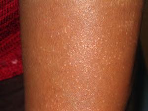
Macule (or patch)
• Flat lesion
<1 cm in diameter (macule)
>1 cm in diameter (patch)
• Circumscribed
• Color change that cannot be appreciated by tactile sensation alone
Papule
• Elevated lesion
• Usually <1 cm in diameter
• Often with other secondary features (e.g., scale, crust)
Classic Disease Image
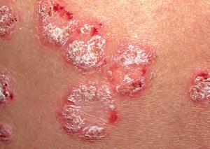
Plaque
• Elevated lesion
• Usually >1 cm in diameter
• Nonvesicular
• Often with other secondary features (e.g., scale, crust)
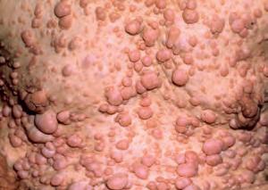
Nodule
• Large elevated lesion
• Usually ≥2 cm in diameter
• Involves the dermis and may extend into the subcutis
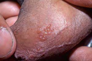
Vesicle
• Small elevated lesion
• <1 cm in diameter
• Filled with clear fluid
• Circumscribed
Classic Diagnoses
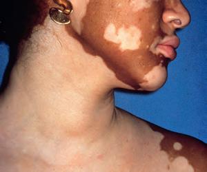
• Vitiligo (photo)
• Café-au-lait spot
• Flat component of exanthems (measles)
• Freckle
• Lentigo
• Lichen nitidus (photo)
• Elevated component of exanthems (measles)
• Melanocytic nevi
• Verruca or molluscum
• Lichen planus
• Guttate psoriasis
• Psoriasis vulgaris (photo)
• Lichen simplex chronicus
• Eczematous plaques
• Granuloma annulare
• Sarcoidosis
• Neurofibromata (photo)
• Basal cell carcinoma
• Cutaneous lymphoma
• Erythema nodosum
• Lipoma
• Herpes simplex infection (photo)
• Varicella zoster infection
• Dermatitis herpetiformis
TABLE 1.1 Primary Morphologic Terms—cont’d
Morphologic Term Salient Features
Bulla • Elevated lesion
• Usually >1 cm in diameter
• Filled with clear fluid
• Circumscribed
Pustule • Elevated lesion
• Usually <1 cm in diameter
• Filled mainly with purulent fluid
• Circumscribed
Classic Disease Image
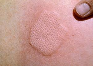
Wheal
• Firm edematous papule or plaque
• Evanescent
• Pruritic
Fitzpatrick Skin Types
Type I Always burns, never tans
Type II Usually burns, then tans
Type III May burn, tans well
Type IV Rarely burns, tans well
Type V Very rarely burns, tans well, brown skin
Type VI Very rarely burns, tans well, dark brown skin
box), which describes skin color based on a response to sun exposure.
Baseline pigmentation also affects cutaneous findings in skin disorders. For example, erythema may be difficult to appreciate in darker skin. Keloids (aberrant scarring) are more common in those with darker skin types. Even after a disease process has resolved, postinflammatory hypopigmentation and postinflammatory hyperpigmentation are more marked (or more evident) in those with darker skin types.
Classic Diagnoses
• Epidermolysis bullosa (photo)
• Bullous drug eruption
• Bullous pemphigoid
• Linear immunoglobulin A disease
• Pemphigus
• Porphyria
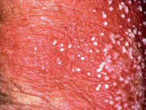
• Pustular psoriasis (photo)
• Acute generalized exanthematous pustulosis
• Candidiasis
• Folliculitis
• Subcorneal pustular dermatosis
• Urticaria (photo)
• Dermatographism
• Urticaria pigmentosa
Cyanosis is also more difficult to appreciate when the skin is more darkly pigmented.
CONFIGURATION AND DISTRIBUTION
A dermatologist must always analyze two closely related properties—configuration and distribution— to find the correct diagnosis. The configuration, or arrangement of skin lesions, includes descriptors such as linear, annular, arciform, clustered, reticulated, dermatomal, and retiform. For example, pruritic and fragile vesicles, arranged in clusters on the elbows and knees, should prompt consideration of dermatitis herpetiformis. Grouped vesicles on an erythematous base, but confined to a single dermatome, mandates consideration of herpes zoster.
Assessment of distribution includes stepping back and observing the anatomic pattern of skin lesions on the body. For example, plaques of psoriasis often favor extensor surfaces—elbows and knees; atopic dermatitis often favors flexural surfaces in older children and adults (antecubital and popliteal fossae), and lichen planus favors other flexural surfaces (wrists and
TABLE 1.2 Morphologic Terms for Secondary Skin Lesions
• Partial loss of the epidermis and superficial dermis
• Often linear
• May be associated with scars from old excoriations
• Vertical loss of epidermis and dermis
• Sharply defined walls (crack)
ankles). It is also important simply to notice if the condition is focal or generalized, unilateral or bilateral, and sharply circumscribed or more difficult to perceive. These factors will ultimately shape the final differential diagnosis.
Terminology that is encountered frequently in dermatology includes discussion of a seborrheic
Classic Diagnoses
• Impetigo (photo)
• Resolving dermatitis
• Resolving vesiculobullous disorders
• Secondary finding in many bullous lesions that have broken (photo depicts pemphigus)
distribution, which includes the head, neck, and upper trunk, areas with a high density of sebaceous oil glands, and use of the term photodistributed, which describes an accentuation in areas exposed to ultraviolet light (e.g., the face, parts of the neck outside of the natural shadow beneath the chin, upper chest, forearms, dorsal hands). Connective tissue disease and
some drug-induced processes may show a photodistributed pattern.
TEMPORAL COURSE
The temporal course is central to any medical history, and dermatology is no exception. All patients presenting with skin lesions should be queried about duration and any change in intensity or distribution over time. For example, some inflammatory conditions, such as measles, pityriasis rubra pilaris, and dermatomyositis, have a notorious cephalocaudal progression over time.
However, the examiner is always at an advantage because the skin is so readily accessible. Any information provided by the patient can be readily compared to observations made on physical examination.
Armed with some basic knowledge of the skin, the clinician can usually determine whether cutaneous lesions are acute, subacute, or chronic by observation. For example, because of the relative transit times of proliferating skin, true scale (not to be confused with crust) requires 2 weeks to develop. Similarly, lichenification, or thickening of the skin, with accentuation of normal skin markings, takes weeks or even months to develop. Therefore, if scale or lichenification is apparent, then the skin problem has been present for more than a few days, no matter what the patient may believe or has noticed.
OTHER HISTORICAL INFORMATION
A complete discourse on the pertinent aspects of history cannot be adequately addressed in this short introduction. Moreover, the finer details are better learned in association with specific diseases. Suffice it to say that there is always a need to take a good history regarding when, where, what, who, and how. The patient may be asked the following questions:
When did the rash (or lesion) develop, or for how long do you remember having the condition?
• When have you had similar eruptions in the past?
• When have you sought care in the past? Where were the first lesions on your body?
• Where did the condition progress to next on the body?
• Where were you when you first noticed the condition (e.g., traveling, at home, at work)?
• Where have you sought care in the past (for records or phone consultation)? What has been done about the condition to date?
• What makes the problem better?
• What makes the problem worse?
• What were you told about the condition by other professionals in the past?
• What was the plan for any follow-up (if it is a known condition)?
• What other relevant past medical histor y do you have?
• What kinds of medical conditions do you see a physician for regularly or take medications for on a regular basis?
Who has weighed in on this problem in the past (e.g., primary care provider, other urgent care or emergency care provider, dermatologist)?
• Who is in charge of your general health care?
• Who would have other information about your skin condition that you would like me to speak with, given your explicit permission? Who can you follow up with regarding this condition (is there a primary care physician [PCP] or dermatologist you have seen, now or in the past)?
How sick is the patient? This is a question an urgent care or emergency provider is uniquely equipped to answer. Perhaps the patient has other issues that take precedence over his or her long-term, noncritical, non–life-threatening skin complaint(s). If so, it may be necessary to acknowledge this skin complaint but defer work until another visit or the patient sees another clinician.
CONCLUSION
In summary, dermatology is a discipline that can definitely be learned. Although one cannot expect to master the entire domain of dermatology to the same degree as a dermatologist, who has completed an entire residency in the subject, any clinician can improve in the approach to patients with skin complaints simply by focusing on the following:
• Etiologic premise. Is this an inflammator y or neoplastic condition?
• Morphology. What morphologic term can be applied to the native lesion(s) of the condition (macular, maculopapular, papulosquamous, vesiculobullous, etc.)? Are there secondary morphologies (excoriated, lichenified, crusted, impetiginized, etc.)?
• Palpation. Are there any textural features, detected by palpation, which may be useful in refining the morphologic descriptors of the lesion(s), such as scaling, crusting, induration, and palpable or nonpalpable purpura?
• Color. What colors are identifiable in association with the condition? If there is erythema present, is it blanching or nonblanching (purpura)? Is the patient’s own background skin tone such that the color of the lesion may be altered or more difficult to perceive?
• Configuration and distribution. What is the configuration of the lesions (linear, annular, reticulated,
retiform, etc.), and how is the condition distributed upon the body (flexural areas, extensor surfaces, photodistributed, etc.)? Is it focal or generalized? Is it unilateral or bilateral? Does it involve mucosal surfaces or not?
• Temporal course. Has the condition evolved over days, weeks, months, or even years?
• Has it changed in its appearance (or its primar y morphology) during that time?
• Was it initially macular or maculopapular and then became bullous?
• Is it waxing and waning over time?
• Is it worsening or improving?
• Other historical information. Clearly, any final questioning and discourse will be affected by the
specific disease categories under consideration. However, it is always important to focus on questioning that is broad enough to describe any items in the general medical history more fully, such as determining when, where, what, who, and how.
Simply thinking like a dermatologist, without learning any additional dermatologic information, will not meaningfully affect the care rendered. If the information in this chapter is kept in mind, particularly as one reads the rest of this text—which will fill in the gaps with regard to basic skin disease—there is little doubt that treating dermatologic ailments will be easier for you, as a clinician, and more rewarding for the patients under your care.
Chapter 2 Diagnostic Techniques
Key Terms
Saucerization biopsy
Tinea corporis
EQUIPMENT NEEDED
This chapter, on diagnostic techniques, not only discusses how to perform the procedures, but also lists the type of equipment needed to perform these techniques.
Purchase of Equipment
There are numerous medical supply houses that supply surgical and diagnostic equipment that can equip an emergency room or an office adequately. Although we do not have any financial interest in this company, we can recommend Delasco, which has a complete line of equipment directed for dermatologists, including anything mentioned in this text.
Core cutaneous surgical equipment
• Scalpel, no 14 blade
• Scalpel handle
• 3-mm disposable punch
• 4-mm disposable punch
• Curved iris scissors
• Suture scissors
• Tissue forceps (e.g., 4-inch iris with teeth)
• Needle holders
Optional cutaneous surgical equipment
• 2-mm disposable punch
• 5-mm disposable punch
• 6-mm disposable punch
• 8-mm disposable punch
• Flexible, double-edged razor blades
Core diagnostic equipment
• Microscope
• Mineral oil
• Mycologic media—Sabouraud agar (plates, tubes or slants)
• 10% to 20% potassium hydroxide (KOH) solution, with or without dimethyl sulfoxide (DMSO)
• Glass slides and coverslips
Optional diagnostic equipment
• Antifungal stains (as alternatives to KOH)
• Chlorazol black fungal stain
• PMS fungal stain
• Swartz Lamkins fungal stain
• Methylene blue Triton X (for Malassezia yeast)
• Dermatophyte test medium (DTM)
• Tzanck stains
• PMS Tzanck stain
• Wright-Giemsa stain
• Wood light (Wood lamp)
WOOD LIGHT
Introduction
The Wood light or Wood lamp emits long-wave UV light (black light) that is filtered by a Wood filter (barium silicate and nickel oxide) that only allows a band of UV light between 320 and 400 nm (peak, 365 nm) to be emitted.
Applications
• Diagnosis of fluorescent species of tinea capitis
• Diagnosis of erythrasma
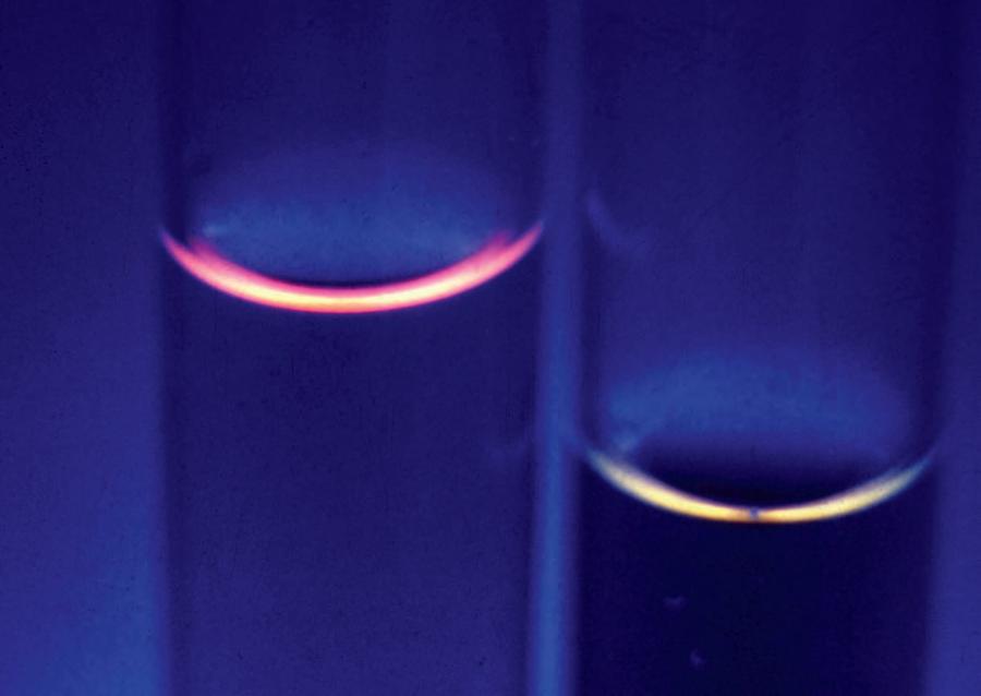
• Examination of porphyrins in urine in porphyrias, such as porphyria cutanea tarda (Fig. 2.1)
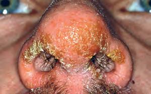
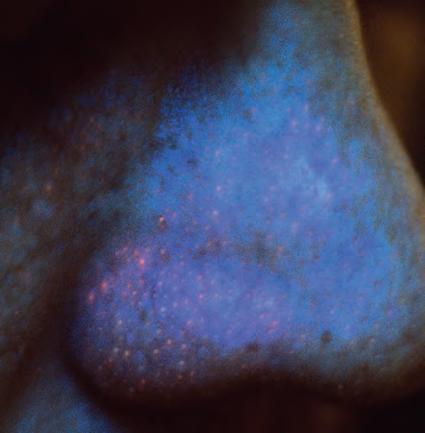
• Examination for Propionibacterium acnes in patients with acne vulgaris (Fig. 2.2)
• Demonstration of hypopigmentation and depigmentation in light-skinned individuals
• Detection of tetracycline deposition in children’s teeth
Supplies
• Wood light (Wood lamp)
• Wood lights are available as small, portable, battery-powered units or as larger units with a cord
Technique
• Completely darken the examination room and allow your eye to accommodate.
• Turn on the lamp and allow it to warm up for 30 to 60 seconds.
• Hold the lamp approximately 4 to 6 inches from examination site.
Interpretation
• A positive examination result for tinea capitis consists of yellow-green or blue-green fluorescence of the broken hair shafts caused by the production of a compound called pteridine. Care must be taken
not to confuse this with fibers and lint from clothing that may fluoresce a white color (due to optical brightening agents) or scale mixed with sebum that may demonstrate a dull yellow color.
• A positive test for erythrasma demonstrates a coral red fluorescence confined to the areas of infection. The color is due to a water-soluble porphyrin (coproporphyrin III) produced by the bacteria Corynebacterium minutissimum. The porphyrins are water-soluble and, if the patient has recently bathed, the test will be negative because the fluorescent porphyrins will be washed out.
• In acne vulgaris, untreated patients will demonstrate follicular coral red fluorescence. In treated patients, if fluorescence is still present, the patient is not using the medication or the Propionibacterium acnes is now resistant to the antibiotics.
• A positive test for urinary porphyrins is the demonstration of coral red fluorescence in the urine. If the amount of uroporphyrins is low, only the meniscus will demonstrate the color, but if the quantitative porphyrin level is high, the entire sample will demonstrate coral red fluorescence.
• Pseudomonas infections show a greenish fluorescence due to the pigment pyoverdin.
• Hypopigmented or depigmented skin in fairskinned individuals is accentuated with a Wood light.
• False-negative results may occur in acne vulgaris and erythrasma if the patient has recently bathed.
• False-positive fluorescent substances include some cosmetics, deodorants, soaps, lint, and petrolatum.
Comments
The Wood light has severe limitations in the diagnosis of tinea capitis because most infections in the United States are due to nonfluorescent species, especially Trichophyton tonsurans. The only species that is commonly fluorescent in the United States is Microsporum canis, which only accounts for about 5% to 10% of cases.
Fig. 2.1. Wood light examination of urine demonstrating coral red fluorescence in a patient with porphyria cutanea tarda. Fluorescence can be accentuated by adding dilute hydrochloric acid. Left, Positive. Right, Negative. (From the Fitzsimons Army Medical Center Collection, Aurora, CO.)
Fig. 2.2. Wood light examination of a patient’s nose demonstrating coral red porphyrins produced by Propionibacterium acnes. Because this patient is under treatment for acne, this finding indicates that the patient is not taking the antibiotic or the organism is resistant to the antibiotic. (From the Fitzsimons Army Medical Center Collection, Aurora, CO.)
POTASSIUM HYDROXIDE PREPARATION
Applications
• Diagnosis of dermatophytes and yeast infections of the skin and mucous membranes
Supplies
• Microscope glass slide
• Coverslip (any size)
• No. 14 or 15 scalpel blade
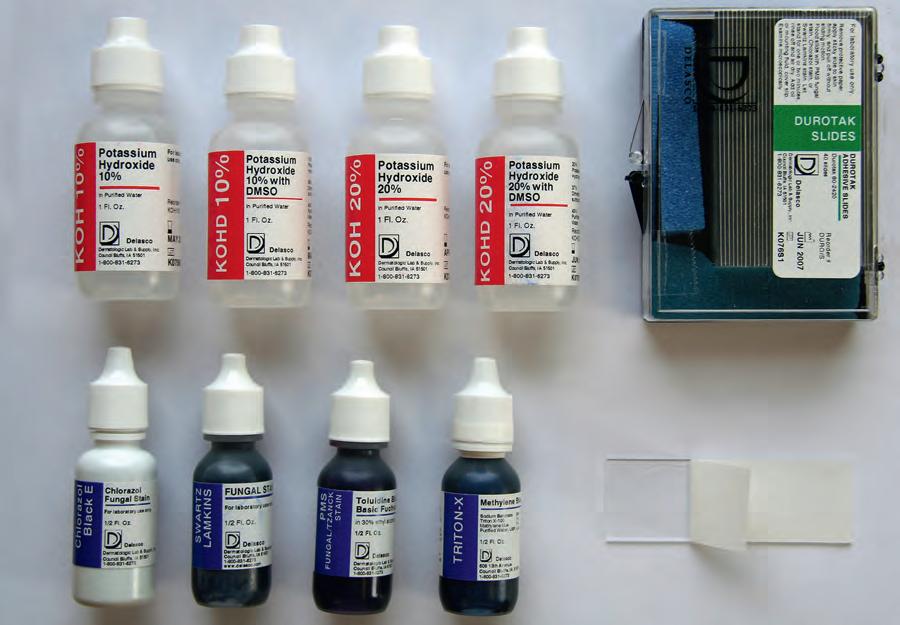
• KOH, 10% to 20% concentration (Fig. 2.3). Variants include the following:
• KOH, 10% to 20%, with DMSO
• KOH, 10%, with DMSO and chlorazol black
• Access to a microscope
Technique
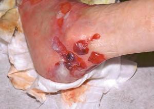
• For tinea corporis, the best samples are from annular edges with scale.
• Wet the area with an alcohol pad or, if in a sensitive area, with water.
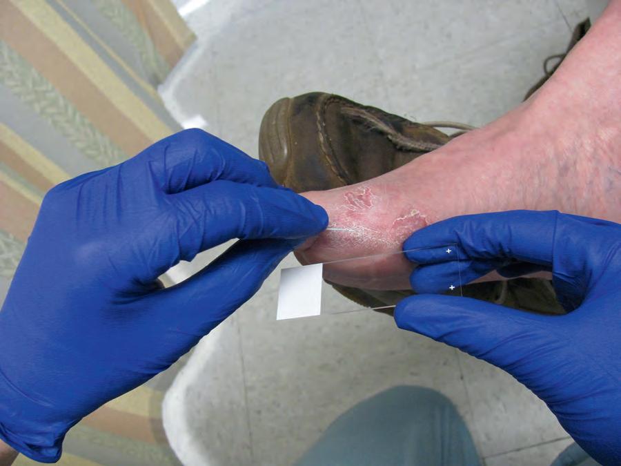
• Gently scrape the site (you should not draw blood) and gather as much scale on the edge of the scalpel blade as possible. It is best to do this on more than one site to ensure that you have enough material (Fig. 2.4).
• Transfer the material on the edge of the scalp to the glass slide and spread it across the slide.
Warning!
Be very careful not to get the KOH on the microscope objectives because KOH can permanently etch the lens. Obviously, anything that will etch glass should not come into contact with the skin of patients, the staff, or yourself.
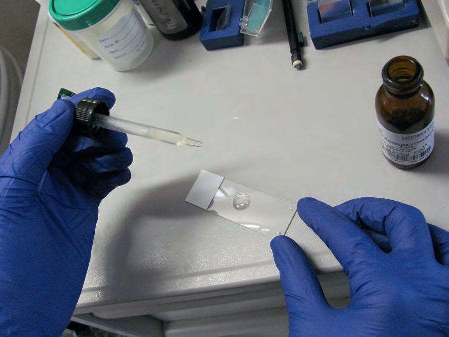
• Apply one or two drops of KOH to the slide (Fig. 2.5) and coverslip.
• If the coverslip is not even, gently press with your finger.
• Blot any excess KOH at the edges of the coverslip.
• If DMSO is used, wait for 1 minute to examine.
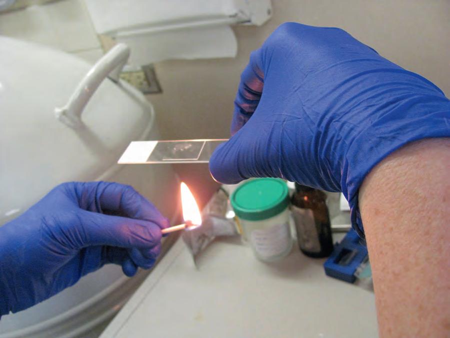
• If DMSO is not used, gently heat (do not boil) the glass slide to enhance clearing of the keratinocytes (Fig. 2.6).
• The hyphae are easier to visualize if the condenser is dropped. The amount that it needs to be dropped is variable and depends on the amount of light used and thickness of the specimen.
Interpretation
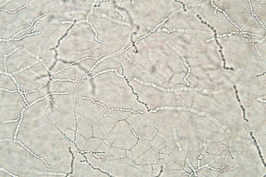
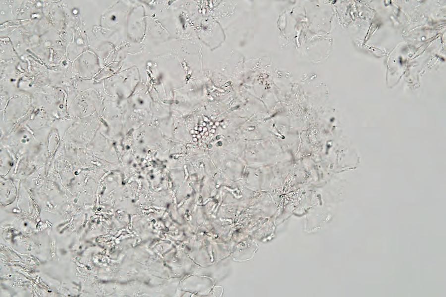
• A positive examination consists of demonstrating hyphae (dermatophyte infections), short hyphae and yeast (tinea versicolor), or yeast (candidiasis; Figs. 2.7 and 2.8). Other fungal species are only rarely seen on the skin or skin appendages.
• Fragments of clothing fibers and refractile material between the keratinocyte walls may lead to confusion. The false hyphae-like structures seen between keratinocytes can be differentiated by their angulated appearance, lack of linear shape for long distances, and tendency to break up. They also do not demonstrate nuclei that can sometimes be seen in hyphae.
• Epithelial lipids and exogenous lipids (e.g., moisturizers) may be difficult to differentiate from yeast. In general, they are more irregular in shape and lack nuclei. In some cases, there is so much exogenous lipid that it is almost impossible to interpret the specimen.
Comments
• Performing KOH examinations of the skin and mucous membrane samples is increasingly becoming a lost art because of Clinical Laboratory Improvement Amendments (CLIA) regulations. However, for those who carry out a KOH examination, or learn how to do it, it is frequently the quickest and is often the more sensitive test when compared to cultures. KOH examinations of mucosal scrapings are easier to interpret than scrapings from skin, hair, and nails, and some health care providers may want to limit this study for mucosal lesions.
Fig. 2.4. After wetting the skin of a patient with suspected tinea pedis, the area is gently scraped with a no. 15 scalpel blade. Because the stratum corneum (scale) is the material of interest, the procedure should not draw blood.
Fig. 2.3. Top row, Fungal KOH (with and without DMSO). Bottom row, Four different types of fungal stains. Far right, Special glass slides with adhesive tape used for the diagnosis of tinea versicolor.
2.5. A single drop of KOH is placed on the scale and then cover-slipped. Note that the physician is wearing gloves— remember that KOH is toxic to the skin.
2.6. After being cover-slipped, the KOH preparation is gently heated over a flame. If the KOH contains DMSO, this step can often be omitted. After heating, the slide is ready to be examined under the microscope.
Fig.
Fig.
2.7. Positive KOH finding of patient with tinea pedis demonstrating numerous linear segmented hyphae. The thin lines represent the cell walls of the keratinocytes (KOH, 400×).
2.8. Positive KOH finding of patient with tinea versicolor demonstrating numerous refractile, clumped yeast and short hyphae (so-called spaghetti and meatballs) (KOH, 400×).