https://ebookmass.com/product/urgent-care-dermatology-
Instant digital products (PDF, ePub, MOBI) ready for you
Symptom to Diagnosis An Evidence Based Guide, Fourth Edition
https://ebookmass.com/product/symptom-to-diagnosis-an-evidence-basedguide-fourth-edition/
ebookmass.com
Nelson Pediatric Symptom-Based Diagnosis 2nd Edition
Robert M. Kliegman
https://ebookmass.com/product/nelson-pediatric-symptom-baseddiagnosis-2nd-edition-robert-m-kliegman/
ebookmass.com
Symptom to Diagnosis: An Evidence Based Guide 4th Edition
Scott D.C. Stern
https://ebookmass.com/product/symptom-to-diagnosis-an-evidence-basedguide-4th-edition-scott-d-c-stern/
ebookmass.com
International Taxation Adnan Islam
https://ebookmass.com/product/international-taxation-adnan-islam/
ebookmass.com
Lunchtime Chronicles: Caribbean Jerk Dahlia Rose & Lunchtime Chronicles
https://ebookmass.com/product/lunchtime-chronicles-caribbean-jerkdahlia-rose-lunchtime-chronicles/
ebookmass.com
Not Alone Sarah K Jackson
https://ebookmass.com/product/not-alone-sarah-k-jackson/
ebookmass.com
Solving Urban Infrastructure Problems Using Smart City Technologies: Handbook on Planning, Design, Development, and Regulation 1st Edition John R. Vacca
https://ebookmass.com/product/solving-urban-infrastructure-problemsusing-smart-city-technologies-handbook-on-planning-design-developmentand-regulation-1st-edition-john-r-vacca/ ebookmass.com
Sunshine and Sammy (Vested Interest: ABC Corp Book 5) Melanie Moreland
https://ebookmass.com/product/sunshine-and-sammy-vested-interest-abccorp-book-5-melanie-moreland/
ebookmass.com
Computer Aided Drug Design (CADD): From Ligand-Based Methods to Structure-Based Approaches Mithun Rudrapal
https://ebookmass.com/product/computer-aided-drug-design-cadd-fromligand-based-methods-to-structure-based-approaches-mithun-rudrapal/
ebookmass.com
The Devils and The Duchess: A Dark College Bully Romance
(Verona Falls University Book 1) Marissa Farrar & S.R. Jones
https://ebookmass.com/product/the-devils-and-the-duchess-a-darkcollege-bully-romance-verona-falls-university-book-1-marissa-farrar-sr-jones/
ebookmass.com
This book is dedicated to my sons, Jeff and Josh -JEF and Misha, Madison, and Morgan – “my three M’s” -WAH
Acknowledgments
The authors would like to thank the outstanding team at Elsevier for their continued effort, support, and patience. Whereas there were many contributors from Elsevier, we would like to especially acknowledge Jim Merritt, Executive Content Strategist, and Suzanne Toppy, Senior Content Strategist, who were critical in getting the initial approval for this textbook. Once the textbook was started, we principally worked with Sarah Barth, Senior Content Strategist, and especially Rebecca “Becca” Gruliow, Director, Content Development. Becca was fantastic in guiding us through the editorial process and deserves special recognition for her patience, diligence, and promptness, and for
just being a great individual to work with. In the final stages of this book, we would like to especially acknowledge the crucial role of of Sharon Corell, our Senior Project Manager, who we worked with on an almost daily basis.
The senior author (JEF) would especially like to acknowledge Linda Belfus, Senior Vice President of Content, for her role in not just this book but for other projects and for bringing us into the Elsevier family. I have worked with Linda for more than 20 years. Linda retired near the end of the editorial process, and I wish her happiness in her retirement.
TABLE 1.1 Primary Morphologic Terms
Morphologic Term Salient Features
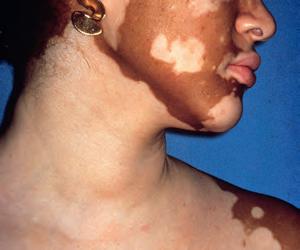
Macule (or patch)
• Flat lesion
<1 cm in diameter (macule)
>1 cm in diameter (patch)
• Circumscribed
• Color change that cannot be appreciated by tactile sensation alone
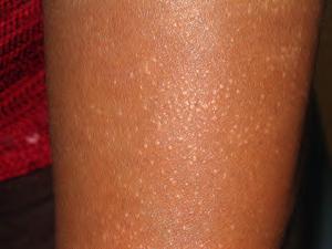
Papule
• Elevated lesion
• Usually <1 cm in diameter
• Often with other secondary features (e.g., scale, crust)
Classic Disease Image
Plaque
• Elevated lesion
• Usually >1 cm in diameter
• Nonvesicular
• Often with other secondary features (e.g., scale, crust)
Nodule
• Large elevated lesion
• Usually ≥2 cm in diameter
• Involves the dermis and may extend into the subcutis
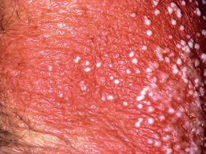
Vesicle
• Small elevated lesion
• <1 cm in diameter
• Filled with clear fluid
• Circumscribed
Classic Diagnoses
• Vitiligo (photo)
• Café-au-lait spot
• Flat component of exanthems (measles)
• Freckle
• Lentigo
• Lichen nitidus (photo)
• Elevated component of exanthems (measles)
• Melanocytic nevi
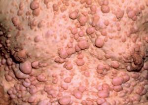
• Verruca or molluscum
• Lichen planus
• Guttate psoriasis
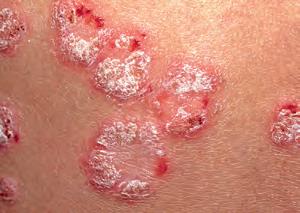
• Psoriasis vulgaris (photo)
• Lichen simplex chronicus
• Eczematous plaques
• Granuloma annulare
• Sarcoidosis
• Neurofibromata (photo)
• Basal cell carcinoma
• Cutaneous lymphoma
• Erythema nodosum
• Lipoma
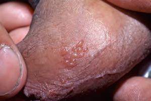
• Herpes simplex infection (photo)
• Varicella zoster infection
• Dermatitis herpetiformis
TABLE 1.1 Primary Morphologic Terms—cont’d
Morphologic Term Salient Features
Bulla • Elevated lesion
• Usually >1 cm in diameter
• Filled with clear fluid
• Circumscribed
Pustule • Elevated lesion
• Usually <1 cm in diameter
• Filled mainly with purulent fluid
• Circumscribed
Classic Disease Image
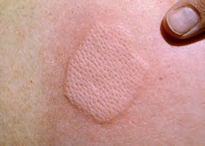
Wheal
• Firm edematous papule or plaque
• Evanescent
• Pruritic
Fitzpatrick Skin Types
Type I Always burns, never tans
Type II Usually burns, then tans
Type III May burn, tans well
Type IV Rarely burns, tans well
Type V Very rarely burns, tans well, brown skin
Type VI Very rarely burns, tans well, dark brown skin
box), which describes skin color based on a response to sun exposure.
Baseline pigmentation also affects cutaneous findings in skin disorders. For example, erythema may be difficult to appreciate in darker skin. Keloids (aberrant scarring) are more common in those with darker skin types. Even after a disease process has resolved, postinflammatory hypopigmentation and postinflammatory hyperpigmentation are more marked (or more evident) in those with darker skin types.
Classic Diagnoses
• Epidermolysis bullosa (photo)
• Bullous drug eruption
• Bullous pemphigoid
• Linear immunoglobulin A disease
• Pemphigus
• Porphyria
• Pustular psoriasis (photo)
• Acute generalized exanthematous pustulosis
• Candidiasis
• Folliculitis
• Subcorneal pustular dermatosis
• Urticaria (photo)
• Dermatographism
• Urticaria pigmentosa
Cyanosis is also more difficult to appreciate when the skin is more darkly pigmented.
CONFIGURATION AND DISTRIBUTION
A dermatologist must always analyze two closely related properties—configuration and distribution— to find the correct diagnosis. The configuration, or arrangement of skin lesions, includes descriptors such as linear, annular, arciform, clustered, reticulated, dermatomal, and retiform. For example, pruritic and fragile vesicles, arranged in clusters on the elbows and knees, should prompt consideration of dermatitis herpetiformis. Grouped vesicles on an erythematous base, but confined to a single dermatome, mandates consideration of herpes zoster.
Assessment of distribution includes stepping back and observing the anatomic pattern of skin lesions on the body. For example, plaques of psoriasis often favor extensor surfaces—elbows and knees; atopic dermatitis often favors flexural surfaces in older children and adults (antecubital and popliteal fossae), and lichen planus favors other flexural surfaces (wrists and
retiform, etc.), and how is the condition distributed upon the body (flexural areas, extensor surfaces, photodistributed, etc.)? Is it focal or generalized? Is it unilateral or bilateral? Does it involve mucosal surfaces or not?
• Temporal course. Has the condition evolved over days, weeks, months, or even years?
• Has it changed in its appearance (or its primar y morphology) during that time?
• Was it initially macular or maculopapular and then became bullous?
• Is it waxing and waning over time?
• Is it worsening or improving?
• Other historical information. Clearly, any final questioning and discourse will be affected by the
specific disease categories under consideration. However, it is always important to focus on questioning that is broad enough to describe any items in the general medical history more fully, such as determining when, where, what, who, and how.
Simply thinking like a dermatologist, without learning any additional dermatologic information, will not meaningfully affect the care rendered. If the information in this chapter is kept in mind, particularly as one reads the rest of this text—which will fill in the gaps with regard to basic skin disease—there is little doubt that treating dermatologic ailments will be easier for you, as a clinician, and more rewarding for the patients under your care.
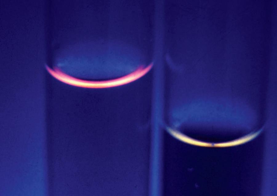
Fig. 2.1. Wood light examination of urine demonstrating coral red fluorescence in a patient with porphyria cutanea tarda. Fluorescence can be accentuated by adding dilute hydrochloric acid. Left, Positive. Right, Negative. (From the Fitzsimons Army Medical Center Collection, Aurora, CO.)
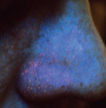
Fig. 2.2. Wood light examination of a patient’s nose demonstrating coral red porphyrins produced by Propionibacterium acnes. Because this patient is under treatment for acne, this finding indicates that the patient is not taking the antibiotic or the organism is resistant to the antibiotic. (From the Fitzsimons Army Medical Center Collection, Aurora, CO.)
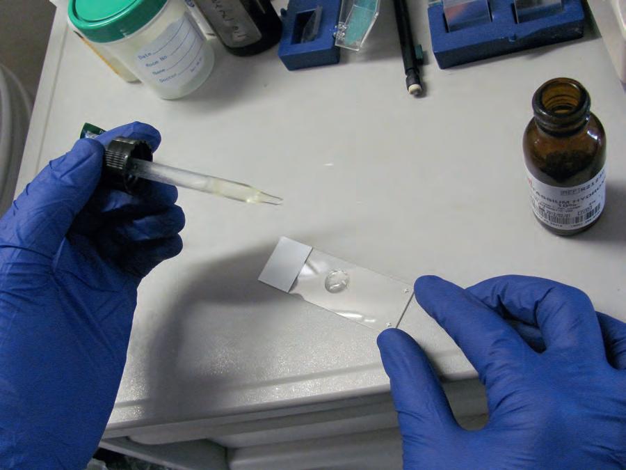
2.5. A single drop of KOH is placed on the scale and then cover-slipped. Note that the physician is wearing gloves— remember that KOH is toxic to the skin.
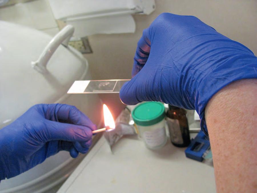
2.6. After being cover-slipped, the KOH preparation is gently heated over a flame. If the KOH contains DMSO, this step can often be omitted. After heating, the slide is ready to be examined under the microscope.
Fig.
Fig.