Effective Software Development for the Enterprise: Beyond Domain Driven Design, Software Architecture, and Extreme Programming 1st Edition Tengiz Tutisani
College of Veterinary Medicine and Biomedical Sciences
Fort Collins, Colorado
A. N. (Nickie) Baird, DVM, MS, DACVS
Associate Editor
Purdue University
College of Veterinary Medicine
West Lafayette, Indiana
Khursheed Mama, DVM, DACVA
Colorado State University College of Veterinary Medicine and Biomedical Sciences
Fort Collins, Colorado
PREFACE TO THE FIRST EDITION
The purpose of this book is to present some fundamental techniques in large animal surgery to both veterinary students and large animal practitioners. It is designed to be brief, discussing only the major steps in a particular operation, and each discussion is accompanied by appropriate illustrations. Most of the techniques presented in this book can be performed without the advantages of a fully equipped large animal hospital or teaching institution.
The book assumes a basic understanding of anatomy and physiology. Those who wish to know more about a particular technique are encouraged to consult the bibliography.
We and our colleagues at the Colorado State University Veterinary Teaching Hospital consider the procedures discussed in this book to be time honored. Some practitioners may perform certain techniques in slightly different ways. We would be happy to receive input about modifications of these techniques for future editions of this book.
All of the drawings in the book are original and based on rough sketches and photographs taken at various points during actual surgery. Occasionally, dissections were performed on cadavers.
The surgical procedures described in this text represent not only our thoughts, but suggestions from many of our colleagues as well. Their help was an important contribution to the production of this book. We are indebted to Dr. Wilbur Aanes, Professor of Surgery, Colorado State University, who unselfishly shared 30 years of his personal experience in large animal surgery with us. We are proud to be able to present in Chapter 10 of this book “Aanes’ Method of Repair of ThirdDegree Perineal Laceration” in the mare, a technique that he pioneered over 15 years ago. We also wish to give credit to the following faculty members at Colorado State University Veterinary Teaching Hospital who willingly gave us advice on the diagrams and manuscript of various techniques discussed in this book: Dr. Leslie Ball, Dr. Bill Bennett, Dr. Bruce Heath, Dr. Tony Knight, Dr. LaRue Johnson, Dr. Gary Rupp, Dr. Ted Stashak, Dr. Gayle Trotter, Dr. James Voss, and Dr.
Mollie Wright. We also wish to express appreciation to Dr. John Baker, Purdue University, and Dr. Charles Wallace, University of Georgia, for their comments on some questions we had. Dr. McIlwraith is also grateful to Dr. John Fessler, Professor of Surgery, Purdue University, for his inspiration and training.
We are particularly grateful to Dr. Robert Kainer, Professor of Anatomy, Colorado State University, for checking the manuscript and the illustrations and advising us on nomenclature. His input impressed upon us the importance of the relationship between the dissection room and the surgery room.
The terrific amount of time and effort involved with the illustrations will be clear to the reader who cares only to leaf through the book. For these illustrations, we are indebted to Mr. Tom McCracken, Director, Office of Biomedical Media, Colorado State University. We are thankful for his expertise, as well as his cooperation and understanding. The diagrams for “Aanes’ Method of Repair of ThirdDegree Perineal Laceration” were done by Mr. John Daughtery, Medical Illustrator, Colorado State University. We must also thank Kathleen Jee, who assisted with various aspects of the artwork. We would also like to thank Messrs. Al Kilminster and Charles Kerlee for taking photographs during the various surgical procedures that were used to assist with the artwork of this text.
The manuscript was typed by Mrs. Helen Mawhiney, Ms. Teresa Repphun, and Mrs. Jan Schmidt. We thank them for their patience and understanding during the many changes we made during the generation of the final manuscript.
We are grateful to the following instrument companies for allowing us to use some of the diagrams from their sales catalogs for inclusion in Chapter 3, “Surgical Instruments”: Schroer Manufacturing Co., Kansas City, MO; Intermountain Veterinary Supply, Denver, CO; Miltex Instrument Co., Lake Success, NY; J. Skyler Manufacturing Co., Inc., Long Island, NY.
The idea for this book was conceived in 1978 when one of us (AST) was approached by Mr. George Mundroff,
Executive Editor, Lea & Febiger. We would like to thank him for his encouragement and guidance. We are also grateful to Mr. Kit Spahr, Jr., Veterinary Editor; Diane Ramanauskas, Copy Editor; Tom Colaiezzi, Production Manager; and Samuel A. Rondinelli, Assistant Production Manager, Lea & Febiger, for their assistance, as well as to
others at the Publisher who assisted in the production of this book.
Colorado
A. Simon Turner C. Wayne McIlwraith Fort Collins,
PREFACE TO THE SECOND EDITION
The second edition of Techniques in Large Animal Surgery is in response to the acceptance of the first edition and the continued need for such a book for both veterinary students and large animal practitioners. In many instances, the techniques are time honored and require no change from 5 years ago. In other instances, however, refinements in technique as well as improved perception of indications, limitations, and complications have made changes appropriate.
A significant change is the addition of Dr. R. Bruce Hull, Professor of Veterinary Clinical Sciences, The Ohio State University, as a contributor. He has carefully analyzed the entire bovine section, and his suggested changes and additions have been incorporated into the text. In addition, two procedures, “teaser bull preparations by penile fixation” and “treatment of vaginal prolapse by fixation to the prepubic tendon,” have been added. We are most grateful in having Dr. Hull’s help and expertise. Among the introductory chapters, the section on anesthesia required the most updating, and we are grateful to our colleague Dr. David Hodgson at Colorado State University for his review and advice. Two new procedures, “superior check ligament desmotomy” and “deep digital flexor tenotomy,” were considered appropriate additions to this edition. We are grateful to Dr. Larry Bramlage, Ohio State University, for his comments and help with the first of these procedures. Many of the other changes in this edition are in response to the book reviews and comments on the first edition returned to Lea & Febiger. To these people, we appreciate your feedback.
A chapter on llama tooth removal was added because of the increased popularity of this species, especially in our own part of the country. Although we only discuss this one technique, it should not be inferred that other operations are unheard of in llamas. We have corrected angular limb deformities, repaired fractures, and performed gastrointestinal surgery, among other procedures, but tooth removal is the most common. Descriptions of these other procedures in llamas are beyond the scope of this book at this stage.
The need for more sophisticated equine techniques prompted us to produce the textbook Equine Surgery: Advanced Techniques in 1987. It is envisioned that the book will be used as a companion to this second edition, to provide a full spectrum of equine procedures, with the well-accepted format of concise text and clear illustrations.
Again, we are thankful to Mr. Tom McCracken, Assistant Professor, Department of Anatomy and Neurobiology, Colorado State University, for his talent in capturing the techniques described in his line drawings. We are also indebted to Helen Acvedo for typing our additions and to Holly Lukens for copyediting. Finally, our thanks again to the excellent staff at Lea & Febiger for the production of this edition.
Simon Turner
C. Wayne McIlwraith Fort Collins, Colorado
A.
PREFACE TO THE THIRD EDITION
The first two editions of Techniques in Large Animal Surgery have been well accepted, much to the credit of Drs. Turner and McIlwraith. They have been excellent texts for the veterinary student and the large animal practitioner. I was fortunate to be able to take on the task when it came time to update the information for a third edition. I am deeply appreciative of the opportunity to take such an excellent text and update it with new information and techniques.
The third edition of Techniques in Large Animal Surgery has been updated in response to the continued need for such a book for both veterinary students and large animal practitioners. There are some techniques that are time tested and continue to be included. There are other techniques that have been refined or replaced, and these are included in the new text.
New information has been included in essentially every chapter. We have made extensive use of tables to simplify the information. The anesthesia section includes new and updated information on sedation and anesthetic agents. The instrument section has been evaluated, adding new
instruments where applicable and removing outdated or unavailable instruments. The section on suture materials has been updated to include new materials. There are new illustrations in the suture pattern section to better aid the practitioner with surgical techniques. The sections on wound management and reconstructive surgery have been increased to provide up-to-date information on wound care. Tables of required instrumentation have been added to all sections of the remaining surgical chapters to aid in surgical planning and preparation.
I am very grateful for our new illustrator Anne Rains; she has done an excellent job and has made my life very easy. I am indebted to Joanna Virgin who has done the lion’s share of the research to make sure this text was as up-to-date and accurate as possible. I could not have done this work without her. Thanks to the folks at Blackwell for their help and assistance in the production of this edition.
Dean A. Hendrickson Fort Collins, Colorado
PREFACE TO THE FOURTH EDITION
The first two editions of Techniques in Large Animal Surgery have been well accepted much to the credit of Drs. Turner and McIlwraith. They have been excellent texts for the veterinary student and the large animal practitioner. I was fortunate in that when it came time to update the information for a third edition, I was able to take on the task; and now we have added a fourth edition.
The fourth edition of Techniques in Large Animal Surgery has been updated in response to the continued need for such a book for both veterinary students and large animal practitioners. As with the third edition, we have gone through the entire text to make sure the information was reliable. The “tried-and-true” procedures have been retained, the outdated procedures have been removed, and new procedures have been added. As we thoroughly researched each of the chapters in the text, we did a major overhaul of the references.
Probably the most important changes in this text are the addition of two authors. Nickie Baird joined me in coauthoring the fourth edition. His expertise in livestock
animal surgery was a perfect fit for this textbook. He brings a great deal of new information to the text and has been a great partner. Dr. Khursheed Mama joined us as the author of Chapter 2, Anesthesia and Fluid Therapy. She did an excellent job updating all of the information.
We added a considerable amount of new information in the text and retained the table format to simplify information. New figures have been added, where needed, to support the updated information.
I am very grateful for Grahm Hendrickson for illustrating the new procedures, as well as Katie Hunsucker and Joy Fuhrman for providing a lot of the background research. Thanks as well to the folks at Wiley for their help and assistance in the production of this edition.
Dean A. Hendrickson Fort Collins, Colorado
A. N. (Nickie) Baird West Lafayette, Indiana
Turner and McIlwraith’s
Techniques in Large Animal Surgery
4th Edition
Chapter 1
PRESURGICAL CONSIDERATIONS
Dean A. Hendrickson, DVM, MS, DACVS
Objectives
1. Discuss some of the presurgical considerations that can affect the success of a procedure, including the physiological state and condition of the patient; the predisposing factors for infection; and the limitations of the surgeon, facilities, and equipment.
2. Describe the methods of asepsis and antisepsis.
3. Describe the classification of different procedures with regard to risk of infection and degree of contamination.
4. Discuss the judicious use of antibiotics and their applications prophylaxis and postoperative infection.
5. Describe proper techniques for surgical site preparation.
Preoperative Evaluation of the Patient
Before a surgical procedure, a physical examination is generally indicated. This applies to both emergency and elective surgery. The following are laboratory tests that are generally indicated for horses based upon animal age and systemic status at our clinic:
• For horses younger than 4 years old and healthy:
• Packed cell volume (PCV)
• Total protein
• Appropriate for horses greater than 4 years old or those that are systemically ill:
• Complete blood count (CBC)
• Chemistry
Exactly where to draw the line on laboratory tests is largely a matter of judgment on the part of the surgeon. Obviously, if the surgery consists of castration of several litters of piglets, then for purely economic reasons laboratory tests prior to surgery may not be performed. In many cases, however, additional tests will be necessary. The following are examples of other optional tests and their indications:
• Electrolyte measurement for right-sided abomasal diseases of the dairy cow
• Urinalysis in the dairy cow to evaluate the presence of ketosis
• Measurement of blood urea nitrogen (BUN) and creatinine if urinary problems are suspected
• Analysis of peritoneal fluid prior to laparotomy for horses with colic
• Full chemistry panels when there are age or systemic considerations
If any laboratory parameters are abnormal, the underlying causes should be investigated and efforts made to correct them. In “elective” surgery this is possible, but it may not be possible in an emergency. The owner should be made aware of any problems prior to subjecting the animal to surgery. Risks are always present in normal elective surgery, and these should be explained to the owner. It is always better to have an early, frank discussion with the owner about the possible risks associated with the surgery than to have the discussion after the risk has been realized.
Fluid replacement should be performed if necessary. In the elective case, the surgical procedure should be postponed if the animal’s physical condition or laboratory parameters are abnormal. In some animals, internal and external parasitism may have to be rectified to achieve this goal.
Medical records should be kept at all times. Obviously this can be difficult in such cases as castration of several litters of piglets. However, record keeping should become
an essential part of the procedure for horses and cattle in a hospital, and herd records should be kept in all other situations. Finally, if the animal is insured, the insurance company must be notified of any surgical procedure; otherwise, the policy may be void.
Surgical Judgment
Surgical judgment cannot be learned overnight by reading a surgery textbook, nor is it necessarily attained by years of experience. The surgeon who continually makes the same mistake will probably never possess good surgical judgment. Not only should the surgeon learn from his own mistakes; he also should learn from the mistakes of others, including those documented in the surgical literature. As part of surgical judgment, the surgeon must ask the following questions:
• Is the surgery necessary?
• What would happen if the surgery were not performed?
• Is the procedure within the capabilities of the surgeon, the facilities, and the technical help?
If the surgeon finds that the procedure is too advanced for his or her capabilities and/or facilities, the surgery should be referred. Some veterinarians have a fear that this will mean loss of the client’s business in the future, but this is rarely the case. If the surgeon explains why the case should be referred elsewhere, most clients will be grateful for such frankness and honesty. It is inexcusable to operate on a patient and then have complications arise due to inadequate training and facilities, when the surgery could easily have been referred to a well-equipped, wellstaffed hospital with specially qualified personnel. Clearly, this rule has exceptions—mainly the emergency patient, which may fare better by undergoing immediate surgery than being subjected to a long trailer ride to another facility.
Many of the procedures described in this book can be done “on the farm.” Some, such as arthrotomy for removal of chip fractures of the carpal and sesamoid bones in horses, should be done in a dust-free operating theater. If clients want these latter procedures to be done “in the field,” they should understand the disastrous consequences of postsurgical infection. The surgeon must be the final judge of whether his facilities or experience are suitable.
Principles of Asepsis and Antisepsis
There are four main determinants for a surgical site infection (SSI): host defense, physiologic derangement, bacterial contamination risk at surgery, and prolonged surgical time.1 Other factors that impact infection of deep structures and organs include hypoalbuminemia and a prior
Classification Description Examples
Clean Gastrointestinal, urinary, or respiratory tract is not entered.
Cleancontaminated
Contaminateddirty
Arthrotomy for removal of a chip fracture of a carpal bone of a horse
Gastrointestinal, respiratory, or urinary tract is entered. There is no spillage of contaminated contents. Abomasopexy for displaced abomasum in the dairy cow
Gross spillage of contaminated body contents or acute inflammation occurs. Wounds Abscesses
Devitalized bowel
operation.2 Perioperative blood loss also contributes to SSI.3 Control methods include aseptic surgical practices as well as identification of the high-risk patient, correction of systemic imbalances prior to surgery, and the proper use of prophylactic antibiotics.
We are sometimes reminded by fellow veterinarians in the field that we must teach undergraduates how to do surgery in the real world. By this they mean that we must ignore aseptic draping and gloving and lower the standard to a “practical” level. This is fallacious in our opinion. Although we recognize that while the ideal may be unattainable in private practice, one should always strive for the highest possible standard; otherwise, the final standard of practice may be so low that the well-being of the patient is at risk, not to mention the reputation of the veterinarian as a surgeon. For this reason, we believe that it behooves us as instructors of undergraduates to teach the best possible methods with regard to asepsis as well as technique.
The extent to which the practice of asepsis or even antisepsis is carried out depends on the classification of the operation, as shown in Table 1.1. This classification may also help the veterinarian decide whether antibiotics are indicated or whether postoperative infection can be anticipated.
Surgical Classifications
Once the surgeon has categorized the surgical procedure, appropriate precautions to avoid postoperative infection can be determined. In all cases, however, the surgical site is prepared properly, including clipping and aseptic scrubbing.
Table 1.1. Surgical classifications.
Whatever category of surgery is performed, clean clothing should be worn. The wearing of surgical gloves is good policy even if only to protect the operator from infectious organisms that may be present at the surgical site. Surgical gowns, gloves, and caps are recommended for clean surgical procedures, although such attire has obvious practical limitations for the large animal surgeon operating in the field. The purpose of this book is to present guidelines rather than to lay down hard-and-fast rules. For example, the decision between wearing caps, gowns, and gloves and wearing just gloves can be made only by the surgeon. Good surgical judgment is required. In general, it is better to be more careful than what may appear necessary in order to be better prepared when problems arise.
Role of Antibiotics
Antibiotics should never be used to cover flaws in surgical technique. The young surgeon is often tempted, sometimes under pressure from the client, to use antibiotics prophylactically. However, the disadvantages of antimicrobial therapy often outweigh its benefits. Extended periods of antimicrobial therapy can select for resistant organisms and adversely affect the gastrointestinal tract by eliminating many of the normal enteric organisms and allowing outgrowths of pathogenic bacteria, such as Clostridia spp., which can result in colitis and diarrhea.4 When selecting an antibiotic regimen, the surgeon should consider the following aspects:
• Does the diagnosis warrant antibiotics?
• Which organisms are most likely to be involved, and what is their in-vitro antimicrobial susceptibility?
• What is the location or likely location of the infection?
• How accessible is the location of the infection to the drug?
• What possible adverse reactions and toxicities to the drug could occur?
• What dosage and duration of treatment are necessary to obtain sufficient concentrations of the drug?
Again, some judgment is required, but suffice it to say, antibiotics should never be substitutes for “surgical conscience.” Surgical conscience consists of the following: dissection along tissue planes, gentleness in handling tissues, adequate hemostasis, selection of the best surgical approach, correct choice of suture material (both size and type), closure of dead space, and short operating time. If the surgeon decides that antibiotics are indicated, special attention should be given to selecting the type of antimicrobial drug, the dosage, and the duration of use. Ample scientific literature indicates that for maximum benefit, antimicrobials should be administered prophylactically prior to surgery and, at the latest, during surgery. Beyond 4 hours postsurgically, the administration of prophylactic antibiotics has little to no effect on the incidence
of postoperative infection.1 The duration of treatment should not exceed 24 hours because most research indicates that antimicrobial use after this period of time does not confer further benefits. If longer duration of antimicrobial coverage is necessary, the full duration of the specific antimicrobial drug selected should be given. This varies depending on the drug; however, in most cases the duration is at least 3 and up to 5 days. If the surgeon is operating on a food animal, there are regulations for withdrawal times from different antimicrobial drugs prior to slaughter that must be taken into account.
If topical antibiotics are used during surgery, they should be nonirritating to the tissues; otherwise, tissue necrosis from cellular damage will outweigh any advantageous effects of the antibiotics. It is also beneficial when using topical antibiotics to use antibiotics that are not generally used systemically.
All equine surgical patients should have tetanus prophylaxis. If the immunization program is doubtful, the horse can receive 1500–3000 units of tetanus antitoxin. Horses on a permanent immunization program that have not had tetanus toxoid within the previous 6 months should receive a booster injection.
Tetanus prophylaxis is generally not provided for food animals, but an immunization program may be considered, especially if a specific predisposition is thought to exist.
Preoperative Planning
The surgeon should be thoroughly familiar with the regional anatomy. In this book we illustrate what we consider to be the important structures in each technique. If more detail is required, a suitable anatomy text should be consulted. Not only should the procedure be planned prior to the surgery, but the surgeon also should visit the dissection room and review local anatomy on cadavers prior to attempting surgery on a client’s animal. We are fortunate in veterinary surgery to have greater access to cadavers than our counterparts in human surgery.
Preparation of the Surgical Site
For the large animal surgeon, preparation of the surgical site can present major problems, especially in the winter and spring when farms can be muddy. Preparation for surgery may have to begin with removal of dirt and manure. Some animals that have been recumbent in mud and filth for various reasons may have to be hosed off. Hair should then be removed, not just from the surgical site, but from an adequate area surrounding the surgical site.
The clipping should be done in a neat square or rectangular shape with straight edges. Surprisingly, this, along with the neatness of the final suture pattern in the skin,
is how the client judges the skill of the surgeon. Clipping may be done initially with a no. 10 clipper blade, and then the finer no. 40 blade may be used. The incision site can be shaved with a straight razor in horses and cattle, but debate exists regarding the benefit or problems associated with this procedure. In sheep and goats, in which the skin is supple and pliable, it is difficult to shave the edges.
Preparation of the surgical site, such as the ventral midline of a horse about to undergo an exploratory laparotomy, may have to be performed when the animal is anesthetized. If surgery is to be done with the animal standing, an initial surgical scrub, followed by the appropriate local anesthetic technique and a final scrub, is standard procedure.
For cattle or pigs, the skin of the surgical site can be prepared for surgery with the aid of a stiff brush. For horses, gauze sponges are recommended. Sheep may require defatting of the skin with alcohol prior to the actual skin scrub. The antiseptic scrub solution used is generally a matter of personal preference. Either povidoneiodine scrub (Betadine Scrub) alternated with a 70% alcohol rinse, or Chlorhexidine alternated with water, can be used. Finally, the skin can be sprayed with povidoneiodine solution (Betadine Solution) and allowed to dry.
Scrubbing of the proposed surgical site is done immediately prior to the operation. Scrubbing should commence at the proposed site of the incision and progress toward the periphery; one must be sure not to come back onto a previously scrubbed area. Some equine surgeons clip and shave the surgical site the night before the surgery, perform an aseptic preparation as previously described, and wrap the limb in a sterile bandage until the next day. A shaving nick made the day before surgery may be a pustule on the day of surgery, however, so this is generally not recommended for anything proximal to the pastern region.
When aseptic surgery is to be performed, an efficient draping system is mandatory. Generally, time taken to drape the animal properly is well spent. The draping of cattle in the standing position can be difficult, especially if the animal decides to move or becomes restless. It can be difficult to secure drapes with towel clamps in the conscious animal because only the operative site is anesthetized. However, if the surgeon applies slow pressure when closing the towel clamps, most animals will tolerate their application, even if the local site is not desensitized. If draping is not done, the surgeon must minimize contact with parts of the animal that have not been scrubbed. The tail must be tied to prevent it from flicking into the surgical field.
Several operations described in this book require the strictest of aseptic technique; sterile, antimicrobial, adhe-
sive, incise drapes are indicated. Characteristics of sterile plastic adhesive drapes include their ability to adhere, their antimicrobial activity, and their clarity when applied to the skin. Probably the most desirable feature is the one first mentioned. With excessive traction or manipulation, some brands of drapes quickly separate from the skin surfaces, and this separation instantly defeats their purpose.
Rubberized drapes are helpful when large amounts of fluids (such as peritoneal and amniotic fluid) are encountered during the procedure. Rubberized drapes are also useful to isolate the bowel or any other organ that is potentially contaminated, to prevent contamination of drapes. Newer fluid-impermeable paper drapes that are disposable make the surgeon’s job even easier.
Postoperative Infection
Prevention of postoperative infection should be the goal of the surgeon, but infection may occur despite all measures taken to prevent it. If infection occurs, the surgeon must decide whether antibiotic treatment is indicated, or whether the animal is strong enough to fight it using its own defense mechanisms. Some surgical wounds require drainage at their most ventral part, whereas others require more aggressive treatment. If, in the judgment of the surgeon, the infection appears to be serious, a Gram stain, culture, and sensitivity testing of the offending microorganism(s) will be indicated. A Gram stain may give the surgeon a better idea of what type of organism is involved and may in turn narrow the selection of antibiotics. Sometimes in-vitro sensitivities have to be ignored because the antibiotic of choice would be prohibitively expensive. This is especially true for adult cattle and horses. A broad-spectrum antibiotic should be given, if possible, as soon as practical.
References
1. Barie, P.S. Modern surgical antibiotic prophylaxis and therapy—less is more. Surgical Infections, 1:23–29, 2000.
2. Haridas, M., and Malangoni, M.A. Predictive factors for surgical site infection in general surgery. Surgery, 144:496–503, 2008.
3. Sorensen, L.T., Hemmingsen, U., Kallehave, F., Wille-Jorgensen, P., Kjoergaard, J., Moller, L.N., and Jorgensen, T. Risk factors for tissue and wound complications in gastrointetstinal surgery. Annals of Surgery, 241:654–658, 2005.
4. Papich, M.G. Antimicrobial therapy for gastrointestinal disease. The Veterinary Clinics of North America. Equine Practice, 19: 645–663, 2003.
Chapter 2
ANESTHESIA AND FLUID THERAPY
Khursheed Mama, DVM, DACVA
Objectives
1. Describe routine regional anesthetic techniques in large animals.
2. Discuss selected species differences in reference to anesthetic techniques.
3. Describe the indications for, advantages of, and disadvantages of general anesthesia in large animal species.
4. Provide a basic discussion of the fundamentals of fluid therapy including methods for ascertaining fluid deficits, acid-base imbalances, and electrolyte abnormalities.
5. Discuss specific fluid therapies in patients undergoing elective surgery and in compromised patients, either with or without preliminary data.
Local and Regional Anesthesia (Analgesia)
Regional anesthesia results from desensitization of sensory nerves to a given area. This may be performed by infiltration into the desired location or by “blocking” sensory nerve(s) innervating a region. Both techniques may be used to desensitize the surgical site. Depending on the required duration of anesthesia, local anesthetic agents including lidocaine hydrochloride (shortest onset and duration), mepivacaine hydrochloride and bupivacaine hydrochloride (longest onset and duration) may be used. Due to cardiovascular toxicity with vascular absorption, bupivacaine use is usually limited to epidural and perineural administration; lidocaine and mepivacaine may be used by any route. Mepivacaine is often selected because of its rapid onset, intermediate duration, and reduced tissue reactivity.6
Regional anesthesia techniques are still commonly used as primary means to facilitate noxious intervention in many ruminant species. Sedation may be used as an adjunct. In horses, while these techniques may be used in sedated patients, they are also commonly used as adjuncts to general anesthesia. A description of selected regional anesthesia techniques follows.
Anesthesia
The purpose of this section is not to present an in-depth discussion of all aspects of anesthesia. Details on the principles of anesthesia, recognition of stages of anesthesia, monitoring during anesthesia, and the pharmacology and physiology associated with anesthesia are well documented in other texts.1–3 Rather, information pertaining to routinely used anesthetic techniques for large animals is provided. The interested reader is referred to additional sources for more in depth information.4,5
Infiltration Analgesia
The principles of infiltration anesthesia are simple and similar for all species. Following definition of the area to be desensitized, local anesthetic is injected at an initial site with a small gauge needle and then a longer needle is inserted through the initial region of desensitization. Repeat injections are usually made through a region that has already been desensitized. When possible, the skin and subcutis should be infiltrated first and then the deeper layers, such as muscle and peritoneum. The injection of significant amounts of local anesthetic into the peritoneal
cavity should be avoided as rapid vascular absorption can result in toxicity. Infiltrating injections should be made in straight lines; “fanning” should be avoided as much as possible because of the potential for tissue trauma.
Infiltration analgesia is commonly used for suturing wounds and for removing cutaneous lesions in all large animal species. It may also be used in the form of a “line block” for laparotomy, in which case the analgesic agent is infiltrated along the line of incision. Although convenient, the infiltration of the analgesic agent into the incision line causes edema in the tissues and may affect wound healing. In this respect, regional anesthetic techniques that are removed from the surgical site are generally preferred.
Techniques of Regional Analgesia
Inverted L Block
This is the simplest technique of regional anesthesia for laparotomy and laparoscopy in large animal species. It may be used to facilitate flank or paramedian interventions. The principles of the technique are illustrated for cattle in Figure 2.1. Local anesthetic agent is administered nonspecifically in the form of an inverted L with a goal
of blocking nerves entering the surgical field. The procedure is facilitated by the use of an 8- to 10-cm, 16- to 18-gauge needle. It is generally recommended that a dose of local anesthetic is limited to 2 mg/kg. However, up to 100 ml of 2% lidocaine has been used for adult horses and cows (4 mg/kg for 500-kg patient). The vertical portion of the inverted L is caudal to the last rib, and the horizontal portion is just ventral to the transverse processes of the lumbar vertebrae. Ten to fifteen minutes should be allowed for the drug to take effect.
Systemic toxicity following inadvertent intravenous administration or absorption from regional sites is reported. Experiments in sheep have shown that convulsions occur in adult sheep at a dose of lidocaine hydrochloride of 5.8 ± 1.8 mg/kg intravenously.7 Sub-convulsive doses of lidocaine hydrochloride produce drowsiness. Above convulsive doses, hypotension, respiratory arrest, and circulatory collapse occur progressively. If convulsions do occur, they can be controlled with an intravenous dose of 0.5 mg/ kg of diazepam (Valium). To minimize the occurrence, it is recommended that diluted local anesthetic is used for smaller sized animals such as sheep and goats.1,8 Toxicity following inadvertent intravenous bupivacaine administration manifests in cardiovascular collapse.
Fig. 2.1. Inverted L block.
Paravertebral Block
While not common, the paravertebral block has been described and utilized to desensitize the flank area for standing procedures in horses.9 It is however more commonly performed in cattle, sheep, and goats.8,10 In ruminants, the thirteenth thoracic nerve (T13), the first and second lumbar nerves (L1 and L2), and the dorsolateral branch of the third lumbar nerve (L3) supply sensory and motor innervation to the skin, fascia, muscles, and peritoneum of the flank. Regional analgesia of these nerves is the basis of the paravertebral block. For practical purposes with flank laparotomy, blocking of the dorsolateral branch of L3 is not generally considered necessary and may be contraindicated because, if one has miscounted the vertebrae, one may actually block L4, where sensory and motor nerve fibers to the hind limbs originate.
Two approaches to performing the paravertebral block have been described for cattle. The first consists of walking the needle off the transverse process, as illustrated in Figure 2.2. As the nerve is most distinct at its interverte-
bral foramen, walking the needle off the transverse process closer to this site allows one to block the nerve before or close to the split into individual dorsal and ventral branches. As the transverse processes slope forward, the transverse process of L1 is used as a landmark to block T13, and the transverse processes of L2 and L3 are similarly used to locate nerves L1 and L2, respectively. When the transverse process has been located, a line is drawn from its cranial edge to the dorsal midline. The site for injection is 3 to 5 cm from the midline (Figure 2.2) caudal to transverse processes of L1, L2 and L3. The transverse process of L1 is difficult to locate in fat animals, in which case the site is estimated relative to the distance between the processes of L2 and L3. Following subcutaneous infiltration of local anesthetic, a 1-inch, 16-gauge needle is inserted to act as a guide in placing a 10-cm, 20-gauge needle perpendicular to the transverse process is encountered. The needle is then walked off the cranial border of the transverse process and advanced 0.75 cm (will generally feel penetration of the intertransverse ligament); and
Fig. 2.2. Paravertebral block.
approximately 10 ml of local anesthetic solution (typically 2% lidocaine or mepivacaine) is administered below the ligament. An additional 5 ml is placed dorsal to the ligament. If the drug has been administered correctly, desensitization will be effective within a few minutes. In testing the block, one must remember that the distribution of the nerves is such that T13 innervates the ventral flank area, whereas L2 innervates the more dorsal region closer to the transverse processes. A temporary lateral deviation of the spine due to muscle paralysis is observed in association with paravertebral analgesia. Vasodilation of surface vessels may also be observed.
An alternate technique favored by some was developed by Magda and modified by Cakala.11 It uses a lateral approach to the nerves and is sometimes referred to as the distal paravertebral or paralumbar approach. The branches of T13, L1, and L2 are blocked close to the ends of the first, second, and fourth transverse processes, respectively, as illustrated in Figure 2.2. The skin is clipped and prepared at the ends of the first, second, and fourth lumbar transverse processes. An 18-gauge needle is inserted under each transverse process towards the midline, and 10 ml of solution is injected. The needle is then withdrawn a short distance and is redirected both cranially and caudally and additional local anesthetic solution is injected. In this fashion, a diffuse region ventral to the transverse process is infiltrated, to block the ventral branch of the nerve. The needle is then redirected slightly dorsal and caudal to the transverse process to block the dorsolateral branch of each nerve. In adult cattle, up to 25 ml of local anesthetic solution has been administered at each site without adverse effect. Because the paralumbar technique does not paralyze the lumbar muscles, lateral deviation of the spine does not occur.
The technique for paravertebral nerve block is the same in sheep and goats as it is in cattle. Up to 5 ml of 1% or 2% lidocaine is recommended for each of the injection sites.1 While the total dose should not exceed 6 mg/kg, a lower dose (2 mg/kg) is usually recommended.
Epidural Analgesia
Epidural analgesia is used frequently to facilitate standing surgical interventions in cattle and horses, for cesarean sections in swine, for urogenital surgery in goats, and for postoperative analgesia. Drug selection varies depending on the goal (e.g., local anesthetics for anesthesia of the area, opioids or alpha-2’s for analgesia without motor blockade) Species differences also influence the choice of epidural drug and need for systemic administration of sedatives or tranquilizers. Sheep can be easily handled and may only require administration of a local anesthetic and physical restraint for some procedures. Goats and pigs tend to be more curious and less amenable to physical restraint so may require concurrent sedation. Site of administration of the epidural drug(s) also varies with species and procedure to be performed and commonly includes either the lumbosacral or caudal epidural space.
The lumbosacral space is commonly used in sheep, goats, and pigs, whereas in cattle and horses the caudal epidural site is more typical and facilitates “standing” procedures as motor control of the hind legs is generally not affected (as it often is with lumbosacral administration of local anesthetic); but the anal sphincter relaxes, and tenesmus and obstetric straining is prevented.
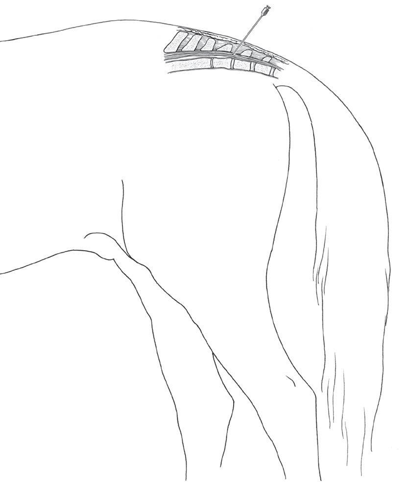
Caudal epidural administration describes the injection of analgesic and anesthetic agents between either the first and second coccygeal vertebrae or in the sacrococcygeal space. The former site is 1–2 inches cranial to the long tail hairs in the horse. To locate the space, the tail is grasped and is moved up and down; the first obvious articulation caudal to the sacrum is the first intercoccygeal space. After clipping and skin preparation, a skin bleb is made with 2% lidocaine using a 2.5-cm, 25-gauge needle, to facilitate needle placement. An 18- or 19-gauge, 3- to 5-cm needle (or a spinal needle) is introduced through the center of the space on the midline at a 45° angle in cattle until its point hits the floor of the spinal canal (Figure 2.3). The needle is then retracted slightly to ensure that the end is not embedded in the intervertebral disc. In the horse, this needle may be inserted at an angle of 30° from a perpendicular line through the vertebrae or at an angle of 60° as illustrated in Figure 2.4. If a regular needle (versus a spinal needle with a stilette) is utilized, aspiration of sterile fluid placed in the needle hub is a practical indicator of entry into the epidural space especially with first-time injections. Additionally, if the needle is correctly placed in the epidural space, there should be no resistance to injection. The bevel of the needle is usually directed cranially. In cattle and small ruminants, 2% lidocaine may be used for epidural anesthesia. Doses are shown in Table 2.1. Injections of 2% lidocaine can also be used in the sacrococcygeal space of sheep and goats to provide caudal epidural analgesia for obstetric procedures. The total volume of drug administered via the caudal epidural space should not exceed 3 ml in sheep and goats and 10 ml in cattle if the goal is to avoid hind limb incoordination or recumbency.12 Alpha-2 agents can be used with 2% lidocaine in cattle to achieve a longer duration of analgesia.1,12,13 In horses, caudal epidural administration of lidocaine has been similarly used for certain procedures (e.g., Caslicks). This provides surgical anesthesia for a duration of 87.2 ± 7.5 minutes, which is considered fairly short for many procedures performed in horses. Volumes greater than 5–7 ml may extend duration of the block but are not typically recommended as they increase the likelihood of recumbency.1,14 As in cattle, alpha-2 agonists such as detomidine, xylazine, and medetomidine are therefore often administered in combination to increase the duration of analgesia and potentially decrease ataxia.6,15 Systemic effects may, however, be observed. Alternative anesthetic/ analgesic combinations are shown in Table 2.2.
To achieve anesthesia for more rostral perineal and hind limb surgical procedures in small ruminants and
pigs, the drug is administered at the lumbosacral space. In sheep and goats, a volume of 1 ml/5–7 kg lean body weight has been suggested depending on the desired cranial extent of the block; recumbency should be expected. While generally contraindicated and challenging to perform in horses, this technique has been used to provide 2–4 hours of analgesia in cattle for laparotomy, pelvic limb surgery, or udder amputation. Recognize that the animal will become recumbent if high volumes of local anesthetic are injected into the lumbosacral space. Accidental spinal injection in any species will extend the block further cranially which could result in respiratory compromise and worsening hypotension from vasodilation.16 Ruminal atony and bloat may also be observed in cattle.
Continuous caudal epidural anesthesia using a commercial epidural catheter kit (continuous epidural tray) is also used in horses and, in some instances, ruminants for repeated delivery of epidural delivery of analgesics and postoperative pain relief.15,17,18 The kit contains a Huberpoint directional needle with stilette (Tuohy spinal needle) inserted through a pilot hole at 45° to the horizontal until one encounters an abrupt reduction in resistance. The catheter is then inserted through the needle, it is advanced a distance beyond the end of the needle, and the needle is withdrawn. Combinations of either a local anesthetic or an alpha-2 adrenergic agonist and morphine administered in the caudal epidural space have been shown to have useful clinical applications for postoperative and long-term pain relief in both humans and animals. Preoperative epidural administration of detomidine (30 μg/kg) and morphine (0.2 mg/kg) provides effective,
long-lasting pain relief and decreases postoperative lameness in horses that undergo bilateral stifle arthroscopy.19
Lumbosacral epidural analgesia has been used in both young and adult pigs and in particular for cesarean section in the sow. This technique is reported to result in maternal immobilization and analgesia, with minimal fetal depression. The lumbosacral space is located at the intersection of the spine with a line drawn through the cranial borders of the ilium. An 18-gauge needle is inserted 1–2 cm caudal to this line in small pigs and 2.5–5 cm caudal to the line in larger animals. The needle is then directed ventrally until it is felt to pass through the dorsal ligament of the vertebrae and into the epidural space. The needle size varies with the size of the pig: 8 cm is used for the pig weighing up to 75 kg; and 15 cm is used for pigs heavier than 75 kg. The dose is about 1 ml/5–10 kg of 2% lidocaine for pelvic limb block; the higher dose rate is used in small pigs; and the smaller dose rate is used in large pigs. Other drug combinations used for epidural anesthesia in swine are listed in Table 2.3.
Although epidural analgesia may have advantages based on a requirement for minimal depression of the central nervous system and decreased expense, its use in swine practice has been limited by the time required to perform the technique and the temperament of the animal.
As a cautionary note, Food and Drug Administration regulations must also be considered in any milk-producing animals and those destined for market. Few analgesics are approved for use in ruminant species and swine, and withdrawal and food residue values are not consistently available.
Fig. 2.3. Bovine epidural anesthesia.
Fig. 2.4. Equine epidural anesthesia. A. Overall view of hindquarters. B. Close-up of caudal vertebra.