Consciences and the Reformation: Scruples over Oaths and Confessions in the Era of Calvin and His Contemporaries (Oxford Studies in Historical Theology) 1st Edition
No part of this publication may be reproduced or transmitted in any form or by any means, electronic or mechanical, including photocopying, recording, or any information storage and retrieval system, without permission in writing from the publisher. Details on how to seek permission, further information about the Publisher’s permissions policies, and our arrangements with organizations such as the Copyright Clearance Center and the Copyright Licensing Agency can be found at our website: www.elsevier.com/permissions
This book and the individual contributions contained in it are protected under copyright by the Publisher (other than as may be noted herein).
Notice
Practitioners and researchers must always rely on their own experience and knowledge in evaluating and using any information, methods, compounds, or experiments described herein. Because of rapid advances in the medical sciences in particular, independent verification of diagnoses and drug dosages should be made. To the fullest extent of the law, no responsibility is assumed by Elsevier, authors, editors, or contributors for any injury and/or damage to persons or property as a matter of products liability, negligence or otherwise, or from any use or operation of any methods, products, instructions, or ideas contained in the material herein.
Library of Congress Control Number: 2018962331
Content Strategist: Lauren Willis
Content Development Manager: Laura Schmidt
Publishing Services Manager: Catherine Albright Jackson
Senior Project Manager: Doug Turner
Designer: Bridget Hoette
When we seek to discover the best in others, we somehow bring out the best in ourselves.
–William Arthur Ward
To all the teachers and students: Writing this textbook has been a journey for us that we could not have done alone. The process has enriched our friendship and shaped our teaching and practice. We hope that the information presented will help you on your professional journey and bring out the best in each other and the patients that we serve.
We dedicate this book to our families for their support, patience, and energy during this long and time-intensive process.
To Greg and the Dunleavy, Johnstone, and Smith families.
Kim
To Ken, Miye, and Tyler.
Amy
CONTRIBUTORS
Kim Dunleavy, PT, PhD, MOMT, OCS, FNAP
Board-Certified Orthopedic Clinical Specialist
Director of Community Engagement and Professional Education
Clinical Associate Professor
Department of Physical Therapy
College of Public Health and Health Professions
University of Florida Gainesville, Florida
Krissa Gorman, PT, DPT*
Physical Therapist
UF Health Rehab Center at the Orthopaedic and Sports Medicine Institute Gainesville, Florida
Kevin Lulofs-MacPherson, PT, DPT, FAAOMPT
Board-Certified Orthopedic Clinical Specialist
Clinical Lecturer
Department of Physical Therapy
College of Public Health and Health Professions
University of Florida Gainesville, Florida
* Contributed Case Study 11.3.
William F. McGehee, PT, PhD
DPT Program Director
Department of Physical Therapy
College of Public Health and Health Professions
University of Florida Gainesville, Florida
Jason Roberts, PT, CSCS
Senior Physical Therapist
Rehabilitation Institutes of Michigan Detroit, Michigan
Equilibrium Studio Bloomfield Hills, Michigan
Amy Kubo Slowik, PT, CSCS
Senior Physical Therapist
Rehabilitation Institute of Michigan Detroit, Michigan
Equilibrium Studio Bloomfield Hills, Michigan
As specialists in human activity and movement, one of the unique and essential roles that physical therapists have is prescribing therapeutic exercise. In the era of chronic health conditions, opioid abuse, and tightening reimbursement, it is becoming increasingly imperative to be able to select, modify, teach, and evaluate exercises chosen specifically for an individual patient in a short amount of time and with limited visits. Countless variations of exercises can be used for rehabilitation, and choosing the appropriate type and intensity of exercise to meet a patient’s needs is complex. Teaching and adjusting therapeutic exercise can be surprisingly challenging, and the complexities are often difficult to navigate without application of theoretical content to practical scenarios.
This text grew out of the challenges of teaching therapeutic exercise in entry-level and continuing education settings, and its presentation developed from questions asked by students who wanted to understand the reasoning behind therapeutic prescriptions more clearly and expand their toolbox of alternative exercises and teaching tips. We watched students struggle with the paradox of viewing the task of choosing and teaching exercise as being simple but at the same time having difficulty fine-tuning their abilities to translate foundational knowledge into appropriate selections and modifications that would help their patients address impairments and regain function. Therapeutic Exercise Prescription addresses this issue by presenting foundational concepts in a summarized form, using examples and illustrations to apply the content to patient scenarios, and providing the reader with compare-and-contrast clinical examples, with an emphasis on safety and precautions, progression, and application of content throughout the book.
The text is divided into two parts, each organized in a different format. The first eight chapters summarize relevant foundational content and concepts, and the last three chapters act as workbooks with detailed exercise descriptions. We have tried to present the content in a succinct and useful format. We have incorporated common patient examples that use the International Classification of Functioning (ICF) model to help the reader engage in clinical reasoning by applying such concepts as precautions, SINS (severity, irritability, nature, and stage), tissue healing, biomechanics, and categories of conditions to clinical scenarios.
The concepts of tissue healing and biomechanics and the application of the ICF model are introduced in Chapters 1 through 3, followed by a summary of relevant examination and exercise parameters in Chapters 4 and 5. In Chapter 6, categories are used to contrast circumstances in which mobility either is lacking (hypomobility) or exceeds normal limits and requires additional neuromuscular support (hypermobility). Contrasting patterns of injury for repetitive strain and trauma or surgical history, also discussed in this chapter, reflects the importance of psychological factors in recovery after musculoskeletal injury. Principles of teaching or relearning movement in the presence of compensations, abnormal movement patterns, or pain are presented in Chapter 7. Chapter 8 covers methods to evaluate the outcomes of therapeutic exercise choices. The concepts in the first eight chapters are illustrated with short clinical examples, including comparisons of different diagnoses, patient characteristics, stages of recovery, progressions, and rationale for choices. The application of content is further supported with videos and illustrations.
The last three chapters, which are presented in a workbook format, deal with the lower extremity, the upper extremity, and the spine. Regional biomechanics and other important concepts are followed by presentations of exercises that are frequently used for rehabilitation purposes and are organized by mobility categories and trauma categories. Exercises are described, along with modifications, progressions, alternatives, teaching tips, and common compensations. Full case studies are presented at the end of each section in the workbooks, with explanations of choices and application of the concepts and principles as a platform to start developing and practicing these skills with patients.
We sincerely hope that the material in this book provides the reader with the opportunity to view exercise prescription from multiple perspectives, integrate clinical reasoning processes using foundational concepts, and in turn, facilitate patient outcomes.
Kim Dunleavy, PT, PhD, MOMT, OCS, FNAP
Amy Kubo Slowik, PT, CSCS
This book would not have been possible without the contributions from a long list of colleagues, friends, and family who have generously provided time and expertise. We hope that we have acknowledged all these individuals who have given so freely of their time and opinions.
Thank you to the contributors to the chapters: Kevin Lulofs-MacPherson, PT, DPT, OCS, FAAOMPT; William McGee, PT, PhD; and Jason Roberts, PT, CSCS; as well as Krissa Gorman, PT, DPT, for the case study in Chapter 11. We are also indebted to the following individuals who reviewed chapters: Mark Bishop, PT, PhD, FAPTA; Alaine Blessman, PT, DPT; Brittany Britt, PT, DPT; Melissa Clarke, PT, DPT; Brooke Denninghoff, PT, DPT; Kelsey Diaz, PT, DPT; Brittany Forster, PT, DPT; Steven George, PT, PhD, FAPTA; Margaret Halsey, PT, DPT; Kelly Hardesty, PT, DPT; Jessica Herring, PT, DPT; Jaclyn Jefferson, PT, DPT; Heather Kalafarski, PT, DPT; Kristi Kirby, PT, DPT; Monica Lim, PT, DPT; Gloria Miller, PT, PhD; Jeffrey Peckins, PT, DPT; Alexandra Perry, PT, DPT; Savannah Smith, PT, DPT; Rachael Studer-Byrnes, PT, DPT, NCS; Scott Venkatamaran, PT, DPT; and especially to Dr. Derrick Sueki, PT, PhD, OCS, who provided comprehensive reviews for the foundational chapters of the book.
We would also like to thank and acknowledge the impact that our colleagues, students, and patients have had on the content and format of this book. The privilege of teaching both entry-level and post-graduate students has shaped our thoughts and guided the presentation of the concepts we have used to illustrate the reasoning processes in the book. The opportunities provided by Equilibrium Pilates Studio and Merrithew Stott Pilates provided the inspiration, structure, and format to build our repertoire while working with clients and teaching rehabilitation Pilates and Pilates certification courses. We acknowledge that the information provided in these courses shaped some of the explanations, exercise alternatives, and teaching tips in the workbook chapters. Our patients have stimulated thoughts and directions, trusted us with their goals, and supported us during this journey. We would also like to thank our mentors and instructors during post-graduate training: Dr. William Bandy, Dr. Rob Tillman, and numerous continuing education instructors.
We also appreciate the support from the departments of physical therapy at Wayne State University and University of
Florida, as well as Kinetix Physical Therapy for providing space and audiovisual support for the media and photographs. This book would not have been possible without the encouragement, support, and vision of Kathy Falk, Jolyn Gower, and Christie Hart, who initiated the book proposal. Thanks to Linda Wood, who provided editorial assistance for the foundational chapters, and to Laura Schmidt, Lauren Willis, and Doug Turner from Elsevier for their help in the later stages of production.
A very special thank you to Erick Sprout, PT, DPT, at Wayne State University and Thad Boucher, PT, DPT, at the University of Florida, who were responsible for a large number of the photographs and videos.
To all the individuals who served as models or assisted with the media shoots, thank you!
• At Wayne State University: Mathew Briscoe, Susan Clayton, Shauna Cook, Ann Duff, Timothy Duff, Dale Knuth, Melissa Kutchek-Potts, Peter Lichtenberg, Selim Mahutomvic, Eric Sanders, Mathew Schwartz, Rachael Stebbins-Billingsley, and Tim Wiater.
• At the University of Florida: Lynne Arthur, Erik Black, Thad Boucher, Christopher Bruscato, Jonathan Castro, Christina Chan-Pong, Sung Cho, Robert Christy, Melissa Clarke, Ashleigh Coffman, Katie Dewar, Josh Elam, John Evanich, Kim Fasano, Abigail Garner, Ashley Gibbens, Macy Gill, Jodi Gilles, Paula Gomes, Rebecca Gordon, Mark Green, Sarah Guidry, Kelly Hardesty, Josh Hare, Jessica Herring, Jaclyn Jefferson, Karen Kilgore, Kristi Kirby, Stephen Lagor, Kevin Lulofs-MacPherson, Mark Miller, Gabriela Nunez, Jeffrey Peckins, Kamryn Pederson-Buck, Ganga Sheth, Brittny Silke, Gregory Smith, Savannah Smith, Minsuk Song, Jeremy Steiner, Alex Wietrzykowski, and Brandon Wilson.
Also thanks to Josh Cooper, Theresa Chmielewski, David Cox, Derek Drake, Cleone Dunleavy, Ian Dunleavy, Nina Dunleavy, Roxy Dunleavy, Mathew Ford, CJ Gonzalez, Anna Heinzman, Jessica MacWilkinson, Janie Ottenbreit, Arthur Smith, and Zari Whittaker, who provided media or served as the subjects for courtesy media contributions.
Introduction to Therapeutic Exercise for Rehabilitation
Kim Dunleavy
OBJECTIVES
Upon completion of this chapter, the reader will be able to:
1. Describe the role of therapeutic exercise in a patient’s rehabilitation process for promoting maximal functional recovery and preventing further injury.
2. Define the components of the patient management model.
3. Describe the phases of healing.
4. Define and provide examples for the terms precautions and contraindications
USE OF THERAPEUTIC EXERCISE FOR REHABILITATION
Therapeutic exercise is an essential component of any rehabilitation program following injury, deconditioning, or disease. This textbook will emphasize the use of therapeutic exercise for musculoskeletal conditions to promote recovery and restoration of function. Therapeutic exercise is often used in conjunction with other interventions and taught in the clinical setting. After discharge from individual management, patients may continue performing the exercises in the clinic or at home, alone, or in organized groups. While some exercises seem simple, patients often have difficulty learning how to perform them correctly. Exercising using poor form will render less than optimal results, substituting strong muscles for the targeted weak muscle and movement patterns that place continued stress on the injured area. Safe, effective therapeutic exercise requires skilled instruction and teaching strategies.
Prescribing exercise is often a complex process and requires constant monitoring and clinical reasoning. As movement specialists, therapists should select the exercises to meet each individual’s needs and adjust the prescription based on their responses. Some essential aspects of therapeutic exercise prescription include (1) setting individualized goals that are repeatedly adjusted as a patient progresses, (2) selecting appropriate exercises based on foundational concepts, (3) assessing the patient’s ability to perform exercise and choosing appropriate dosage, (4) teaching patients to perform the exercises safely and efficiently, and (5) monitoring and evaluating responses to exercise. The therapist also functions as a coach, encouraging patients to reach their highest potential. There are countless variations of exercises, and therapists have the unique opportunity to create exercise programs that are matched to the patient’s needs rather than only using established exercise protocols. Protocols provide helpful
5. Define SINS (severity, irritability, nature, stage) components and explain how information from subjective complaints, patient history, and diagnoses are used to guide the choice of therapeutic exercise and modify techniques.
6. Define and explain types of exercise.
guidelines and healing time frames, but applying the principles of safe exercise prescription will allow the therapist to prescribe tailored, inspiring exercise programs for their patients. This text is not intended to be an all-inclusive, exhaustive list of exercises and does not include rigid guidelines. Rather, the aim of the content and concepts and their application to case study examples is to provide a framework for the thought processes and decision-making required for exercise prescription, instruction, and evaluation.
PATIENT MANAGEMENT MODEL
Elements of the Patient Management Model described in the American Physical Therapy Association’s Guide to Physical Therapy Practice will be used in this text ( Table 1.1 ), with the major focus on the Intervention category.1 The physical therapist will perform an examination including taking a history, a review of systems, and performing tests and measures to determine the extent of impairments and activity or participation limitations. Some examination techniques are covered in Chapter 4 ; however, because this is not the emphasis of this textbook, the reader is referred to other textbooks such as Magee’s Orthopedic Physical Assessment, 2 Magee and Sueki’s Orthopedic Physical Assessment Atlas and Video,3 Reese and Bandy’s Joint Range of Motion and Muscle Length Tested , 4 and Netter’s Orthopaedic Clinical Examination: An Evidence-Based Approach 5 for details on specific examination techniques. The evaluation process involves interpreting results from the examination while considering pathology to formulate a physical therapy diagnosis. The medical diagnosis is often described using standardized International Classification of Disease (ICD) codes,6 which are now used in all healthcare settings for data records.
The diagnosis helps describe patient problems and suggests a possible prognosis for recovery based on evidence and knowledge of a typical pathology or injury. The diagnosis also assists with identifying movements, forces, or positions that should be completely avoided (contraindications ) or limited and monitored (precautions ). Identifying contraindications and precautions is the key to patient safety and must occur before performing certain tests, including initial exercise testing.
This text will emphasize using knowledge of pathologyspecific information related to disease, disorders, or injuries and results of tests and measures to develop therapeutic exercise interventions. During the evaluation process, the physical therapist identifies the relevant impairments contributing to the patient’s functional deficits or disability. The text will use the International Classification of Functioning, Disability and Health (ICF) model as a framework.7 The impairments (problems with body functions) that are hypothesized to have direct influence on the patient’s activity limitations (difficulties in performing tasks) and participation restrictions (difficulties
with activities related to life situations) affecting the patient’s ability to interact socially) are then prioritized, and goals are set to direct treatment. All the evaluation findings, including the impairments, activity limitations, and participation restrictions, assist with determining a more detailed physical therapy diagnosis, a patient problem list, and patient-specific goals (Fig. 1.1 and Box 1.1). These relationships are discussed in Chapter 3.
In addition to the diagnosis, the therapist should also consider the relative severity, irritability, nature, and the stage of the condition to guide choice of interventions (see the section on SINS (Severity, Irritability, Nature, Stage) later in the chapter). The extent and relationship of impairments and activity limitations are considered when developing short-term and long-term goals and guide choices regarding the type of therapeutic exercise and exercise parameters, which will be discussed in Chapter 5. Finally, the patient’s impairments and functional activity restrictions will help determine what type of outcome measure (discussed in Chapter 8) will be used to reflect improvement at specific intervals or before discharge.
TABLE 1.1 Elements of the Patient Management Model for Therapeutic Exercise Prescription1
Element Process
Examination Process of collecting data from the patient using tests and measures including:
• Subjective history
• Systems review
• Test and measures
• Functional assessment
Evaluation Clinical judgment and interpretation of results of tests and measures
Intervention Choice, delivery, and adjustment of exercise
Skilled education and coaching of patient performance
Outcomes
Measurement of results including resolution of impairments and improvement in functional ability or ability to participate in society
Result
Diagnosis
Precautions and contraindications are determined, along with deciding if there is a need to refer or consult with other medical providers
Determining relative priorities and linkages among impairments, activity limitations, and participation restrictions (see Box 1.1 for definitions and Chapter 3 for description of components)
Formulating a prognosis (potential for improvement)
Developing timelines, goals, and a plan of care
Individual exercise prescription is devised from initial evaluation results, then continuously monitored to determine if the exercises should be progressed or modified based on tolerance
Outcomes allow comparison to normative data and to results for patients with similar dysfunction
Outcomes also assist with decisions to progress to the next phase and for reflection on the effectiveness of the intervention
1.1 Impairments, activity limitations, and participation restrictions. (From Hengeveld E, Banks K. Maitland Peripheral Manipulation. 5th ed. Edinburgh: Churchill Livingstone Elsevier; 2014)
BOX 1.1 Impairments, Activity Limitations, and Participation Restrictions7
Impairments: Problems with body functions (physiological) or structure (anatomical)
Participation restrictions: Difficulties an individual has with involvement in life situations (social interaction, work, family, or community activities)
CONTRAINDICATIONS AND PRECAUTIONS
One of the most important tasks is to identify any contraindications (resulting in stopping the examination and referring to other providers) or precautions ( requirements to prevent further or new damage of tissue) (Table 1.2 and Box 1.2 ) . Contraindications and precautions should be determined early in the evaluation process. A thorough review of symptoms and medical history should be used to rule out any serious pathology or reasons for referral to other professionals. 8 Contraindications are elements indicating that severe
TABLE 1.2 Examples of Precautions and Contraindications
Type of
Precautions Diagnosis Precautions
Cardiovascular Contraindications and Precautions
Cardiovascular contraindications
Acute angina
Severe hypertension
Cardiovascular precautions Hypertension (150/90)
Postsurgical Precautions
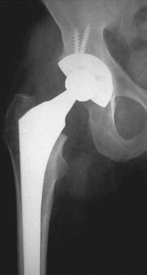
Total hip replacement precautions
Posterior total hip replacementa
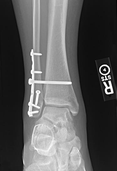
Weight bearing Bi-malleolar fracture treated with internal fixationb
Do not exercise or discontinue exercise with signs of extreme fatigue, sweating, shoulder or chest pain
Exercise is contraindicated if systolic blood pressure is above 200 mm Hg or below 80 mm Hg or if diastolic blood pressure is greater than 100 mm Hg8,9
Discontinue exercise with signs of extreme fatigue, sweating, shoulder or chest pain
Discontinue exercise if blood pressure increases >10 mm Hg
Avoid hip flexion >90 degrees, adduction crossing midline, and internal rotation
Methods/Actions
Do not start to exercise or discontinue; refer for medical attention
Do not start to exercise; refer for medical attention
Monitor cardiovascular responses before, during, and after exercise
Non–weight bearing for 8 weeks, cast boot, and partial weight bearing (50%) after sufficient healing is present at 8 weeks
Choose exercises that do not involve hinging at the hip past 90 degrees
No gluteal or hamstring stretching past 90 degrees of hip flexion or flexion-adduction combinations
Care with functional transitions (sit-to-stand, transfers to the floor)
Exercise equipment settings may require adjustments (raise seat on stationary bike)
See Fig. 1.2
Use scale to ensure amount of force
Use assistive device or parallel bars to reduce and distribute forces
TABLE 1.2 Examples of Precautions and Contraindications—cont’d
Type of
Precautions
Diagnosis
Fall prevention High fall risk after bedrest
Posttrauma Contraindications
Screen for contraindications related to structural damage
Monitor dizziness or lightheadedness during changes in position
Fall prevention precautions
Avoid specific directions of force
Ankle lateral ligament moderate tear
Full examination to rule in or out the possibility of fracture in the presence of swelling, inability to bear weight, extreme pain, or positive screening tests
Possible ligamentous laxity after whiplash
Possible vertebral artery syndrome
Avoid valgus forces during weight-bearing exercises
Methods/Actions
Monitor blood pressure pre-exercise and with transitions
Monitor patient symptoms during transitions or changes in position
Guarding and gait belt if any symptoms or blood pressure drops with transitions
Choose single-plane motion in sagittal plane to avoid localized forces to the medial structures
Limit tensile forces Achilles tendon partial tear
Avoid full stretch of ligaments (plantarflexion and inversion: the direction of force causing the injury)d
Avoid full stretch of tendon with forces exceeding tissue strength
Ranges and movements that do not stress lateral ligaments (neutral starting position, mid-position, moving into full eversion) with exercise against elastic resistance, avoiding the full stretch of the ligament into inversion and plantarflexion—the position that resulted in the injury (see Clinical Examples 2.1 and 2.2)
Use bracing or taping to assist with limiting movement in full range (see Clinical Examples 2.1 and 2.2)
Limit range to mid-position of the tendon length (see Clinical Example 2.3)
Limit amount of force (see stress-strain curve in Chapter 2)
aFigure from Patton K, Thibodeau G. The Human Body in Health and Disease. 6th ed. St. Louis, MO: Elsevier; 2014, p 193.
bFigure from Rynders SD, Hart JA. Orthopaedics for Physician Assistants. St. Louis, MO: Elsevier Saunders; 2013, Fig 8.15.
cFigure from Manske RC. Fundamental Orthopedic Management for the Physical Therapist Assistant. 4th ed. St. Louis, MO: Mosby; 2016, Fig. 9.1.
dFigure from Manske RC. Fundamental Orthopedic Management for the Physical Therapist Assistant. 4th ed. St. Louis, MO: Mosby; 2016, Fig. 17.1.
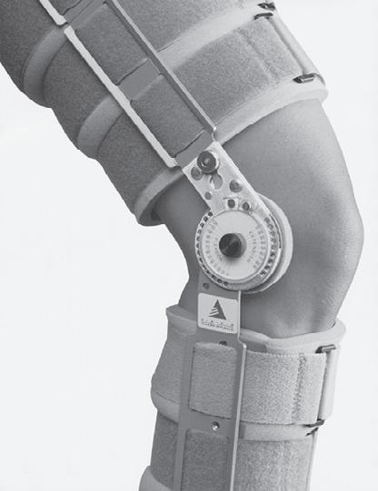
Fig. 1.2 Exercises for total hip replacement. (Cameron MH, Monroe LG. Physical Rehabilitation Evidence-based Examination, Evaluation and Intervention. Philadelphia: Saunders; 2007, Fig. 10.5.)
BOX 1.2 Contraindications and Precautions
Contraindication : Direction and degree of motion, extent of force, or other force-related factors that should be completely avoided to prevent further damage and allow structures to heal. Cardiovascular contra indications are used to prevent excessive stress on the cardiovascular system.
Precaution: Direction and degree of motion, amount of force, or other force-related factors that should be limited in order to allow structures to heal. Precautions can also be related to the need to rule out more serious damage or to evaluate responses to exercise. Other precautions are to prevent damage, injury, or adverse event, such as fall prevention.
consequences could result from movement or exercise. As an example, if a patient has symptoms consistent with acute angina or extremely high blood pressure, all exercise would be contraindicated and the patient would be referred for medical attention. If a patient has moderate spinal osteoporosis, spinal flexion against resistance or for long durations is contraindicated, but exercise in supported positions with low resistance may be possible.
Identifying precautions during the subjective history can result in modifying or avoiding examination procedures. An example of a precaution is limiting hip flexion to no more than 90 degrees in patients who recently had a total hip replacement with a posterior surgical approach to avoid stretching the posterior hip capsule. Therefore, the physical therapist would not attempt to measure hip flexion range of motion beyond 90 degrees during this patient’s initial
BOX 1.3 SINS
The acronym SINS is useful for judging the intensity and difficulty of the initial exercise.
• S = Severity (greater severity – use more caution in the choice of exercise)
• I = Irritability (greater irritability – use more caution in the choice of exercise)
• N = Nature (observe precautions and contraindications related to diagnosis and comorbidities, elements of the pathology that influence type of forces allowed or avoided)
• S = Stage (phase of healing – use to determine type of exercise and focus on impairments or functional recovery)
evaluation. The precautions and contraindications are formulated based on subjective information reported by the patient, knowledge of pathology, and the possible identification of additional diagnoses or pathologies derived from the patient’s medical and surgical histories (see Table 1.2 ). There are common precautions that are based on postsurgical healing requirements for incisions, while others are related to the type and extent of tissue damage. Fall and weight-bearing precautions prevent injury (see Table 1.2 ). Contraindications and precautions can also be related to comorbidities (other medical conditions) such as hypertension or cardiac disease. 8,9,10 While most contraindications and precautions are extremely important during the early stages of healing, precautions related to medical comorbidity are followed throughout the rehabilitation process.
SEVERITY, IRRITABILITY, NATURE, STAGE
The acronym SINS (Box 1.3) can be used to help determine the intensity and difficulty of the initial choice of exercise. In the subjective interview, the therapist collects information from the patient, including medical history, relevant medical tests, symptoms, and the relationships with movement. The therapist will use the interview findings as well as intake forms and validated tools to supplement the patient interview and determine an initial hypothesis to help plan further examination. The SINS acronym can assist with an early clinical judgment of whether the examination needs to be cautious or can be somewhat more aggressive.
Severity
Severity is usually determined by the amount of pain reported, but it can also be based on the extent of physical damage. The greater the severity of the symptoms or damage, the more cautious the initial exercise choice should be and the slower the exercise progression (Clinical Example 1.1). For example, a patient who is recovering after a fracture that required surgical fixation will be progressed cautiously, taking into account the amount of bone healing. A patient with a mild ankle sprain can be progressed faster than a patient with a moderate ankle ligament tear.
Irritability
The concept of irritability describes how easily the patient’s symptoms are aggravated (Clinical Example 1.2). A patient who has easily reproducible symptoms will benefit from a conservative initial exercise plan and slow progression until his or her symptoms are not as responsive to specific movements or positions. Conversely, a patient with low irritability may be tested at a higher level of exercise or progressed more rapidly. Irritability can be defined as the degree of activity resulting in symptoms (pain, tingling, numbness, or other subjective complaints) and the length of time taken for symptoms to subside.11 The clinical judgment of irritability also takes into account the severity
of the complaints. Questions that will help with the judgment about the degree of irritability include:
1. How easily are symptoms reproduced?
If mild activity (specific movement, exercise, or position) results in increased symptoms, the condition can be regarded as a highly irritable. Symptoms that are only reproduced with more aggressive activity would be regarded as less irritable.
2. How long do the symptoms last?
If the symptoms increase substantially and last for a long time after being exacerbated, the symptoms can be regarded as more irritable.
CLINICAL EXAMPLE 1.1 Severity: Degrees of Ankle Ligament Tears
Patient A
Patient A is a 26-year-old man who sustained a mild ankle sprain (Grade I; see Fig. A) while jogging. He was referred to physical therapy for progressive exercise.
Evaluation: Low Severity
The patient may be able to start weight-bearing exercises in a double-leg standing position almost immediately but should be monitored closely for swelling and pain.
Patient B
Patient B is a 30-year-old woman who sustained a complete ankle ligament tear (Grade III; see Fig. B) during an accident 3 months ago. The ligament was surgically repaired and she was non–weight bearing on crutches with the ankle immobilized in a cast for 6 weeks, followed by protection using a walking boot for 2 weeks. The patient was referred to physical therapy for progressive exercise 8 weeks after surgery and was allowed to begin partial weight-bearing exercises.
Evaluation: High Severity
The repaired ligaments should still be protected from high levels of force until there is sufficient tissue healing and muscle support. This is particularly important in the planes of movement that would increase tension on the ligaments and pull on the fracture. Initially, non–weight-bearing exercises are preferred in order to limit forces and protect the patient from poor balance responses after using crutches for 6 weeks. Equipment such as the leg press machine is used to initiate weight bearing with reduced vertical compression forces.
CLINICAL EXAMPLE 1.2 Irritability
Two patients, A and B, complain of neck pain and occasional referred pain and tingling into the upper extremity at their initial physical therapy appointment. They report the following symptoms on their second visit.
Patient A
Patient A reports increased neck and tingling pain down the arm that started 4 hours after the exercise session; the symptoms are still at the increased level 3 days later.
Evaluation
Patient A is exhibiting signs of high irritability and would need a more conservative approach. The patient would be asked to report any symptoms that increase after the exercise session. If any pain or other symptoms are reproduced after the session, exercise choices would be reevaluated and the type and dosage changed.
A B
(A) Partial tear of the anterior talo-fibular ligament. A mild Grade I tear may only result in some of these fibers being disrupted. (B) Complete tear of all three ligaments of the ankle (anterior and posterior talofibular ligament, calcaneofibular ligament) results in complete instability. (From Manske RC. Fundamental Orthopedic Management for the Physical Therapist Assistant. 4th ed. St. Louis, MO: Mosby; 2016, Fig. 17.2.)
Comparison of Patients A and B
Both patients will require the same precautions to allow ligament healing (i.e., limit the length of the lateral ankle ligaments and the amount of force on the ligament; avoid full inversion and full plantarflexion). However, the severity of the damage is greater in Patient B than in Patient A, and thus the surgical repair requires strict precautions with immobilization for 2 months and slow progression once the cast is removed. Patient A will be able to start progressive movement in the sagittal plane and limited movement into eversion as soon as the effusion resolves, often within 3–5 days.
Patient B
Patient B reports slightly increased neck pain while performing the exercise in the clinic but that the pain was immediately reduced after the exercise was stopped; the patient has not returned to the clinic since.
Evaluation
Patient B is exhibiting signs of low irritability.
Comparison of Patients A and B
Patient A’s symptoms can be characterized as more irritable than those of Patient B. Therefore exercise choices are more conservative, progressed more cautiously, and if symptoms are exacerbated, exercise would be adjusted promptly.
Nature
The nature of the problem is related to the medical and physical therapy diagnosis and is determined from the medical history, the type of symptoms, diagnosis, and prognosis. The information is collated from a combination of symptoms, general patterns, and specific tests. The medical and surgical history is vital when considering precautions, appropriate exercise choices, and timelines for progression. Specific modifications for postsurgical protocols are based on the physician’s recommendations as well as the therapist’s consideration of the extent of damage, tissue type, and comorbidities. Two categories related to the type of trauma—repetitive strain and major traumatic injury (see Chapters 2 and 6 )—and two categories related to extremes of mobility (see Chapters 3 and 6 ) will be described later in the text.
CLINICAL
EXAMPLE
1.3
The impact of disease will also influence the choice of exercise. For example, if a patient has rheumatoid arthritis with major joint destruction and chronic edema, lower forces and limited weight-bearing exercise is indicated (Clinical Example 1.3). The therapist will also determine impairments that influence function (discussed in Chapters 3 and 4).
Stage
Stage can refer to the phases of inflammation, tissue repair, and functional recovery or describe the progression of a disease process. Disease processes are often described in terms of stage; for example, Stage IV breast cancer is more advanced than Stage I breast cancer. The phases of tissue repair will play a significant role in guiding the exercise goals, selection, and progression. Stages often follow time guidelines, but exercise management can be initiated at any stage, and the progression
of Problem: Influence of Diagnosis and Extent of Pathology on Exercise Choices
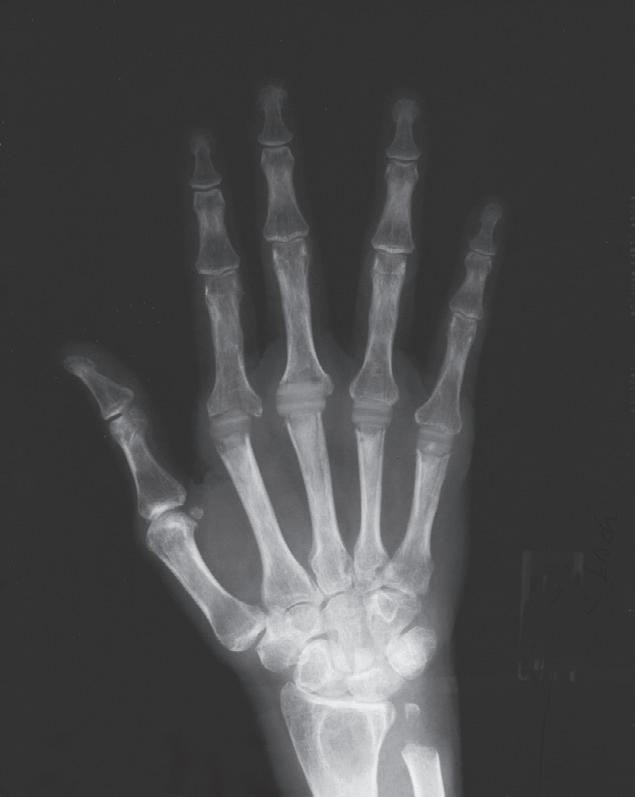
Patient A
A 60-year-old journalist with a history of chronic rheumatoid arthritis was referred for management of hand and wrist dysfunction , which lead to difficulty with dressing, bathing, writing, and grasping objects. Radiographs show joint damage resulting from synovial inflammation and loss of cartilage with joint space narrowing, subluxations, and deformities. She has had a recent exacerbation of her symptoms, and there is some redness, swelling, and heat in the hand. She is scheduled for joint replacements. The goal of physical therapy is to teach the patient active exercise, joint protection techniques, and ways to modify her activities of daily living that will limit further progression of deformity.
Precautions
The evaluation requires a conservative approach because rheumatoid arthritis, which is a systemic connective tissue disease, has a high risk of initiating inflammatory responses. In addition, the anti-inflammatory medications the patient is taking contribute to weakness of the connective tissue. Thus active movement is evaluated without any overpressure and with special consideration to the patient’s tolerance level.
Minimal force is applied to the wrist joints during the examination and no hands-on techniques are used on the fingers until the heat and redness have dissipated.
Exercise Implications
Severe damage to the ligaments and joint structures restricts the amount of force and directions of force that can be used, and exercise will focus on improving functional movement using joint protection techniques. Splinting is used initially with the exercise emphasis on proximal shoulder, elbow, and wrist motion until the inflammatory processes are less active. Once the inflammatory process has decreased, exercise choices that assist movement with lower forces, such as aquatic therapy, would be useful.
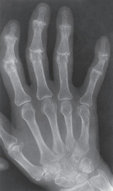
(1) Normal hand radiograph. (2) Hand radiograph for a patient with rheumatoid arthritis, similar to Patient A. Note the metacarpal phalangeal joint destruction and ulnar drift in the index through fifth fingers with extreme damage and dislocation at the second and third metacarpal phalangeal joints. The examination would need to be extremely cautious, with minimal forces on the fingers to limit further damage. (From Eisenberg R. Comprehensive Radiographic Pathlogy. 5th ed. St. Louis: Mosby; 2012, Fig. 4.28.)
CLINICAL EXAMPLE 1.3 Nature of Problem: Influence of Diagnosis and Extent of Pathology on Exercise Choices—cont’d
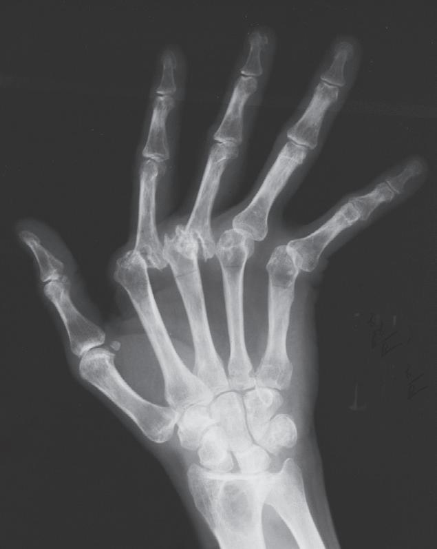
Patient B
A 53-year-old administrative assistant was referred to the clinic for management of pain in her hands. She has been diagnosed with osteoarthritis.
Precautions
Forces that increase the angulation of the index or fifth fingers would be adjusted in this patient. Excessive force is limited for the proximal and distal interphalangeal joints.
Exercise Implications
Active or passive movement may be limited by bony changes. Functional movement and exercise would need to be evaluated, keeping in mind the need to distribute forces and avoid excessive angulation.
Comparison of Patients A and B
The amount of force applied to both patients would need to be monitored, but Patient A’s inflammatory disease condition requires very strict precautions and extremely limited loads. Patient B might be able to start with functional movement reeducation and with exercises at a slightly higher level than Patient A. Both patients will need to be monitored for joint effusion and exacerbation of symptoms, but the expectation for the speed of exercise progression would be higher for Patient B.
3
Radiograph showing osteoarthritic changes similar to Patient B. There is generalized osteopenia with joint space narrowing at the proximal and distal interphalangeal joints and osteophytic changes. While range of motion at the second and third proximal interphalangeal joints and distal fifth interphalangeal joint may be restricted, there is less need for caution here than with Patient A, and exercise would be progressed as tolerated. (From Eisenberg R. Comprehensive Radiographic Pathlogy 5th ed. St. Louis: Mosby; 2012, Fig. 4.23.)
through stages varies. This chapter will briefly summarize each phase. Chapter 2 will cover the goals for each phase, the characteristics of various tissue types, and how these characteristics impact exercise prescription.
Phase I: Inflammation (Immediately to Approximately 10 Days)
Inflammation is the normal initial response to injury and is necessary for healing; it is typified by redness, swelling, increased warmth, and pain. Resolving the inflammation as soon as possible can help prevent the ill effects of prolonged immobility and disuse. The forces that the tissue is normally designed to withstand may need to be decreased to allow resolution of the inflammatory process. Exercises during this phase can be used to reduce inflammatory by-products while avoiding increasing the inflammatory responses. Chronic inflammation related to inflammatory arthritis, infection, or connective tissue disorders may continue for longer than 10 days, and there are occasions where aggravation of a chronic condition results in low-grade chronic inflammation for longer periods.
Phase II: Tissue Repair (10 Days to
According to Tissue Type)
3 Months, Variable
Fibroblasts lay down tissue to repair the injured site and macrophages remove waste tissue. The new tissue is organized in response to stresses placed on it by a slow progression of either stress or strain (usually separately). Too much stress can reinitiate the inflammatory process and result in damage to the newly formed tissue. Exercises during this phase should address impairments.
Phase III: Functional Recovery (6 Weeks to 2 Years, Variable by Tissue Type)
Mature tissue is able to withstand the demands of functional activities. Exercises during this phase help restore the patient’s functional deficits, or activity and participation limitations.
TYPES OF EXERCISE
The types of exercise are characterized by the amount of muscle activation and by the assistance provided for movement or the specificity of exercise to address impairments while providing optimal support for healing (Box 1.4).
Passive
Passive movement is performed by someone other than the patient (Video 1.1), by a mechanical device, or using gravity. Patients can also perform passive movement using another limb, without active muscle contraction of the affected area. Passive range of motion exercises are used to maintain range of motion in two instances: if a patient is unable to actively move the body part or if muscle activity at the time is likely to cause further damage.
Active-Assisted
Active-assisted exercise is conducted by the patient using some assistance from the other limb (Video 1.2), another person, mechanical devices, or gravity (Video 1.3). Active-assisted exercise may be used to help the patient achieve full range of motion when weakness or pain is limiting the range of motion or as a progression during the recovery when limited muscle force on structures is preferable.
BOX 1.4 Types of Exercise
Passive: Movement is performed by the therapist or a mechanical device or by the patient without active muscle contraction
Active: Movements are performed by the patient, with or without assistance or resistance
• Active-assisted: Movement is performed by the patient with assistance from therapist, mechanical devices, or the patient using another limb
• Active: Performed without any assistance from the therapist or mechanical devices, against gravity or external forces
• Isometrics: Muscle contractions performed without joint motion
• Isotonic concentric: Performed with joint motion with a shortening muscle contraction
• Isotonic eccentric: Performed with joint motion with a lengthening contraction
• Resisted: Movement against gravity or external resistance, such as weights
Active
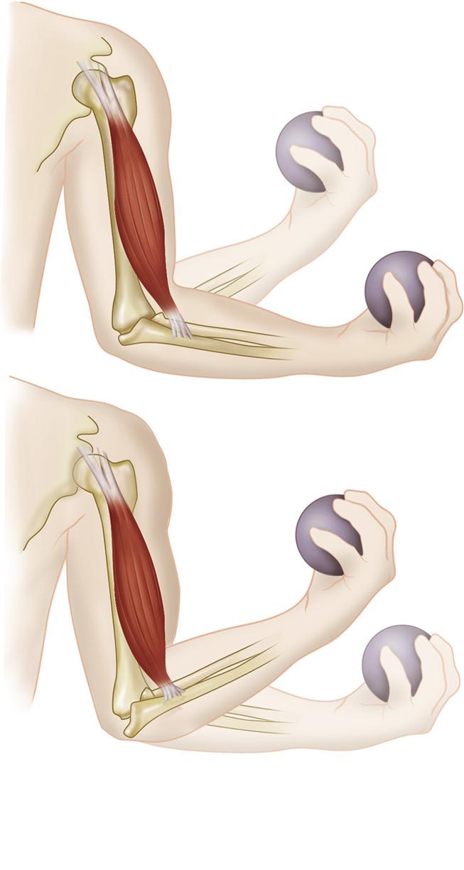
Fig. 1.3 (A) Isotonic (concentric and eccentric) contractions. (B) Isometric contractions. (From Thibodeau GA. Structure and Function of the Body. 14th ed. St. Louis, MO: Elsevier; 2012.)
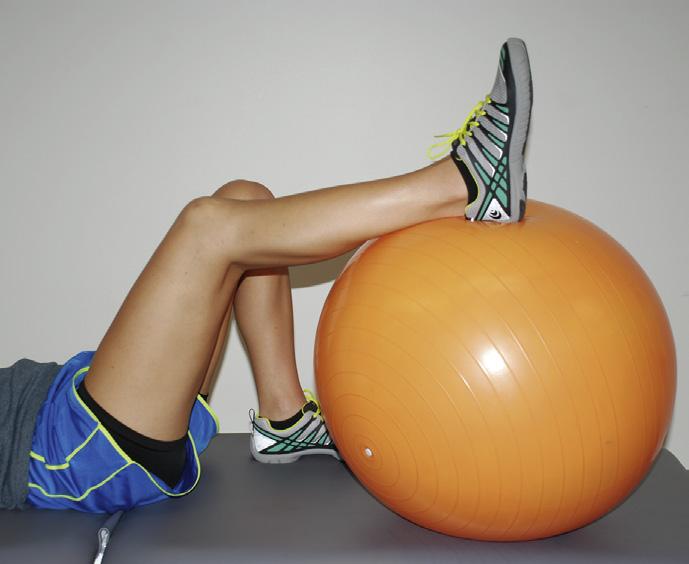
Active exercise is performed by the patient without any assistance from the therapist or mechanical devices (Videos 1.3 and 1.4).
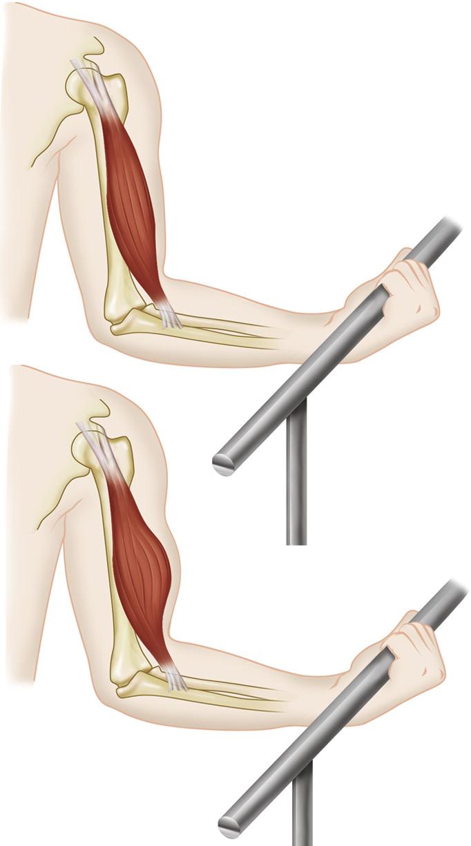
Resisted
The patient moves limbs or the body against some form of resistance such as gravity, external forces from free weights, pulley systems (Video 1.5), mechanical equipment, or therapist-applied manual resistance. Isokinetic exercise is a type of resisted exercise using specialized equipment to maintain a constant velocity of motion while the resistance adjusts throughout the range of motion.
TYPES OF MUSCLE CONTRACTION
Isometric
Isometric contractions are performed without joint motion and the muscle length remains constant. The use of isometric contractions may be indicated when there is joint damage and joint motion is contraindicated or likely to increase pain (Video 1.6), during early strengthening when the limb is supported, or to promote circulation through alternating contractions. Isometric contractions are also required for some functional activities ( Fig. 1.3 ).
Isotonic: Concentric
Isotonic contractions are performed with joint motion and the muscle length changes. A concentric contraction occurs with a shortening action of the muscle and results in joint motion (Video 1.7 and 1.8).
Isotonic: Eccentric
Eccentric contractions are also a form of isotonic contraction and occur when the muscle controls movement against
SUMMARY
• Precautions and contraindications related to tissue damage and comorbidities need to be established and followed throughout the course of the evaluation and treatment.
• Elements that influence choice of exercise include severity, irritability, nature, and stage (including phase of inflammation, tissue repair, and functional recovery). This information is gathered from the subjective history and the medical background.
REVIEW QUESTIONS
1. Differentiate between a precaution and contraindication and provide an example of each.
2. List and describe the four factors used to determine how cautious or aggressive the initial exercise and examination process will be.
3. Differentiate among passive, active-assisted, and active movement.
REFERENCES
1. American Physical Therapy Association. Guide to Physical Therapy Practice. http://guidetoptpractice.apta.org.
3. Magee DJ, Sueki D Orthopedic Physical Assessment Atlas and Video: Selected Special Tests and Movements. St. Louis: Elsevier Saunders; 2011.
4. Reese NB, Bandy WD Joint Range of Motion and Muscle Length Testing. 3rd ed. St. Louis: Elsevier Saunders; 2017.
5. Cleland J, Koppenhaver S, Su J Netter’s Orthopaedic Clinical Examination: An Evidence-Based Approach. 3rd ed. Philadelphia: Elsevier; 2016.
6. World Health Organization. International Classification of Diseases http://www.who.int/classifications/icd/en/
resistance (including gravity) by lengthening or slowing the movement (Videos 1.2 and 1.5). Eccentric contractions occur during functional activities to control, counterbalance, or resist motion. The noncontractile components of the muscle (tendon, connective tissue components of muscle fibers) absorb a greater amount of the tensile forces than do concentric contractions.
• Evaluation of SINS can assist with deciding on how cautious or aggressive the exercise approach will be before starting the examination.
• Type of exercise is defined based on amount of muscle activity and type of contraction.
4. Explain the difference between isometric and isotonic muscle contractions.
5. Explain the difference between concentric and eccentric movement.
7. World Health Organization. Towards a Common Language for Functioning, Disability and Health ICF. 2002. http://www.who.int/ classifications/icf/icfbeginnersguide.pdf?ua=1.
8. Boissonnault WG. Primary Care for the Physical Therapist. Examination and Triage. 2nd ed. St. Louis, MO: Elsevier Saunders; 2011.
9. American College of Sports Medicine. ACSM Guidelines for Exercise Testing and Prescription. 9th ed. Philadelphia: American College of Sports Medicine Lippincott Williams and Wilkins; 2014.
10. Smirnova I. The cardiovascular system. In: Goodman CC, Fuller KS, eds. Pathology: Implications for the Physical Therapist. 4th ed. St. Louis, MO: Elsevier Saunders; 2015.
11. Hengeveld E, Banks K Maitland’s Peripheral Manipulation. 5th ed. Edinburgh: Churchill Livingstone Elsevier; 2014.
Review of Foundational Concepts
Kim Dunleavy
OBJECTIVES
Upon completion of this chapter, the reader will be able to:
1. Compare the causes, signs, and symptoms of traumatic versus repetitive strain injuries.
2. Describe the implications of the history of the damage on the examination and treatment using exercise.
3. List and describe the phases of tissue repair and recovery and factors for promotion of optimal recovery.
TISSUE DAMAGE: TRAUMATIC VERSUS REPETITIVE STRAIN INJURIES
Musculoskeletal injuries can be categorized as either traumatic or repetitive strain injuries (Box 2.1 and Table 2.1). Traumatic injuries occur when major forces are applied beyond the body part’s capacity to withstand (Fig. 2.1). The individual can identify a specific incident when the injury occurred and often complains of immediate functional difficulty. Signs include inflammation (swelling, pain, redness) and inability to use the region. The sooner the symptoms appear, the greater the likelihood of major damage. Examples of traumatic injuries are fractures, cartilage tears, tendon ruptures, or complete muscle tears. Traumatic injuries are related to excessive load (stress), excessive tensile force (strain), or combinations of both types of forces. These injuries often occur with high-speed, uncontrolled movements, or when the individual is unable to avoid or withstand external force (Fig. 2.1A). Another possible mechanism of injury is when a patient attempts to move one body part in one direction while stabilizing another body part, such as when the foot is firmly planted and the individual attempts to turn or move the body in another direction (Fig. 2.1B).
BOX 2.1 Traumatic Versus Repetitive Strain Injury
• Traumatic injury: An injury resulting from a major force in a single, identifiable event.
• Repetitive strain injury: An injury occurring over time, from the cumulative effect of repeated minor trauma to an area.
4. Describe how tissue healing concepts for bone, muscle, tendon, ligament, and cartilage influence goals for exercise therapy.
5. Describe the forces that should be limited or applied through exercise therapy to allow optimal tissue recovery.
6. Describe the biomechanical concepts that will influence functional movement and therapeutic exercise techniques during exercise therapy rehabilitation.
TABLE 2.1 Traumatic Versus Repetitive Strain Injury
Traumatic Injury (or Resultant Surgery)
Extent of forces causing damage
Major forces (speed, torque, localized direction)
History Sudden onset: History of an identifiable incident
Signs of inflammation Signs of inflammation (redness, heat, swelling, pain) appear immediately or shortly after incident, constant pain and limited function
Implications for evaluation
Need to identify extent of tissue damage (for possible referral) and to identify precautions and contraindications
After surgical management, evaluation may need to be adjusted based on surgical technique and postoperative contraindications and precautions
Repetitive Strain Injuries
Minor forces (repeated, concentrated)
Gradual onset: slow build up and multiple minor incidents, OR patient is not able to identify an incident
Low-grade inflammation or no visible redness, heat, swelling; pain experienced with movement, specific positions, or after onset of movement not constant, symptoms can be due to fibrosis
Need to identify extent of damage and possible contributing factors
TABLE 2.1 Traumatic Versus Repetitive Strain Injury—cont’d
Traumatic Injury (or Resultant Surgery)
Repetitive Strain Injuries
Implication for choice of exercise
Contraindications and precautions determine which exercises cannot be used during Phases I and II and would be progressed slowly throughout recovery
Post injury recovery or surgical guidelines may be used
More complex choices related to initial starting point for exercise program
Exercise program will need to address contributing factors
Biomechanical alignment analysis and constant observation during progression is important. Functional movement reeducation will be important to prevent recurrence
Assess need for external support (bracing/taping/ orthotic/assistive device)
2.2 Repetitive strain injuries. There is often no specific incident for repetitive strain injuries: patients report a slow onset over a period of time or after an intense period of activity. There are also contributing impairments that contribute to microfailure. Throwing injuries, such as shoulder impingement, labral tears, and anterior laxity of the shoulder, are examples of repetitive strain injuries. (Courtesy of Derek Drake and David Cox.)
Repetitive strain injuries occur over time when minor forces are applied to an area of the body and the structures are unable to an area of the body and the structures are unable to withstand the repeated forces. Repetitive injuries result from an accumulation of forces due to overuse, insufficient time to replenish tissue strength, or the concentration of force on one area of a joint, region, or tissue. One or more factors, such as lack of joint range of motion or flexibility, insufficient muscle strength or endurance, coordination inefficiencies, or structural malalignments, can contribute to excessive force concentration (Fig. 2.2). Forces contributing to repetitive strain injuries include (1) compressive forces, (2) angulation, (3) rotation, (4) tensile strain, or combinations of force directions. Examples of repetitive strain injuries are stress fractures, cartilage degeneration in osteoarthritis, or tendinopathy (Videos 2.1 and 2.2).
ABILITY TO WITHSTAND FORCES (STRESSSTRAIN RELATIONSHIPS)
Each tissue has the capacity to withstand load (amount of external force or forces applied to the tissue). The larger the cross-sectional area, the greater the area available to distribute force and the more force the tissue can withstand. Stress is defined as the amount of force per cross-sectional area, while strain describes the tensile forces resulting in change in length (Fig. 2.3 and Box 2.2). The direction of the load can be described as a compressive (causing tissue to condense) or tensile (causing tissue to lengthen or expand) force.
Fig. 2.1 Traumatic injury involves either (A) impact or (B) rapid movement away from a stabilized body segment (yellow arrow) with localized stress such as angulation (red arrows). ([A] Courtesy of Derek Drake and David Cox.)
Fig.
Beginning of movement
Physiological ROM
injury (Phase I to II)
Plasticity Total failure
Physiological loading 1–2 degree injury
Fig. 2.3 Stress-strain curve showing microfailure and macrofailure. (Fig. 1.1 from Magee DJ, Zachazewski J, Quillen S. Scientific Foundations and Principles of Practice in Musculoskeletal Rehabilitation. St. Louis, MO: Saunders Elsevier; 2007.)
BOX 2.2 Stress-Strain Definitions
• Load: The amount of external stress applied to tissue
• Stress: The ability to withstand a given amount of force per cross-sectional area
• Strain: The ability to resist lengthening.
• Elasticity: The ability to return to original length after application of force
• Plasticity: Ability to deform with permanent changes (sometimes often desired for improvements in mobility)
• Failure: Tissue damage
• Microfailure of small amounts of tissue occurs after the yield point in the plastic range
• Macrofailure of entire tissue complex occurs after the failure point with rupture
Each tissue has a different stress-strain relationship depending on the material properties of the tissue. Tissue also has some ability to deform and return to its initial position (elasticity), ability to deform over time and retain the new position (plasticity), or slowly return to its initial position (viscoelasticity) (see Fig. 2.3). All of these properties are determined by (1) the alignment of the collagen fibers, (2) the percentage of elastin versus collagen fibers, (3) the amount of ground substance, and
The stress-strain curve varies slightly for different tissues but essentially consists of the same components: a toe phase where the connective tissue slack is taken up, followed by a linear relationship between the amount of stress and the strain until the maximum elasticity (yield point) is reached. Up to this point, if the stress is removed, the tissue will return to its previous length without any permanent changes. If the stress is increased past this point, plastic or permanent changes will occur. In some instances, tissue plasticity is desirable, such as with stretching or joint mobilization. The threshold or failure point may be reached with lower stress or strain in healing tissue or after immobilization (see Fig. 2.3).1,2 The stress and strain concepts are important considerations for establishing precautions to effectively limit further damage and when adjusting exercises to provide sufficient forces for promoting optimal regeneration. If the tissue reaches the endpoint of the ability to withstand the forces, failure will occur. Partial damage of tissue results Some microfailure
(4) the strength of cross-links. Each tissue differs in the extent to which it can deform and return to its normal length.1–3 Collagen can return to normal after reaching of 2–3% of initial length, while increases of 4–6% result in microfailure, and 6–10% is likely to result in macrofailure (see Fig. 2.3).1,3
in microfailure, while macrofailure is a complete tissue tear. Microfailure can occur after the yield point when the loads reach the plastic region of the stress-strain curve and tissue is unable to return to the initial length after the force is removed. Application of repeated forces without sufficient time for the tissue to recover and mild damage after a traumatic incident can both result in failure of some fibers and are both described as microfailure. Macrofailure occurs when the failure point is reached and permanent rupture of collagen occurs.1,2
Ligament, tendon, and muscle tears are often classified according to the extent of damage, such as Grade I (mild), Grade II (moderate), and Grade III (complete disruption) (Box 2.3).4,5 Grade I damage is the result of microfailure. More fibers reach the failure point in Grade II. In Grade III, complete macrofailure and disruption occurs.4 The greater the tissue damage, the greater the inflammatory response and the risk of further structural damage to the affected or nearby tissues.
After diagnosis of the location and degree of damage, appropriate medical or surgical management should follow.
Precautions or contraindications are determined based on the extent of damage and the need to protect healing structures. The process of orthopedic physical therapy examination and diagnosis is described extensively in other texts.4–6 The extent of the damage is considered when determining the severity of the injury (Clinical Example 2.1; see Chapter 1), which guides the aggressiveness of the exercise choices and progression. If tissue is partially torn, the amount of stress or strain that will result in further damage is less than for intact tissue. The anatomical plane and tissue type are used to establish precautions related to which directions of movement and forces are safe and promote optimal healing (see Box 2.1 and Clinical Example 2.1; Videos 2.3 and 2.4).
BOX 2.3 Classifications of Tissue Damage
• Grade I: Mild tear (microfailure)
• Grade II: Moderate tear
• Grade III: Complete disruption (macrofailure)
CLINICAL EXAMPLE 2.1 precautions During healing after Grades I–III ankle Sprain
The types of precautions will depend on the structure and function of the damaged tissue. The degree and length of time required for precautions increase based on the extent of tissue damage (see the following figures).
Grade I Ankle Sprain:
Stress and strain need to be limited more for Grade III tears. (Image on left from Mansfield P, Neumann D. Essentials of Kinesiology for the Physical Therapist Assistant 2nd ed. St. Louis, MO: Mosby; 2014.)
• Brace or taping is used to limit inversion for 6 weeks for walking, up to 3 months or longer for running and sports.
• Avoid placing the anterolateral ligaments in the fully stretched position (full combined plantarflexion and inversion). Midrange plantarflexion and dorsiflexion or isometrics are used to assist with resolution of the inflammatory by-products.
• While the ligament heals, full stretch of the ligaments in plantarflexion and inversion and outer range of the peronei is avoided, especially with high forces in weight-bearing positions. Often the position and direction of force of the injury is the same as the directions that need to be avoided.
Continued
CLINICAL EXAMPLE
Direction of force to be avoided the most is the combination of plantarflexion and inversion for an anteriorlateral ligament sprain.
Grade II Ankle Ligament Tear:
• Avoid any stress on the ligament; the ankle is maintained in neutral position using a cast boot until sufficient healing (approximately 6–8 weeks), followed by air cast or ankle brace (consistently for additional 6 weeks, longer for running and sports). Isometric contractions are indicated to assist with resolution of inflammatory by-products.
TISSUE REPAIR AND FACTORS FOR PROMOTING OPTIMAL RECOVERY
The degree of healing and repair are important considerations when selecting exercises and setting appropriate parameters. The phases of tissue repair will direct the goals, type, intensity, and progression of exercises. General guidelines for exercise selection, progression, and goals are presented here; however, personal factors (age, genetics, comorbidities) will influence both the initial starting point and the rate of progression through exercise programs.
Phases of Tissue Repair and Recovery
Therapeutic exercises can enhance tissue repair and recovery. Understanding the events, time frames, and objectives of each phase will guide exercise selection and progression. It is
• Take care with excessive stress or strain for additional 6–8 weeks Exercise is introduced and progressed in the sagittal plane first before the frontal plane.
Grade III Ankle Ligament Tear:
• No stress on the ligament until sufficient healing. Treatment is either full immobilization or surgical management. Weight-bearing precautions: Non–weight-bearing for 6–8 weeks with cast boot; partial weight-bearing with cast boot for additional 6 weeks.
• Continued movement limitation precautions similar to Grade II after allowed to bear weight at 3 months.
• Care with excessive stress or strain for additional 6–8 weeks dependent on muscle activation and function recovery.
the therapist’s responsibility to prescribe exercises during the appropriate time frame that will promote resolution of inflammatory by-products, provide sufficient forces to stimulate healing, and provide activities to challenge neuromuscular recovery while respecting the limits of each tissue.
Phase I: Inflammation (Immediately to 3–10 Days)
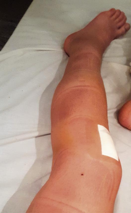
The inflammatory process is the body’s response to damage, infection, or abnormal cellular mechanisms, and is a necessary process for healing and repair (Box 2.4). Inflammation is the first response to tissue damage and is responsible for removing damaged tissue and initiating cellular mechanisms to promote repair and avoid further damage (Box 2.5).1 The early process of tissue damage includes bleeding and fluid release into joint (effusion) and extracellular spaces (edema) (Fig. 2.4), which also results in redness and heat in the inflamed area (Fig. 2.5).
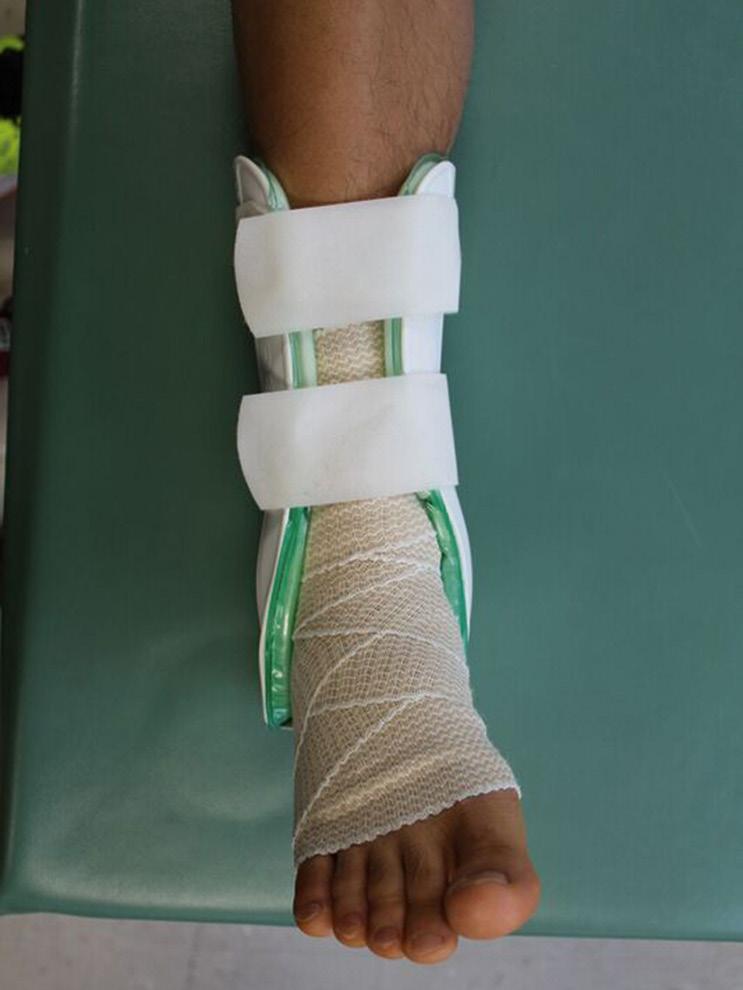
Ankle taping to limit inversion.
An Aircast brace is used to limit frontal plane motion while still allowing sagittal plane movement. The compressive bandage is used to assist resolving edema.
BOX 2.4 Symptoms of Inflammation
• Edema: An accumulation of excessive extracellular tissue fluid outside a joint
• Effusion: Increased intraarticular fluid in a synovial joint
• Heat and redness: Increased local vasodilation in region
• Pain: From damage to tissue, inflammatory mediators, hypoxia, pressure from edema and effusion
BOX 2.5 phase I Goals
• Limit inflammation and protect the injured tissue
• Assist with removal of by-products of inflammation (edema, effusion, metabolites), increase circulation to the area, and decrease pain related to hypoxia, muscle spasm, and vasoactive mediators
• Exercise can minimize the effects of immobility or deconditioning without placing excessive stress on injured structures.
A. NORMAL
B. EDEMA
(high protein content, and may contain some white and red cells)
Hydrostatic pressure
Colloid osmotic pressure
Plasma proteins
No net fluid or protein leakage
Increased interendothelial spaces
Vasodilation and stasis
Fluid and protein leakage
Fig. 2.4 Edema resulting from increased blood flow to damaged tissue. (A) Under normal circumstances, capillaries contain blood plasma and cells with a continuous exchange of fluid and nutrients based on the osmotic and hydrostatic pressure balance within the vessel and the extracellular space. (B) After trauma there is a rapid increase in blood flow, and the inflammatory responses extend the vasodilation and stress on the endothelium due to the increase in hydrostatic pressure. Water escapes into the surrounding extracellular space, resulting in effusion. If there is more severe trauma plasma proteins leak into the extracellular space, and, finally, blood cells leak into the surrounding tissue. The resulting increase in colloid osmotic pressure further increases fluid motion. The external pressure on the blood vessels also causes pain. (From Kumar et al., Robbins and Cotran Pathologic Basis of Disease. 8th ed. Philadelphia, PA: Saunders Elsevier, 2010.)
Fig. 2.5 (A) Immediate and secondary responses to traumatic injury. (B) Bruising, edema, and effusion after an anterior cruciate ligament (ACL) reconstruction.
TABLE 2.2 Goals of Exercise During phase I (acute Inflammation)
Goals
Protect injured structures
Increase circulation to region
Assist with removal of edema and effusion
Maintain muscle activation without excessive stress on injured structures
Exercise Choices
Avoid placing excessive load (stress or strain) on injured tissue where microfailure or macrofailure is likely to occur (see stress-strain curve)
Additional protection may be provided by use of assistive devices, bracing, or taping
Active low-intensity exercise of other joints around the injured region
Alternating active muscle contraction within available range (low intensity)
Isometric muscle contraction for short durations followed by relaxation
Generalized movement of the affected limb or trunk as well as localized muscle contractions listed above will help with motion of edema and effusion
Small-range active movement without stress on injured structures to maintain range or assist with removal of effusion. Decreasing effusion helps restore range if caused by increased intraarticular volume or pain at the end of range
Elements to Consider
Motion without strain may be possible in planes other than the plane controlled by the injured tissue
Muscle activity will assist with venous return, particularly if the limb is elevated
Only used if likelihood of bleeding is small
Active contractions are likely to provide mechanical “pumping” actions for circulation as well as promote movement of extracellular edema or intraarticular effusion
Motion of proximal or distal joint or other areas of the body can increase overall circulatory flow and challenge
Motion may increase immediately if effusion is the cause of limited range
Minimize effects of lack of activity or immobilization
Minimizing muscle atrophy and cardiovascular effects of immobility for the uninvolved regions and general body systems; may need to be adapted to follow precautions and allow adequate healing of the affected structure
Stimulation of nociceptive pathways occurs with the tissue damage and continues with the release of inflammatory mediators such as histamine and bradykinin (Fig. 2.6). Inflammatory responses also include a secondary vasoconstriction to reduce bleeding and muscle spasm to avoid further motion and damage. The inflammatory cycle is intended to restrict further damage and is completed by the removal of the damaged cellular debris and stimulation of fibroblastic activity. Scar tissue is formed to repair the structures.
Allow movement in other regions and joints, include equipment and unloaded exercise such as bike, aquatic therapy, upper body ergometer, upper body and trunk exercise, or resistance exercise with uninvolved body regions
During Phase I, the primary rehabilitation goals are to limit excessive forces to protect the damaged area and assist with resolution of inflammatory by-products (Table 2.2). Exercises can target the injured area or uninvolved areas and can assist with promoting circulation to the area, limiting pain, and assisting with resolution of edema.
Phase I exercises directed toward the injured joint should avoid excessive loads (stress) or stretching (strain), or the inflammatory phase can reinitiated. Movement in directions
Fig. 2.6 Secondary responses to traumatic injury.1
that do not place stress on the injured tissue is used to assist with removal of edema and effusion. The patient can exercise uninvolved areas to help deter the effects of disuse or inactivity, or, if the therapist has identified compensations or other areas of dysfunction likely to impact recovery, these impairments can also be addressed during Phase I.
If excessive load or tension is placed on tissue during Phase I, bleeding may continue and edema and effusion may increase, resulting in excessive hematoma formation. Extensive bleeding results in a greater likelihood of excessive fibrotic tissue and pressure on surrounding tissue, further increasing pain responses. The therapist should monitor the patient’s injured region for signs of inflammation: increased redness, pain, heat, swelling, or bruising. These signs indicate the need for caution and further evaluation. If present, the precautions should be strictly enforced, and load or tension on the area during exercises and activities of daily living reassessed and modified accordingly.
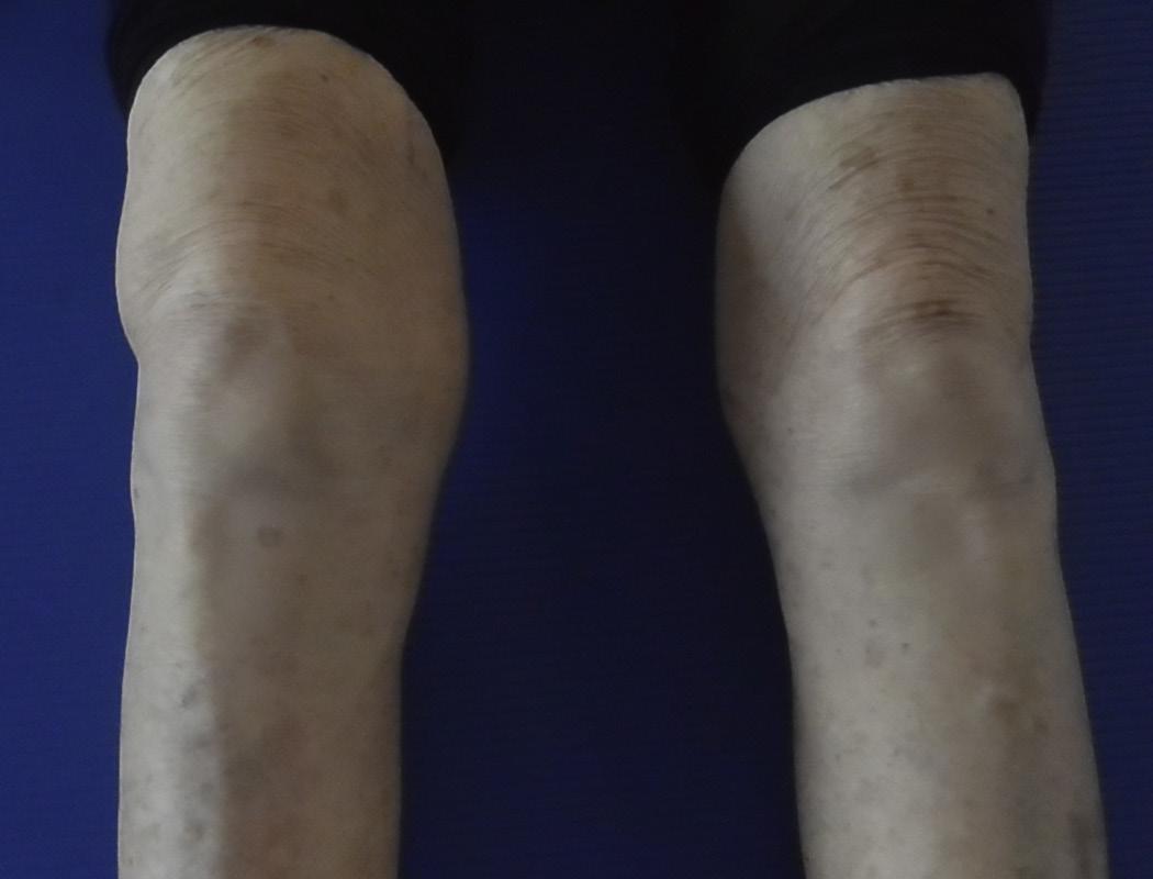
While the damaged tissue must be protected following acute inflammation, chronic inflammation related to repetitive strain injury calls for identifying and training to correct faulty biomechanics and compensations. If excessive forces on the joint are not resolved or other factors limit resolution of inflammation, chronic inflammation can result. Chronic inflammation due to excessive forces can lead to edema, effusion, and prolonged proliferation of fibroblasts, causing thickened connective tissue and mobility limitations (Fig. 2.7 and Box 2.6).2 Pain, deconditioning, and compensations can also further complicate and contribute to excess stress and decreased ability to withstand strain.
Not all patients will enter the rehabilitation process while in Phase I of healing. During the initial evaluation, the physical therapist should determine where the patient’s injury falls
BOX 2.6 Differences Between acute and Chronic Inflammation
• Acute inflammation occurs with immediate trauma.
• Chronic inflammation can be a result of accumulation of forces, repeated microtrauma, or inadequate resolution of acute inflammation.
CLINICAL EXAMPLE 2.2 phase II and phase III Exercise for an ankle Ligament Injury
precautions
As the patient progresses to Phase II, the injured tissue is still protected in the plane of the ligament. Stress is increased slowly in shortened positions without placing strain on the ligament, and impairments such as diminished neuromuscular activation and endurance are addressed.
Phase II exercises are the same for all grades of injury; however, contraindications for stress and strain are in place for 6–8 weeks for Grade II injuries and 8–12 weeks for Grade III ligament tears. Phase II rehabilitation is only started once sufficient healing has taken place to lift complete contraindications for motion in the frontal plane or in the line of the ligament. Precautions are still required until further healing has taken place.
Exercise Choice
Exercise choices are related to the need to protect the ligament from excess strain and to progressive return to function.
A patient with a Grade I ankle sprain is progressed relatively rapidly while a patient with a Grade II or III injury is progressed slowly once there is adequate
healing. Exercises address impairments (limited mobility from the effusion, decreased muscle activation) and are designed to help protect the ligament using proprioceptive training and neuromuscular coordination.
Exercises are progressed from non–weight-bearing (Video 2.3) to weightbearing (Video 2.4) in Phase II to promote frontal plane stability and balance reactions (Video 2.5), with additional proprioceptive input or support from taping or bracing if needed.
In Phase III, activities are progressed to meet goals to return to the individual’s pre-injury function. Exercises are chosen to address activity limitations and impairments taking into account personal factors (athletic hobbies) and environmental needs (uneven surfaces). Running in place (Video 2.6) forward and backward is introduced before sideways (Video 2.7) or cutting. Changing directions rapidly and running on uneven surfaces is added last in the progression. The timelines to progress to Phase II are slower for more severe ligament damage in Grades II and III damage. The relative speed of exercise progression is also slower with Grades II and III injuries as the threshold for damage has decreased due to the injury and the immobilization/lack of normal activity.
in the tissue healing process, before prescribing the appropriate type and intensity of exercise based on the state of the injured tissue. Each tissue has unique healing time frames, and these time frames can be influenced by other factors such as age, genetics, nutrition, medication, and comorbidities1–3 (Clinical Example 2.2). Continued
Fig. 2.7 Chronic inflammation, effusion, and thickening in a patient with knee osteoarthritis.
0–6 weeks
Grade I
Phases of rehabilitation for Grades I–III ankle ligament injury. Precautions for a Grade II or III tear are in place during early stages (red box). Rehabilitation (yellow box) may be started in Phase I or II, depending on the extent of inflammation.
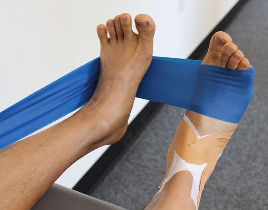
Isometric eversion activation, endurance, and strengthening using elastic resistance (Phase I and II).
Eversion muscle activation in supine against elastic resistance through isometrics or small-range to neutral exercise is used in Phase I to promote muscle activation. The peronei are often stretched and may be painful in the early phases after an inversion injury; therefore, muscle activation is used to improve circulation, remove inflammatory by-products, and decrease pain.
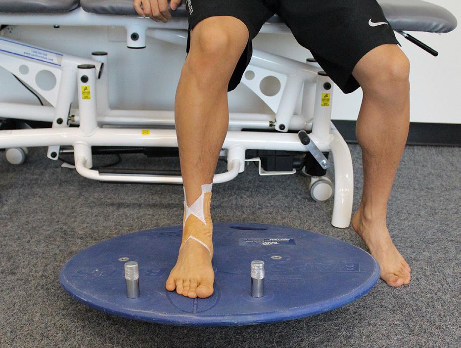
BAPS board strengthening progression (Phase II)
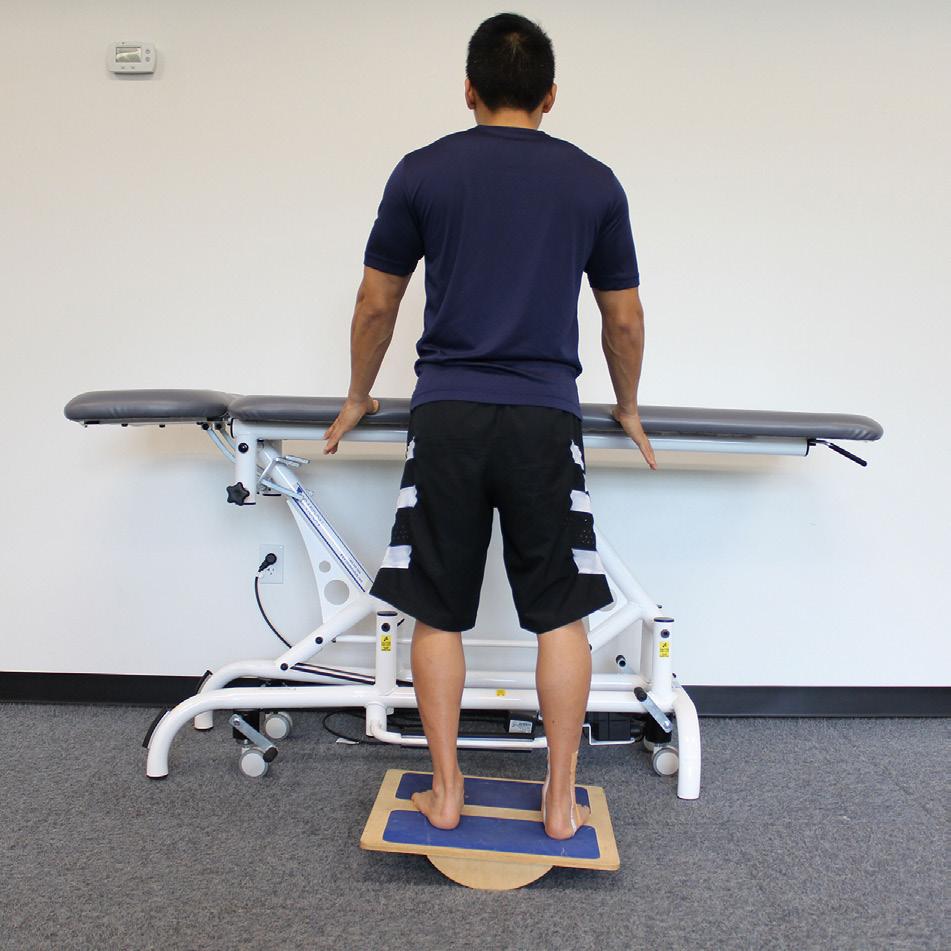
In Phase II, the resistance may be increased, and isotonic control is promoted using a balance board with a weight.
Double-leg balance activities on a balance board (Phase II).
Double-leg balance activities are started with some hand support. The balance board can be placed to allow plantar and dorsiflexion range first, followed by controlled weight shift from side to side to activate the lateral muscles. Weight transference from foot to foot is started in the sagittal plane or without movement in space by running on the spot (Video 2.6). Movement is progressed as the patient tolerates the activity, with slower progression for lateral movements and changes of direction (Video 2.7; see Fig. 3.6B). Transitional movements, such as cutting and changing direction to train the neuromuscular responses for weight acceptance and change of directions at high speeds, are included in late Phase III.
Phase II: Tissue Repair and Early Tissue Regeneration (5–10 Days to 3 Months, Depending on Tissue Type)
The primary mechanism for tissue repair occurs through fibrous tissue bridging the gap in damaged tissue. The amount of fibrous tissue is proportional to the amount of damage and the length of time that fibroblasts remain active. Phase II normally begins around 5–10 days after injury and can last through 3 months, depending on the tissue type (Box 2.7 and Table 2.3). The rate of production of new collagen fibers is greatest between 7 and 14 days post injury.1 The repair phase will overlap with the removal of inflammatory products.
The ability to monitor and evaluate a patient’s tolerance to load is an essential skill for therapists. Therapists should verify that there is adequate distribution of forces, graded load, and activity, and monitor responses to exercise (delayed-onset pain, edema, effusion, or other abnormal symptoms). Adverse responses warrant constant reevaluation and reduction of exercise load. If excessive load or tension is placed on tissue during Phase II, damage to the immature collagen or regenerated tissue results in inflammation and increased fibrosis. The subsequent edema and pain can also result in the patient
• Address impairments
• Provide optimal stimulus for tissue regeneration
• Minimize the effects of inactivity or immobilization
• Protect injured structures
• Improve muscle function without placing undue stress on injured tissues
TABLE 2.3 Goals for healing in phase II
Goals Exercise Choices
avoiding use of the region and compensating with other movement strategies. This can cause muscle atrophy, weaker connective tissue, and, ironically, further stresses on the damaged region. If additional stress occurs and damage of the immature collagen or the injured tissue ensues, the inflammatory cycle is reinitiated (see Phase I, Fig. 2.8).
The organization and strength of fibrous tissue is remodeled in response to stress applied to the structures during Phases II and III.1–3 Phase II exercises should promote tissue repair with targeted, smaller loads specific to the tissue type and function without reaching the ultimate failure point of the tissue. In addition, impairments such as muscle atrophy, joint restrictions, muscle tightness, pain, and compensatory mechanisms should be addressed during this phase (see Fig. 2.8 and Clinical Example 2.2). Exercises may target the impairments that are in the primary, injured area or in secondary regions that are contributing to biomechanical accumulation of forces.
Phase III: Tissue Maturation and Remodeling (6 Weeks to 2 Years, Depending on Tissue Type)
Phase III involves maturation and remodeling of the involved tissue as well as removal of excessive fibrotic tissue (Table 2.4 and Box 2.8). Tissue is remodeled by organizing the direction of collagen fibers to promote tissue capable of withstanding the forces needed for function. This process occurs in response to the appropriate type and direction of force for a particular tissue (see the section “Factors Influencing Healing: Tissue Characteristics”). The entire process may take up to 2 years, depending on the type of tissue.1–3 If necessary, Phase III treatment should
Protect injured structures Avoid placing excessive load on injured tissue or stretching to lengths where microfailure or macrofailure is likely to occur (see stress-strain curve).
Load (stress) is increased slowly in planes of movement of the injured structures, and range is limited to avoid maximum tension (strain).
Improve muscle function without excessive stress on injured structures
Minimize effects of inactivity or immobilization
Address specific impairments
Provide optimal stimulus for regeneration
Target other joints around the injured region with active, moderate-intensity exercise to assist with overall aerobic conditioning, improving muscle function and distributing forces.
Mobility, strength, specific muscular endurance, aerobic conditioning, coordination, or balance exercise
Elements to Consider
Load can be increased using isometric contractions or contractions in the inner or mid-range of injured structures to avoid extreme strain.
Midrange is determined by available range and not necessarily by normative values.
Other diagnoses, comorbidities, and higher severity or irritability are indications for stricter monitoring and extending periods of contraindications and precautions.
Positioning and choice of exercise can be adjusted to avoid excessive stress on injured structure with slow reapplication of stress.
Allow and progress movement in other regions and joints.
Tissue will regenerate in response to appropriate loads and strains placed on the structure. Initial loads should be light and directed in a way that places little stress on the healing tissue.
Monitor for adverse responses (delayed-onset muscle soreness, pain, effusion, or regression of improvements).