controversy. It seems that the pendulum continues to swing. From the early years of CT in the 1980s when few radiology professionals had even a basic idea of the dose their scanners delivered to the early 2000s when information surfaced concerning the effects of low-dose radiation on atomic bomb survivors who were irradiated as children prompted a heightened concern among healthcare professionals and the public. More recently, the pendulum seems to be moving back to a more moderate position with a number of strong voices in the field questioning the validity and applicability of extrapolating data from atomic bomb survivors to relatively low-dose radiologic studies. To increase the reader’s understanding of this hot-button issue, a summary of the debate has been included in the chapter on radiation dosimetry.
Lois E. Romans, RT, (R)(CT) Michigan Medicine, University of Michigan Ann Arbor, Michigan
User’s Guide
This User’s Guide introduces you to the helpful features of Computed Tomography for Technologists: A Comprehensive Text, second edition, that enable you to quickly master new concepts and put your new skills into practice.
Chapter features to increase understanding and enhance retention of the material include:
Examples of Exam Protocols are included for each major anatomical area.
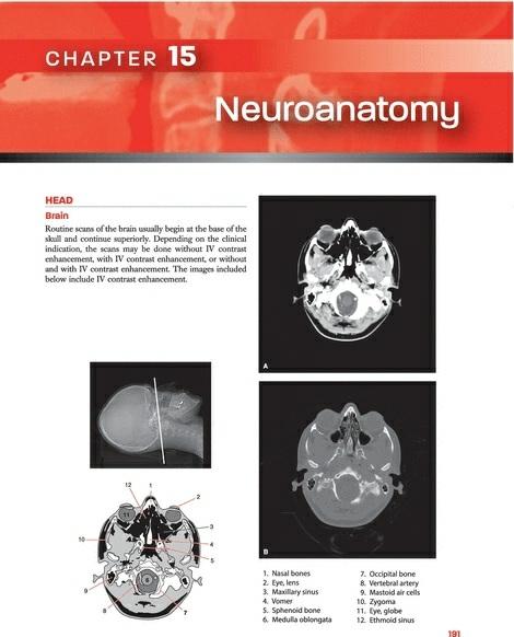
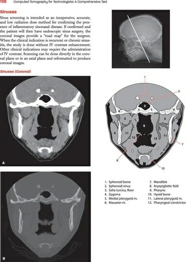
CT Cross-Sectional Slices accompanied by shaded diagrams and a reference image are featured in the Cross-Sectional Anatomy section of the book.
Reviewers
The publisher, author, and editors gratefully acknowledge the valuable contributions made by the following professionals who reviewed this text:
Matthew G. Aagesen, MD
Musculoskeletal Fellow
University of Michigan Health System
Ann Arbor, Michigan
Sulaiman D. Aldoohan, PhD, DABR
Assistant Professor
Department of Radiological Sciences
College of Medicine
University of Oklahoma Health Sciences Center
Oklahoma City, Oklahoma
Jonathan Baldwin, MS, CNMT, RT(CT)
Assistant Professor and Clinical Coordinator Nuclear Medicine
Statistical and Research Design Consultant
College of Allied Health
University of Oklahoma Health Sciences Center
Oklahoma City, Oklahoma
Jeff L. Berry, MS, RT (R) (CT)
Associate Professor, Radiography Program Director
Director of Advanced Imaging and Continuing Education
College of Allied Health
University of Oklahoma Health Sciences Center
Oklahoma City, Oklahoma
Jonathan R. Dillman, MD
Abdominal Radiology Fellow
University of Michigan Health System
Ann Arbor, Michigan
Lois Doody, Med
Instructor
Medical Radiography Program
BCIT
Vancouver, British Columbia
Susan K. Dumler, MS, RT (R)(M)(CT)(MR)
Lecturer
Fort Hays State University
Lecturer and Clinical Coordinator
Department of Allied Health
Fort Hays State University
Hays, Kansas
John W. Eichinger, MSRS, (R)(CT) ARRT
Program Director-Radiologic Technology
Technical College of the LowCountry
Beaufort, South Carolina
James H. Ellis, MD, FACR
Professor of Radiology
University of Michigan Health System
Ann Arbor, Michigan
Frances Gilman, MS, RT, R, CT, MR, CV, ARRT
Assistant Professor and Chair
Department of Radiologic Sciences
Thomas Jefferson University
Acknowledgments
I cannot overstate the contributions of Dr. James Ellis from the University of Michigan Radiology Department. I routinely handed him a sow’s ear and he unfailingly amazed me by returning a silk purse. His meticulous review went far beyond my wildest expectation. His thoughtful suggestions and patient guidance improved every aspect of the manuscript. The second edition greatly benefited by the expertise of Jeff Berry, Radiography Program Director, University of Oklahoma Health Sciences Center. His extensive educational experience ensured that each concept presented was consumable by the typical student and that new developments in the field were included. Special thanks to Dr. Saabry Osmany for his help in editing the chapter on PET/CT and for supplying PET/CT images. I gratefully acknowledge the expertise of diagnostic physicists Emmanuel Christodoulou and Mitch Goodsitt, who helped me make sense of the complex data available on radiation dose. Thanks to my dear friend and fellow technologist Renee Maas for support, encouragement, and willingness to be my model when I needed photographs of a patient in a scanner. Thanks to 3D lab technologist Melissa Muck, who supplied outstanding images for the chapter on postprocessing techniques. My gratitude to the many CT technologists at the University of Michigan that helped me to find just the right images: Ronnie Williams, Ricky Higa, John Rowe, and Eric Wizauer. Thanks to Lisa Modelski for keeping me organized. Finally, my thanks to the exceptionally talented artist, Jonathan Dimes, who listened to my ramblings and somehow produced exactly what was in my mind’s eye.
Table of Contents
Preface
User’s Guide
Reviewers
Acknowledgments
SECTION I: Physics and Instrumentation
CHAPTER 1 Basic Principles of CT
CHAPTER 2 Data Acquisition
CHAPTER 3 Image Reconstruction
CHAPTER 4 Image Display
CHAPTER 5 Methods of Data Acquisition
CHAPTER 6 Image Quality
CHAPTER 7 Quality Assurance
CHAPTER 8 Post-processing
CHAPTER 9 Data Management
SECTION II: Patient Care
CHAPTER 10 Patient Communication
CHAPTER 11 Patient Preparation
CHAPTER 12 Contrast Agents
whether a trip to the repair shop is warranted. The physics presented in this section will allow technologists to move past the rote learning of examination protocols and allow them to grasp why we do what we do. They will understand the connection between the choices they make selecting scan parameters and the radiation dose delivered to the patient. It will not prepare them for a career as a physicist. For those readers who desire a more in-depth understanding of CT physics, there are many textbooks from which to choose.
Conventional radiographs depict a three-dimensional object as a twodimensional image. This results in overlying tissues being superimposed on the image, a major limitation of conventional radiography. Computed tomography (CT) overcomes this problem by scanning thin sections of the body with a narrow x-ray beam that rotates around the body, producing images of each cross section. Another limitation of the conventional radiograph is its inability to distinguish between two tissues with similar densities. The unique physics of CT allow for the differentiation between
adjust the opening based on the operator’s selection.
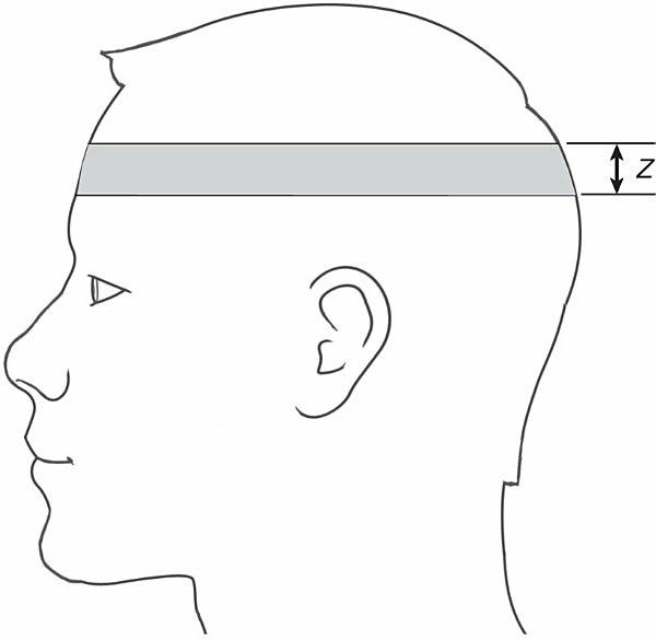
FIGURE 1-1 The thickness of the cross-sectional slice is referred to as its Z axis.
The data that form the CT slice are further sectioned into elements: width is indicated by X, while height is indicated by Y (Fig. 1-2). Each one of these two-dimensional squares is a pixel (picture element). A composite of thousands of pixels creates the CT image that displays on the CT monitor. If the Z axis is taken into account, the result is a cube, rather than a square. This cube is referred to as a voxel (volume element).
FIGURE 1-2 The data that form the CT slice are sectioned into elements.
A matrix is the grid formed from the rows and columns of pixels. In CT, the most common matrix size is 512. This size translates to 512 rows of pixels down and 512 columns of pixels across. The total number of pixels in a matrix is the product of the number of rows and the number of columns, in this case 512 × 512 (262,144). Because the outside perimeter of the square is held constant, a larger matrix size (i.e., 1,024 as opposed to 512) will contain smaller individual pixels. Each pixel contains information that the system