https://ebookmass.com/product/the-temporal-lobe-gabrielemiceli/
Instant digital products (PDF, ePub, MOBI) ready for you
Download now and discover formats that fit your needs...
Temporal Gifts Whitney Hill
https://ebookmass.com/product/temporal-gifts-whitney-hill/
ebookmass.com
Temporal Gifts Whitney Hill
https://ebookmass.com/product/temporal-gifts-whitney-hill-2/
ebookmass.com
The Black Mediterranean: Bodies, Borders and Citizenship Gabriele Proglio
https://ebookmass.com/product/the-black-mediterranean-bodies-bordersand-citizenship-gabriele-proglio/
ebookmass.com
Gone Like Yesterday: A Novel Janelle M. Williams
https://ebookmass.com/product/gone-like-yesterday-a-novel-janelle-mwilliams/
ebookmass.com
The Bone Track Sara E. Johnson https://ebookmass.com/product/the-bone-track-sara-e-johnson/
ebookmass.com
Sweet Wild of Mine Laurel Kerr
https://ebookmass.com/product/sweet-wild-of-mine-laurel-kerr-2/
ebookmass.com
Introduction to Dependent Types with Idris: Encoding Program Proofs in Types 1st Edition Boro Sitnikovski https://ebookmass.com/product/introduction-to-dependent-types-withidris-encoding-program-proofs-in-types-1st-edition-boro-sitnikovski-2/
ebookmass.com
Pearl of the Desert: A History of Palmyra Rubina Raja
https://ebookmass.com/product/pearl-of-the-desert-a-history-ofpalmyra-rubina-raja/
ebookmass.com
Staging Trauma 1st ed. Edition Miriam Haughton https://ebookmass.com/product/staging-trauma-1st-ed-edition-miriamhaughton/
ebookmass.com
The teachings of the Jehovah's Witnesses : [a comparison of the tenets of the Jehovah's Witnesses with traditional Christian doctrines] Gerstner
https://ebookmass.com/product/the-teachings-of-the-jehovahs-witnessesa-comparison-of-the-tenets-of-the-jehovahs-witnesses-with-traditionalchristian-doctrines-gerstner/ ebookmass.com
ELSEVIER
Radarweg29,POBox211,1000AEAmsterdam,Netherlands TheBoulevard,LangfordLane,Kidlington,OxfordOX51GB,UnitedKingdom 50HampshireStreet,5thFloor,Cambridge,MA02139,UnitedStates
Copyright©2022ElsevierB.V.Allrightsreserved.
Nopartofthispublicationmaybereproducedortransmittedinanyformorbyanymeans,electronicor mechanical,includingphotocopying,recording,oranyinformationstorageandretrievalsystem,without permissioninwritingfromthepublisher.Detailsonhowtoseekpermission,furtherinformationaboutthe Publisher’spermissionspoliciesandourarrangementswithorganizationssuchastheCopyrightClearanceCenter andtheCopyrightLicensingAgencycanbefoundatourwebsite: www.elsevier.com/permissions.
ThisbookandtheindividualcontributionscontainedinitareprotectedundercopyrightbythePublisher(other than asmaybenotedherein).
Notices
Knowledgeandbestpracticeinthisfieldareconstantlychanging.Asnewresearchandexperiencebroadenour understanding,changesinresearchmethods,professionalpractices,ormedicaltreatmentmaybecome necessary.
Practitionersandresearchersmustalwaysrelyontheirownexperienceandknowledgeinevaluatingandusingany information,methods,compounds,orexperimentsdescribedherein.Inusingsuchinformationormethodsthey shouldbemindfuloftheirownsafetyandthesafetyofothers,includingpartiesforwhomtheyhaveaprofessional responsibility.
Withrespecttoanydrugorpharmaceuticalproductsidentified,readersareadvisedtocheckthemostcurrent informationprovided(i)onproceduresfeaturedor(ii)bythemanufacturerofeachproducttobeadministered,to verifytherecommendeddoseorformula,themethodanddurationofadministration,andcontraindications.Itis theresponsibilityofpractitioners,relyingontheirownexperienceandknowledgeoftheirpatients,tomake diagnoses,todeterminedosagesandthebesttreatmentforeachindividualpatient,andtotakeallappropriate safetyprecautions.
Tothefullestextentofthelaw,neitherthePublishernortheauthors,contributors,oreditorsassumeanyliability foranyinjuryand/ordamagetopersonsorpropertyasamatterofproductsliability,negligenceorotherwise,or fromanyuseoroperationofanymethods,products,instructions,orideascontainedinthematerialherein.
ISBN:978-0-12-823493-8
ForinformationonallElsevierpublications visitourwebsiteat https://www.elsevier.com/books-and-journals
Publisher: Nikki Levy
EditorialProjectManager: KristiAnderson
ProductionProjectManager: PunithavathyGovindaradjane
CoverDesigner: MatthewLimbert
HandbookofClinicalNeurology3rdSeries
Availabletitles
Vol.79,Thehumanhypothalamus:Basicandclinicalaspects,PartI,D.F.Swaab,ed.ISBN9780444513571
Vol.80,Thehumanhypothalamus:Basicandclinicalaspects,PartII,D.F.Swaab,ed.ISBN9780444514905
Vol.81,Pain,F.CerveroandT.S.Jensen,eds.ISBN9780444519016
Vol.82,Motorneuronedisordersandrelateddiseases,A.A.EisenandP.J.Shaw,eds.ISBN9780444518941
Vol.83,Parkinson’sdiseaseandrelateddisorders,PartI,W.C.KollerandE.Melamed,eds.ISBN9780444519009
Vol.84,Parkinson’sdiseaseandrelateddisorders,PartII,W.C.KollerandE.Melamed,eds.ISBN9780444528933
Vol.85,HIV/AIDSandthenervoussystem,P.PortegiesandJ.Berger,eds.ISBN9780444520104
Vol.86,Myopathies,F.L.MastagliaandD.HiltonJones,eds.ISBN9780444518996
Vol.87,Malformationsofthenervoussystem,H.B.SarnatandP.Curatolo,eds.ISBN9780444518965
Vol.88,Neuropsychologyandbehaviouralneurology,G.GoldenbergandB.C.Miller,eds.ISBN9780444518972
Vol.89,Dementias,C.DuyckaertsandI.Litvan,eds.ISBN9780444518989
Vol.90,Disordersofconsciousness,G.B.YoungandE.F.M.Wijdicks,eds.ISBN9780444518958
Vol.91,Neuromuscularjunctiondisorders,A.G.Engel,ed.ISBN9780444520081 Vol.92,Stroke – PartI:Basicandepidemiologicalaspects,M.Fisher,ed.ISBN9780444520036 Vol.93,Stroke – PartII:Clinicalmanifestationsandpathogenesis,M.Fisher,ed.ISBN9780444520043 Vol.94,Stroke – PartIII:Investigationsandmanagement,M.Fisher,ed.ISBN9780444520050 Vol.95,Historyofneurology,S.Finger,F.BollerandK.L.Tyler,eds.ISBN9780444520081 Vol.96,Bacterialinfectionsofthecentralnervoussystem,K.L.RoosandA.R.Tunkel,eds.ISBN9780444520159 Vol.97,Headache,G.NappiandM.A.Moskowitz,eds.ISBN9780444521392 Vol.98,SleepdisordersPartI,P.MontagnaandS.Chokroverty,eds.ISBN9780444520067 Vol.99,SleepdisordersPartII,P.MontagnaandS.Chokroverty,eds.ISBN9780444520074 Vol.100,Hyperkineticmovementdisorders,W.J.WeinerandE.Tolosa,eds.ISBN9780444520142 Vol.101,Musculardystrophies,A.AmatoandR.C.Griggs,eds.ISBN9780080450315 Vol.102,Neuro-ophthalmology,C.KennardandR.J.Leigh,eds.ISBN9780444529039 Vol.103,Ataxicdisorders,S.H.SubramonyandA.Durr,eds.ISBN9780444518927 Vol.104,Neuro-oncologyPartI,W.GrisoldandR.Sofietti,eds.ISBN9780444521385 Vol.105,Neuro-oncologyPartII,W.GrisoldandR.Sofietti,eds.ISBN9780444535023 Vol.106,Neurobiologyofpsychiatricdisorders,T.SchlaepferandC.B.Nemeroff,eds.ISBN9780444520029 Vol.107,EpilepsyPartI,H.StefanandW.H.Theodore,eds.ISBN9780444528988 Vol.108,EpilepsyPartII,H.StefanandW.H.Theodore,eds.ISBN9780444528995 Vol.109,Spinalcordinjury,J.VerhaagenandJ.W.McDonaldIII,eds.ISBN9780444521378 Vol.110,Neurologicalrehabilitation,M.BarnesandD.C.Good,eds.ISBN9780444529015 Vol.111,PediatricneurologyPartI,O.Dulac,M.LassondeandH.B.Sarnat,eds.ISBN9780444528919 Vol.112,PediatricneurologyPartII,O.Dulac,M.LassondeandH.B.Sarnat,eds.ISBN9780444529107 Vol.113,PediatricneurologyPartIII,O.Dulac,M.LassondeandH.B.Sarnat,eds.ISBN9780444595652 Vol.114,Neuroparasitologyandtropicalneurology,H.H.Garcia,H.B.TanowitzandO.H.DelBrutto,eds. ISBN9780444534903
Vol.115,Peripheralnervedisorders,G.SaidandC.Krarup,eds.ISBN9780444529022 Vol.116,Brainstimulation,A.M.LozanoandM.Hallett,eds.ISBN9780444534972 Vol.117,Autonomicnervoussystem,R.M.BuijsandD.F.Swaab,eds.ISBN9780444534910 Vol.118,Ethicalandlegalissuesinneurology,J.L.BernatandH.R.Beresford,eds.ISBN9780444535016 Vol.119,NeurologicaspectsofsystemicdiseasePartI,J.BillerandJ.M.Ferro,eds.ISBN9780702040863 Vol.120,NeurologicaspectsofsystemicdiseasePartII,J.BillerandJ.M.Ferro,eds.ISBN9780702040870 Vol.121,NeurologicaspectsofsystemicdiseasePartIII,J.BillerandJ.M.Ferro,eds.ISBN9780702040887 Vol.122,Multiplesclerosisandrelateddisorders,D.S.Goodin,ed.ISBN9780444520012 Vol.123,Neurovirology,A.C.TselisandJ.Booss,eds.ISBN9780444534880 Vol.124,Clinicalneuroendocrinology,E.Fliers,M.KorbonitsandJ.A.Romijn,eds.ISBN9780444596024 Vol.125,Alcoholandthenervoussystem,E.V.SullivanandA.Pfefferbaum,eds.ISBN9780444626196 Vol.126,Diabetesandthenervoussystem,D.W.ZochodneandR.A.Malik,eds.ISBN9780444534804 Vol.127,TraumaticbraininjuryPartI,J.H.GrafmanandA.M.Salazar,eds.ISBN9780444528926 Vol.128,TraumaticbraininjuryPartII,J.H.GrafmanandA.M.Salazar,eds.ISBN9780444635211
Vol.129,Thehumanauditorysystem:Fundamentalorganizationandclinicaldisorders,G.G.CelesiaandG.Hickok,eds. ISBN9780444626301
Vol.130,Neurologyofsexualandbladderdisorders,D.B.VodušekandF.Boller,eds.ISBN9780444632470
Vol.131,Occupationalneurology,M.LottiandM.L.Bleecker,eds.ISBN9780444626271
Vol.132,Neurocutaneoussyndromes,M.P.IslamandE.S.Roach,eds.ISBN9780444627025
Vol.133,Autoimmuneneurology,S.J.PittockandA.Vincent,eds.ISBN9780444634320
Vol.134,Gliomas,M.S.BergerandM.Weller,eds.ISBN9780128029978
Vol.135,NeuroimagingPartI,J.C.MasdeuandR.G.González,eds.ISBN9780444534859
Vol.136,NeuroimagingPartII,J.C.MasdeuandR.G.González,eds.ISBN9780444534866
Vol.137,Neuro-otology,J.M.FurmanandT.Lempert,eds.ISBN9780444634375
Vol.138,Neuroepidemiology,C.Rosano,M.A.IkramandM.Ganguli,eds.ISBN9780128029732 Vol.139,Functionalneurologicdisorders,M.Hallett,J.StoneandA.Carson,eds.ISBN9780128017722
Vol.140,CriticalcareneurologyPartI,E.F.M.WijdicksandA.H.Kramer,eds.ISBN9780444636003
Vol.141,CriticalcareneurologyPartII,E.F.M.WijdicksandA.H.Kramer,eds.ISBN9780444635990
Vol.142,Wilsondisease,A.CzłonkowskaandM.L.Schilsky,eds.ISBN9780444636003
Vol.143,Arteriovenousandcavernousmalformations,R.F.Spetzler,K.MoonandR.O.Almefty,eds.ISBN9780444636409
Vol.144,Huntingtondisease,A.S.FeiginandK.E.Anderson,eds.ISBN9780128018934 Vol.145,Neuropathology,G.G.KovacsandI.Alafuzoff,eds.ISBN9780128023952 Vol.146,Cerebrospinalfluidinneurologicdisorders,F.Deisenhammer,C.E.TeunissenandH.Tumani,eds. ISBN9780128042793
Vol.147,NeurogeneticsPartI,D.H.Geschwind,H.L.PaulsonandC.Klein,eds.ISBN9780444632333 Vol.148,NeurogeneticsPartII,D.H.Geschwind,H.L.PaulsonandC.Klein,eds.ISBN9780444640765 Vol.149,Metastaticdiseasesofthenervoussystem,D.SchiffandM.J.vandenBent,eds.ISBN9780128111611 Vol.150,Brainbankinginneurologicandpsychiatricdiseases,I.HuitingaandM.J.Webster,eds.ISBN9780444636393 Vol.151,Theparietallobe,G.VallarandH.B.Coslett,eds.ISBN9780444636225 Vol.152,TheneurologyofHIVinfection,B.J.Brew,ed.ISBN9780444638496 Vol.153,Humanpriondiseases,M.PocchiariandJ.C.Manson,eds.ISBN9780444639455 Vol.154,Thecerebellum:Fromembryologytodiagnosticinvestigations,M.MantoandT.A.G.M.Huisman,eds. ISBN9780444639561 Vol.155,Thecerebellum:Disordersandtreatment,M.MantoandT.A.G.M.Huisman,eds.ISBN9780444641892 Vol.156,Thermoregulation:FrombasicneurosciencetoclinicalneurologyPartI,A.A.Romanovsky,ed.ISBN9780444639127 Vol.157,Thermoregulation:FrombasicneurosciencetoclinicalneurologyPartII,A.A.Romanovsky,ed.ISBN9780444640741
Vol.158,Sportsneurology,B.HainlineandR.A.Stern,eds.ISBN9780444639547
Vol.159,Balance,gait,andfalls,B.L.DayandS.R.Lord,eds.ISBN9780444639165
Vol.160,Clinicalneurophysiology:Basisandtechnicalaspects,K.H.LevinandP.Chauvel,eds.ISBN9780444640321
Vol.161,Clinicalneurophysiology:Diseasesanddisorders,K.H.LevinandP.Chauvel,eds.ISBN9780444641427
Vol.162,Neonatalneurology,L.S.DeVriesandH.C.Glass,eds.ISBN9780444640291
Vol.163,Thefrontallobes,M.D’EspositoandJ.H.Grafman,eds.ISBN9780128042816
Vol.164,Smellandtaste,RichardL.Doty,ed.ISBN9780444638557
Vol.165,Psychopharmacologyofneurologicdisease,V.I.ReusandD.Lindqvist,eds.ISBN9780444640123
Vol.166,Cingulatecortex,B.A.Vogt,ed.ISBN9780444641960
Vol.167,Geriatricneurology,S.T.DeKoskyandS.Asthana,eds.ISBN9780128047668
Vol.168,Brain-computerinterfaces,N.F.RamseyandJ.delR.Millán,eds.ISBN9780444639349
Vol.169,Meningiomas,PartI,M.W.McDermott,ed.ISBN9780128042809
Vol.170,Meningiomas,PartII,M.W.McDermott,ed.ISBN9780128221983
Vol.171,Neurologyandpregnancy:Pathophysiologyandpatientcare,E.A.P.Steegers,M.J.CipollaandE.C.Miller,eds. ISBN9780444642394
Vol.172,Neurologyandpregnancy:Neuro-obstetricdisorders,E.A.P.Steegers,M.J.CipollaandE.C.Miller,eds. ISBN9780444642400
Vol.173,Neurocognitivedevelopment:Normativedevelopment,A.Gallagher,C.Bulteau,D.CohenandJ.L.Michaud,eds. ISBN9780444641502
Vol.174,Neurocognitivedevelopment:Disordersanddisabilities,A.Gallagher,C.Bulteau,D.CohenandJ.L.Michaud,eds. ISBN9780444641489
Vol.175,Sexdifferencesinneurologyandpsychiatry,R.Lanzenberger,G.S.Kranz,andI.Savic,eds.ISBN9780444641236 Vol.176,Interventionalneuroradiology,S.W.HettsandD.L.Cooke,eds.ISBN9780444640345
Vol.177,Heartandneurologicdisease,J.Biller,ed.ISBN9780128198148
Vol.178,Neurologyofvisionandvisualdisorders,J.J.S.BartonandA.Leff,eds.ISBN9780128213773
Vol.179,Thehumanhypothalamus:Anteriorregion,D.F.Swaab,F.Kreier,P.J.Lucassen,A.SalehiandR.M.Buijs,eds. ISBN9780128199756
Vol.180,Thehumanhypothalamus:Middleandposteriorregion,D.F.Swaab,F.Kreier,P.J.Lucassen,A.SalehiandR.M.Buijs, eds.ISBN9780128201077
Vol.181,Thehumanhypothalamus:Neuroendocrinedisorders,D.F.Swaab,R.M.Buijs,P.J.Lucassen,A.SalehiandF.Kreier, eds.ISBN9780128206836
Vol.182,Thehumanhypothalamus:Neuropsychiatricdisorders,D.F.Swaab,R.M.Buijs,F.Kreier,P.J.Lucassen,andA.Salehi, eds.ISBN9780128199732
Vol.183,Disordersofemotioninneurologicdisease,K.M.HeilmanandS.E.Nadeau,eds.ISBN9780128222904
Vol.184,Neuroplasticity:Frombenchtobedside,A.Quartarone,M.F.Ghilardi,andF.Boller,eds.ISBN9780128194102
Vol.185,Aphasia,A.E.HillisandJ.Fridriksson,eds.ISBN9780128233849
Vol.186,Intraoperativeneuromonitoring,M.R.NuwerandD.B.MacDonald,eds.ISBN9780128198261
Allvolumesinthe3rdSeriesofthe HandbookofClinicalNeurology arepublishedelectronically, onScienceDirect: http://www.sciencedirect.com/science/handbooks/00729752
HANDBOOKOFCLINICAL NEUROLOGY SeriesEditors MICHAELJ.AMINOFF,FRANÇOISBOLLER,ANDDICKF.SWAAB
VOLUME187 Contributors K.Amunts
InstituteofNeuroscienceandMedicine,INM-1, ResearchCentreJuelich,Juelich;C&OVogtInstitutefor BrainResearch,UniversityHospitalD€ usseldorf, MedicalFaculty,Heinrich-HeineUniversity,D€ usseldorf, Germany
P.Bartolomeo
SorbonneUniversite,InstitutduCerveau-ParisBrain Institute-ICM,INSERM,CNRS,APHP,H^ opitaldela PitieSalp^ etrière,Paris,France
J.J.S.Barton
DivisionofNeuro-ophthalmology,Departmentsof Medicine(Neurology),OphthalmologyandVisual Sciences,Psychology,UniversityofBritishColumbia, Vancouver,BC,Canada
S.Benetti
CenterforMind/BrainSciences-CIMeC,Universityof Trento,Trento,Italy
F.Bielle
SorbonneUniversite,INSERM,CNRS,UMRS1127, ParisBrainInstitute,ICM;NeuropathologyDepartment, H^ opitauxUniversitairesLaPitieSalp^ etrière-Charles Foix,AP-HP,Paris,France
C.G.Bien
DepartmentofEpileptology(KrankenhausMara), BielefeldUniversity,Bielefeld;LaboratoryKrone,Bad Salzuflen,Germany
S.Boluda
SorbonneUniversite,INSERM,CNRS,UMRS1127, ParisBrainInstitute,ICM;NeuropathologyDepartment, H^ opitauxUniversitairesLaPitieSalp^ etrière-Charles Foix,AP-HP,Paris,France
V.Borghesani
Centrederecherchedel'Institutuniversitairedegeriatrie deMontreal;DepartmentofPsychology,Universitede Montreal,Montreal,QC,Canada
D.Bottari
IMTSchoolforAdvancedStudiesLucca,Lucca, Italy
M.Bowren,Jr.
DivisionofNeuropsychologyandCognitive Neuroscience,DepartmentsofNeurologyand PsychologicalandBrainSciences,UniversityofIowa, IowaCity,IA,UnitedStates
A.Caccia
CenterforMind/BrainSciences-CIMeC,Universityof Trento,Rovereto;DepartmentofPsychology,University ofMilano-Bicocca,Milan,Italy
F.Cacciamani
BordeauxPopulationHealth,UniversityofBordeaux, Bordeaux,France
G.A.Carlesimo
DepartmentofSystemsMedicine,TorVergata University;ClinicalandBehavioralNeurology Laboratory,I.R.C.C.S.SantaLuciaFoundation,Rome, Italy
M.Catani
Natbrainlab,DepartmentofForensicand NeurodevelopmentalSciences;Departmentof NeuroimagingSciences,InstituteofPsychiatry, PsychologyandNeuroscience,London,United Kingdom
S.W.C.Chang
DepartmentofPsychology;Interdepartmental NeuroscienceProgram,YaleUniversity,NewHaven, CT,UnitedStates
S.Clemenceau
DepartmentofNeurosurgery,LaPitie-Salp^ etrière UniversityHospital,Paris,France
L.Cohen
ParisBrainInstitute,H^ opitaldelaPitie-Salp^ etrière, Paris,France
O.Collignon
CenterforMind/BrainSciences-CIMeC,Universityof Trento,Trento,Italy;InstituteforResearchin PsychologyandNeuroscience,FacultyofPsychology andEducationalScience,UCLouvain,Louvain-laNeuve,Belgium
L.Cousyn
AP-HP,DepartmentofNeurologyandDepartmentof ClinicalNeurophysiology,EpilepsyandEEGUnit, ReferenceCenterforRareEpilepsies,Pitie-Salp^ etrière Hospital;SorbonneUniversite,ParisBrainInstitute, Team “DynamicsofNeuronalNetworksandNeuronal Excitability ”,Paris,France
V.deAguiar
CenterforLanguageandCognitionGroningen; DepartmentofNeurolinguisticsandLanguage Development,UniversityofGroningen,Groningen,The Netherlands
J.DeLeon
MemoryandAgingCenter,DepartmentofNeurology; DepartmentofNeurology,DyslexiaCenter,University ofCalifornia,SanFrancisco,CA,UnitedStates
M.Denos
RehabilitationUnit,NeurosciencesDepartment,H^ opital delaPitie-Salp^ etrière,Paris,France
J.Domínguez-Borràs
DepartmentofClinicalPsychologyandPsychobiology &InstituteofNeurosciences,UniversityofBarcelona, Barcelona,Spain
F.Doricchi
DepartmentofPsychology, “LaSapienza” University; LaboratoryofNeuropsychologyofAttention,I.R.C.C.S. SantaLuciaFoundation,Rome,Italy
V.Frazzini
AP-HP,DepartmentofNeurologyandDepartmentof ClinicalNeurophysiology,EpilepsyandEEGUnit, ReferenceCenterforRareEpilepsies,Pitie-Salp^ etrière Hospital;SorbonneUniversite,ParisBrainInstitute, Team “DynamicsofNeuronalNetworksandNeuronal Excitability ”,Paris,France
M.L.Gorno-Tempini
MemoryandAgingCenter,DepartmentofNeurology; DepartmentofNeurology,DyslexiaCenter,University ofCalifornia,SanFrancisco,CA,UnitedStates
A.E.Hillis
DepartmentofNeurology,JohnsHopkinsUniversity SchoolofMedicine,Baltimore,MD,UnitedStates
J.Kaminski
DivisionofNeuropsychologyandCognitive Neuroscience,DepartmentsofNeurologyand PsychologicalandBrainSciences,UniversityofIowa, IowaCity,IA,UnitedStates
O.Kedo InstituteofNeuroscienceandMedicine,INM-1, ResearchCentreJuelich,Juelich,Germany
B.Z.Mahon
DepartmentofPsychology,CarnegieMellonUniversity, Pittsburgh,PA,UnitedStates
K.Manzel
DivisionofNeuropsychologyandCognitive Neuroscience,DepartmentsofNeurologyand PsychologicalandBrainSciences,UniversityofIowa, IowaCity,IA,UnitedStates
R.C.Martin
DepartmentofPsychologicalSciences,RiceUniversity, Houston,TX,UnitedStates
B.Mathon
DepartmentofNeurosurgery,LaPitie-Salp^ etrière UniversityHospital;SorbonneUniversity;ParisBrain Institute,Paris,France
O.C.Meisner
DepartmentofPsychology;Interdepartmental NeuroscienceProgram,YaleUniversity,NewHaven, CT,UnitedStates
G.Miceli CenterforMind/BrainSciences-CIMeC,Universityof Trento,Rovereto;CentroInterdisciplinareLinceo ‘Beniamino Segre’—AccademiadeiLincei,Rome,Italy
R.Migliaccio
ParisBrainInstitute,INSERMU1127;Departmentof Neurology,Institutdelamemoireetdelamaladie d’Alzheimer,H^ opitaldelaPitie-Salp^ etrière,Paris, France
A.Nair
DepartmentofPsychology,YaleUniversity,NewHaven, CT,UnitedStates
V.Navarro
AP-HP,DepartmentofNeurologyandDepartmentof ClinicalNeurophysiology,EpilepsyandEEGUnit, ReferenceCenterforRareEpilepsies,Pitie-Salp^ etrière Hospital;SorbonneUniversite,ParisBrainInstitute, Team “DynamicsofNeuronalNetworksandNeuronal Excitability ”,Paris,France
C.Papagno
CenterforMind/BrainSciences-CIMeCandCenterfor NeurocognitiveRehabilitation,UniversityofTrento, Rovereto,Italy
F.Pavani
CenterforMind/BrainSciences-CIMeC,Universityof Trento,Rovereto,Italy
S.Pollmann
DepartmentofPsychologyandCenterforBehavioral BrainSciences,Otto-von-Guericke-University, Magdeburg,Germany
A.Rofes
CenterforLanguageandCognitionGroningen; DepartmentofNeurolinguisticsandLanguage Development,UniversityofGroningen,Groningen, TheNetherlands
S.Samson
DepartmentofPsychology,UniversityofLille,Lille; EpilepsyUnit,NeurosciencesDepartment,H^ opitaldela Pitie-Salp^ etrière,Paris,France
T.S€ ark€ am € o
DepartmentofPsychologyandLogopedics,University ofHelsinki,Helsinki,Finland
W.X.Schneider
DepartmentofPsychologyandCenterforCognitive InteractionTechnology,BielefeldUniversity,Bielefeld, Germany
D.Seilhean
SorbonneUniversite,INSERM,CNRS,UMRS1127, ParisBrainInstitute,ICM;NeuropathologyDepartment, H^ opitauxUniversitairesLaPitieSalp^ etrière-Charles Foix,AP-HP,Paris,France
C.Semenza DepartmentofNeuroscience,PadovaNeuroscience Center,UniversityofPadova,Padova,Italy
A.J.Sihvonen
SchoolofHealthandRehabilitationSciences, QueenslandAphasiaResearchCentre,TheUniversityof Queensland,Herston,QLD,Australia;Departmentof PsychologyandLogopedics,UniversityofHelsinki, Helsinki,Finland
A.Spagna DepartmentofPsychology,ColumbiaUniversity,New YorkCity,NY,UnitedStates
D.Tranel
DivisionofNeuropsychologyandCognitive Neuroscience,DepartmentsofNeurologyand PsychologicalandBrainSciences,UniversityofIowa, IowaCity,IA,UnitedStates
D.M.Ubellacker DepartmentofNeurology,JohnsHopkinsUniversity SchoolofMedicine,Baltimore,MD,UnitedStates
P.Vuilleumier
DepartmentofNeuroscienceandCenterforAffective Sciences,UniversityofGeneva,Geneva,Switzerland
Q.Yue DepartmentofPsychology,VanderbiltUniversity, Nashville,TN,UnitedStates
D.Zachlod InstituteofNeuroscienceandMedicine,INM-1, ResearchCentreJuelich,Juelich,Germany
Preface Thisvolumeofthe HandbookofClinicalNeurology offersclinicalandresearchprofessionalsinthefieldof neuroscienceanoverviewandupdateoncurrenthypothesesregardingthecorrelationsbetweenthetemporallobe andnormalandpathologicbehavior.
Anatomically,thetemporallobeisdefinedbyitslocationventraltotheSylvianfissure,occupyingthemiddlecranial fossa.Likeotherbrainlobes,itisnotafunctionallyunitarystructure.Themultiplerolesofdistincttemporalregionsare definedbytheirconnectivitywithotherbrainsystems,suchastheperisylvianlanguagenetworksandthefrontoparietal attentionsystems,andbythesensory(visual,auditory,andolfactory)inputstheyreceive.Themedial,evolutionarily moreancientportionsofthetemporallobeplayimportantrolesinmemory,emotion,andsocialcognition.
Inthepresentvolume,SectionI(Chapters1 and 2)laystheanatomicfoundationsfortheissuesdiscussedinthe following chapters.Itprovidesahistoricalperspectiveandcutting-edgeupdatesonthecytoarchitecturalorganization ofthetemporallobeandonitsconnectionswithotherstructuresofthecentralnervoussystem.
SectionII(Chapters3–22)providesanoverviewoftheroleofthetemporallobeinauditoryprocessing,visual perception, visuospatialprocessing,auditoryandvisuallanguagecomprehensionandproduction,facerecognition, socialcognition,emotion,andmemoryprocesses.Eachchapterreviewsavailableevidenceontheclinicalpictures thatemergefollowingdamagetothetemporallobe(auditoryagnosia,amusia,disordersofauditory/spokenandof visual/writtenlanguage,visuospatialneglect,prosopagnosia,coloragnosia,disordersofshort-termandlong-term memory,anddisordersofemotionandsocialcognition).Sincethetemporallobeprovidestheneuralunderpinnings ofauditoryandvisualpathwaysandiscriticalformultisensoryintegration,studiesofindividualswithcongenitalor earlysensorydeprivationarealsoreviewed,astheyinvestigatetheneuroplasticitymechanismsinvolvedintheremediation/compensationofinbornorearly-acquireddisordersofmodality-specificperception/recognition.Chaptersalso provideanoverviewofthecurrentneurofunctionalandneurophysiologicevidencefromneurotypicalparticipantsand brain-damagedindividualstopinpointconvergingevidence,divergingevidence,andgapsbetweenthetwodata sources.Eventhoughcontrastingdatacontinuetoexist,functionalneuroimagingstudiesincontrolpopulations and(mostlystructural)investigationsinbrain-damagedpersonswillhavetoconvergeultimately.Pointingoutthe outstandingdiscrepanciesinsomekeyareaswillhopefullystimulatefurtherresearchaimedatovercomingthem.
SectionIII(Chapters23–29)providesanupdateonconditionsthataffectspecificallyorpredominantlythetemporal lobe, with specialemphasisontemporallobeepilepsy,limbicencephalitis,andneurodegenerativediseases(frontotemporaldementiaandAlzheimerdisease).Thesechapterspointouthowspecificgeneticdefectsorauto-antibodies directedagainstaneuronalproteinmayprogressivelydisturbthetemporalnetworksandresultinspecificpathologic behaviors.Thechaptersalsoreviewthecurrentdiagnosticandtherapeuticapproachesandpointoutfuturedirections forclinicalandbasicresearch.
Seenasawhole,chapterscontributetothesamegoal areviewoftheevidenceonthefunctionsofthetemporallobe andonthedysfunctionsensuingfromdamage.However,theydifferwidelyintermsofthefocusoftheirnarrative,thus showcasingtheheterogeneityoftheexperimentalapproaches.Whilesomechaptersprovideclinicalupdateson specificsigns,symptoms,orclinicalconditions,othersconcentrateonbasicresearchanditsimplicationsfortheinterpretationofclinicalpictures.Yetothersfocusonneuroplasticity,biomarkers,intracerebralrecordings,andsoon. However,andregardlessofthespecificapproachtakenbyeachcontributor,consideringthevolumeinitsentirety allowsonetopinpointsomeoverarchingissuesthatreflectthestateoftheartandcouldstimulatefutureresearch. Themostobviousissueisthattherole(s)ofthetemporallobecanonlybeunderstoodif(eachportionof)thisregion isconsiderednotinisolationbutasacomponentofanumberoflarge-scalenetworkstowhichitcontributestoa differentextentandwithdifferentroles.Thisappliesnotonlytotheso-calledhigh-levelfunctionsbutalsotobasic sensoryandperceptualskillsandtotheemotionaldomain.Luckily,currentneuropsychologic,neurophysiologic, andneuroimagingmethodsallowaddressingtheoreticalandclinicalissuesatalevelofcomplexitythat,albeitstill
less-than-ideal,wouldhavebeenunimaginable20orsoyearsago.Technicaladvancespermitinvivoinvestigationsto focussimultaneouslyonmultiplecorticalareasaswellasontheirwhitematterconnections.Hence,itispossibleto investigateinthesameexperimentalsettingnotjustonefacetofaspecificcognitiveskillbutalsoitsintegrationwith othercognitiveabilitiesandtheunderlyingneurofunctionalinteractionsthatmakeitpossible.Asaconsequence, researchquestionsarenolongerrestrictedtoestablishingwhetherthetemporallobe(oraspecificportionthereof) ispartoftheneuralnetworkinvolvedinacognitivefunctionbutcanbebroadenedtoaddressthequalitativeand quantitativeinteractionsbetweenthetemporallobeandotherregionsinthatfunction.
Afurtherissueistightlyinterwovenwiththatdiscussedinthepreviousparagraph.Sincethe1970s,studiesonthe neuralrepresentationofcognitive/linguisticfunctionshavetypicallystartedfromtheselectionofaspecificcategoryor behavioralpartitionidentifiedbyinvestigationsinrelatedfields(psychology,psycholinguistics,linguistics,etc.),such asphoneme,noun,verb,semanticcategory,extrapersonalspace,andworkingmemory.Studieshavethencollected evidenceofdeviantbehaviorinbrain-damagedindividuals,inordertocorrelatethesecategoriesandpartitionswith specificneuralsubstrates.Thisapproach,heraldedbycognitiveneuropsychology,hasyieldedverysignificantresults inpromptingincreasinglydetailedfunctionalmodelsofcognitiveabilitiesandhasbeenmoderatelysuccessfulin clarifyingbasicanatomoclinicalcorrelations,largelybasedonstructuralMRIdata.Resultsofcurrentstudiesthatadopt increasinglysophisticatedtechniques(e.g.,electrocorticalrecordingsfrommultielectrodegrids,andhigh-fieldfMRI) suggestthatthisapproachshouldnotbetakenforgranted.Theissueinthiscaseconcernstheextenttowhichresearch questionsshouldstillfocusexclusivelyonpsychologically/linguisticallyderivednotionssuchasthosementioned earlier,asopposedtousingunitsofanalysisdrivenbymoreneurallybasedevidence.Thisquestionisraisedbyrecent observations.Forexample,alinguistic “category” mightbebestcharacterizednotbythefactthatitsmemberssharea setofabstractfeaturesbutbythefactthattheyelicitthesameorverysimilarpatternsofactivityinspecific(shared?) neuralnetworks.Adoptingthislatteroptionwouldnotonlyshifttheemphasisofneurofunctionalstudiesfurthertoward theneuralsidebutalsochallengelong-lastingtheoreticalconstraintsonthetypesofissuesthatdeservetobesystematicallyinvestigatedinstudiesontheneuralcorrelatesofcognition,language,andbehavior.
Asafinalnote,wewouldliketoemphasizethatcollatingthechaptersforthisvolumehastakenlongerthan expected,duetolimitationsanddelaysimposedbytheCOVID-19pandemic.Forthisreason,weareparticularly gratefultoallthecontributorswhoacceptedtosharethiscomplicatedadventurewithus.WearealsogratefultoMike Parkinsonforhispatience,availability,andprofessionalism,andtotheserieseditorsfortheirsupport.Wehopethatthe volumewillbeusefultoprofessionalswhooperateindifferentareasoftheneurologicandpsychologicsciencesand thatitwillprovidefoodforthoughtandstimulationforfurtherclinicalandbasicresearch.
GabrieleMiceli PaoloBartolomeo VincentNavarro
Foreword Thefirmbeliefthatsomepartsofthebrainareassociatedwithspecificbehaviorsismorerecentthanonemightthink. Ofcourse,therewasthepioneeringworkofGallandBroca,butmanypeopleresistedtheidea.LouisGratiolet (1815–1865)iscreditedwithattributingtheircurrentnamestothecerebrallobes,buthestronglydisagreedwithBroca onthesubjectofcerebrallocalizationalthoughtheybothcamefromthesamesmalltowninSouthwesternFrance (SteFoylaGrande).KarlLashley(1890–1958),oneofthemostcitedfiguresinthefieldofpsychology,introduced, aslateasthe1950s,theprinciplesof massaction and equipotentiality,implyingthatlossoffunctionafterbraininjury depends on theamountoftissuedestroyed,notonitslocation,andthatonepartofthecortexcantakeoverthefunction ofotherdamagedparts.Interestinwhatisnowknownasneuropsychologydeclinedalsobecausetheanatomo-clinical methodhadreacheditslimits.Renewedattentionwasduetotheentryintothefieldofallieddisciplinessuchas cognitivepsychology,neurolinguistics,and,aboveall,neuroimaging.
Therefore,someyearsago,the HandbookofClinicalNeurology undertookthetaskofpresentingbrain–behavior correlationsofspecificpartsofthecentralnervoussystem.Thefirstvolumeofthekindwastheonededicatedtothe parietallobe,thencamethecerebellum,thefrontallobe,andthecingulatecortex.Thehypothalamusholdsaspecial placeinthatrespectbecauseitwascoveredinthefirsttwovolumesofthecurrentseriesofthe Handbook andhasvery recentlybeenthesubjectoffourvolumes,allofthemeditedbyDickSwaab,coeditorofthecurrentseries.Wenow proudlypresentthisvolumeofthe Handbook devotedinitsentiretyandforthefirsttimetothetemporallobe.
Thevolumeincludesthreesections.Followingchaptersonthehistory,cytoarchitecture,andmainconnectionsof thetemporallobe,thesecondsectionpresentsvariousaspectsoftheirfunctionswithemphasisonlanguage,emotions, andmemory.Ineachcase,theevidenceisbasedonobservationsofsubjectswithacquireddeficitsaswellasstudiesof individualswithcongenitallesionsincludingearlysensorydeprivation.Muchemphasisisgiventomodernexploratory techniques,particularlyimagingandneurophysiologicstudiesthatinvolvenormalsubjects.Thefinalsectiondeals withsomeofthemainconditionsthataffectthetemporallobesincludingAlzheimerdiseaseandrelateddisorders. Threeofthechaptersinthissectiondealwithtemporallobeepilepsy(TLE)andincludediscussionsofspecialproblems,includingdiagnosis,clinicalfeatures,andneuropsychologicstudiesofpatientswiththisdiagnosis.TheconcludingchapterevaluatescriticallytheopportunitiesforsurgicalinterventioninTLE.
Wecongratulateandaremostgratefultothethreeeditorsofthepresentvolume:GabrieleMiceli,CenterforMind/ BrainSciences,UniversityofTrento,Rovereto,Italy;PaoloBartolomeo,ParisBrainInstitute,SorbonneUniversite, Pitie-Salp^ etrièreUniversityHospital,Paris;andVincentNavarro,EpilepsyandEEGUnit,ReferenceCenterforRare Epilepsies,DepartmentofNeurologyandDepartmentofClinicalNeurophysiology,Pitie-Salp^ etrièreUniversity HospitalParis,France.Togethertheyhavebroughtaremarkablesetofauthors,thusassuringtherightmixofcontinuity andhighlyupdatedinformationconcerningdisordersofthetemporallobes.
Asserieseditors,wereviewedallthechaptersinthevolumeandmadesuggestionsforimprovement,butweare delightedthatthevolumeeditorsandchapterauthorsproducedsuchscholarlyandcomprehensiveaccountsofdifferent aspectsofthetopic.Wehopethatthevolumewillappealtocliniciansasastate-of-the-artreferencethatsummarizesthe clinicalfeaturesandmanagementofpatientswithtemporallobedisorders.Wealsohopethatjuniorresearcherswill findwithinitthefoundationsfornewapproachestothestudyofthecomplexissuesinvolved.
Inadditiontotheprintversion,thevolumesareavailableelectronicallyonElsevier ’sScienceDirectwebsite: https:// www.sciencedir ect.com/.ScienceDirectispopularamongreadersandwillimprovethebooks’ accessibility.Indeed,all thevolumesinthepresentHCNseriesareavailableelectronicallyonthiswebsite.Thisshouldmakethemevenmore accessibletoreadersandalsofacilitatesearchesforspecificinformation.
Asalways,itisapleasuretothankElsevier(ourpublisher)andinparticularMichaelParkinsoninScotland,Nikki LevyandKristiAndersoninSanDiego,andPunithavathyGovindaradjaneatElsevierGlobalBookProductionin Chennai,fortheirassistanceinthedevelopmentandproductionofthe HandbookofClinicalNeurology.
MichaelJ.Aminoff Franc ¸ oisBoller DickSwaab
HandbookofClinicalNeurology, Vol.187(3rdseries)
TheTemporalLobe
G.Miceli,P.Bartolomeo,andV.Navarro,Editors https://doi.org/10.1016/B978-0-12-823493-8.00001-8 Copyright©2022ElsevierB.V.Allrightsreserved
Theconnectionalanatomyofthetemporallobe MARCOCATANI1,2*
1Natbrainlab,DepartmentofForensicandNeurodevelopmentalSciences,InstituteofPsychiatry, PsychologyandNeuroscience,London,UnitedKingdom
2DepartmentofNeuroimagingSciences,InstituteofPsychiatry,PsychologyandNeuroscience,London,UnitedKingdom
Abstract
Theideaofatemporallobeseparatedfromtherestofthehemispherebyreasonofitsuniquestructuraland functionalpropertiesisaclinicallyusefulartifact.Whilethetemporallobecanbesafelydefinedasthe portionofthecerebrumlodgedinthemiddlecranialfossa,thepatternofitsconnectionsisamorerevealing descriptionofitsfunctionalsubdivisionsandspecificcontributiontohighercognitivefunctions.This chapterprovidesanhistoricaloverviewoftheanatomyofthetemporallobeandanupdatedframework oftemporallobeconnectionsbasedontractographystudiesofhumanandnonhumanprimatesandpatients withbraindisorders.Comparedtomonkeys,thehumantemporallobeshowsarelativelyincreasedconnectivitywithperisylvianfrontalandparietalregionsandasetofuniqueintrinsicconnections,whichmay havesupportedtheevolutionofworkingmemory,semanticrepresentation,andlanguageinourspecies. Conversely,thedecreasedvolumeoftheanterior(limbic)interhemispherictemporalconnectionsin humansisrelatedtoareducedrelianceonolfactionandapartialtransferenceoffunctionsfromtheanterior commissuretotheposteriorcorpuscallosum.Overallthenoveldatafromtractographysuggestarevision ofcurrentdualstreammodelsforvisualandauditoryprocessing.
NOTESONHISTORY1 Thestoryofthetemporallobeisreallytwostories,that ofthelimbicsystemthatcontributestoitsanteromedial subdivisionandthatoftheisocortexthatliesbelowthe lateralfissure.Thelimbicsystemstorybeginsin1587 whenGiulioCesareAranzidescribedthehippocampus anditsanatomicalproximitytotheneighboringsectors ofthelateralventriclesandthewhitematterofthefornix (Aranzi,1587).In1664ThomasWillisusedforthe first timetheterm “limbus” toindicateasemicircular convolutionborderingthediencephalon;hislimbicconvolutionincludedthecingulategyruscirclingaround thecorpuscallosumandtheparahippocampalgyrus
(PHG)locatedinthemedialaspectofthetemporallobe (Willis,1664).Alongthemedialsurfaceofthetemporal lobe,justanteriortotheheadofthehippocampus,aprotrudingconvolutioncaughttheeyeofVicqd’Azyrwho referredtoitas uncus inhis1786atlas(Vicqd’Azyr, 1786).CarlBurdachnamed amygdala the subcortical graymatterovoidresponsibleforthepeculiarbulging shapeoftheuncus(Burdach,1819–26).Theanatomical proximityandconnectivitypatternbetweenthemedial temporalstructuresandtheolfactoryregionsledthe GermanbrothersGottfriedandLudolphTreviranus (TreviranusandTreviranus,1816)toconsiderapossible roleofthehippocampusinmemoryfunctions.Bythe endofthe19thcentury,aseriesofcomparativeand
1AbbreviationsusedinthechapterarelistedattheendofthechapterbeforeReferencessection.
*Correspondenceto:MarcoCatani,Natbrainlab,DepartmentofForensicandNeurodevelopmentalSciences,Instituteof Psychiatry,PsychologyandNeuroscience,16DeCrespignyPark,LondonSE58AF,UnitedKingdom.Tel:+44-207-848-0984, Fax:+44-207-848-0650,E-mail:marco.1.catani@iop.kcl.ac.uk
experimentalstudiesinanimalsandclinical –anatomical observationsinpatientswithbrainlesionsconsolidated theconceptofalimbicsystemcomposedofcorticalareas andsubcorticalnucleiconnectedbywhitematterfibersof thefornix,mammillothalamictract,thalamicprojections, uncinatefasciculus,andcingulum.Thismodelbecame knowninEuropeasthe grandlobelimbique (Broca, 1878)orthe limbicsystem (Bechterew,1900; Jakob, 1906),andmuchlaterasthe Papezcircuit intheUnited States(Papez,1937).Ofnote,alllimbicconnections, exceptforthemammillothalamictract,originateorterminateinthetemporallobe(foranextendedreviewofthe historyofthelimbicsystem,see Catanietal.,2013).
Whilethetemporallimbiccomponentsarerelatively preservedamongvertebrates,itisonlyinprimatesthat thetemporallobeemergesasaclearlyidentifiablesubdivisionofthecerebralhemisphereduetotheremarkable expansionoftheisocortexthatoccursinmammalian evolution(Gloor,1997).Thefirsttorealizesuchadistinctiveanatomicalfeatureofthehumanbrainwas FranciscusSylvius,whodescribedadeepfissureseparatingasmallerventralpartofthecerebrum thetemporallobe fromtherestofthecerebralhemisphere (Bartholin,1641).Inthe18thcentury,someanatomists proposed athreepartitionofthecerebralhemispheresin whichthetemporalandparietallobeswerelumped togetherintothe “middlelobe,” flankedbyananterior andaposteriorlobe.Whiletheborderbetweentheanteriorandthemiddlelobehadsomeanatomicalgrounds theanteriorlobeoccupiestheanteriorfossaandthecentralsulcusisaclearlandmarkinallbrains theborder betweenthemiddleandtheposteriorlobewasparticularlydifficulttodefine.In1786,Vicqd’Azyrsuggested thepreoccipitalnotch,whichheadmittedlyrecognized asafaintanatomicalfeatureinmostofthebrainshe examined,asapossiblelandmarkfortheseparation betweenthemiddleandtheposteriorlobe,acriterion thatsurprisinglyisstillinuse.Hewasalsoresponsible forreplacingthetermsanterior,middle,andposterior lobeswiththenamesfrontal,parietal,andoccipital borrowedfromtheoverlyingbonesoftheskull.
Asprogressiveclinicalevidenceforaprominentrole inlanguagecomprehension(Wernicke,1874),behavior (BrownandSchafer,1888),andmemoryfunctions (Bechterew,1900)accumulatedinthesecondhalfof the 19thcentury,theconceptofatemporallobedistinguishedfromaposterioroccipitallobecompletelydedicatedtovisualfunctionsandaparietallobededicatedto sensorimotorandvisuospatialfunctionsbecamewidely accepted.But,experimentalsupportfortheauditory functionofthetemporallobewassurprisinglyslower inemerging(Finger,1994)comparedtothatofother senses,perhapsbecauseofthedifficultyofexamining hearinginanimals(ClarkeandO’Malley,1996).Ittook David Ferrieralmost10yearsofelectricalstimulation
andcorticalablationinmonkeystoconvincehiscolleaguesofthelocalizationofauditoryfunctionsinthe superiorregionofthetemporallobe.Thefinalproof wasdeliveredbyFerrierin1881attheInternational MedicalCongressheldinLondonwherehepublicly demonstratedthatoneofhismonkeyswithbilateralablationofthesuperiortemporalgyruswassodeafthatit didnotevenshowastartleresponsetothesoundofa gunfiredclosedtoitsear(FerrierandYeo,1884 ).In 1895, vonMonakowreportedthataftercorticalablationofthetemporalauditoryareainacat,retrograde degenerationwasevidentinthepartofthethalamus thatBurdachdescribedin1822asthe corpusgeniculatuminternum laterrenamedthemedialgeniculate nucleus(MGN)( vonMonakow,1895 ).In1896, Flechsig discoveredthattheareasofthetemporal auditorycortexwheremostoftheMGNfibersproject toarealreadymyelinatedina2-month-oldnewborn brain(Flechsig,1896).Hence,justlikehedidfortheother lobes,Flechsigseparatedtheearlymyelinatedareasofthe temporallobelocatedinthetransversegyrusdescribed byHeschl whichhetermedthe “primordial” fieldsor accordingtocontemporarynomenclatureprimaryauditoryareas fromthelatemyelinatedassociativetemporalareas.Thetemporalareashavebeensubsequently mappedintoamultitudeofsmallerareaswhosenumber variesaccordingtotheanimalmodelandthemethod adoptedtoidentifythebordersbetweencorticalfields (see Chapter2)(foranextendedreviewofthehistory of theauditorycortexanditsconnections,see Finger, 1994; Jones,2011).
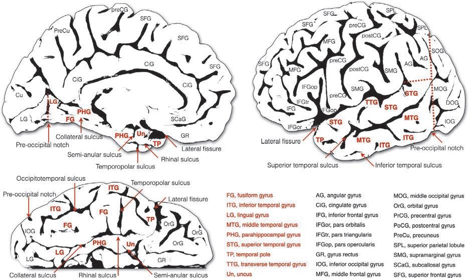
SURFACEANATOMY Externally,thetemporallobehasasuperior,lateral,and inferior(ventral)surface(Fig.1.1).Thesuperiortemporal surfaceisburiedwithinthesylvanfissureandforms thetemporaloperculumthatcoverstheinsula.Thissurfaceisflatexceptforitscentralportionthatisoccupied bythebulgingtransversegyrusofHeschl.Thisgyrus, whichisoftenduplicated,isseparatedfromtherestof thesuperiortemporalsurfacebytheanteriorandposteriortransversetemporalsulci.Thesurfaceofthesuperior temporalgyrusthatliesanteriorandposteriortothe transversetemporalgyrusofHeschlformstheplanum polareandplanumtemporale,respectively.
Onthelateralsurface,thesuperior,middle,andinferiortemporalgyricourselongitudinallyandconverge anteriorlytoformthetemporalpole.Thesuperiorand middletemporalgyriareoftenwellseparatedbyacontinuoussuperiortemporalsulcus.Inhumansthemiddle andinferiortemporalgyriareseparatedbytheinferior temporalsulcus,whichisoftendifficulttorecognize asasinglelineargroove.
Fig.1.1. Surfaceanatomyofthetemporallobe.Medial(upperleft),lateral(upperright),andventral(lowerleft)surfacesofthe temporallobe.Temporalgyriarelabeledinred.ModifiedfromCataniM,Dell’AcquaF(inpress).Atlasofhumanbrainpathways, Elsevier.
TEMPORALLOBETRACTS Mostoftheinferiorsurfaceofthetemporallobeiscoveredbytheventralportionoftheinferiortemporalgyrus, thefusiformgyrus,andthePHG.Allthecortexofthefusiformgyrusbelongstothetemporallobeexceptforitsmost posteriortip,whichispartoftheoccipitallobe.Theprincipalsulcioftheinferiorsurfacearethelateraloccipitotemporalsulcusandthecollateralsulcus,whichseparate thefusiformgyrusfromtheinferiortemporalgyrusand thePHG,respectively.Theanteriorpartofthecollateral sulcusisalsotermedrhinalsulcus.Smallersulcicanoften berecognizedontheinferiorandmedialsurface,suchas thetemporopolarsulcusthatdefinestheposteriorborder ofthetemporalpoleandthesemianularsulcus.Thelatter sulcusisoftenusedasanexternalanatomicallandmarkto separatethePHGcoveringtheheadofthehippocampus fromtheuncus.
WhiletheSylvianfissurerepresentsanaturalanatomicallandmarkbetweenthetemporallobeandboththe frontalandanteriorparietallobes,theposteriorborders betweenthetemporal,occipital,andparietallobesare arbitraryasoftenthesuperior,middle,andinferiortemporalgyricontinueintotheangulargyrusoftheparietallobe andthemiddleandinferioroccipitalgyri.Thisanatomical continuityishighlightedbythefrequentuseoftheterm “temporo-occipital-parietaljunction” intheneuroimagingliterature.
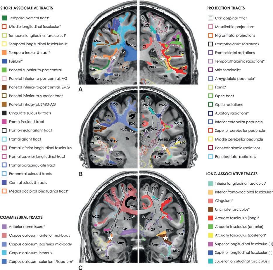
Oncoronalsections,thetemporallobeappearslikea smallcauliflowercomposedofasingleshortstemand smallfloretsthatcorrespondtothefivelongitudinaltemporalgyri(Fig.1.2A–C).Itstemporalstemoriginates fromtheflooroftheexternal-extremecapsuleandin itsnarrowestportioniscontainedwithinaspacedelimitedmediallybythelateralventricleandlaterallybythe inferiorbranchoftheperi-insularsulcus.Exceptfor thecingulum,thefornix,andthestriaterminalis,thetemporalstemcontainsmostoftheprojectiontractsfrom thethalamus(temporothalamic),MGN(auditoryradiations),andbasalganglia,thecommissuralfibersofthe corpuscallosum(tapetum)andanteriorcommissure, andthelargeassociationfibersofthelongandposterior segmentsofthearcuatefasciculus,uncinatefasciculus, andinferiorfronto-occipitalfasciculus(IFOF).Short associationtractsconnectingthetemporalcortextothe insulaarealsoenclosedintheposteriorandtheanterior portionsofthetemporalstem.Withinthetemporalstem mostofthesefibersrunparalleltothelateralventricle beforetakingamoreradialcourseoutwardlytoward thecortexofeachtemporalgyrus.Therestofthewhite matterbetweenthetemporalstemandthetemporalcortexisoccupiedbytheinferiorlongitudinalfasciculus (ILF)andtheshort-associationtemporaltracts.
Fig.1.2. Coronalslicescuttingthroughtheposterior(A),middle(B),andanterior(C)temporallobe.Thetemporalstemisindicatedwitha whitestar.Theshortassociativeandcommissuraltractsaredisplayedinthe leftimages whilethemajorprojectionand longassociationtractsareonthe right.Alltemporalconnectionsareindicatedwithan asterisk.ModifiedfromCataniM,Dell’AcquaF(inpress).Atlasofhumanbrainpathways,Elsevier.
Projectionpathways Thetemporallobehasextensiveconnectionswiththe inferiorcolliculus,theMGN,thepulvinarandthedorsomedialnucleusofthethalamus,thestriatumandclaustrum,thehypothalamicnuclei,andthemammillary bodies.Thetemporalprojectionscanbeseparatedinto anonlimbicgroupencompassingtheauditoryandtemporothalamicradiations,andthepathwaysofthelimbic system.
The auditoryradiations arethemainafferentsensory pathwaytothetemporallobe.Theyareresponsiblefor
relayingauditoryinformationfromtheMGNandinferiorcolliculustothetemporalcortex.Inthebrainstem, thelaterallemniscusconveysauditoryinputsfromthe cochlearnucleiandtheolivarycomplextotheinferior colliculus,whichinturnprojectstotheMGN.Thecolliculogeniculatefibersarevisibleontheposterioraspect ofthemesencephalonasanobliqueprotrusioncalledthe inferiorbrachium(Naidich,2020).Somefibersfrom theinferiorcolliculuscontinuedirectlywithoutstopping andmergewiththosefromthemedialgeniculatetoform theauditoryradiations.Afterashorthorizontalcourse
withinthemostposteriorregionoftheinternalcapsule, theauditoryradiationspassinferiortotheputamenand enterintothetemporalstem.Here,mostoftheauditory radiationscontinuelaterallyandanteriorlywithin Heschl’sgyrusandterminateinthecortexoftheearly auditoryareas.Theinformationcarriedbytheacoustic radiationstotheprimaryauditorycortexispartially segregatedaccordingtothespatialorganizationofthe corticalneuronsdecodingsoundsofdifferentfrequency (tonotopy)orintensity(ampliotopy).Inthesuperiortemporalcortex,severaltonotopicareashavebeendescribed inthecortexofHeschl’sgyrusandsurroundingcortexof thesuperiortemporalgyrus(Talavageetal.,2004).The acousticradiationsalsocontainfibersthatruninthe reversedirectionfromthetemporalcortextotheMGN andinferiorcolliculus(Pickles,2015).Thiscentrifugal systemmediatesatop-downmodulatorycontrolused bythetemporalcortextoenhancetheresponseof subcorticalnucleitosalientstimuli.
Theprimaryauditorycortexreceivesthemajorityof theauditoryinputsfromtheMGNandinferiorcolliculus, whoseprojectionsbecomeprogressivelyweakerinmore anteriorandlateraltemporalregions.Thisreductionis offsetbyarelativeincreaseinfiberdensityfromthemultisensorythalamicnucleithatconveybothauditoryand visualinformation(Hackett,2015).Theseconnections are collectivelytermed temporothalamicfibers andtheir initialcourseisdifferentaccordingtotheirlateralor medialorigininthethalamus.Thethalamicfibersthat originatefromthelateralpulvinarnucleirunparallelto theauditoryradiationsandhaveasublenticularcourse. Theseconnectionsareindicatedbysomeauthorsas thetemporopulvinarbundleofArnold(Gloor,1997). The connectionsfromthemedialnucleiofthepulvinar andmidlinethalamicnucleirunthroughthestratum zonale,athinlayerofwhitematterthatcoverstheventricularsurfaceofthethalamus(Fig.1.2Bright).These fiberscurvearoundtheposteriorandinferiorsurfaces ofthethalamusandmergewiththeinferiorfibersof theauditoryradiationsbeforeenteringthetemporallobe. Bothmedialandlateraldivisionsofthetemporothalamic projectionsarereciprocalandruntogetherwiththefibers ofthestriaterminaliswithinthewhitematterthatform thelateralwallandroofofthelateralventricle.
Tractographyandpostmortemstudiesusinghistology andbluntdissectionhavebeenusedtovisualizethe acousticradiations,althoughitisoftendifficultto obtaintheirentirereconstructionduetothelimitations ofthesetechniques(Burgeletal.,2006; Behrensetal., 2007; Maffeietal.,2015,2017).Tractographyreconstructionsofthemostdorsalandlateralacousticprojectionsarepossibleonlywithadvanceddiffusionmethods duetothecomplexityofthewhitematterregionlateral tothethalamusandposteriortothelenticularnucleus
(Behrensetal.,2007; Maffeietal.,2015, Maffeietal., 2019).Here,theacousticradiationsdirectinglaterally towardHeschl’sgyruscrosswithalargegroupofassociationandprojectionfiberscoursingaroundtheposterior insulainaperpendiculardirection(e.g.,arcuatefiber, middlelongitudinalfasciculus(MLF),IFOF).Other acousticradiationsdirectedtowardmoreanteriortemporal regionscanbeeasilyvisualizedalsowithtractography algorithmsbasedonthetensormodel.
Theorganizationoftheacousticandtemporothalamic projectionsisbothserialandparallelasmanyfibersproject directlytodownstreamtemporalregions(Pickles,2015). ThedirectacousticprojectionsfromtheMGNtotheamygdala,forexample,undergochangesinresponsetofear conditioningandthesechangesareresponsiblefortheformationofmemoriesrelatedtobothsafeandthreatening auditorycues(Ferraraetal.,2017).Inhumans,arelative temporalcortexamygdalahyperactivationtosadand hypoactivationtohappysoundshavebeenreportedin infantsborntodepressedmothers.Thismaysuggestaneuralbiastowardemotionallynegativestimuli,mediatedby theeffectofperinatalexposuretomaternaldepressionon theearlydevelopmentoftheauditoryandtemporothalamic radiations(Craigetal.,2022).
The crossedbilateraldistributionoftheauditorypathwaysandtheirparallelorganizationwithinthetemporal lobecouldalsoexplainthedifferentmanifestationsassociatedwithdamagetoauditorypathways.Hearingdisorderscommonlyassociatedwithdamagetotheacoustic radiationsandtheirsubcortical/corticalterminations includereduced(corticaldeafness)orincreased(hyperacusis)perceptualawarenesstosound.Otherauditory phenomenaassociatedwithacousticradiationdamageincludetinnitus,auditoryhallucinations,andpalinacousis(perseverationofthehearingsensationafter cessationoftheexternalstimulus).Theunderlying mechanismsresponsiblefortheseclinicalmanifestationsmayvaryandincludesubcorticalorcortical damagedisconnectionresultinginthehypoactivityor hyperactivityofcentripetaland/orcentrifugalnetworks. Aselectivedamageofthenucleiand/orfibersinvolved inprocessingwordsoundsmanifestswithpureword deafness(Maffeietal.,2017).
The twomajorsubcorticallimbicgreystructuresof thetemporallobehavetheirownsystemofafferent andefferentconnectionsthattheyusetoreceivesensory stimulidirectlyfromearlysensoryareasorindirectly fromassociationareasortheinsula.Theinformation thatreachesthehippocampusandamygdalaisthen furtherelaboratedandconsolidatedintomemorytraces thatcaninfluenceourcognitivefunctionsandvisceral bodyresponses(Catanietal.,2013).
The fibersofthefornixarisefromthehippocampusof eachside,runthroughthefimbria(i.e.,thetwolegsofthe
fornix),andjoinbeneaththespleniumofthecorpus callosumtoformthebodyofthefornix.Otherfimbrial fiberscontinuemedially,crossthemidlinethroughthe hippocampalcommissure,andprojecttothecontralateralhippocampus.Mostofthefiberswithinthebody ofthefornixrunanteriorlybeneaththebodyofthecorpuscallosumtowardtheanteriorcommissurewithout crossingthemidline.Astheyapproachtheanteriorcommissure,theydivideintoleftandrightanteriorcolumns andleftandrightposteriorcolumns.Theanteriorcolumnsenterthehypothalamusandprojecttotheseptal andhypothalamicnucleiandthenucleusaccumbens. Theposteriorcolumnsofthefornixcurveventrallyin frontoftheinterventricularforamenofMonroeand posteriortotheanteriorcommissurebeforeterminating intothemammillarybody(postcommissuralfornix), adjacentareasofthehypothalamus,andanteriorthalamicnucleus.Allthecomponentsofthefornixcanbe dissectedwithdiffusiontractography(Catanietal., 2002, 2013).
The threemainfibersystemsoftheamygdalaarethe lateralolfactorystria,the striaterminalis (ordorsal amygdalofugalpathway),andthe amygdaloidpeduncle (orventralamygdalofugalpathway).Theamygdaloid peduncleistheprimaryoutputoftheamygdalatoother structuresofthelimbicsystemlocatedoutsidethetemporallobe,suchastheanteriorhypothalamicnuclei, thestriatum particularlydensearetheterminationsto thenucleusaccumbens,thesubgenualanteriorcingulate cortexandseptalnuclei,themedialdorsalnucleusof thethalamus,andthebasalforebrain(Canterasetal., 1995; Kamalietal.,2016).Theamygdalaconnections are ofparticularinterestwithregardtounderstanding humanstress-relatedpsychiatricdisease(Lebowand Chen,2016).
Commissuralpathways Thehumantemporallobehasauniquelyrichpattern ofcommissuralconnections,beingtheonlyregionconnectedbyallthreecommissuresofthecerebralhemispheres,namely,theposteriorcorpuscallosum,the anteriorcommissure,andthehippocampalcommissure.
Thecallosalfibersfromtheposterioristhmusandthe spleniumrunwithinthewhitematterthatformsthedorsolateralwallofthelateralventricles.Fortheirlaminar arrangementalongthetemporalstem,thetemporalcallosalfibersaregenerallyindicatedwiththeterm tapetum (fromLatin,meaningcarpet)(CataniandThiebautde Schotten,2012).Thecallosalfibersreachthebeltand parabeltassociativeauditorycortex,thepolymodal temporalcortex,andpartofthetemporalvisualcortex.
Theremainingpartofthetemporalcortex,theamygdala,andthehippocampusareconnectedbytheanterior
commissureandthesmallerhippocampalcommissure. Onthemidlinesagittalsection,thefibersoftheanterior commissureoccupyacircularregionofdiameterless than2Sq.mmdelimitedbytheanteriorandposteriorcolumnsofthefornix,theseptumpellucidum,andthethird ventricle.Fromhereitsfibersdirectlaterallyanddownwardlypassingthroughtheventralportionoftheglobus pallidus.Astheanteriorcommissureleavestheregion betweentheamygdalaandtheputamenandentersthe anteriorpartofthetemporalstem,itsplitsintotwo branches.Theanteriorbranch,whichmergeswiththe fibersoftheamygdaloidpeduncle,projectstotheamygdalaandthecortexofthetemporalpole.Theposterior branchcourseswithinthetemporalstemandprojects totheventraltemporalcortexandtheanteriortwo-thirds oftheinferior,middle,andsuperiortemporalgyri (Catanietal.,2002).Axonaltracinginmonkeysandtractography inhumanshavebeenusedtolocatetheposition ofthetemporalfiberswithinthemidsagittalsectionof thecorpuscallosum(SchmahmannandPandya,2006; Parketal.,2008; CataniandThiebautdeSchotten, 2012).Studiesinthemonkeybrainindicateacertain degree ofoverlapbetweentheterritoriesoftheanterior commissureandthecorpuscallosum(Zeki,1973; JouandetandGazzaniga,1979).However,theprojection territories ofthetwocommissuresareprobablydifferent inhumansfortheproportionalvolumereductionofthe anteriorcommissureinhumanevolutioncomparedto OldWorldmonkeys(Barrettetal.,2020).Thesecomparative anatomydifferencesareprobablylinkedtoa reducedimportanceofolfactioninhumancognition paralleledbyanincreasedinter-hemisphericconnectivity betweenthoseposteriortemporalareasthathavesupportedtheemergenceofouruniquecognitiveabilities.
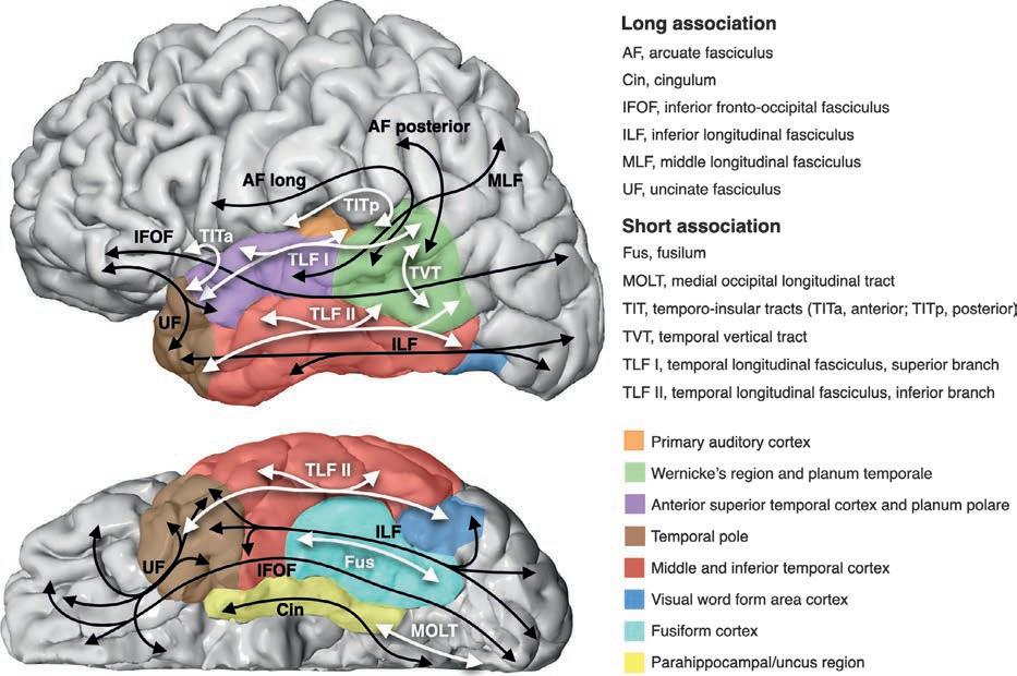
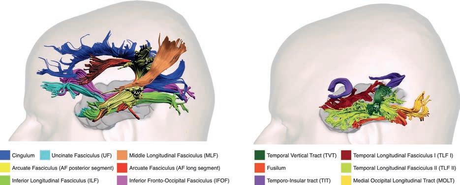
Associationpathways Theassociationpathwaysofthetemporallobecanbe dividedintotwolargegroups(Figs.1.3 and 1.4).The first groupincludesthelonginterlobarassociationpathwaysthathavebeentraditionallydescribedinhumans withpostmortemdissectiontechniquesandrecently revisitedwithtractographymethods(Fig.1.4 left).The arcuate fasciculus,theuncinatefasciculus,theILF,the IFOF,theMLF,andthecingulumarethemajorinterlobar pathwaysofthetemporallobe.Asecondgroupincludes thesmallerinterlobarfibersconnectingthetemporallobe totheneighboringcortexandtheintralobartemporal fibers(Fig.1.4 right).Amongtheshortinterlobarfibers, only theU-shapedtemporoinsulartractsandthenewly describedmedialoccipitallongitudinaltract(MOLT) (Beyhetal.,2022)willbediscussedinmoredetail.Three major systemsofintralobarfibershavebeenrecently
Fig.1.3. Diagramoftheorganizationofthemajorassociationpathwaysofthetemporallobe.Longinterlobarassociationtractsare indicatedwith blacklines,whileshortinterlobarandintralobarfibersareindicatedwith whitelines Coloredcorticalregions correspondtobroadlydefinedanatomical/functionalareasofthehumantemporallobe.
Fig.1.4. Tractographyreconstructionsofthetemporalassociationpathways. Left,majorinterlobarassociationpathways. Right, shorterinterlobarandintralobartemporaltracts.
identifiedwithtractography.Theseincludethetemporal longitudinalfasciculus(TLF)IandII,thefusilum,and thetemporalverticaltract(TVT).
ARCUATEFASCICULUS Theposteriorregionsofthesuperiorandmiddletemporalgyrusareconnectedtotheperisylvianparietaland
frontalcortexbythearcuatefasciculus.Thearcuate fasciculusiscomposedofthreesegmentslodgedinthe whitemattersurroundingthedorsalandposteriorsectionsofthecircularsulcusoftheinsula.Ofthethree segments,onlytheanteriorsegmentdoesnotoriginate orterminateinthetemporallobe(Catanietal.,2005). Tractographystudiessuggestthatboththelongand
posteriorsegmentofthearcuatefasciculushaveapartiallyoverlappingoriginorterminationintheposterior temporalregions.Afterleavingthewhitematterofthe superiorandmiddletemporalgyri,thefibersofbothlong andposteriorsegmentsgatherinthetemporalstemlaterallytothetemporothalamicprojectionsandopticradiations,beforebendingaroundtheposteriorsectionofthe circularsulcusoftheinsula(Fig.1.2AandBright).Here, the mostlateralfibersoftheposteriorsegmentcontinue dorsallyandarchlaterallytowardthecortexoftheangulargyrusandtheposteriorcortexofthesupramarginal gyrus.Thefibersofthelongsegmentcontinuetoarch aroundthecircularsulcusoftheinsulaandastheyenter theparietallobetheyfollowahorizontalcoursemedialto theanteriorsegmentofthearcuatefasciculus.Asthe longsegmentfiberspassthecentralsulcustheycurvelaterallytoreachtheinferiorcortexoftheprecentralgyrus andtheposteriorcortexoftheinferiorandmiddlefrontal gyri.ItcanbearguedthatsomeU-shapedfibersofthe posteriorsegmentofthearcuatefasciculusrepresent shortinterlobartemporoparietalfibers(Fig.1.2).
The temporalconnectionsofthearcuatefasciculus havechangedsignificantlyinprimateevolutioncompared,forexample,toothertemporalconnectionsof thelimbicsystem(e.g.,uncinatefasciculus).Nonhuman primatesshareasubcomponentofthelongsegmentof thearcuatefasciculuswithhumans(Rillingetal., 2008; Barrettetal.,2020),whichprojectstotheposterior superi ortemporalgyrusandisinvolvedinacoustic spatiotemporalprocessingandstimulusidentification (AboitizandGarcía,2009).However,inhumans,the long segmentofthearcuatefasciculusisproportionally largercomparedtothemonkeybrain(Barrettetal.,2020) and projectsmoreextensivelytothesuperiorandmiddle temporalgyri(Catanietal.,2005).Theexpandedanatomy ofthelongsegmentallowsadirectlinkbetween perisylvianregionsinvolvedwithauditorymemory (RauscheckerandScott,2009; Schulzeetal.,2012), wordlearning(López-Barrosoetal.,2013),andsyntax (Wilsonetal.,2011).Similarly,theposteriorsegment of thearcuatefasciculusconnectsareasoftheinferior parietallobuleandtheposteriortemporalgyrusthat havegreatlyexpandedinhumansfortheacquisition ofevolutionarynovelcognitiveskillssuchassentence comprehensionandrepetition(Forkeletal.,2020) and complexpragmaticcommunication(Cataniand B ambini,2014).
UNCINATEFASCICULUS Theanteriorflooroftheexternal-extremecapsuleisan obligatorypassageforallfibersconnectingtheanterior temporallobetothefrontallobe.Thesefibersarecollectivelygroupedintoasinglehook-shapedbundle,the
uncinatefasciculus.Theoriginoftheuncinatefasciculus islimitedtotheanteriorthirdoftheiso-andparalimbic temporalcortex.Astheyleavethetemporalpole,the uncinatefibersgatherintoasinglebundlethatoccupies almosttheentireanteriortemporalstem.Here,theuncinatefasciculusliesventraltotheIFOF,medialtothe smallanteriortemporoinsulartractanddorsaltotheamygdaloidpeduncleandtemporothalamicfibers(Fig.1.2C). Intheexternal-extremecapsule,theuncinatefasciculus continuestoruninparallelwiththeIFOFandasboth tractsenterthefrontallobe,theuncinatefasciculussplits intwobranches.Thelateralbrancharchesabruptly towardtheinferiorfrontalgyruswhereitsfibersterminateinthefrontalopercularandlateralcortex.The medialbranchoftheuncinatefasciculuscontinues towardthefrontalpoleandtheventromedialfrontal cortex(Catanietal.,2002).
In animalstudies,disconnectionoftheuncinate fasciculuscausesimpairmentofobject-rewardassociationlearningandreducedperformancesinmemory tasksinvolvingtemporallycomplexvisualinformation (GaffanandWilson,2008).Inanumberofneurodevelopmental andneurologicaldisorders,damagetothe uncinatefasciculushasbeenreportedtocorrelatewith severityofdeficitsinemotionalprocessing(Catani et al.,2016; D’Anna etal.,2016),behavioralinhibition (Craigetal.,2009),andimpairedobjectnaming (Catanietal.,2013).
The lackofhuman-simianspeciesdifferencesinthe relativevolumeoftheuncinatefasciculus( Barrett et al.,2020 )indicatesasharedanatomicalsubstrate for thesetemporalfibersdedicatedtoaspectsof memoryreward,andsocialbehavior( Catanietal., 2013 ; Rolls,2015 ).
INFERIORLONGITUDINALFASCICULUS TheILFisthelargestwhitematterpathwayconnecting theoccipitaltothetemporallobeanditcontainsfibersof differentlength(Catanietal.,2003).Intheoccipitallobe, the ILForiginatesfromextrastriateareasonthedorsolateraloccipitalcortex(e.g.,descendingoccipitalgyrus) andtheventralsurfaceoftheposteriorlingualandfusiformgyri(Catanietal.,2003).Thesebranchesrunanteriorly parallelandlateraltothefibersofthespleniumand opticradiationand,attheleveloftheposteriorhornof thelateralventricle,gatherintoasinglebundle.Unlike theIFOFanduncinatefasciculus,theILFneveroccupies thewhitematterofthetemporalstem.Inthetemporal lobe,theILFcontinuesanteriorlyandprojectstotheinferiortemporalgyri,thefusiformgyrus,andthetemporal pole.Itslongestfibersprojectdirectlytothehippocampusandamygdala.TheroleoftheILFistherefore centralinallactivitiesinvolvingprocessingcomplex
visualinformation,fromobjects,faces,andwordperceptiontoemotionrecognitionandsemantics(ffytcheetal., 2010).Someofthebehavioralandcognitivedeficits descri bedinpatientswithanteriortemporallobedamage areduetodisconnectionoftheILFfibersthatprevent visualinputstoreachthelimbic,paralimbic,andtemporopolarcortex.
INFERIORFRONTO-OCCIPITALFASCICULUS Ofalllongtemporalinterlobarassociationpathways,the IFOFistheonlytractcoursingthroughthetemporallobe withoutprojectingtoanyofitscorticalregions.Thistract enterstheanteriortemporallobefromthemostventral portionoftheexternal-extremecapsuleandrunslongitudinallywithinthecentralportionofthetemporalstem. Someauthorshavereportedthatasignificantnumber offibersbranchofftheIFOFandprojecttoWernicke’s region(Sauretal.,2008).Thetraditionalliteraturebased on axonaltracinginthemonkeybraindenytheexistence ofadirectconnectionbetweenoccipitalandfrontal lobes;instead,theterm “extremecapsuletract” isused toindicateconnectionsbetweenfrontalandposterolateraltemporalcortex(SchmahmannandPandya, 2006).RecentstudiessuggesttheevidenceofIFOF connectionsalsointhemonkeybrain(Marsetal.,2016).
While thefunctionsofthistractremainlargely unknown(Forkeletal.,2014)comparedtoothertracts, its greaterproportionalvolumeinhumanscomparedto monkeys(Barrettetal.,2020)mayhavefacilitateddirect frontalaccesstovisualinputsandtop-downcontrolof earlyvisualprocessingforfunctionslikefaceandobject perception(ffytcheandCatani,2005)andreading (Vanderauweraetal.,2018).Asignificantrightwardvolume hemisphericasymmetryoftheIFOF(Thiebautde Schottenetal.,2011)andtheassociationofarightIFOF disconnectioninleftspatialneglect(Urbanskietal., 2008)confirmaroleofthistractinspatialawareness.
MIDDLELONGITUDINALFASCICULUS TheMLForiginatesinthesuperiortemporalgyrusand projectstobothinferiorandsuperiorparietallobules (SchmahmannandPandya,2006).Initiallydescribed in themonkeybrainasaverticalconnectionbetween inferiorparietalandsuperiortemporallobes,theanatomicaldefinitionofthistractinhumansisstilldebated. Tractographyreconstructionsshowabundleoffibers thatoriginatewithinthesuperiortemporalgyrusand gatheratthelevelofHeschl’sgyrusbeforeentering thetemporalstem.Here,theMLFrunsparalleltothe arcuatefasciculusandlateralanddorsaltotheinferior fronto-occipitalfasciculus(Fig.1.2AandBleft).As its fibersentertheparietallobe,somebendlaterallyto
reachtheangulargyruswhileotherscontinuetoward theposteriorcortexofthesuperiorparietallobuleand precuneus.
CINGULUM ThecingulumisaC-shapedtractthatcanbedividedinto adorsal(cingulate)andaventral(parahippocampal) component.Thedorsalfibersofthecingulumformthe corewhitematterofthecingulategyrus;theyoriginate notonlyfromthecingulategyruscortexbutalsofrom theneighboringcortexofthemedialfrontalandparietal lobes.Asthefibersofthedorsalcingulumcurvebehind thespleniumofthecorpuscallosum,theyenterintothe PHGwheretheyrunparallelandventraltothefimbriae ofthefornixandthehippocampaltail.Inthetemporal lobe,thecingulumbranchesofftotheparahippocampal isocortex,themoreanteriorparalimbicandlimbiccortex,andtheamygdala.LiketheILF,thecingulumiscomposedoffibersofdifferentlengthandthedorsaland ventralcomponentscorrespondtodistinctfunctional divisionsofthelimbicsystem(Catanietal.,2013). The dorsalcingulumisthemainconnectionofthedorsomedialdefault-modenetworkconsistingofagroup ofregionswhoseactivitydecreasesduringgoal-directed tasks(RaichleandSnyder,2007).Theventralcingulum connects areasoftheprecuneus,retrosplenialcortex,and medialtemporallobeinvolvedinmemoryandspatial orientation.Theshortestfibersoftheventralcingulum carryinformationbetweentheposteriorretrosplenialcortexandtheparahippocampalareawhereasthelongest fibersreachthesubiculumofthehippocampalformation (Beyhetal.,2022).
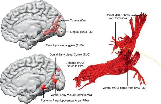
MEDIALOCCIPITALLONGITUDINALTRACT Acoherentwhitematterbundlethatrunsbetweenthe peripheralvisualrepresentationwithinthemedialoccipitalcortexandtheposteriorPHGhasbeenrecently reportedbyourgroupusingtractography(Beyhetal., 2022).Consideringitsmediallocationandcoursealong theposterior–anterioraxis,werefertothispathwayas themedialoccipitallongitudinaltract(MOLT).The MOLThasadorsal(cuneal)andaventral(lingual)componentandbothprojectontotheposteriorparahippocampalplacearea(PPA)(Fig.1.5).FibersoftheMOLTwere identifiedinearlytractographystudiesbutwereconsideredaspartoftheventralcingulum(Catanietal.,2002) andILF(Catanietal.,2003).OurviewisthattheMOLT is anindependenttractwithauniquerolecrucialfor visuospatiallearning(Beyhetal.,2022).TheMOLT collects “raw” visualinformationfromperipheralearly visualcortex(EVC)andconveysitthePPAwhereacompletemapofthevisualsceneisgenerated.Inthiscontext, theMOLTmaycarryfeedforwardandfeedbackspatial
Fig.1.5. Tractographyreconstructionofthemedialoccipitallongitudinaltract(MOLT).TheMOLTisanoccipitotemporalwhite matterpathwaythatstemsfromtheanteriorcuneus(Cu)andlingualgyrus(LG)andterminatesintheposteriorparahippocampal gyrus(PHG).Inthemedialoccipitallobe,itprojectsontoperipheralvisualfieldrepresentationswithintheearlyvisualcortex (EVC),whileitstemporallobeterminationsoverlapwiththeposteriorparahippocampalplacearea(PPA).The dashedyellow line indicatesthecalcarinesulcus.ModifiedfromBeyhA,Dell’AcquaF,CancemiDetal.(2022).Themedialoccipitallongitudinaltractsupportsearlystageencodingofvisuospatialinformation. CommunBiol 5:318.
informationbetweentheEVCandthePPA,thereby servingasapathwayforre-entrantvisualinformation (HochsteinandAhissar,2002)thatsupportsamultistage encodingandlearningofthevisualscene.Assuch,early activationswithinthePPA(Bastinetal.,2013a)maycorrespondtoaninitialmappingofthe “gistofthescene” which,viafeedbacktotheEVC,refinesthelater,detailed mappingofobjectconfiguration.Thisrefinedinformation,stillinaretinotopicspace,couldthenbecombined withparietalandretrosplenialspatialinformationatlonger latencies(Bastinetal.,2013b),andtranslatedintoanallocentricframeofreference.Thishigherorder,viewpointinvariantspatialinformationwouldultimatelyreachmore anteriormedialtemporalregionsincludingthehippocampalformation,andmoredistantfrontalregions(Van Hoesen,1982; Kravitzetal.,2011; DaltonandMaguire, 2017).Inbothhemispheres,thelingualgyruscomponent oftheMOLThasalargervolumeanddistributestoa wideroccipitalsurfacecomparedtothecunealcomponent (Beyhetal.,2022).ThisdistinctionbecomesmoreimportantwhenweconsiderthattheMOLTmediatesastronger overallconnectivitybetweenthePPAandventralEVC (lingualgyrus,uppervisualfield)comparedwiththedorsalEVC(cuneus,lowervisualfield).Thisimbalanceisin linewithafunctionalbiaswithinthePPA,whichcontains alargerrepresentationoftheuppervisualfield.The MOLTalsoexhibitsarighthemispherelateralization, whichisanobservationthatfitsexistingliteraturereportingsuchahemisphericbiasinthespatiallearningfunctionssupportedbytheparietalandmedialtemporal
lobes.Further,thislateralizationmayexplainthehigher frequencyofvisuospatiallearningdeficitsfollowing posteriorrighthemispherelesions.
U-SHAPEDTEMPOROINSULARINTERLOBARFIBERS Shortinterlobarfibersconnectthesuperiortemporal gyrustoadjacentregionsoftheinsula(Mesulamand Mufson,1982).Inthehumanbrain,temporoinsularfibers havebeendissectedpostmortem(Nachtergaeleetal., 2019)andtwogroupscanbeidentifiedwithtractography (Fig.1.4right).Theanteriortemporoinsularfibersoriginatefromthecortexoftheplanumpolareand,through theextremecapsule,reachthelimeninsulaeandthe shortinsulargyri.Theanteriorfibersareoftendissected togetherandconfusedwiththefibersoftheuncinatefasciculus.Theposteriortemporoinsularfibersoriginate fromHeschl’sgyrusandplanumtemporaleandafter formingalooparoundthelateralfissureterminatein thelonginsulargyri.Theseconnectionsareoftendissectedtogetherwiththemostmedialfibersofthearcuate fasciculus.Theroleofthetemporoinsularconnectionsis unknowninhumansbuttheirpatternofdistributionas revealedbytractographymirrorsthefindingsfromaxonal tracingstudiesinthemonkeybrain(Mesulamand Mufson,1982).Thus,basedonthefindingsinmonkeys, theposteriorgroupmaydirectlyrelayauditoryinformationfromtheearlyauditorycortextotheposteriorinsular regionsandbymeansofreciprocalconnectionsallowthe insulatomodulateauditoryinputstotheearlyauditory
cortex.Consideringtheprominentroleofthehumananteriortemporalcortexinsemanticprocessingandsingle wordcomprehension,thefunctionoftheanteriorgroup maydivergeinhumansascomparedtomonkeys.
TEMPORALINTRALOBARTRACTS Thetemporalintralobarfiberscanbegroupedaccordingtotheirorientationintolongitudinalandvertical pathways( Fig.1.4 right).
The temporallongitudinalfasciculus(TLF)isasystemofshortandlongfibersbetweentheanteriorandposteriorregionsofthesuperior,middle,andinferior temporalgyri(CataniandDawson,2016; Maffeietal., 2017; CataniandForkel,2019).Itsmostdorsalbranch, the TLFI,runswithinthewhitematterofthesuperior temporalgyruswhereitformsachainofconnections originatinginHeschl’sgyrusandprogressinginboth anteriorandposteriordirectionstowardtheplanum polareandplanumtemporale,respectively.Longerfibers connecttheposteriorandanteriortemporalregions(Fig. 1.2A–C). TLFIIisalargertractcomparedtoTLFIand itsfibersarecontainedwithinthemiddleandinferiortemporalgyri(Fig.1.2BandCleft).SomefibersoftheTLF havebeenpreviouslyvisualizedinthemonkeybrain usingaxonaltracingmethods(SchmahmannandPanda, 2006)andinhumanswithtractography(Catanietal., 2013)buttheywereconsideredasbeingpartofthe MLF ortheILF.
TractographyanalysisinhumansrevealedasignificantleftwardasymmetryforTLFI,butnotforTLFII. TheexistenceofTLFinhumansisinlinewithpreviously proposedmodelsofdualstreamsforvisualandauditory processingbasedonanimalexperimentsandappliedto humancognition(Kravitzetal.,2013; DeWittand Rauschecker,2012).Accordingtothesemodels,the ventral streamsarecomposedofcorticalareasthatprocessvisualandauditoryinformationforobjectidentificationwhereastheareasofthedorsalstreamsspecialize inspatiallocalizationofobjectsandsounds.Inthemonkeybrain,theareasofbothauditoryandvisualventral streamsaredistributedalongthecortexofthesuperior andinferiortemporalgyriandsuperiortemporalsulcus. Theconceptof “stream ” impliestheexistenceofa networkinwhichspecializedneuronsarespatiallysegregatedincorticalareasbutreciprocallyconnectedby short-andlong-rangefibers(Kravitzetal.,2013).
In general,thegoaloftheventralstreamsistoprocess thearrayoflightandsoundthatenterourorgansofperceptionandderiveacoherentrepresentationoftheobjectsthat havegeneratedit,thatis,totransformlocalsensoryrepresentationsintomultisensorypercepts(Mesulam,1998; Farah,2000; DeWittandRauschecker,2012).Wesuggest thatinhumanstheTLFfibershavearolesimilartothe connectionsdescribedinmonkeys.Specifically,TLF
Imediatesreciprocalconnectionsbetweentheareasof theventralauditorystreamwhereasTLFIIhasasimilar functionfortheventralvisualstream.TLFistherefore responsiblefortheflowofsensoryinformationthattransformlocalauditoryandvisualperceptsintoglobalrepresentations.Thisflownecessitatesneuronalmechanisms suchashierarchicalprocessingandcombinationsensitivity thattransformstonotopic(orretinotopic)perceptionin earlytemporalandoccipitalsensoryareasintosemantic representationintheanteriortemporallobe(Binderetal., 2009).Tractographyhasbeenusedtodemonstratedamage to TLFIinapatientwithpureworddeafnessduetostroke (Maffeietal.,2017).Furthermore,inpatientswithprimary progressiveaphasia,abnormalitiesofTLFIandIIcorrelate withimpairmentinsinglewordcomprehensionandobject namingbutnotsentencecomprehension.
Intheventralaspectofthetemporallobe,the fusilum representsanintrinsicsystemoffibersconnectingdifferentregionsofthefusiformgyrus.Thefusilummayrepresentthecoreventralnetworkspecializedinword (Epelbaumetal.,2008)andface(Foxetal.,2008)processingwithsomedegreeofhemisphericfunctionalspecialization(leftfusilumforwordsandrightfusilumforfaces) (L€ udersetal.,1991; AlbonicoandBarton,2017).Lesions to thefusilumcanmanifestwithalexia(Epelbaumetal., 2008)andassociativeprosopagnosia(Foxetal.,2008).
The TVTiscomposedofdenseU-shapedfibers betweentheSTGandMTG.Thistractarchesbeneath theconvexityofthesuperiortemporalsulcusandispresentonlyinthemostposteriortemporalregion.Preliminarycomparativetractographyanalysissuggeststhatthe TVTisabsentinthemonkeybrainwhileinhumansisa left-lateralizedtract.Hence,theTVTrepresentsthemajor intrinsicconnectionsystemwithinWernicke’sareainthe lefthemisphereanditsdamageisprobablycorrelated withtheseverityofbothauditoryandwrittencomprehensiondeficitsthatcharacterizeposterioraphasias.
ABBREVIATIONS AF,arcuatefasciculus;EVC,earlyvisualcortex;IFOF, inferiorfronto-occipitalfasciculus;ILF,inferiorlongitudinalfasciculus;LG,lingualgyrus;MGN,medialgeniculatenucleus;MLF,middlelongitudinalfasciculus; MOLT,medialoccipitallongitudinaltract;PHG, parahippocampalgyrus;PPA,parahippocampal placearea;TIT,temporoinsulartract;TLF,temporallongitudinalfasciculus;TVT,temporalverticaltract;UF, uncinatefasciculus.
REFERENCES AboitizF,Garcı´aR(2009).Mergingofphonologicalandgesturalcircuitsinearlylanguageevolution.RevNeurosci 20: 71–84.
AlbonicoA,BartonJ(2017).Faceperceptioninpurealexia: complementarycontributionsoftheleftfusiformgyrus tofacialidentityandfacialspeechprocessing.Cortex 96: 59–72.
AranziGC(1587).Dehumanofoetulibertertioeditus,ac recognitus,ejusdemanatomicarumobservationumliber, acDetumoribussecundumlocosaffectoslibernuncprimum editi,apudJacobunBrechtanum,Venetiis[Translatedin ClarkeE,O’MalleyCD(eds.)(1996).Thehumanbrain andspinalcord:ahistoricalstudyillustratedbywritings fromantiquitytothetwentiethcentury,secondedn.San Francisco,CA:NormanPublishing].
BarrettRLC,DawsonM,DyrbyTBetal.(2020).Differences infrontalnetworkanatomyacrossprimatespecies. JNeurosci 40:2094–2107.
BartholinC(1641).Institutionesanatomicae,Lug.Batavorum, ApudFranciscumHackium,Leiden.
BastinJetal.(2013a).Temporalcomponentsintheparahippocampalplacearearevealedbyhumanintracerebral recordings.JNeurosci 33:10123–10131. BastinJetal.(2013b).Timingofposteriorparahippocampal gyrusactivityrevealsmultiplesceneprocessingstages. HumBrainMapp 34:1357–1370.
BechterewW(1900).Demonstrationeinesgehirnsmit ZestorungdervorderenundinnerenTheilederHirnrinde beiderSchlafenlappen.NeurolCentralbl 20:990–991.
BehrensTEJ,BergHJ,JbabdiSetal.(2007).Probabilisticdiffusiontractographywithmultiplefibreorientations:what canwegain?Neuroimage 34:144–155.
BeyhA,Dell’AcquaF,CancemiDetal.(2022).Themedial occipitallongitudinaltractsupportsearlystageencoding ofvisuospatialinformation.CommunBiol 5:318.
BinderJR,DesaiRH,GravesWWetal.(2009).Whereisthe semanticsystem?Acriticalreviewandmeta-analysisof 120functionalneuroimagingstudies.CerebCortex 19: 2767–2796.
BrocaP(1878).Anatomiecompareedescirconvolutions cerebrales:legrandlobelimbique.RevAnthropol 1: 385–498.
BrownS,Sch€ aferEA(1888).Aninvestigationintothefunctionsoftheoccipitalandtemporallobesofthemonkey’s brain.PhilosTransRSocLondB 303–327. BurdachCF(1819–26).VomBaueundLebendesGehirns, Dyk,Leipzig. BurgelU,AmuntsK,HoemkeLetal.(2006).Whitematter fibertractsofthehumanbrain:three-dimensionalmapping atmicroscopicresolution,topographyandintersubjectvariability.Neuroimage 29:1092–1105. CanterasNS,SimerlyRB,SwansonLW(1995). Organizationofprojectionsfromthemedialnucleusof theamygdala:aPHALstudyintherat.JCompNeurol 360:213 –245.
CataniM,BambiniV(2014).Amodelforsocialcommunicationandlanguageevolutionanddevelopment(SCALED). CurrOpinNeurobiol 28:165–171.
CataniM,DawsonMS(2016).Languageprocessing,developmentandevolution.In:PMConn(Ed.),Conn’stranslational neuroscience.AcademicPress,pp.679–692.
CataniM,ForkelSJ(2019).Diffusionimagingmethodsin languagesciences.In:TheOxfordhandbookofneurolinguistics,OxfordUniversityPress,pp.212–227.
CataniM,ThiebautdeSchottenM(2012).Atlasofhuman brainconnections,OxfordUniversityPress,Oxford.
CataniM,HowardRJ,PajevicSetal.(2002).Virtualinvivo interactivedissectionofwhitematterfasciculiinthehuman brain.Neuroimage 17:77–94.
CataniM,JonesDK,DonatoRetal.(2003).Occipitotemporalconnectionsinthehumanbrain.Brain 126: 2093–2107.
CataniM,JonesDK,ffytcheDH(2005).Perisylvianlanguage networksofthehumanbrain.AnnNeurol 57:8–16.
CataniM,Dell’AcquaF,ThiebautdeSchottenM(2013). Arevisedlimbicsystemmodelformemory,emotionand behaviour.NeurosciBiobehavRev 37:1724–1737.
CataniM,Dell’AcquaF,BudisavljevicSetal.(2016).Frontal networksinadultswithautismspectrumdisorder.Brain 139:616–630.
ClarkeE,O’MalleyCD(1996).Thehumanbrainandspinal cord:ahistoricalstudyillustratedbywritingsfromantiquitytothetwentiethcentury,NormanPublishing,San Francisco,CA.
CraigMC,CataniM,DeeleyQetal.(2009).Alteredconnectionsontheroadtopsychopathy.MolPsychiatry 14: 946–953.
CraigMC,SethnaV,GudbrandsenMetal.(2022).Birthofthe blues—theeffectsofprenataldepressionontheinfant brain.PsycholMed (inpress).
DaltonMA,MaguireEA(2017).Thepre/parasubiculum:a hippocampalhubforscene-basedcognition?CurrOpin BehavSci 17:34–40.
D’AnnaL,MesulamMM,ThiebautDeSchottenMetal. (2016).Frontotemporalnetworksandbehavioralsymptomsinprimaryprogressiveaphasia.Neurology 86: 1393–1399.
DeWittI,RauscheckerJP(2012).Phonemeandwordrecognitionintheauditoryventralstream.ProcNatlAcadSciUS A 109:E505–E514.
EpelbaumE,PinelP,GaillardRetal.(2008).Purealexiaasa disconnectionsyndrome:newdiffusionimagingevidence foranoldconcept.Cortex 44:962–974. FarahMJ(2000).Thecognitiveneuroscienceofvision, BlackwellPublishing..
FerraraNC,CullenPK,PullinsSPetal.(2017).Inputfromthe medialgeniculatenucleusmodulatesamygdalaencoding offearmemorydiscrimination.LearnMem 24:414–421. FerrierD,YeoGF(1884).XIX.Arecordofexperimentsonthe effectsoflesionofdifferentregionsofthecerebralhemispheres.PhilosTransRSocLond 479–564. ffytcheDH,CataniM(2005).Beyondlocalization:from hodologytofunction.PhilosTransRSocBBiolSci 360:767–779.
ffytcheDH,BlomJD,CataniM(2010).Disordersofvisual perception.JNeurolNeurosurgPsychiatry 81:1280–1287. FingerS(1994).Originsofneuroscience,OxfordUniversity Press,Oxford. FlechsigPE(1896).GehirnundSteele,Veit,Leipzig.
ForkelSJ,ThiebautdeSchottenM,KawadlerJMetal.(2014). Theanatomyoffronto-occipitalconnectionsfromearly bluntdissectionstocontemporarytractography.Cortex 56:73–84.
ForkelSJ,RogalskiE,DrossinosSanchoNetal.(2020). Anatomicalevidenceofanindirectpathwayforwordrepetition.Neurology 94:594–606.
FoxCJ,IariaG,BartonJJS(2008).Disconnectioninprosopagnosiaandfaceprocessing.Cortex 44:996–1009.
GaffanD,WilsonC(2008).Medialtemporalandprefrontal function:recentbehaviouraldisconnectionstudiesinthe macaquemonkey.Cortex 44:928–935.
GloorP(1997).Thetemporallobeandthelimbicsystem, OxfordUniversityPress.
HackettT.A.Anatomicorganizationoftheauditorycortex.In Handbook ofclinicalneurology,thirdseries,vol.129 (2015)Thehumanauditorysystem.ChelseaGGand HickokGeditors.Elsevier:Amsterdam,Netherlands HochsteinS,AhissarM(2002).Viewfromthetop:hierarchies and reversehierarchiesinthevisualsystem.Neuron 36: 791–804.
JakobC(1906).Nuevacontribucio ´ na ´ lafisio-patologı´adelos lo ´ bulosfrontales.LaSemMed 13:1325–1329.
JonesEG(2011).Thehistoricaldevelopmentofideas abouttheauditorycortex.In:JAWiner,CESchreiner (Eds.),Theauditorycortex.SpringerUS,Boston,MA, pp.1–40.
JouandetML,GazzanigaMS(1979).Corticalfieldoforiginof theanteriorcommissureoftherhesusmonkey.ExpNeurol 66:381–397.
KamaliA,SairHI,BlitzAMetal.(2016).Revealingtheventralamygdalofugalpathwayofthehumanlimbicsystem usinghighspatialresolutiondiffusiontensortractography. BrainStructFunct 221:3561–3569.
KravitzDJ,SaleemKS,BakerCIetal.(2013).Theventral visualpathway:anexpandedneuralframeworkforthe processingofobjectquality.TrendsCognSci 17:26–49. KravitzDJ,SaleemKS,BakerCIetal.(2011).Anewneural frameworkforvisuospatialprocessing.NatRevNeurosci 12:217–230.
LebowM,ChenA(2016).Overshadowedbytheamygdala: thebednucleusofthestriaterminalisemergesaskeyto psychiatricdisorders.MolPsychiatry 21:450–463.
Lo ´ pez-BarrosoD,CataniM,RipollesPetal.(2013).Word learningismediatedbytheleftarcuatefasciculus.Proc NatlAcadSciUSA 110:13168–13173.
LudersH,LesserRP,HahnJetal.(1991).Basaltemporallanguagearea.Brain 114:743–754.
MaffeiC,SoriaG,Prats-GalinoAetal.(2015).Imaging white-matterpathwaysoftheauditorysystemwithdiffusionimagingtractography.In:GGChelsea,GHickok (Eds.),Thehumanauditorysystem.Handbookofclinical neurology,thirdseries. vol.129.Elsevier,Amsterdam, Netherlands.
MaffeiC,CapassoR,CazzolliGetal.(2017).Pureword deafnessfollowinglefttemporaldamage:behavioraland neuroanatomicalevidencefromanewcase.Cortex 97: 240–254.
MaffeiC,SarubboS,JovicichJ(2019).Amissingconnection: areviewofthemacrostructuralanatomyandtractography oftheacousticradiation.FrontNeuroanat 13. MarsRB,FoxleyS,VerhagenLetal.(2016).Theextreme capsulefibercomplexinhumansandmacaquemonkeys: acomparativediffusionMRItractographystudy.Brain StructFunct 221:4059–4071.
MesulamMM(1998).Fromsensationtocognition.Brain 121: 1013–1052.
MesulamMM,MufsonEJ(1982).Insulaoftheoldworld monkey.III:efferentcorticaloutputandcommentson function.JCompNeurol 212:38–52.
NachtergaeleP,RadwanA,SwinnenSetal.(2019).The temporoinsularprojectionsystem:ananatomicalstudy. JNeurosurg 132: 615–623.
NaidichT.Brainstem.InGray’sanatomy(42edn.)Standing,S. (editor-in-chief).(2020),pp.512-39.Elsevier,Amsterdam. PapezJW(1937).Aproposedmechanismofemotion.Arch Neurol Psychiatry 38:725–743.
ParkHJ,KimJJ,LeeSKetal.(2008).Corpuscallosalconnectionmappingusingcorticalgraymatterparcellationand DT-MRI.HumBrainMapp 29:503–516.
PicklesJO(2015).Auditorypathways:anatomyandphysiology.In:GGChelsea,GHickok(Eds.),Thehumanauditory system.Handbookofclinicalneurology. vol.129.Elsevier, Amsterdam,Netherlands.
RaichleME,SnyderAZ(2007).Adefaultmodeofbrainfunction:abriefhistoryofanevolvingidea.Neuroimage 37: 1083–1090.
RauscheckerJP,ScottSK(2009).Mapsandstreamsinthe auditorycortex:nonhumanprimatesilluminatehuman speechprocessing.NatNeurosci 12:718–724.
RillingJK,GlasserMF,PreussTMetal.(2008).Theevolution ofthearcuatefasciculusrevealedwithcomparativeDTI. NatNeurosci 11:426–428.
RollsET(2015).Limbicsystemsforemotionandformemory, butnosinglelimbicsystem.Cortex 62:119–157. SaurD,KreherBW,SchnellSetal.(2008).Ventralanddorsal pathwaysforlanguage.ProcNatlAcadSciUSA 105: 18035–18040.
SchmahmannJ,PandyaD(2006).Fiberpathwaysofthebrain, OxfordUniversityPress.
SchulzeK,Vargha-KhademF,MishkinM(2012).Testofa motortheoryoflong-termauditorymemory.ProcNatl AcadSciUSA 109:7121–7125.
TalavageTM,SerenoMI,MelcherJRetal.(2004).Tonotopic organizationinhumanauditorycortexrevealedbyprogressionsoffrequencysensitivity.JNeurophysiol 91: 1282–1296.
ThiebautdeSchottenM,ffytcheDH,BizziAetal.(2011). Atlasinglocation,asymmetryandinter-subjectvariability ofwhitemattertractsinthehumanbrainwithMRdiffusion tractography.Neuroimage 54:49–59.
TreviranusGR,TreviranusLC(1816).VermischteSchriften anatomischenundphysiologischenInhalts,BeyJ.F. R € ower.
UrbanskiM,ThiebautdeSchottenM,RodrigoSetal.(2008). Brainnetworksofspatialawareness:evidencefrom
diffusiontensorimagingtractography.JNeurolNeurosurg Psychiatry 79:598–601.
VanderauweraJ,DeVosA,ForkelSJetal.(2018).Neural organizationofventralwhitemattertractsparallelsthe initialstepsofreadingdevelopment:aDTItractography study.BrainLang 183:32–40. VanHoesenGW(1982).Theparahippocampalgyrus: newobservationsregardingitscorticalconnectioninthe monkey.TrendsNeurosci 5:345–350. Vicqd’AzyrF(1786).Traite’d’anatomieetdephysiologie. vonMonakowC(1895).ExperimentelleundpathologischanatomischeUntersuchungen € uberdieHaubenregion, denSehhugelunddieRegiosubthalamica,nebst BeitragenzurKentnissfruherworbenerGross-und
Kleinhirndefekte.ArchPsychiatrNervenkr 27:1–128. 386–478.
WernickeC(1874).DerAphasischeSymptomencomplex.Ein psychologischeStudieaufanatomischerBasis,Cohn& Weigert,Breslau. WillisT(1664).CerebriAnatome,Martyn&Allestry, London. WilsonSW,GalantucciS,TartagliaMCetal.(2011). Syntacticprocessingdependsondorsallanguagetracts. Neuron 72:397–403. ZekiSM(1973).Comparisonofthecorticaldegenerationin thevisualregionsofthetemporallobeofthemonkey followingsectionoftheanteriorcommissureandthe splenium.JCompNeurol 148:167–176.