SURVIVAL GUIDE
Second Edition
Anthony Pane MBBS MMedSc FRANZCO PhD Neuro-Ophthalmologist, Queensland Eye Institute, Brisbane, Australia
Neil R Miller MD FACS
Professor of Ophthalmology, Neurology and Neurosurgery & Frank B Walsh Professor of Neuro-Ophthalmology, Wilmer Eye Institute, Johns Hopkins Hospital, Baltimore, MD, USA
Michael Burdon BSc MB BS MRCP FRCOphth President, Royal College of Ophthalmologists, UK Consultant Neuro-Ophthalmologist, Selly Oak Hospital, Birmingham, UK
For additional online content visit ExpertConsult.com
© 2018, Elsevier Limited. All rights reserved.
First edition 2007
Second edition 2018
The right of Anthony Pane, Neil R Miller and Michael Burdon to be identified as authors of this work has been asserted by them in accordance with the Copyright, Designs and Patents Act 1988.
No part of this publication may be reproduced or transmitted in any form or by any means, electronic or mechanical, including photocopying, recording, or any information storage and retrieval system, without permission in writing from the publisher. Details on how to seek permission, further information about the Publisher’s permissions policies and our arrangements with organizations such as the Copyright Clearance Center and the Copyright Licensing Agency, can be found at our website: www.elsevier.com/permissions
This book and the individual contributions contained in it are protected under copyright by the Publisher (other than as may be noted herein).
Notices
Knowledge and best practice in this field are constantly changing. As new research and experience broaden our understanding, changes in research methods, professional practices, or medical treatment may become necessary.
Practitioners and researchers must always rely on their own experience and knowledge in evaluating and using any information, methods, compounds, or experiments described herein. In using such information or methods they should be mindful of their own safety and the safety of others, including parties for whom they have a professional responsibility.
With respect to any drug or pharmaceutical products identified, readers are advised to check the most current information provided (i) on procedures featured or (ii) by the manufacturer of each product to be administered, to verify the recommended dose or formula, the method and duration of administration and contraindications. It is the responsibility of practitioners, relying on their own experience and knowledge of their patients, to make diagnoses, to determine dosages and the best treatment for each individual patient and to take all appropriate safety precautions.
To the fullest extent of the law, neither the Publisher nor the authors, contributors or editors, assume any liability for any injury and/or damage to persons or property as a matter of products liability, negligence or otherwise, or from any use or operation of any methods, products, instructions or ideas contained in the material herein.
ISBN: 978 0 7020 7267 3
Printed in China
Last digit is the print number: 9 8 7
Content Strategist: Laurence Hunter
Content Development Specialist: Fiona Conn
Project Manager: Louisa Talbott
Design: Miles Hitchen
Illustration Manager: Lesley Frazier
Illustrator: Marie Dean
The publisher’s policy is to use paper manufactured from sustainable forests
Video contents
2 Blurred vision or field loss
Video 2.1 The Mirror Test for non-organic blindness. Note that despite supposedly being blind, this patient with non-organic blindness cannot suppress her eye movements when the large mirror is moved back and forth/up and down.
Video 2.2 The Optokinetic Test for non-organic blindness. Note that despite supposedly being blind, the patient has optokinetic responses to a rotating target. If the patient claimed unilateral blindness (in this case, the left eye), one begins by testing the patient with both eyes open. Once normal responses are noted, the patient’s right eye is suddenly covered. Persistence of an optokinetic response in the left eye indicates intact vision in that eye.
Video 2.3 Testing proprioception in a patient with non-organic blindness. Even a blind patient should be able to touch a finger to the nose or bring both fingers together using proprioception as noted in this video in which the subject has both eyes patched. Failure to perform this task suggests non-organic visual loss.
5 Double vision
Video 5.1 Restrictive myopathy – tight left medial rectus muscle in thyroid eye disease.
Video 5.2 Global paretic myopathy due to chronic progressive external ophthalmoplegia (CPEO).
Video 5.3 Ocular myasthenia, misdiagnosed as a decompensating exophoria.
Video 5.4 Ocular myasthenia, at first thought to be a decompensating congenital fourth nerve palsy.
Video 5.5 Right partial third nerve palsy due to a large basilar artery aneurysm.
Video 5.6 Horizontal diplopia.
Video 5.7 More detailed examination reveals a right partial third nerve palsy with aberrant regeneration.
Video 5.8 Patient diagnosed as having a decompensated exophoria.
Video 5.9 A complete left third nerve palsy with pupil involvement.
Video 5.10 Diplopia due to a right exotropia.
Video 5.11 Acquired (traumatic) left fourth nerve palsy due to a motor vehicle accident.
Video 5.12 Congenital left fourth nerve palsy.
Video 5.13 Left sixth nerve palsy from an internal carotid artery aneurysm.
Video contents
Video 5.14 Left cavernous sinus syndrome due to lung cancer metastasis.
Video 5.15 Miller Fisher syndrome (MFS).
Video 5.16 Bilateral but asymmetric internuclear ophthalmoplegia (INO) in a young woman with multiple sclerosis.
Video 5.17 Right hypertropic skew deviation due to a midbrain stroke.
Video 5.18 Dorsal midbrain syndrome due to hemorrhage into a pinealoma.
Video 5.19 Congenital bilateral horizontal gaze palsy due to Moebius syndrome.
Video 5.20 Acquired left conjugate horizontal gaze palsy – left sixth nerve nucleus lesion due to pontine stroke.
Video 5.21 Vertical gaze palsy (downgaze worse than upgaze) due to midbrain stroke.
Video 5.22 Progressive supranuclear palsy (PSP).
7 Abnormal movement or orientation of the visual world
Video 7.1 Horizontal jerk nystagmus.
Video 7.2 Upbeat nystagmus.
Video 7.3 Downbeat nystagmus.
Video 7.4 Acquired horizontal pendular nystagmus.
Video 7.5 Torsional jerk nystagmus.
Video 7.6 See-saw nystagmus.
Video 7.7 Periodic alternating nystagmus in a patient with a Chiari malformation.
Video 7.8 Gaze-evoked nystagmus in a child with a Chiari malformation.
Video 7.9 Superior oblique myokymia in a healthy young woman.
Video 7.10 Square-wave jerks.
Video 7.11 Macro-square-wave jerks.
Video 7.12 Macrosaccadic oscillations.
Video 7.13 Opsoclonus in a woman with an ovarian carcinoma.
Video 7.14 Ocular flutter.
Video 7.15 Monocular (left) pendular nystagmus in a patient with long-standing multiple sclerosis (MS).
Video 7.16 Convergence-retraction nystagmus in a young woman with a dorsal midbrain lesion.
Video 7.17 Pupillary light-near dissociation in a man with a pineal region tumor.
Video 7.18 Rebound nystagmus in a patient with a fourth ventricular epidermoid cyst.
Video 7.19 Rebound nystagmus in a patient with long-standing multiple sclerosis (MS).
Video 7.20 Voluntary nystagmus in a healthy young man.
Video 7.21 Downbeat nystagmus before and after treatment with memantine.
8 Abnormal eye movements without visual symptoms
Video 8.1 Acquired left horizontal gaze palsy in a middle-aged woman.
Video 8.2 Horizontal “motor” jerk nystagmus in a middle-aged man.
Video 8.3 Horizontal “sensory” pendular nystagmus in a small child.
Video 8.4 Spasmus nutans in a child with an optic chiasmal glioma.
Video 8.5 Square-wave jerks in a patient with Parkinson disease.
9 Unequal pupils
Video 9.1 Infrared video of a patient with a left Horner syndrome.
Video 9.2 Tonic pupil (Adie syndrome).
10 Ptosis
Video 10.1 Cogan lid twitches in three patients with myasthenia gravis.
Preface
Whether you are a busy optometrist who primarily performs refractions, an ophthalmologist who sees patients with cataracts or glaucoma, or a neurologist who sees a lot of patients with headache, you never know when a patient with a potentially vision- or lifethreatening disorder will come to your clinic with a visual problem. Over the many years that we have practiced neuro-ophthalmology, we have encountered many patients who became permanently blind or neurologically impaired or who died because their otherwise skilled and well-meaning ophthalmologists, optometrists, or neurologists failed to recognize that they had a potentially devastating but treatable neuro-ophthalmic condition. This, despite the existence of many excellent and detailed neuro-ophthalmology texts. The problem is that none of these texts are written for the vast majority of practitioners who have no particular interest or expertise in neuro-ophthalmology. In addition, most of these texts are diagnosis-based and, therefore, only helpful once the diagnosis had been made. However, in our opinion, the three most difficult challenges for most practitioners are to recognize that their patient has a “neuro-ophth” problem in the first place, then to make the correct diagnosis, and, finally, to provide appropriate treatment in a timely fashion.
To address these challenges, we set out ten years ago to write a simple, practical clinical guide to benefit practitioners and their trainees. We made the guide symptom-based; i.e. listen to the patient’s concern (e.g. “I see double”; “the vision in my left eye is slowly worsening”), turn to the appropriate chapter (e.g. Chapter 5: Double vision; Chapter 2: Blurred vision or field loss), and let the book guide you every step of the way to the correct diagnosis and treatment without presuming that you have any previous neuroophthalmic training.
In the ten years since the book’s publication, there have been many advances in our ability to diagnosis and treat neuroophthalmic conditions. Accordingly, this second edition of
Preface
The Neuro-Ophthalmology Survival Guide provides an updated but still carefully structured approach. Specifically, it tells you what questions to ask, what to look for during the examination, what diagnostic tools might be useful to make the correct diagnosis, and, depending on the diagnosis, what the management options are, all using clear and simple bullet points and flowcharts. Also unique to this book are 60 videos showing various important eye movement and pupil abnormalities and some important examination techniques.
Neuro-ophthalmology is the “needle in the haystack” in the busy clinics of all practitioners who see patients with visual complaints. We hope this book helps you avoid sitting on too many of these needles.
Examination
2. Every new eye patient complaining of blurred vision should have:
• confrontation field testing (peripheral and central)
• a “swinging torch test” for a relative afferent pupillary defect (RAPD) before dilation
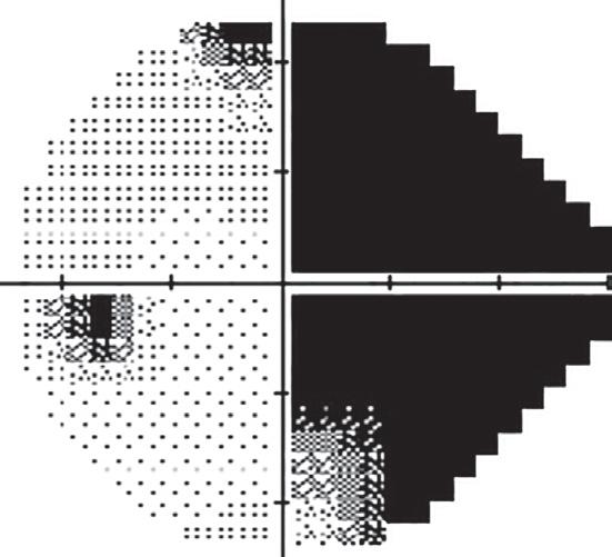
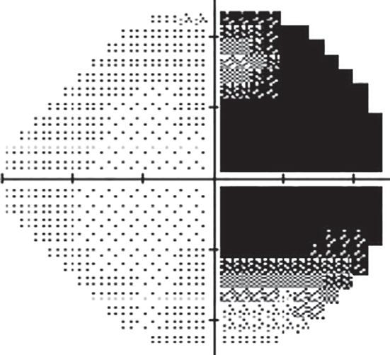
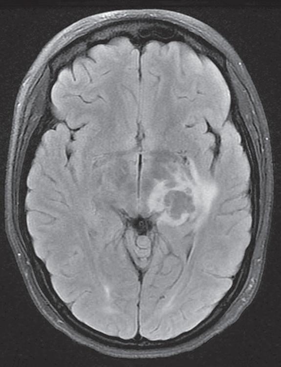
• perimetry if either of these is abnormal, the patient describes a field defect, or the degree of visual loss is not consistent with the ocular examination (Fig. 1.2)
1.2 This 16-year-old girl presented to the ophthalmology department complaining of headaches, blurred vision in her right eye and flashing lights. Visual acuity was 20/20 in each eye and intraocular examination was normal. No visual field testing was performed, and the patient was told her symptoms were due to migraine. Re-examination by another doctor revealed a right homonymous hemianopia (A, B) that was easily detected with confrontation testing; C a left thalamic mass lesion was diagnosed on MRI; further investigation showed this to be a cryptococcal abscess.
Blurred vision or field loss
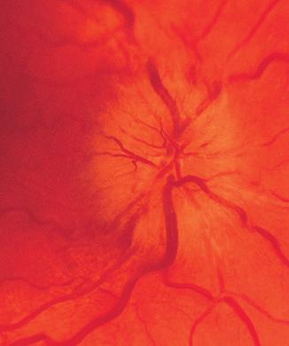
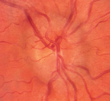
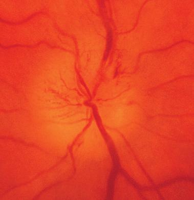
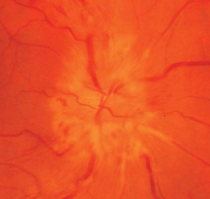
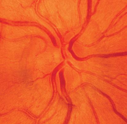
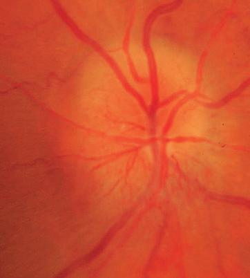
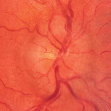
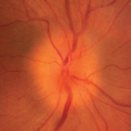
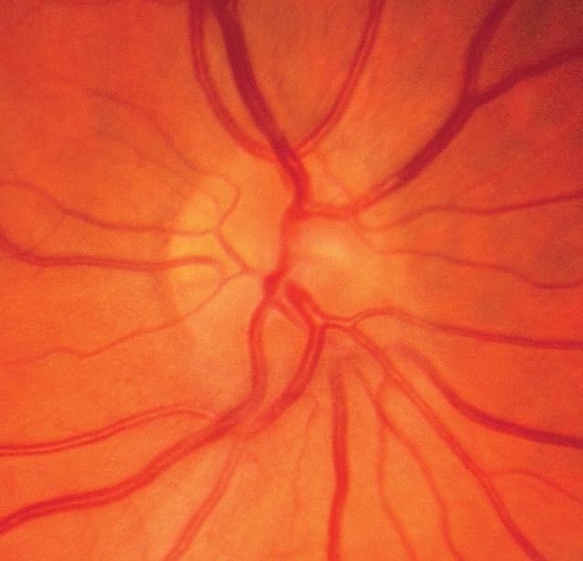
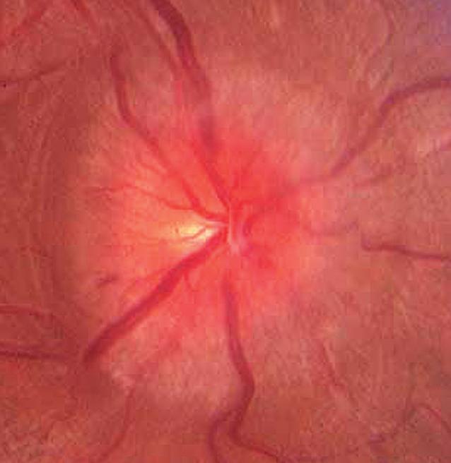
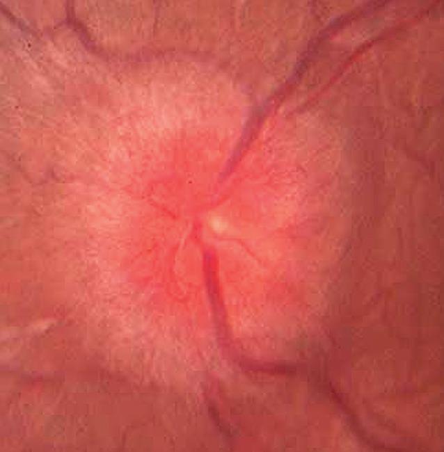
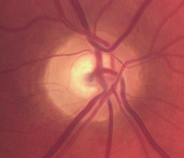
3. You can never diagnose the cause of optic nerve dysfunction just by looking at the disc (Fig. 1.3).
1.3 Hundreds of different diseases can present with a similar optic disc appearance. For example, these swollen discs have been caused by A sarcoid optic neuritis, B optic nerve infiltration by lymphoma, C non-arteritic AION, D papilledema, E Leber’s hereditary optic neuropathy, F cat scratch disease, G idiopathic optic neuritis and H optic nerve sheath meningioma. In no case was the disc appearance diagnostic; diagnosis was made on careful history, other examination, perimetry and other investigations as suggested in the management flowchart on p. 31.
H
4. All patients with non-traumatic ACUTE optic nerve dysfunction who do not meet all the clinical diagnostic criteria for either:
• typical optic neuritis (p. 35), or
• anterior ischemic optic neuropathy (AION) (p. 36) require urgent referral to a neuro-ophthalmologist (or, if this is not possible, urgent investigation as suggested on p. 38) (Fig. 1.4).
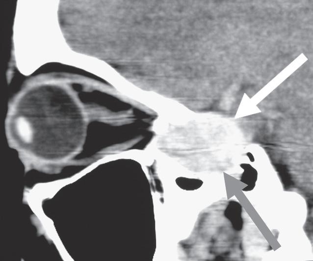
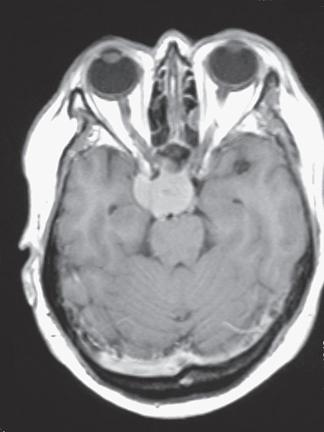
1.4 Not all acute optic neuropathies in young adults are optic neuritis! A This 32-yearold woman presented with progressive visual loss in the right eye over 3 weeks (right 20/60), pain behind the right eye, a right RAPD and normal optic discs. Her ophthalmologist diagnosed “retrobulbar optic neuritis” and reassured the patient that her vision would return spontaneously. Three months later, vision had worsened (20/200) and optic atrophy had developed; B MRI showed this large nasal tumor compressing the right optic nerve. Vision did not improve after removal of the tumor. Visual outcome would probably have been better with earlier diagnosis. For how to safely diagnose typical optic neuritis, see p. 35.
1.4
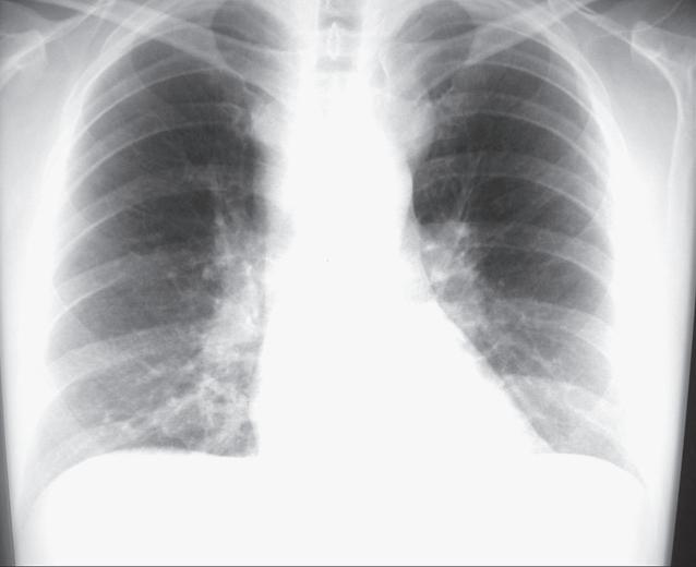
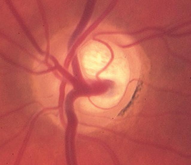
Not all acute optic neuropathies with a swollen disc are AION! This 55-year-old hypertensive man complained of progressive loss of vision in his right eye over 7 days. His ophthalmologist found C right visual acuity to be 20/40, a right RAPD and right optic disc swelling. A right inferior altitudinal scotoma was detected on perimetry. The ophthalmologist diagnosed “anterior ischemic optic neuropathy” and advised the patient that there was no treatment. Ten weeks later, right visual acuity (VA) had deteriorated to 20/400 and the right disc had become pale; further investigation revealed an increased serum angiotensin converting enzyme (ACE) and D hilar lymphadenopathy on chest x-ray. Biopsy of a lower eyelid conjunctival granuloma confirmed the diagnosis of sarcoidosis. Because diagnosis was delayed, right VA only returned to 20/80 with steroid treatment. For how to safely diagnose AION, see p. 36.
D
5. All patients with CHRONIC optic nerve dysfunction who do not meet all the clinical diagnostic criteria for glaucomatous optic neuropathy (p. 37) require referral to a neuro-ophthalmologist (or, if this is not possible, investigation as suggested on p. 38) (Fig. 1.5).
1.5 Not all chronic optic neuropathy with a “cupped” disc is glaucoma! This 48-year-old patient asked her optometrist for a change of glasses because of blurred vision. The optometrist found A, B visual acuity right 20/30, left 20/60, intraocular pressures of right 25, left 29, and bilateral disc “cupping”, and referred the patient to an ophthalmologist for treatment of possible glaucoma. Perimetry was attempted but fields were said to be “unreliable”; the ophthalmologist commenced glaucoma eyedrops. One year later, visual acuity had decreased further to right 20/60, left 20/200, and optic disc pallor was noted; C MRI revealed a large suprasellar meningioma. Visual outcome would probably have been better with earlier diagnosis. For how to safely diagnose glaucoma, see p. 37.
6. Amblyopia is a specific diagnosis, with specific diagnostic features; never use a history of “lazy eye” as the explanation for worsening vision. Features of optic nerve disease should be absent and a demonstrable cause for the amblyopia should be present (Fig. 1.6).
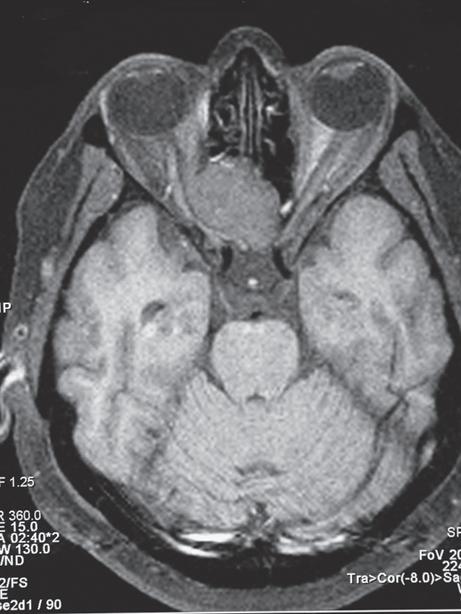
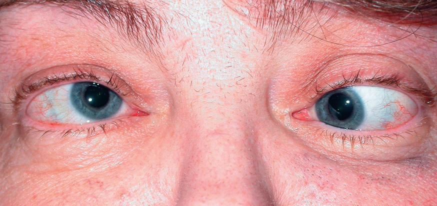
A1.6 A 38-year-old woman presented to her ophthalmologist complaining of blurred vision in her left eye and the left eye “turning in”. She said that the left eye had always been “a bit lazy” so the ophthalmologist recorded “left amblyopia” as the cause of the blurred vision without checking for an RAPD. The ophthalmologist diagnosed “decompensated congenital esotropia” and attributed the poor vision in the left eye (20/80) to “strabismic amblyopia”. Eventually another ophthalmologist investigated the patient and found A, B a large internal carotid artery aneurysm causing compressive optic neuropathy and sixth nerve palsy. Unfortunately, by this time, the left eye was blind.
9. Just because both eyes look completely normal and there is no RAPD do not assume the patient has non-organic (“functional”) visual loss; only diagnose this if you have thoroughly examined the patient, performed perimetry and have been able to “trick” the patient into demonstrating completely normal vision (see p. 34) (Fig. 1.9).
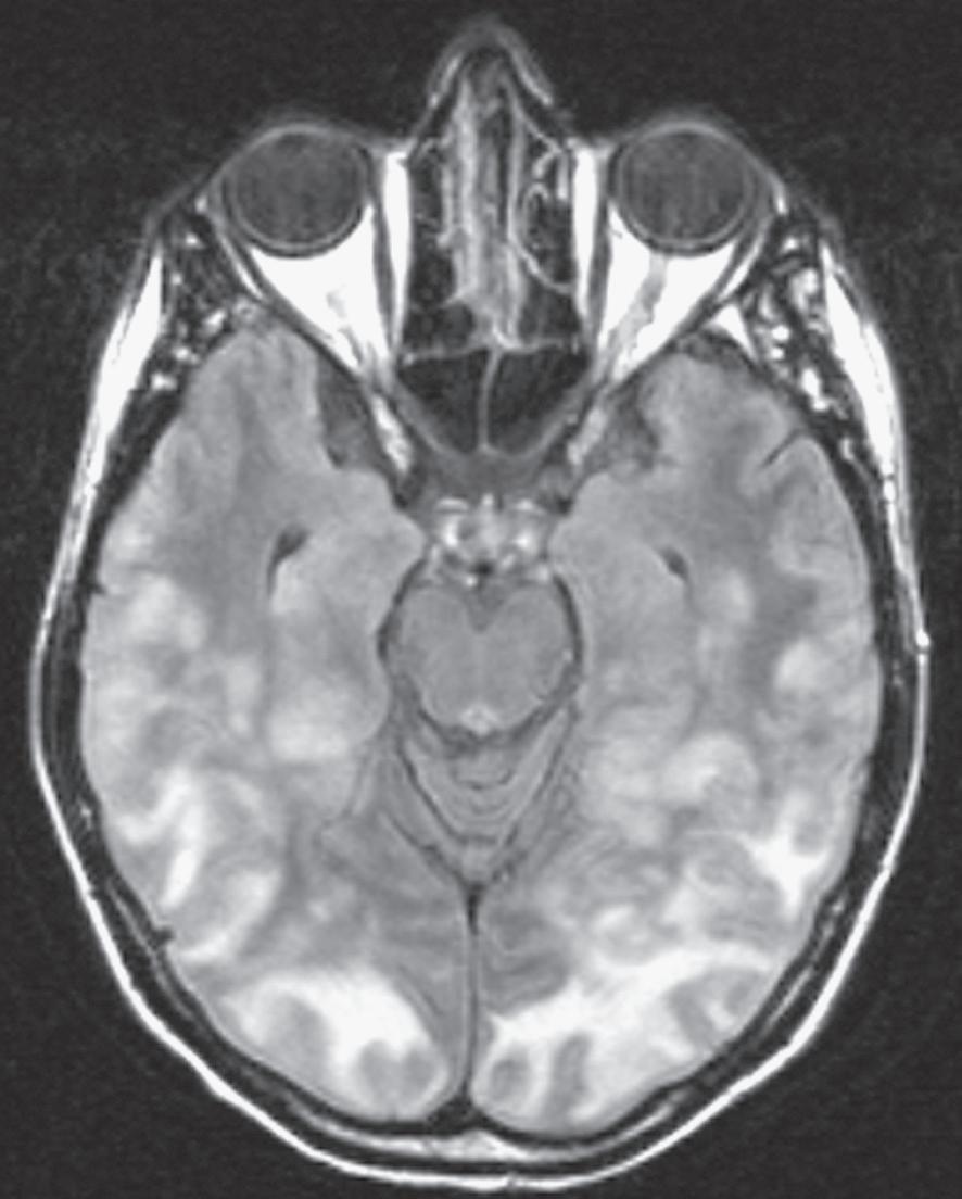
1.9 This 33-year-old man was thought by his ophthalmologist to have “functional visual loss”. Despite complaining of poor vision in both eyes, he had normal pupil reactions and normal optic disc and retinal examination. The patient was referred to a neuroophthalmologist; MRI revealed posterior leukoencephalopathy affecting the optic radiations bilaterally.
Bilateral disc swelling
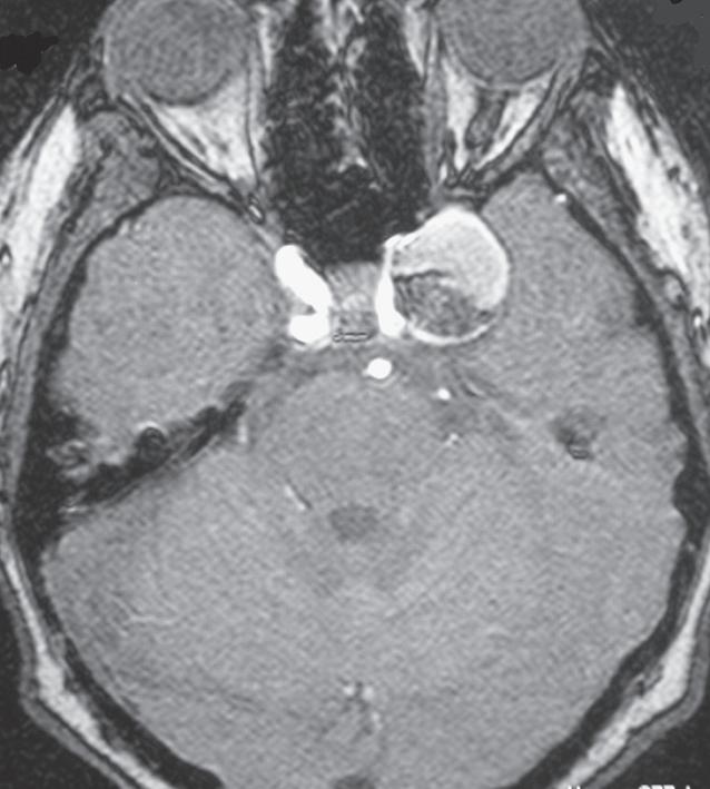
10. Bilateral disc swelling could be papilledema (disc swelling due to raised intracranial pressure). The first investigation should be urgent (same-day) magnetic resonance imaging (MRI) plus magnetic resonance venography (MRV) to exclude a brain tumor or dural venous sinus thrombosis (see p. 120) (Fig. 1.10).
1.10 This 38-year-old man presented to an ophthalmic emergency department complaining of blurred vision and headaches. Examination revealed visual acuity 20/20 right and left and bilateral moderate disc swelling A, B. Because vision was good and there were “no other neurologic signs”, the patient was allowed to go home and was scheduled for neuro-imaging as an outpatient. Two days later, while driving, the patient experienced a generalized seizure, resulting in severe injuries to himself and another driver. MRI revealed a brain tumor which was found at surgery to be an astrocytoma. For how to safely manage disc swelling with normal vision, see p. 110.
13. All cases of unexplained double vision (in which you are unable to make a definite diagnosis) require neuro-ophthalmic referral, urgently if of acute onset. There could be a serious underlying cause such as aneurysm, tumor or myasthenia (Fig. 1.13).
A
B
1.13 This 42-year-old woman initially presented to her local ophthalmologist complaining of gradual-onset horizontal double vision. At first examination, she had a small esotropia in primary position; however, eye movements were noted to be “full” (no restriction was seen in any direction). The ophthalmologist diagnosed “decompensated esophoria” and prescribed prism to be ground into the patient’s spectacles. Although this relieved the diplopia, the amount of prism required gradually increased over the next 18 months until the spectacles were too heavy for the patient to wear comfortably; at this stage, it was noted that abduction was visibly limited in both eyes. MRI revealed a large clival tumor that on biopsy was found to be a chordoma A. The tumor had caused gradually progressive bilateral sixth nerve palsies that initially presented as a small comitant esotropia with no visible limitation of abduction. Further tumor spread eventually led to a vertical deviation being superimposed on the esotropia B, due to partial third nerve palsy.
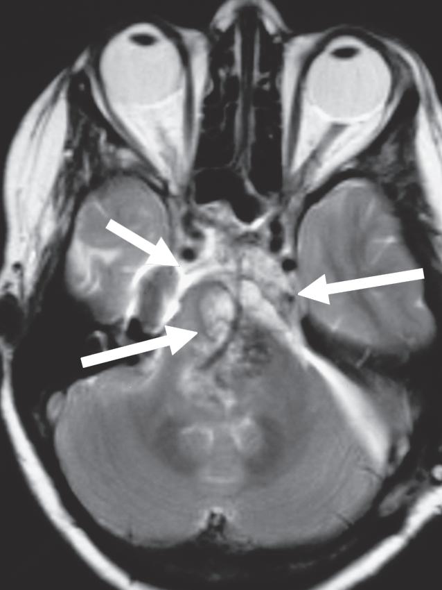
14. All patients with partial or complete third nerve palsy require urgent MRI and magnetic resonance angiography (MRA) to exclude an aneurysm, except patients who meet all the clinical diagnostic criteria for ischemic third nerve palsy (p. 177) (Fig. 1.14).
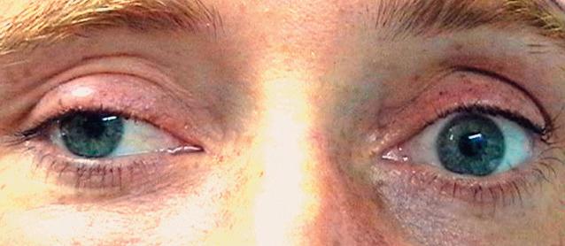
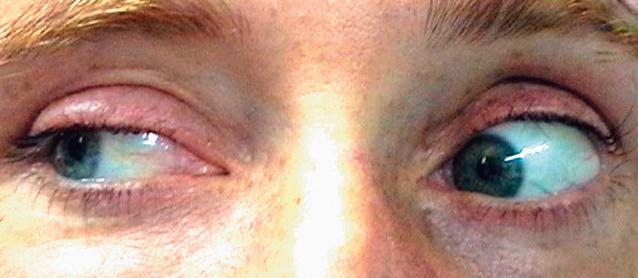
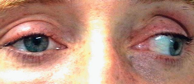
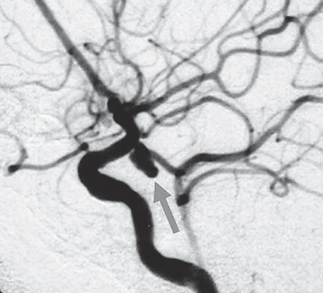
1.14 This 35-year-old diabetic woman presented with acute-onset horizontal diplopia. Her ophthalmologist noted a right exotropia, limitation of adduction of the right eye with normal elevation and depression, and a mild right ptosis A–C The pupils were equal in size and both were briskly reactive to light. The ophthalmologist diagnosed a partial right third nerve palsy. He thought that this was “ischemic” in origin because the right pupil was “spared” and the patient was a diabetic. “To rule out any other cause”, a CT scan of the brain was obtained that was normal; the patient was reassured the diplopia would resolve spontaneously and a return appointment was made for 3 months’ time. The following day, the patient collapsed at home; an urgent MRI and MRA showed a right posterior communicating artery aneurysm that had ruptured, causing subarachnoid hemorrhage; this was confirmed on angiography D. The patient underwent urgent neurosurgical intervention and survived but was left with a dense left hemiplegia. Earlier diagnosis of the aneurysm before it ruptured could have prevented her serious permanent disability. For how to safely diagnose ischemic third nerve palsy, see p. 177.