All rights reserved. No part of this publication may be reproduced, stored in a retrieval system, or transmitted, in any form or by any means, electronic, mechanical, photocopying, recording or otherwise, except as permitted by law. Advice on how to obtain permission to reuse material from this title is available at http://www.wiley.com/go/permissions.
The right of Luigi O. Massa and J. Anthony von Fraunhofer to be identified as the authors of this work has been asserted in accordance with law.
Registered Office
John Wiley & Sons, Inc., 111 River Street, Hoboken, NJ 07030, USA
Editorial Office
111 River Street, Hoboken, NJ 07030, USA
For details of our global editorial offices, customer services, and more information about Wiley products visit us at www.wiley.com.
Wiley also publishes its books in a variety of electronic formats and by print‐on‐demand. Some content that appears in standard print versions of this book may not be available in other formats.
Limit of Liability/Disclaimer of Warranty
The contents of this work are intended to further general scientific research, understanding, and discussion only and are not intended and should not be relied upon as recommending or promoting scientific method, diagnosis, or treatment by physicians for any particular patient. In view of ongoing research, equipment modifications, changes in governmental regulations, and the constant flow of information relating to the use of medicines, equipment, and devices, the reader is urged to review and evaluate the information provided in the package insert or instructions for each medicine, equipment, or device for, among other things, any changes in the instructions or indication of usage and for added warnings and precautions. While the publisher and authors have used their best efforts in preparing this work, they make no representations or warranties with respect to the accuracy or completeness of the contents of this work and specifically disclaim all warranties, including without limitation any implied warranties of merchantability or fitness for a particular purpose. No warranty may be created or extended by sales representatives, written sales materials or promotional statements for this work. The fact that an organization, website, or product is referred to in this work as a citation and/or potential source of further information does not mean that the publisher and authors endorse the information or services the organization, website, or product may provide or recommendations it may make. This work is sold with the understanding that the publisher is not engaged in rendering professional services. The advice and strategies contained herein may not be suitable for your situation. You should consult with a specialist where appropriate. Further, readers should be aware that websites listed in this work may have changed or disappeared between when this work was written and when it is read. Neither the publisher nor authors shall be liable for any loss of profit or any other commercial damages, including but not limited to special, incidental, consequential, or other damages.
Library of Congress Cataloging‐in‐Publication Data
Names: Massa, Luigi O., author. | Von Fraunhofer, J. A. (Joseph Anthony), author. | American Dental Association, issuing body.
Title: The ADA practical guide to dental implants / Luigi O. Massa, J. Anthony von Fraunhofer.
Other titles: American Dental Association practical guide to dental implants | Practical guide series (American Dental Association)
Description: First edition. | Hoboken, NJ : Wiley-Blackwell, 2021. | Series: ADA practical guide | Includes bibliographical references and index.
Identifiers: LCCN 2021007918 (print) | LCCN 2021007919 (ebook) | ISBN 9781119630692 (paperback) | ISBN 9781119630661 (adobe pdf) | ISBN 9781119630685 (epub)
Set in 9.5/12pt Palatino by SPi Global, Pondicherry, India
Preface
Dentistry has a venerable history. Although prosthodontics has been practiced for several thousand years, the science of dentistry and dental care have made their greatest advances over the past 100+ years. What started out with the ground‐breaking work of Greene Vardiman Black (1836–1915), reached its current extraordinary achievements and capabilities with a variety of innovations in the basic sciences, biomaterials science, radiography, dental armamentaria. . . and the dental implant.
The modern dental implant is based on the pioneering work of the Swedish orthopedic surgeon, Per‐Ingvar Brånemark, in 1952.
Basically, a dental implant is a surgical fixture placed into the jawbone where it fuses with bone or osseointegrates over the span of a few months. Thus, the dental implant becomes a replacement for the root of a missing tooth such that it can support a replacement tooth or bridge. In fact, dental implants are now considered the standard of care for most prosthetic replacements of missing teeth.
The great advantage of an osseointegrated dental implant is that it is remarkably stable, mimics a natural tooth and will function independently of adjacent teeth. The success rate for dental implantology is now close to 98%, making dental implants the most successful of any restorative dental treatment.
This book was written in response to numerous requests to make available a practical guide to dental implants that functions as a virtual “how‐to” manual for the dentist. What we have tried to do is discuss the many different aspects of dental implantology, even that difficult subject of economics, and give examples of each treatment modality covered in the text. We have also provided literature references so that the interested reader can delve more deeply into any subject that catches their interest.
We hope we have succeeded in our efforts and that this book will prove to be a useful guide and help to dentists as they embark upon the exciting task of placing and restoring dental implants for their patients.
Luigi O. Massa New Braunfels, TX
J. Anthony von Fraunhofer Boerne, TX
Why Dental Implants?
1
Why dental implants? There is one simple answer: there is an overwhelming need. Within the last one to two generations, there have been vast societal changes, including the fact that people are now living longer with greater motivation to maintain the function and esthetics of their natural teeth. It was common for people just 60 or so years ago to lose most, if not all, of their teeth well before retirement age. As a result, dentistry prior to the 1960s was largely focused on providing restorations for carious teeth and fabricating removable appliances such as removable partial dentures (RPDs) and complete dentures (CDs) as the final dental solutions for missing teeth.
Partial and Complete Edentulism in the
Twenty‐First Century
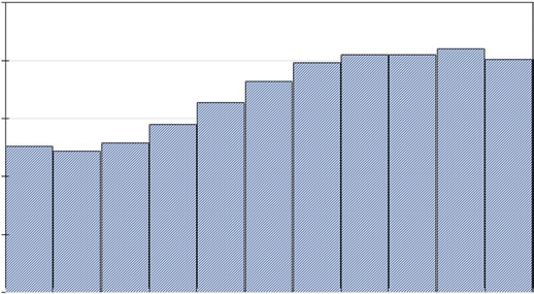
The population is aging and, by 2030, more that 20% of the U. S. population will be aged 65 years or older, Fig. 1.1 [1].
These projected data indicate that within 10–12 years, about 20% of the population will be “senior citizens,” namely 65 years or older [1]. Although advances in medicine and pharmacology, together with improved nutrition, dietary awareness and exercise, have significantly improved the average life expectancy, the outlook for maintained and even improved dental hygiene as well as overall oral health still looks bleak. In fact, partial or complete edentulism is increasing. Whereas fluoridation has markedly reduced dental caries [2, 3], the prevalence of tooth loss through periodontal disease, enamel erosion, wear, trauma and disease (e.g., cancer) is growing [4–7].
Figure 1.1 Projected aging of the United States. (Source: Based on United States Census Bureau. Release Number CB20‐99: 65 and Older Population Grows Rapidly as Baby Boomers Age. Washington, DC, June 25, 2020).
Figure 1.2 Prevalence of edentulism by race and ethnicity in adults ≥65 years [9]. (Source: Based on Centers for Disease Control and Prevention. Edentulism and tooth retention. Atlanta, Ga., September 10, 2019).
According to the American College of Prosthodontics, more than 35 million Americans are edentulous, and 178 million people are missing at least one tooth and these numbers are expected to grow over the next two decades [8]. What is distressing about these statistics is that edentulism affects our most vulnerable populations – the aging and the economically disadvantaged, Fig. 1.2. In the geriatric population, for example, the ratio of edentulous to dentate individuals is 2 : 1, with about 23 million being completely edentulous and some 12 million are edentulous in one arch. About 90% of edentulous patients have dentures and some 15% of edentulous patients will have dentures made each year [8].
The consequences of partial or complete edentulism are well‐known and include many facets of the quality of life (QoL) as well as facial appearance, self‐image and self‐confidence. Overall, health consequences of edentulism encompass significant nutritional changes, digestive issues, obesity, diabetes, and coronary artery disease to name but a few.
The Reality of Dental Implants
Although there have been minor variations over the past few years, the current life expectancy for the U.S. population in 2020 is 78.93 years [10], and we can anticipate increases in tooth failures. Vertical root fractures, endodontic failures, restorative failures, and periodontal disease may result in tooth loss. In contrast to the practice of dentistry in the nineteenth and twentieth centuries, modern dentistry focuses on the replacement of lost teeth utilizing implants, combined with comprehensive analysis of function and esthetics.
In modern dentistry, the dental implant is the best tooth replacement option for nearly all situations where a tooth is missing or is failing. The primary reason for this is the extremely high success rate achieved with dental implants. Saving teeth at all costs is no longer the norm because of the unpredictability of the longevity of heroic dentistry. In other words, preserving bone and tissue regeneration are now considered to be more important than trying to prolong tooth retention. This approach not only promotes bone healing and preservation but ensures that implants are placed in a predictable and solid bony environment with a high rate of success.
The consensus regarding dental implants within the international dental community can be summarized in Table 1.1. Whereas the order of the comments may vary with the individual clinician, most would agree that these comments are valid and pertinent.
Implants and the Edentulous Patient
Over 32 million people in the U.S. wear partial or CDs [11] and approximately 33% of these patients complain that their dentures fit poorly, tend to loosen or dislodge during activities such as chewing and laughing, and/or there is pain on mastication. Flat ridges and/or shallow palatal vaults add to denture retention and instability problems and most dentists are aware that the mandibular CD presents retention issues.
Table 1.1 Advantages of dental implants.
• Implant dentistry is the future of dentistry.
• There is copious scientific literature on dental implantology.
• The 95–97% success rate of dental implants makes them an extremely predictable treatment.
• There is an overwhelming need for tooth replacement and predictable treatment of failing teeth.
• Implant‐retained prosthetics are a very satisfactory solution to the growing prevalence of edentulism in our aging population.
Limitations and/or restrictions on diet, especially which foods can or cannot be eaten, also play a major role in the decision to seek dental implants. It is likely that a significant percentage of those patients experiencing pain or discomfort on chewing will not use their dentures during eating. Due to the decreased mastication forces associated with dentures, edentulous patients have been found to consume less food and have lower intakes of protein, intrinsic and milk sugars, non‐starch polysaccharides (fibrous matter), calcium, non‐heme iron, niacin and vitamin C than dentate people [12]. These dietary deficiencies often have significant adverse effects on overall health and wellbeing, as well as their QoL.
Many patients will resort to utilizing denture adhesives to aid in retention. These adhesives may lead to further problems as they are extremely difficult to remove from the tissues. Impaired speech patterns as well as halitosis (oral malodor or “denture breath”) are frequent complaints among denture wearers, even when the fit of the denture is not a significant issue.
It follows from the above, that patients seek dental implant therapy for a number of reasons, including the following:
• Function
• Esthetics
• Comfort
• Confidence
• Facial appearance
General dental practitioners can address these issues and assist the patient in achieving oral health and functionality lost through missing teeth.
There are two major implant treatment modalities for the edentulous patient:
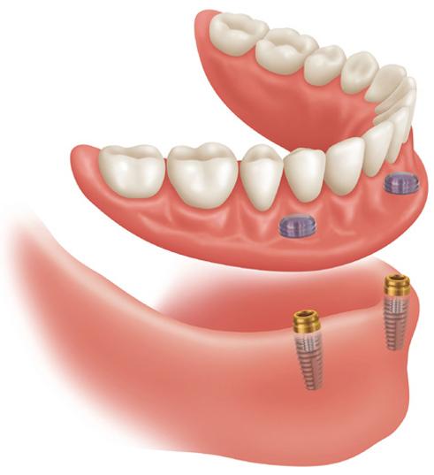
1. Implant over‐dentures. Implant overdentures are removable appliances which are both implant and tissue‐borne prostheses. They utilize an abutment and a denture attachment for the retention (Fig. 1.3). These appliances solve several major problems with traditional dentures by allowing:
• Increased masticatory forces
• Increased retention to potentially eliminate the need for denture adhesives
• Removal of palatal coverage for patients who cannot tolerate the denture due to their gag reflex
An implant‐supported denture is a satisfactory and viable economic alternative to the traditional CD.
• Screw‐retained fixed implant bridges. Fixed implant bridges are implant‐borne prostheses which are not removable by the patient. They are manufactured in zirconia or in acrylic overlaying a chrome‐cobalt or titanium bar. These appliances give patients the greatest masticatory forces and are more appealing to most patients because they are fixed in place.
Implants for Single Crowns and Bridgework
As stated above, 178 million people in the U.S. are missing at least one tooth [11]. Before the use of dental implants, fixed partial dentures (bridges) or RPDs were utilized. One major problem with these treatment modalities is that fewer teeth
Figure 1.3 Implant‐supported overdenture. Source: Courtesy of Zest Anchor.
are taking the load. For example, in the case of a four‐unit fixed bridge, only two teeth are carrying the load of the four teeth it restores because the pontics provide no functional support.
The advantages for placing an implant and restoring it to replace a missing, free‐standing tooth are summarized in Table 1.2.
Table 1.2 Advantages of implants replacing individual teeth.
No unnecessary preparation of adjacent teeth for a traditional bridge
Long‐term prognosis better than for a traditional bridge [13, 14]
Long‐term cost is less than for a traditional bridge
Significantly better retention of prostheses, including RPD’s
In the authors’ opinion, ease of dental hygiene is improved with implants as opposed to a traditional bridge
Greater long‐term patient satisfaction
Dentists are accustomed to replacing multiple missing teeth with a RPD. In fact, RPDs have been a viable treatment option for decades. While they serve a recognizable and useful purpose, they do require some skill and much experience in regard to their design and fabrication.
Despite their many advantages, which include relatively low cost, RPDs have some major drawbacks. In particular, they can lead to increased ridge resorption, especially with appliances fabricated with non‐metallic bases, i.e., what are commonly known as “flippers.”
Whereas RPDs with polymeric (usually acrylic) clasps are somewhat “kinder” to supporting teeth, metallic clasps and rests will commonly traumatize the
clasped teeth over time, notably causing wear and abrasion. This destructive action is due to clasps riding up and down the anchor teeth due to flexure of the RPD during mastication or parafunctional activities. Poor fit and/or repetitive vertical (and lateral) movements due to cyclic loading will not only exacerbate wear and abrasion of the abutment teeth but increase ridge resorption.
Another problem with RPDs, especially those with polymeric bases and poorly‐fitting appliances, is that food particles may often be trapped beneath the denture. This can lead to mucosal irritation, periodontal problems and, possibly, to decay of the supporting teeth. Further, staining of the acrylic “gum work” of the RPD as well as odor necessitates repeated and careful cleaning of the RPD on at least a daily basis to ensure a hygienic appliance and absence of halitosis. Depending upon diet and beverage consumption as well as smoking, there is often the need for more frequent cleaning of the RPD. Failure to remove the RPD and clean teeth and RPD separately compromises effective hygiene of both teeth and RPD.
There are, of course, some disadvantages to the use of implants to replace multiple teeth, Table 1.3.
Table 1.3 Disadvantages of implants vs traditional bridgework and RPDs.
Short‐term cost is higher than for a traditional bridge or RPD Surgery is required Generally, treatment time is longer – 4–8 months.
Implants vs Endodontic Treatment
Although general dentists receive training in endodontics during their education, many prefer not to provide root canal therapy, particularly when surgical intervention is required. There are several reasons for this reluctance to perform surgical endodontics, not the least is the general perception of patients that “root canal therapy” is an unpleasant, long‐drawn out procedure that can be uncomfortable at best and at worst is painful. In fact, to a great many patients, the words “root canal therapy” are synonymous with any procedure or experience that is to be avoided at almost any cost.
In contrast, non‐surgical endodontic treatment is a predictable treatment choice if certain conditions are met. First, there must remain enough sound tooth structure to achieve a 2 mm ferrule effect 360° around the tooth. This will ensure long‐term stability of restorative treatments. Secondly, a cause‐and‐effect should be established when diagnosing a symptomatic tooth. For example, caries approximating a pulp horn with symptoms lead to a clear diagnosis of irreversible pulpitis. Conversely, a symptomatic tooth with no caries present leads to a less predictable treatment outcome until and unless a definitive diagnosis can be achieved.
When there is need for “root canal therapy,” the operator must have available a specialized armamentarium of instruments and restorative materials. However, it must be stated that the available instrumentation and endodontic sealer cements have improved dramatically over the past 20 or so years. Further, it is generally recognized that the time and expertise required to perform endodontic
surgery increases almost exponentially with the number of tooth roots/canals to be treated. Additionally, when canals are sclerosed or calcified, there is increased difficulty in ensuring a clean and extirpated pulpal chamber and root canals.
Finally, teeth that have received extensive endodontic therapy tend to embrittle over time and are subject to failure under loading. Further, it is difficult to achieve a complete hermetic seal of a root canal so that apical leakage and ingress of bacteria, blood and other matter into the treated canal can occur over time. Coronal migration of tissue fluids and bacteria leaking into the treated root canal over time can have many untoward consequences, including dentinal staining, breakdown of sealer cements and restorations, pain and discomfort as well as infection. Due to risks associated with endodontically treated teeth, dentists are often reluctant to use these teeth as abutments for both FPDs and RPDs.
In contrast, the success rate of dental implants is 95–97%. This is far higher than treatment of symptomatic teeth with marginal ridge fractures and endodontic retreatment. These success rates must be considered when discussing treatment options, particularly when relative costs, patient time‐commitment to treatment as well as patient discomfort are considered in addressing the question of root canal therapy vs placement of an implant.
Conclusions
Having presented the overall case for dental implants, specific factors regarding the placement and clinical application of implants will be covered in detail in the following chapters. Nevertheless, modern dentistry now recognizes that dental implants are the standard of care for prosthetic replacement of missing teeth. This is because they can readily and conveniently address some otherwise seemingly intractable problems in traditional restorative dentistry. Further, the advances in implant technology and dental science have progressed so markedly since the first days of the Brånemark concept that the outcome of dental implant placement has a success rate over 95%.
The final word should be that the ground‐breaking concept of Per‐Ingvar Brånemark has transformed dentistry and dental treatment for even the most challenging cases.
References
1. United States Census Bureau (2020). 65 and Older Population Grows Rapidly as Baby Boomers Age. Release Number CB20‐99. https://www.census.gov/newsroom/press‐releases/2020/65‐older‐population‐grows.html (accessed 17 December 2020).
2. Medjedovic, E., Medjedovic, S., Deljo, D. et al. (2015). Impact of fluoride on dental health quality. Mater. Sociomed. 27 (6): 395–398.
3. Centers for Disease Control and Prevention (2001). Recommendations for using fluoride to prevent and control dental caries in the United States. MMWR Recomm. Rep. 50 (RR‐14): 1–42.
4. Martinez‐Canut, P. (2015). Predictors of tooth loss due to periodontal disease in patients following long‐term periodontal maintenance. J. Clin. Periodontol. 42 (12): 1115–1125.
5. Loomansa, B., Opdamb, N., Attinc, T. et al. (2017). Severe tooth wear: European consensus statement on management guidelines. J. Adhes. Dent. 19: 111–119.
6. Bartlett, D.A. (2005). The role of erosion in tooth wear: etiology, prevention and management. Int. Dent. J. 55: 277–284.
7. Michaud, D.S., Fu, Z., Jian Shi, J. et al. (2017). Periodontal disease, tooth loss, and cancer risk. Epidemiol. Rev. 39 (1): 49–58.
8. American College of Prosthodontists (2020). Facts and figures. https://www.gotoapro. org/facts‐figures (accessed 31 July 2020).
9. Centers for Disease Control and Prevention (2019). Edentulism and tooth retention. September 10. https://www.cdc.gov/oralhealth/publications/OHSR‐2019‐edentulism‐tooth‐retention.html (accessed 27 December 2020).
10. Macrotrends (2020). U.S. Life Expectancy 1950–2020. https://www.macrotrends.net/ countries/USA/united‐states/life‐expectancy (accessed 31 July 2020).
11. Statista Research Department (2020). Usage of dentures in the U.S. http://www. statista.com/statistics/275484/us‐households‐usage‐of‐dentures (accessed 31 July 2020).
12. Jauhiainen, L., Männistö, S., Ylöstalo, P. et al. (2017). Food consumption and nutrient intake in relation to denture use in 55‐ to 84‐year‐old men and women — results of a population based survey. J. Nutr. Health Aging 21: 492–500.
13. Ravidà, A., Tattan, M., Askar, H. et al. (2019). Comparison of three different types of implant‐supported fixed dental prostheses: a long‐term retrospective study of clinical outcomes and cost‐effectiveness. Clin. Oral Implants Res. 30 (4): 295–305.
14. Oh, S.‐H., Kim, Y., Park, J.‐Y. et al. (2016). Comparison of fixed implant‐supported prostheses, removable implant‐supported prostheses, and complete dentures: patient satisfaction and oral health‐related quality of life. Clin. Oral Implants Res. 27 (2): e31–e37.
A Brief History of Dental Implants
2
Dentistry has a venerable history in that prosthodontics has been practiced for several thousand years. Fine examples of dental bridgework dating from around 700 BCE were crafted by the Etruscans of Central Italy (now Tuscany) and fixed partial dentures are known to have been fabricated by the Maya of Central America as far back as 700 CE [1–4]. There are many well‐known figures in history, for example, Queen Elizabeth I of England, King Henry II of France, George Washington of the United States and Winston Churchill of the United Kingdom, all of whom either wore removable partial dentures (RPDs) or complete dentures (CDs) [3].
The “father” of dentistry is generally acknowledged to be the French physician Pierre Fauchard (1678–1761) [2] whereas most historians and dentists credit Dr. Greene Vardiman Black (1836–1915) [2, 5, 6] as the “father of modern dentistry.” There are a number of other pioneers in dentistry, including the illustrious Scottish surgeon John Hunter, an early advocate of careful observation and scientific observation in medicine. Not only did Hunter collaborate with his former student Edward Jenner, the pioneer of the smallpox vaccine, but he also dabbled (unsuccessfully) with transplanting teeth, possibly following on from the work of Ambroise Paré (1510–1590). Paré is recognized as the “Father of Modern Surgery” and, interestingly, as the “Foster Father of Dental Surgery.” Interestingly, Paré referred to transplanting of teeth as early as 1564.
Despite its venerable history, the greatest advances in dentistry have really only occurred within the latter half of the twentieth century and, notably, the past 50–60 years. Many influences have transformed dentistry from an ancient quasi‐craft into the evidence‐based technological science it is today, including the innovative work of early dental practitioners, advances in oral medicine, oral surgery and restorative dental techniques, together with an astonishing array of
scientific and technology‐driven progress in dental science and dental biomaterials. Table 2.1 indicates a significant number of innovations that have changed modern dentistry, one of which is the endosseous dental implant.
The confluence of the advances in the basic sciences, dental biomaterials and clinical technique possibly reached their apex in the endosseous dental implant, perhaps the most successful dental restorative technique ever devised. Virtually no other dental procedure has achieved the long‐term success rate found over the past 15–20 years with dental implants.
Replacing Missing Teeth
The efforts of Ambroise Paré, John Hunter and others to replace missing teeth through implantation of sound teeth from donors were the initial attempts to address this need in patients. Dentures, as such, were not available for the general populace back in the fifteenth and sixteenth centuries and only the very wealthy could avail themselves of transplanted teeth or the rudimentary dentures of that period. Charles Allen of York, England, the author of the first English book solely on dentistry [7] was very dismissive of tooth transplantation.
Patients seemed to accept the limited durability of transplanted teeth and transplantation, probably due to clever publicity and hucksterism, became almost a craze on the European Continent, in England and even America in the late eighteenth century. Sadly, through the sixteenth, seventeenth, and eighteenth centuries, paupers often sold their teeth for cash to earn a little money and the heroine Fantine in Victor Hugo’s Les Misérables (1852) was forced to sell her hair, then her incisors and finally her “virtue” in order to survive. Despite its lack of
success and almost total disregard of the basic precepts of oral hygiene, tooth transplanting continued well into the nineteenth century. In fact, barrels of teeth extracted from casualties in the American Civil War were regularly shipped to England, and presumably Europe, for both transplantation and to be used in constructing dentures.
This situation changed with the advent of dental schools, the establishment of professional standards and the growing awareness of the general public that dentistry, dental care and oral hygiene were important not only to the oral cavity but also to systemic health. Nevertheless, despite the venerable history of dentures and the remarkable success of modern CDs, FPDs, and RPDs, many patients simply do not like the fact that they must resort to prostheses to preserve masticatory efficiency and maintain facial esthetics. As any dental professional recognizes, there are myriad reasons that patients complain about their dentures. Many complaints, arising from poor denture fit, discomfort, inadequate retention and even pain, are completely understandable and often justified whereas others arise from a basic dislike of a “foreign body” in the mouth. Further, the need for careful oral hygiene and meticulous cleaning of removable appliances is commonly viewed as an unwelcome chore if not an imposition. The perception of many patients is that all of these factors, combined with many others, contribute to the steadily growing appeal of a dental implant that appears to be permanent, painless, and “maintenance‐free.”
Dental Implants
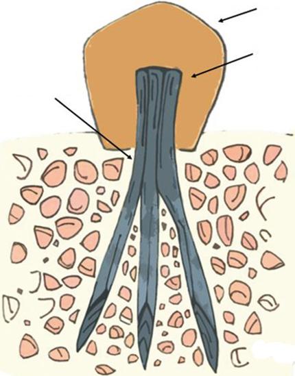
A major problem with CDs, especially for the mandible, is poor retention, often exacerbated by residual alveolar bone above basal cortical bone. Resilient linings, denture creams and other retention aids may alleviate the problem on a temporary basis but rarely “cure” retention or stability issues. One approach to addressing such concerns during the 1970s and, subsequently, was the subperiosteal implant which comprised a metallic framework that closely fit and sat directly on the bone of the mandible.
Subperiosteal Implants
The basic concept of the subperiosteal implant was that a CD rested on abutments that projected through the mucosa, Fig. 2.1. Consequently, masticatory and other stresses were transmitted directly to the supporting bone rather than to the oral mucosa as with conventional CDs. This approach enabled the surgeon to trim the basal bone of any projections or spicules of residual bone to ensure a good fit for the framework but also, incidentally, could help reduce or eliminate any painful sore spots for the final CD.
Fabricating a subperiosteal implant, however, was a long and rather involved procedure. The mandibular mucosa had to be reflected and an impression made of the exposed bone. A wax pattern was then designed on the gypsum cast and used as the pattern for a chrome‐cobalt cast framework. In a subsequent procedure, the mucosa was reflected again, and the framework placed on the exposed bone before the mucosa was restored in position and healing allowed to
start. After healing, a CD could be fabricated and seated on the abutments projecting through the mucosa. There were three principal varieties of subperiosteal implant: full mandibular, full maxillary, and unilateral or single‐unit implants. The latter were smaller than full arch implants and had only one protruding abutment. They were particularly useful when used as terminal abutments for edentulous quadrants, i.e., free‐end saddle retention aids.
Although subperiosteal implants were effective, the overall procedure was lengthy and involved a great deal of discomfort for the patient and often were subject to various complications [8, 9]. Further, a high degree of surgical skill was required and necessitated a close collaboration between the surgeon, prosthodontist, and the laboratory technician to ensure optimal clinical results. Provided care was exercised in patient selection, there was good overlying soft tissue and no residual alveolar bone, the prognosis could be very good with reasonably high short‐term success rates.
Endodontic Implants
For many years, the most established and longest established implant was the endodontic endosseous pin implant, also known as the endodontic stabilizer, and was particularly useful for rigidly anchoring a mobile tooth to bone. Tooth mobility can have many causes, including an unfavorable crown‐to‐root ratio, gum and alveolar recession, bruxism and an unbalanced occlusion.
The basis of this approach was that a pin was inserted through the root canal into the underlying bone such that it was anchored in bone but with upper end projecting into the mouth and upon which, a crown or RPD was fabricated [10].
Typically, the lower end of the pin did not penetrate the cortical plate of the mandible or the antral or nasal floors of the maxilla. Indications for endodontic implants included treatment of root fractures, external or internal root resorption and when better support and stability was required for FPD or RPD abutments.
Cast Framework
Abutment Post
Mucosa
Figure 2.1 The subperiosteal implant.
Although the clinical use of endodontic endosseous pin implants is less common due to the advent of the modern endosseous implant, they were successful, with few contraindications for their use provided correct clinical procedures were followed [11].
Endosseous Implants
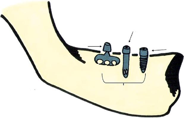
Endosseous implants, also known as intra‐osseous and endosteal implants, have been in clinical use since the 1960s [12]. There are four main categories of endosseous implant, namely pins, spirals, blades, and screws. Regardless of implant design, endosseous implants are used in edentulous areas where there is sufficient healthy bone to accommodate the implant. Selection criteria for the use of implants are discussed in later chapters of this book.
The first successful endosseous implants were the Formiggini spiral screw implant and, subsequently, the Cherchève spiral‐post implant, the former dating from 1947 and the latter from the 1960s to 1970 [4, 12]. The most sophisticated, and successful, Cherchève implant consisted of a double hollow spiral mounted on a square post. After the bone was trephined to create a cavity, the implant was placed beneath the alveolar ridge with the shank or post extending into the oral cavity and, upon which, the final prosthesis was constructed. The problem with these early implants was that trephining of the bone created a gap or space between the abutment post and the host hard and soft tissues, and this could sometimes present problems.
Many workers developed modifications of the spiral implant during the late 1960s and early 1970s. These largely comprised self‐tapping screw implants, often with a vent or port below the threaded portion to permit fibrous tissues and, hopefully, bone to grow through the aperture and promote retention. Although many of these screw implants were successful, numerous failures occurred as the result of tissue irritation, frank infection and epithelial downgrowth preventing adequate retention and sometimes complete evulsion of the implant. Commonly, poor bony attachment to these implants caused stability to be a problem.
An alternative approach to screw‐type endosseous implants was the tripodal pin concept which dated from the same period. In essence, thin tantalum pins were inserted into bone at roughly 120° angulations and the exposed ends of the pins were bonded together using acrylic resin, Fig. 2.2.
The implanted tripodal system could then be used as a bridge abutment or to support a single‐unit prosthesis. Although pin implants had certain applications, they did not possess long‐term retention, generally were not self‐supporting and the pins often were easily displaced or removed over time.
A major development in endosseous implants was the blade or blade vent implant designed by Linkow in 1968 and subsequently modified by Linkow and others over the period 1970–1971 [12–14]. Blade implants were originally designed for use in areas where there were knife‐edge alveolar ridges, situations where screw‐type implants are contra‐indicated. These implants can be used in virtually all maxillary and mandibular edentulous areas, provided there is sufficient residual alveolar process. Because the implants have greater mesio‐distal dimensions than their vertical heights, the design combines maximal
Prosthesis
stability, especially against lateral forces, while minimizing risk to underlying tissues and structures.
Clinically, bone often grew through the vents of the blade implants so that the early success rate was very high although the long‐term prognosis was lower, especially with maxillary placements. Various problems were associated with blade implants, particularly the difficulty in achieving an ideal gingival relationship with that crown when used to support a single crown. There were also problems with thin ridges such that any bony destruction could result in implant loss. Apparently, fewer problems were found with blades used to support a denture base although stability was a problem with unilateral mandibular free‐end saddles.
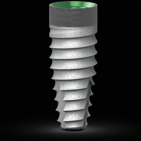
The modern “screw” implant derives from the pioneering work of Stefano Tramonte [15] in Italy and Per‐Ingvar Brånemark in Sweden [16, 17], both of whom advocated the use of titanium for dental implants.
The excellent physical properties and outstanding biocompatibility of titanium were the driving force for this application. In particular, Brånemark described the clinically observed close apposition and adherence of bone with titanium, which he termed osseointegration. Since then, a wide variety of “screw” or tooth root‐shaped endosseous implants have come into clinical use, Fig. 2.3 and Fig. 2.4 and they have achieved remarkable clinical success such that they are now considered important components of the restorative dentistry armamentarium. However, the clinical success of dental implants requires good clinical technique, accurate placement and careful patient selection with good bone quality (see later chapters in this book).
Acrylic Resin Bonding of Pins
Bone
Tantalum Pin
Figure 2.2 Schematic diagram of tripodal pin concept.
Of central importance for any metal within the oral cavity is corrosion resistance as well as mechanical strength. Consequently, the vast majority of modern dental implants are fabricated from titanium and its alloys, notably Ti‐6Al‐4V, the so‐called 6‐4 alloy, although CP (commercial purity) Titanium and alloys such as Ti‐13Cu‐4.5Ni have also been evaluated. Most implants are fabricated using powder metallurgy, typically hot isostatic pressing HIP technology.
Blade
Cylinder
Screw
Jawbone
Implants Inser ted into Jawbone
Figure 2.3 Different types of implants.
Figure 2.4 Modern screw‐type implant. (Source: Courtesy of Biohorizons).
The efficacy and rate of osseointegration of bone and implant has been enhanced by techniques such as designing the implant with a screw profile, providing a micro‐texture to the implant surface as well as coating the surface with hydroxyapatite (HA). More recently, a novel approach to dental implantology has been to coat the implant surface with a nanometer‐thick layer of protein containing a bisphosphonate drug. Animal studies indicate that the bone surrounding the implant becomes denser and stronger, ensuring a more durable implant‐tissue interface.
References
1. Wynbrandt, J. (2000). The Excruciating History of Dentistry. New York: St. Martin’s Griffin.
2. Woodforde, J. (1968). The Strange Story of False Teeth. London: Routledge & Kegan Paul.
3. James, P. and Thorpe, N. (2015). Ancient Inventions. New York: Ballantine Books.
4. Abraham, C.M. (2014). A brief historical perspective on dental implants, their surface coatings and treatments. Open Dent. J. 8: 50–55.
5. Wolff, M.S., Allen, K., and Kaim, J. (2007). A 100‐year journey from GV Black to minimal surgical intervention. Compend. Contin. Educ. Dent. 28 (3): 130–134.
6. Jain, S. and Jain, H. (2017). Legendary Hero: Dr. G.V. Black (1836–1915). Clin. Diagn. Res. 11 (5): ZB01–ZB04.
7. Allen, C. (1685). The Operator for the Teeth. John White: York.
9. Schou, S., Pallesen, L., Hjørting‐Hansen, E. et al. (2000). A 41‐year history of a mandibular subperiosteal implant. Clin. Oral Implants Res. 11 (2): 171–178.
10. Orlay, H. (1960). Endodontic splinting treatment in periodontal disease. Br. Dent. J. 108: 118–121.
11. Gutmann, J.L. and Levermann, V.M. (2013). Endodontic endosseous implants (diodontic or through the tooth implants). ENDO (Lond. Engl.) 7 (4): 299–304.
12. von Fraunhofer, J.A. (1975). Oral implants. In: Scientific Aspects of Dental Materials (ed. J.A. von Fraunhofer). London: Butterworths.
13. Linkow, L.I. (1970). Endosseous blade‐vent implants: a two‐year report. J. Prosth. Dent. 23 (4): 441–448.
14. Linkow, L.I., Weiss, C.M. and Weiss, L.B. et al. (1973). Oral Implant, USP 3,729,825. 1 May 1973.
15. Pasqualini, M.E., Tramonte, S.U., and Linkow, L.I. (2016). Half a century of function a retrospective analysis of Tramonte endosteal screw dental implants that lasted 50 and 36 years. A case report. J. Dental Oral Health 2 (7): 051–058.
16. Moberg, L.E., Sagulin, G.‐B., Per‐Åke Köndell, P.‐A. et al. (2001). Brånemark System® and ITI Dental Implant System® for treatment of mandibular edentulism. A comparative randomized study: 3‐year follow‐up. Clin. Oral Implants Res. 12: 450–461.
17. Maló, P., Rangert, B., and Nobre, M. (2003). Implants for completely edentulous mandibles: a retrospective clinical study. Clin. Implant Dent. Relat. Res. 5 (Suppl.1): 2–9.