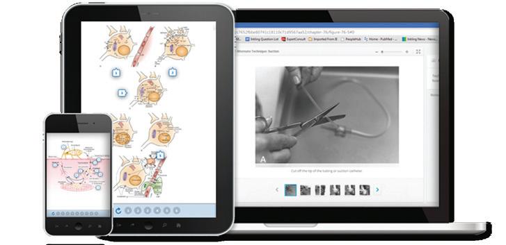
Any screen. Any time. Anywhere.
email expertconsult.help@elsevier.com call 1-800-401-9962 (inside the US) call +1-314-447-8200 (outside the US)
Taylor & Hoyt’s
PEDIATRIC OPHTHALMOLOGY AND STRABISMUS
© 2017, Elsevier Inc. All rights reserved.
First edition 1990
Second edition 1997
Third edition 2005
Fourth edition 2013
No part of this publication may be reproduced or transmitted in any form or by any means, electronic or mechanical, including photocopying, recording, or any information storage and retrieval system, without permission in writing from the publisher. Details on how to seek permission, further information about the Publisher’s permissions policies and our arrangements with organizations such as the Copyright Clearance Center and the Copyright Licensing Agency, can be found at our website: www.elsevier.com/permissions
This book and the individual contributions contained in it are protected under copyright by the Publisher (other than as may be noted herein).
Figure 1.1 is copyright Gillian Lee Illustrations
The authors of chapters 42, 46, 48 and 50 retain copyright of their chapters
The following figures are copyright Addenbrooke’s Hospital – 52.5, 52.13
Videos 86.2–86.5 are from George Spaeth et al., Ophthalmic Surgery: Principles and Practice, 4th Edition.
© Elsevier Saunders 2014
Notices
Knowledge and best practice in this field are constantly changing. As new research and experience broaden our understanding, changes in research methods, professional practices, or medical treatment may become necessary.
Practitioners and researchers must always rely on their own experience and knowledge in evaluating and using any information, methods, compounds, or experiments described herein. In using such information or methods they should be mindful of their own safety and the safety of others, including parties for whom they have a professional responsibility.
With respect to any drug or pharmaceutical products identified, readers are advised to check the most current information provided (i) on procedures featured or (ii) by the manufacturer of each product to be administered, to verify the recommended dose or formula, the method and duration of administration, and contraindications. It is the responsibility of practitioners, relying on their own experience and knowledge of their patients, to make diagnoses, to determine dosages and the best treatment for each individual patient, and to take all appropriate safety precautions.
To the fullest extent of the law, neither the Publisher nor the authors, contributors, or editors, assume any liability for any injury and/or damage to persons or property as a matter of products liability, negligence or otherwise, or from any use or operation of any methods, products, instructions, or ideas contained in the material herein.
ISBN:
Print: 978-0-7020-6616-0
E-book: 978-0-7020-6617-7
Inkling: 978-0-7020-6618-4
Printed in China
Foreword
1 A history of pediatric ophthalmology and strabismus 1
David S Taylor
Section 1: Epidemiology, growth and development
2 Epidemiology and the worldwide impact of visual impairment in children 7
Jugnoo S Rahi and Clare E Gilbert
3 Clinical embryology and development of the eye 17
John R B Grigg and Robyn V Jamieson
4 Developmental biology of the eye 25
David R FitzPatrick
5 Clinical aspects of normal and abnormal visual development and delayed visual maturation 32
Ronald M Hansen and Anne B Fulton
6 Milestones and normative data 40
Hans Ulrik Møller and Dorte Ancher Larsen
Section 2: Core practice
7 Examination, history and special tests in pediatric ophthalmology 50 G Robert LaRoche
8 Managing refractive errors in children 60
Amy K Hutchinson and Buddy Russell
9 Visual electrophysiology: how it can help you and your patient 68
Dorothy A Thompson and Alki Liasis
10 Imaging the child’s eye, orbit, and visual pathways 76
Daniel J Salchow and Nadja Kadom
11 Genetics and pediatric ophthalmology 94 Panagiotis I Sergouniotis and Graeme C M Black
Section 3: Infections, allergic and external eye disorders
12 Congenital infections of the eye 101 Luis Amaya
13 Conjunctivitis of the newborn 109 Megan Geloneck and Gil Binenbaum
14 Preseptal and orbital cellulitis 113 Richard L Scawn and Jimmy M Uddin
15 Endophthalmitis 124
Donal Brosnahan
16 External eye disease and the oculocutaneous disorders 130
Stephen J Tuft
17 Ocular manifestations of HIV/AIDS in children 156 Emmett T Cunningham Jr and Philippe Kestelyn
Section 4: Systematic pediatric ophthalmology
Part 1: Disorders of the eye as a whole
18 Disorders of the eye as a whole 163 Sunju Park and Elias I Traboulsi
Part 2: Lids, brows and oculoplastics
19 Lids: congenital and acquired abnormalities practical management 175
Robert C Kersten and Richard Collin
20 Lid and orbital infantile periocular hemangiomas (capillary hemangiomas) and other vascular diseases 188
Christopher J Lyons
Part 3: Orbit and lacrimal
21 Lacrimal system 200
Caroline J MacEwen and Una O’Colmain
22 The management of orbital disease in children
Alan A McNab
23 Neurogenic tumors
Peter J Dolman and Yvonne Chung
24 Orbital rhabdomyosarcoma
Carol L Shields and Jerry A Shields
25 Other mesenchymal abnormalities
Christopher J Lyons
26 Metastatic, secondary and lacrimal gland tumors
Alan A McNab and Christopher J Lyons
27 Histiocytic, hematopoietic, and lymphoproliferative disorders
Timothy John Sullivan
28 Craniofacial abnormalities
Joanna Black and John Crompton
29 Cystic lesions and ectopias
Alan A McNab
30 Inflammatory disorders
Alan A McNab and Christopher J Lyons
Part 4: External disease and anterior segment
31 Conjunctiva and subconjunctival tissue
Venkatesh Prajna and Perumalsamy Vijayalakshmi
32 Conjunctival tumors
Jill Razor Wells and Hans Grossniklaus
33 Anterior segment developmental anomalies including aniridia
Ken K Nischal 34 Corneal abnormalities in childhood
Stephen J Tuft
35 Corneal dystrophies
Hans Ulrik Møller and Barry Lee
36 The lens
Jay Self and Christopher Lloyd
38
Maria Papadopoulos and Sir Peng Tee Khaw
Chapter 19 Lids: congenital and acquired abnormalities − practical management
19.1 Insertion of prosthetic eye into postenucleation socket
Robert C Kersten and Richard Collin
Chapter 21 Lacrimal system
21.1 Probing in a young child
Caroline J MacEwen and Una O’Colmain
Chapter 31 Conjunctiva and subconjunctival tissue
31.1 A worm being removed from beneath the lateral rectus muscle
Venkatesh Prajna and Perumalsamy Vijayalakshmi
Chapter 32 Conjunctival tumors
32.1 Corneal or conjunctival tumor being treated
Jill Razor Wells and Hans Grossniklaus
Chapter 36 The lens
36.1 Subluxed lensectomy
Jay Self and Christopher Lloyd
Chapter 37 Childhood cataracts
37.1 Cataract extraction and primary intraocular lens implantation in a one-year-old child
37.2 In-the-bag secondary intraocular lens impantation in a two-year-old
Scott R Lambert
Chapter 38 Childhood glaucoma
38.1 Trabeculotomy
38.2 Goniotomy
Sir Peng Tee Khaw, Maria Papadopoulos and John L Brookes
Chapter 45 Current treatment of retinopathy of prematurity
45.1 Avastin injection for retinopathy of prematurity in neonate
Joshua Robinson and G Baker Hubbard
Chapter 81 Vertical strabismus
81.1 Dissociated vertical divergence versus inferior oblique overaction
81.2 Right orbital floor fracture simulating left superior oblique palsy
81.3 Primary inferior oblique muscle overaction
81.4 Pulley heterotopia
Burton J Kushner
List of Contributors
Joanna Black MB, BS
Head of Eye Department and Senior Clinical Lecturer
Adelaide Women’s and Children’s Hospital Adelaide, SA, Australia
Richard J C Bowman MA, MD, FRCOphth Consultant Ophthalmologist
Great Ormond Street Hospital London, UK
John A Bradbury FRCS, FRCOphth Consultant Ophthalmologist Department of Ophthalmology
Bradford Royal Infirmary Bradford, UK
Michael C Brodsky MD
Professor
Ophthalmology and Neurology
Mayo Clinic Rochester, MN, USA
Donal Brosnahan MB, DCH, FRCOphth Consultant Ophthalmic Surgeon
Royal Victoria Eye and Ear Hospital Dublin, Ireland
Alejandra de Alba Campomanes MD, MPH Associate Professor of Ophthalmology and Pediatrics Department of Ophthalmology
University of California San Francisco, CA, USA
Jayne E Camuglia MBBS, BSc Pediatric Ophthalmology Fellow Children’s Health Queensland Associate Lecturer
University of Queensland Brisbane, QLD, Australia
Susan M Carden MBBS, FRANZCO, FRACS, PhD
Senior Lecturer
Department of Pediatrics
University of Melbourne; Consultant Ophthalmologist
Royal Children’s Hospital and Royal Victorian Eye and Ear Hospital Melbourne, VIC, Australia
Giovanni Castano MD
Pediatric Ophthalmology Consultant Department of Ophthalmology
Fundacion Santa Fe de Bogota University Hospital Bogota, Colombia
Ingele Casteels MD, PhD Professor of Ophthalmology
University Hospitals Leuven Leuven, Belgium
Yvonne Chung MBBS, M Med (Ophth), FAMS (Spore)
Consultant
Ophthalmology
Singapore National Eye Centre
Singapore
Michael P Clarke MB BChir, FRCS, FRCOphth, PhD
Newcastle Eye Centre
Newcastle upon Tyne NHS Hospitals
NHS Foundation Trust
Newcastle upon Tyne, UK
David K Coats MD
Professor of Ophthalmology and Pediatrics
Cullen Eye Institute
Baylor College of Medicine; Chief of Ophthalmology
Texas Children’s Hospital
Houston, TX, USA
Richard Collin FRCS, FRCOphth
Senior Consultant Oculoplastic Surgeon, Moorfields Eye Hospital; Honorary Professor of Ophthalmology
UCL, London, UK
John Crompton MBBS, FRANZCO, FRACS Professor
Ophthalmology and Visual Sciences
University of Adelaide
Royal Adelaide Hospital Adelaide, SA, Australia
Emmett T Cunningham Jr, MD, PhD, MPH Director
The Uveitis Service
The Department of Ophthalmology
California Pacific Medical Center; Adjunct Clinical Professor of Ophthalmology
The Stanford University School of Medicine; Research Associate
The Francis I. Proctor Foundation, UCSF School of Medicine; Partner
West Coast Retina Medical Group San Francisco, CA, USA
Joseph L Demer MD, PhD
Arthur L. Rosenbaum Professor of Pediatric Ophthalmology
Professor of Neurology
Chief, Pediatric Ophthalmology and Strabismus Division
Director, Ocular Motility Laboratories
Stein Eye Institute and Departments of Ophthalmology and Neurology
University of California
Los Angeles, CA, USA
Hélène Dollfus MD, PhD
Professor Medical Genetics
Strasbourg University Hospital Strasbourg, France
Peter J Dolman MD, FRCSC
Clinical Professor
Division Head of Oculoplastics and Orbit, Director of Fellowship Programs, Department of Ophthalmology and Visual Sciences
University of British Columbia Vancouver, Canada
Sean P Donahue MD, PhD
Sam and Darthea Coleman Chair in Ophthalmology and Visual Sciences
Chief Pediatric Ophthalmology
Monroe Carrell Jr. Children’s Hospital at Vanderbilt Vanderbilt University Nashville, TN, USA
Clive Edelsten MA, MRCP, FRCOphth Consultant Medical Ophthalmologist
Rheumatology
Great Ormond Street Hospital London, UK
Alistair R Fielder FRCS, FRCP, FRCOphth
Professor Emeritus of Ophthalmology Department of Optometry and Visual Science City University London, UK
David R. FitzPatrick MB, ChB, MD, FRCP
Professor
MRC Human Genetics Unit
MRC Institute of Genetic and Molecular Medicine Edinburgh, UK
Anne B Fulton MD
Professor Ophthalmology
Harvard Medical School; Senior Associate in Ophthalmology
Boston Children’s Hospital Boston, MA, USA
Brenda L Gallie MD, FRCSC Head Retinoblastoma Program
Ophthalmology and Vision Science Hospital for Sick Children Toronto, Canada
Megan Geloneck MD
The Children’s Hospital of Philadelphia Pediatric Ophthalmology and Strabismus Philadelphia, PA, USA
Clare E Gilbert MB ChB, FRCOphth, MD, MSc
Professor of International Eye Health Department of Clinical Research
London School of Hygiene and Tropical Medicine London, UK
Christy Giligson BSc, OC(C)
Senior Teaching Orthoptist Department of Ophthalmology
BC Children’s Hospital Vancouver, Canada
Glen A Gole MD, FRANZCO, FRACS
Professor and Director of Ophthalmology
Departments of Paediatrics/Child Health and Ophthalmology
Queensland Children’s Hospital Brisbane, QLD, Australia
William V Good MD
Senior Scientist
Kettlewell Eye Research Institute San Francisco, CA, USA
John R B Grigg MB, BS, MD, FRANZCO, FRACS
Associate Professor
Head Discipline of Ophthalmology
Sydney Medical School
Eye Genetics Research Group Save Sight Institute
University of Sydney Sydney, NSW, Australia
Hans Grossniklaus MD, MBA Professor of Ophthalmology and Pathology Ophthalmology
Emory University School of Medicine Atlanta, GA, USA
Patrick Hamel MD, FRCSC Ophthalmologist
Department of Ophthalmology
CHU Sainte-Justine Montreal, Canada
Sheryl M Handler MD
Pediatric Ophthalmology Encino, CA, USA
Ronald M Hansen PhD Research Associate Ophthalmology
Boston Children’s Hospital; Assistant Professor Ophthalmology
Harvard Medical School Boston, MA, USA
Gena Heidary MD, PhD Director
Pediatric Neuro-ophthalmology Service
Boston Children’s Hospital Boston, MA, USA
Assistant Professor in Ophthalmology
Harvard Medical School Boston, MA, USA
Richard W Hertle MD, FAAO, FACS, FAAP Director
Vision Center
Akron Children’s Hospital Akron, OH, USA
Göran Darius Hildebrand BM, BCH, MD, MPhil, FEBO, FRCS, FRCOphth
Consultant Ophthalmic Surgeon
Head, Paediatric Ophthalmology Service
Oxford Eye Hospital
John Radcliffe Hospital Oxford, UK
Graham E Holder BSc, MSc, PhD
Professor Director of Electrophysiology
Moorfields Eye Hospital London, UK
Creig S Hoyt MD, MA
Emeritus Professor and Chair Department of Ophthalmology University of California San Francisco, CA, USA
G Baker Hubbard MD
Thomas M. Aaberg Professor of Ophthalmology
The Emory Eye Center
Emory University School of Medicine Atlanta, GA, USA
Amy K Hutchinson MD
Professor of Ophthalmology
Emory University School of Medicine Atlanta, GA, USA
Saurabh Jain MBBS, MS, FRCOphth Consultant Ophthalmic Surgeon Department of Ophthalmology
Royal Free Hospital London, UK
Robyn V Jamieson MBBS (Hons I), PhD, FRACP, HGSA
Associate Professor Head, Eye Genetics Research Group Disciplines of Paediatrics, Genetic Medicine, and Ophthalmology
Sydney Medical School; Children’s Medical Research Institute University of Sydney; The Children’s Hospital at Westmead; Save Sight Institute Sydney, NSW, Australia
Hanne Jensen MD, DMSci Consultant Department of Ophthalmology Rigshospitalet-Glostrup Copenhagen, Denmark
Nadja Kadom MD
Associate Professor of Radiology
Emory University School of Medicine Atlanta, GA, USA
Ramesh Kekunnaya MD, FRCS Head, Child Sight Institute Residency Program Coordinator
Jasti V. Ramanamma Children’s Eye Care Center
L. V. Prasad Eye Institute
Hyderabad, India
Robert C Kersten MD
Professor of Clinical Ophthalmology Department of Ophthalmology
University of California San Francisco, CA, USA
Philippe Kestelyn MD, PhD, MPH Professor in Ophthalmology
Head and Chair, Department of Ophthalmology
Ghent University Hospital
Ghent, Belgium
Jan E E Keunen MD, PhD, FEBO
Professor of Ophthalmology
Radboud University Medical Center Nijmegen, Netherlands
Professor Sir Peng Tee Khaw PhD FRCP FRCS FRCOphth FRCPath FCOptom Hon DSc FRSB
FARVO FMedSci
Professor of Glaucoma and Ocular Healing Consultant Ophthalmic Surgeon
The National Institute for Health Research Biomedical Research Centre at Moorfields Eye Hospital NHS Foundation Trust and UCL Institute of Ophthalmology London, UK
Chong Ae Kim MD, PhD
Associate Professor
Pediatrics
Instituto da Criança São Paulo, Brazil
Jan Koopman MSc, PhD
Amsterdam – Rehabilitation and Advice
Royal Dutch Visio, Centre of Expertise for Blind and Partially Sighted People Amsterdam, Netherlands
Stephen P Kraft MD, FRCSC
Professor
Ophthalmology and Vision Sciences University of Toronto Toronto, Canada
Burton J Kushner MD
Professor Emeritus
Ophthalmology and Visual Sciences University of Wisconsin Madison, WI, USA
Scott R Lambert MD
Professor of Ophthalmology
Stanford University School of Medicine, Stanford, CA, USA
G Robert LaRoche MD, FRCSC
Professor
Ophthalmology and Vision Sciences
Dalhousie University Halifax, Canada
Dorte Ancher Larsen MD
Consultant
Department of Pediatric Ophthalmology
Aarhus University Hospital Aarhus, Denmark
Andrew G Lee MD
Chair, Department of Ophthalmology
Houston Methodist Hospital; Professor of Ophthalmology, Neurology and Neurosurgery
Weill Cornell Medical College; Adjunct Professor
Ophthalmology
Baylor College of Medicine; University of Texas Medical Branch; University of Texas MD Anderson Cancer Center; Houston, TX, USA
Barry Lee MD
Consultant
Ophthalmology
Piedmont Hospital
Eye Consultants of Atlanta Atlanta, GA, USA
Phoebe Lenhart MD
Assistant Professor of Ophthalmology
Emory University School of Medicine Atlanta, GA, USA
Alki Liasis BSc (Hons), PhD
Consultant Clinical Scientist
Clinical and Academic Department of Ophthalmology
Great Ormond Street Hospital for Children London, UK
Grant T Liu MD
Neuro-ophthalmology Service
Division of Ophthalmology
The Children’s Hospital of Philadelphia; Departments of Neurology and Ophthalmology Perelman School of Medicine University of Pennsylvania Philadelphia, PA, USA
Christopher Lloyd MB DO FRCS FRCOphth
Consultant Paediatric Ophthalmologist
Clinical and Academic Department of Ophthalmology
Great Ormond Street Hospital for Children, London;
Hon Professor Paediatric Ophthalmoloogy
Manchester Academic Health Sciences Centre Manchester, UK
Christopher J Lyons MB FRCS FRCOphth, FRCSC
Professor
Department of Ophthalmology and Visual Sciences
University of British Columbia Vancouver BC
Carey A Matsuba BSc, MDCM, MHSc
Clinical Assistant Professor Paediatrics
University of British Columbia Vancouver, Canada
Caroline J MacEwen MB ChB, MD, FRCS, FRCOphth, FFSEM, FRCPE
Professor
Ophthalmology Department University of Dundee Dundee, UK
Alan A McNab FRANZCO, FRCOphth, DMedSc
Head of Clinic
Orbital Plastic and Lacrimal Clinic
Royal Victorian Eye and Ear Hospital East Melbourne, VIC, Australia
Vaishali Mehta BMedSc (Hons) in Orthoptics Teaching Orthoptist
Ophthalmology and Orthoptics
BC Children’s Hospital Vancouver, Canada
Michel Michaelides BSc, MB, BS, MD(Res), FRCOphth, FACS
Professor of Ophthalmology and Consultant
Ophthalmic Surgeon
UCL Institute of Ophthalmology and Moorfields
Eye Hospital
London, UK
Daniel Mojon MD, FEBO, EMHSA
Director
Eye Clinic
Airport Medical Center
Zürich-Airport
Zürich, Switzerland
Professor of Ophthalmology
University of Bern, Switzerland
Consultant
University Eye Clinic
Kepler University, Linz, Austria
Hans Ulrik Møller PhD
Consultant Pediatric Ophthalmologist Associate Professor
Pediatric Ophthalmology
Aarhus University Hospital
Aarhus, Denmark; Viborg Hospital
Viborg, Denmark
Anthony T Moore MA, FRCS, FRCOphth
Michal Vilensky Endowed Chair in Ophthalmology Department of Ophthalmology
University of California San Francisco
San Francisco, CA, USA
Andrew A M Morris BM, BCh, PhD, FRCPCH
Willink Biochemical Genetics Unit
Manchester Centre for Genomic Medicine
Central Manchester University Hospitals NHS Foundation Trust Manchester, UK
Nancy J Newman MD
LeoDelle Jolley Professor of Ophthalmology; Professor of Ophthalmology and Neurology; Instructor in Neurological Surgery; Director of Neuro-Ophthalmology
Emory University School of Medicine
Atlanta, GA, USA
Ken K Nischal MD, FRCOphth
Professor and Director
Pediatric Ophthalmology, Strabismus and Adult Motility
Children’s Hospital of Pittsburgh
University of Pittsburgh Medical Center Pittsburgh, PA, USA
Una O’Colmain, MB, BCh, BAO, FRCOphth Department of Ophthalmology
Ninewells Hospital Dundee, UK
Anna R O’Connor PhD, BMedSci (Hons) Directorate of Orthoptics and Vision Science
University of Liverpool Liverpool, UK
Michael O’Keefe MB, FRCS, FRCSE
Consultant Surgeon
Paediatric Ophthalmology
Children’s University Hospital Dublin, Ireland
Scott E Olitsky, MD
Chief of Ophthalmology
Children’s Mercy Hospital; Professor of Ophthalmology
University of Missouri – Kansas City School of Medicine;
Clinical Associate Professor of Ophthalmology
University of Kansas School of Medicine Kansas City, MO, USA
Luis H Ospina MD
Assistant Professor of Pediatric Ophthalmology and Neuro-ophthalmology
CHU Sainte-Justine University of Montreal Montreal, Canada
Darren T Oystreck MMedSci, OC(C)
Orthoptist and Chair
Clinical Vision Science
IWK Health Centre
Faculty of Health Professions Dalhousie University Halifax, Canada
Maria Papadopoulos MBBS, FRCOphth Glaucoma Service
Moorfields Eye Hospital London, UK
Sunju Park MD
Fellow in Pediatric Ophthalmology
Department of Pediatric Ophthalmology and Strabismus
Center for Genetic Eye Disease
Cole Eye Institute
Cleveland Clinic Cleveland, OH, USA
Evelyn A Paysse MD
Professor of Ophthalmology and Pediatrics
Cullen Eye Institute
Baylor College of Medicine
Texas Children’s Hospital Houston, TX, USA
Jason H Peragallo MD
Assistant Professor of Ophthalmology and Pediatrics
Emory University School of Medicine Atlanta, GA, USA
Erika Mota Pereira MD
Visiting Assistant Professor of Ophthalmology, University of Texas Southwestern Medical Center, Dallas, Texas, USA,
Consultant Pediatric Ophthalmologist Federal University of Minas Gerais Belo Horizonte, Minas Gerais, Brazil
Rachel F Pilling MB, ChB, MA (Med Eth Law), FRCOphth
Consultant Paediatric and Special Needs Ophthalmologist
Department of Ophthalmology
Bradford Teaching Hospitals NHS Trust Bradford, UK
Stacy Pineles MD, MS
Associate Professor of Ophthalmology Department of Ophthalmology
University of California, Los Angeles Los Angeles, CA, USA
Venkatesh Prajna FRCOphth Department of Cornea Aravind Eye Hospital Madurai, India
Frank Antony Proudlock BSc, MSc, PhD
Senior Lecturer
Neuroscience, Psychology and Behaviour University of Leicester Leicester, UK
Narman Puvanachandra MB, BChir, MA (Cantab), FRCOphth
Paediatric Ophthalmology Department Norfolk and Norwich University Teaching Hospital Norwich, UK
Anthony G Quinn FRANZCO, FRCOphth, DCH Consultant Ophthalmologist
West of England Eye Unit
Royal Devon and Exeter Hospital Exeter, UK
Graham E Quinn MD, MSCE Professor
The Children’s Hospital of Philadelphia University of Pennsylvania School of Medicine Philadelphia, PA, USA
Jugnoo S Rahi MBBS, MSc, PhD, FRCOphth Professor of Ophthalmic Epidemiology Lifecourse Epidemiology and Biostatistics Section Institute of Child Health UCL London, UK
Michael X Repka MD, MBA
David L. Guyton MD and Feduniak Family Professor of Ophthalmology Professor of Pediatrics Ophthalmology
Johns Hopkins University Baltimore, MD, USA
Joshua Robinson MD Assistant Professor
The Emory Eye Center
Emory University School of Medicine Atlanta, GA, USA
Buddy Russell COMT, FCLSA, FSLS Contact Lens Technologist Ophthalmology
Emory University Eye Center Atlanta, GA, USA
Luis Carlos Ferreira de Sá MD Consultant, Pediatric Ophthalmology Pediatrics
Instituto da Criança, University of Sao Paulo Medical School
São Paulo, Brazil
Virender Sachdeva MS, DNB Associate Ophthalmologist
Nimmagada Prasad Children’s Eye Care Centre Department of Pediatric Ophthalmology, Strabismus and Neuro-ophthalmology
L. V. Prasad Eye Institute
GMRV Campus
Visakhapatnam, India
Daniel J Salchow MD
Professor of Ophthalmology
Department of Ophthalmology
Charité – Universitätsmedizin Berlin Berlin, Germany
Richard L Scawn BSc, MBBS, FRCOphth Consultant
Adnexal Unit
Moorfields Eye Hospital London, UK
Nicoline Schalij-Delfos MD, PhD
Professor Paediatric Ophthalmologist
Department of Ophthalmology
Leiden University Medical Center
Leiden, Netherlands
Mary J van Schooneveld MD, PhD
Ophthalmologist
Ophthalmology
Academic Medical Centre
Amsterdam, Netherlands
Jay Self BM, FRCOphth, PhD
Associate Professor
Vision Sciences
University of Southampton Southampton, UK
Panagiotis I Sergouniotis MD, PhD
Academic Clinical Fellow
Manchester Royal Eye Hospital and University of Manchester Manchester, UK
Carol L Shields MD
Co-Director
Ocular Oncology Service at Wills Eye Hospital Philadelphia, PA, USA
Jerry A Shields MD
Co-Director
Ocular Oncology Service at Wills Eye Hospital Philadelphia, PA, USA
John J Sloper MA, DPhil, FRCS, FRCOphth Consultant
Strabismus and Paediatric Service
Moorfields Eye Hospital London, UK
Martin P Snead MA, MD, FRCS, DO, FRCOphth Vitreoretinal Service and Vitreoretinal Research Group
University of Cambridge
Addenbrooke’s Hospital Cambridge, UK
Sameh E Soliman MD, PhD, MSc
Lecturer
Ophthalmology
Faculty of Medicine
University of Alexandria Alexandria, Egypt
Timothy John Sullivan MBBS, FRANZCO, FRACS, FRCOphth
Professor of Ophthalmology
Ophthalmology
University of Queensland
Brisbane, QLD, Australia
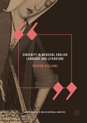
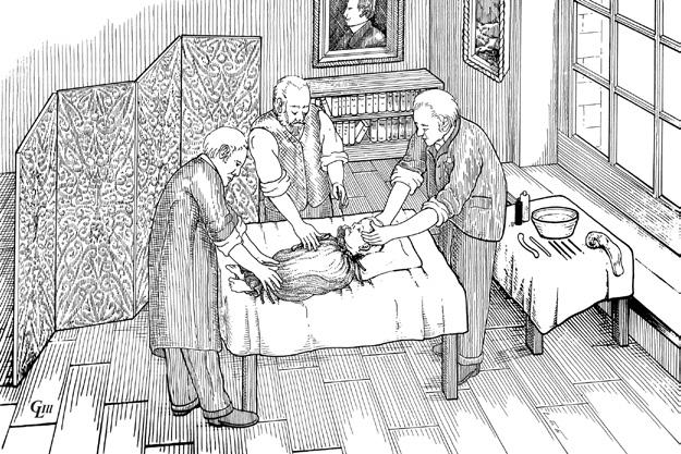
1.1 Cataract Surgery as practised by John Cunningham Saunders in 1811. A small team would hold the child still; restrained, the surgery would be over in a flash, the child hopefully young enough and drugged enough with liquor or opium to forget. William Mackenzie of Glasgow said, in answer to the question of when to operate, “The answer decidedly is to operate in infancy”. He was aware that late operation was accompanied by a worse visual outlook and more difficult surgery. © Gillian Lee Illustrations.

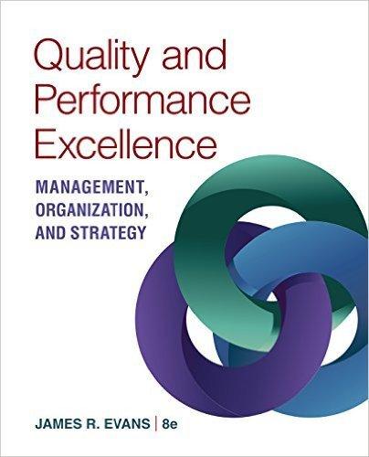
Fig. 1.2 In 1769 the London Dispensary was opened at Holborn by George Armstrong, accepting child out-patients but closed soon after. Similar institutions offered help to children during the chaos of the French Revolution but the first hospital for the treatment of sick children was the Hôpital des Enfants-Malades founded in 1802 on the Rue de Sèvres in Paris (above). Hospitals in Frankfurt, Munich, Hamburg, St. Petersburg soon followed. The Hospital for Sick Children, Great Ormond Street, London was the first in the English speaking world; it opened on the St Valentine’s day in 1852. The child mortality rate at that time was truly shocking: of 50,000 persons dying annually in London, 21,000 were children under 10. From Lereboullet, Pierre. L’Hopital Des Enfants-Malades (1802-1913). Paris Médical. 1913–14;1:3–19. Written as the hospital was being demolished.
a profound social influence. Great Ormond Street (GOS) Hospital was later founded in an adjacent street. L’Hôpital des Enfants-Malades was founded in 1802 in Paris (Fig. 1.2), becoming the first effective children’s hospital1; others in Europe soon followed.
Children’s hospitals in Philadelphia (1855) and Toronto (1875), on a similar model to GOS, were for children and their pediatricians – this was the vanguard of scientific medicine. The ophthalmologists there learnt pediatric skills and embraced scientific medicine. Early GOS ophthalmologists included Robert Marcus Gunn, Edward Nettleship, George Coats, and Philip Doyne.
The 20th century: ophthalmology super-specialization in North America
By the early 20th century, the major specialties, including ophthalmology, were established. Specialist societies were often conjoined: the American Academy of Ophthalmology only separated from its partner, Otolaryngology, in 1979.
Dr Frank Costenbader of Washington DC, who had trained in ophthalmology and otolaryngology, developed particular expertise in children’s eye disorders. In 1943, he limited his practice to children, becoming the first pediatric ophthalmologist.
After World War II, the pace of specialization and the quality of training quickened. Ophthalmologists trained as super-specialists through dedicated fellowships, replacing the previous apprenticeship system, i.e. “sitting at the feet” of a well-known teacher.
Dr Costenbader’s first trainee, Dr Marshall Parks, became the United States’ most prominent pediatric ophthalmologist with wide pediatric expertise. This first fellowship program was followed by many others run by Hermann Burian, Arthur Jampolsky, Phil Knapp, Gunter von Noorden, Martin Urist; and in Canada by Jack Crawford in Toronto, William G Pearce in Alberta, and John Pratt-Johnson and Andrew Q McCormick, both in Vancouver. Their fellows included most of the best-known names today; North American Pediatric Ophthalmology is what it is today because of their clinical activity, research, teaching and collegial behavior.
Fig.
Nutt9 stated, “Uniocular cataracts are better left un-treated, except for cosmetic reasons … the visual result is always bad … the chances are that the affected eye is abnormal in some other respect.” Jules François10 wrote, “Everyone knows the uselessness of operating on unilateral congenital cataract.” The general opinion everywhere then was against any intervention. Along with the growing understanding of amblyopia, things changed. von Noorden and colleagues11 wrote “It may be well worth while a trial to aspirate a unilateral mature congenital cataract (that is not accompanied by other anomalies) within the first days of life to be followed by immediate contact lens correction.” No doubt there were some skeptics but not all were. Frey et al.,12 a group in Washington DC, operated on and fitted a contact lens that was worn for up to 12 hours daily for 21 idiopathic unilateral congenital cataracts. Occlusion of the phakic eye was attempted. Three of the youngest, with good contact lens wear but unspecified occlusion, achieved 20/40 or better. The team wrote, “… the dictum of extreme conservatism in the management of monocular cataracts in children needs to be re-evaluated.”
At this point, several groups were trying to unlock the door to successful treatment. A group in London13 managed 23 cases with unilateral congenital cataract with surgery, contact lens, and graded occlusion. They agreed that later surgery gave worse visual results. They were looking towards very early surgery, contact lens wear, and graded occlusion, but they found that “… correction before 4 months of age produces less visual loss but we cannot specify the function within this period.”
Creig Hoyt’s group14 in San Francisco preceded their study by talking with neonatologists about detecting neonatal cataracts and referring them early: they received cases almost as soon as they were born and published a case series of eight neonates with monocular cataract treated with immediate surgery, vigorous occlusion, and contact lenses. Five developed visual acuities of LogMAR 0.18 (6/9, 20/30, 0.67), three with LogMAR 0.6 (6/24, 20/80, 0.25). This was a stunning achievement, marred only by the time it took the skeptics, including myself, to believe it! It required great expertise and teamwork, the parents being pivotal. Many other cases were then similarly managed by individuals or teams, and good visual results weren’t unusual and occasional cases even had high levels of binocular vision.
A debate arose regarding the risks and benefits for the affected baby. It was now possible to achieve good vision in unilateral congenital cataract. However, since the operated eye might become the best and one most used, the risks of glaucoma and other complications are high in the first weeks of life, and sympathetic ophthalmitis has been recorded, these risks may, in the parent’s view, outweigh the benefits of having a “spare eye.” That parental informed choice was paramount took some time to be accepted, and the weighing of risks and benefits became the basic currency of the contract with a patient.
Government funding has been behind many of the largest and best collaborative studies in many fields of science. In 1997, the Pediatric Eye Disease Investigator Group (PEDIG), a collaborative network facilitating multi-center clinical research in childhood eye disorders was formed in the USA. It has over 100 participant centers and has resulted in the publication of nearly 100 well-constructed collaborative studies. Another study, also government-funded by the National Eye Institute, is the Infant Aphakia Treatment Study (IATS), which asks
vital questions on cataract management with intraocular and corneal contact lenses in 114 affected patients with long follow-up.
Although centrally funded research in children’s eye diseases is most important in many other countries, much is essentially the result of diversely funded groups of collaborators. In the UK, for instance, these include the European Eye Epidemiology Consortium, the British Isles Congenital Cataract Interest Group, the British Childhood Onset Hereditary Retinal Disorders Network, the 1958 British Birth Cohort, the British Ophthalmological and Paediatric Surveillance Units, and the British Childhood Visual Impairment Interest Group.
Orthoptists and pediatric ophthalmology
Mary Maddox, the daughter of Ernest Maddox, an ophthalmologist with a deep interest in eye muscles, optics, refraction, and binocular vision, started a clinic in London in 1928, and was soon joined by Sheila Mayou, the daughter of a London ophthalmologist. At Moorfields Eye Hospital, High Holborn, they started a public clinic and school of orthoptics. Beryl Mayou travelled extensively during World War II and afterwards ran a training course, launching orthoptics in Brazil. They and other orthoptists, Vivien MacLennan, Sylvia Jackson, Joyce Mein, Jona Yoxall, and Barbara Lee in the UK, Birgitta Neikter in Sweden, Catherine Turbayne Lunn and Geraldine Tillson in Canada, and others in North America, France, Holland, South Africa, Australia, and Japan showed how orthoptists were vital colleagues in diagnosis and management of patients with strabismus, to the great benefit of patients and cost savings to service providers.
Early publications on pediatric ophthalmology and strabismus
In 1583, the first ophthalmology textbook, Augendienst, by Georg Bartisch, included the management of strabismus. The first strabismus surgery was claimed in 1751 by the English charlatan (and brilliant physiologist?) “Chevalier” John Taylor, but is generally attributed to Johann Dieffenbach in Berlin in 1839, whose tenotomy was practiced by George Critchett in 1855. A small book entitled Squinting by Carsten Holthouse was published in London in 1858. In 1903, Claud Worth published Squint: its Causes, Pathology and Treatment. It ran to six editions under his authorship and, read today, it is brilliant. Worth was succeeded by Bernard Chavasse on this book. Chevasse worked with the physiologist Charles Sherrington, embracing physiological concepts based on reciprocal innervation and reflexes. After World War II, James Hamilton Doggart published the first book on pediatric ophthalmology, Diseases of Children’s Eyes (Fig. 1.3): it was meticulously organized and beautifully illustrated.
T Keith Lyle, a strabismologist and neuro-ophthalmologist, and a prolific author and lecturer, collaborated with The Hon Geoffrey Bridgman on the ninth edition of Worth and Chavasse’s book and he also wrote prolifically on strabismus. Kenneth Wybar was the author of the strabismus section of Duke-Elder’s System of Ophthalmology.
Epidemiology and the worldwide impact of visual impairment in children
Jugnoo S Rahi and Clare E Gilbert
Chapter contents
INTRODUCTION
SPECIFIC ISSUES IN THE EPIDEMIOLOGICAL STUDY OF VISUAL IMPAIRMENT IN CHILDHOOD FRAMING THE QUESTION
POTENTIAL SOURCES OF INFORMATION ABOUT VISUAL IMPAIRMENT
IMPACT OF VISUAL IMPAIRMENT
VISUAL IMPAIRMENT IN THE BROADER CONTEXT OF CHILDHOOD DISABILITY
PREVENTION OF VISUAL IMPAIRMENT AND BLINDNESS IN CHILDHOOD: VISION 2020
THE ROLE OF OPHTHALMIC PROFESSIONALS IN PREVENTION OF CHILDHOOD VISUAL IMPAIRMENT REFERENCES
Introduction
This chapter considers important issues about epidemiological studies of childhood visual impairment (VI), severe visual impairment (SVI), or blindness (Boxes 2.1 and 2.2); we synthesize current data to provide a global picture of the frequency, causes, and prevention of VI and blindness in childhood.
Specific issues in the epidemiological study of visual impairment in childhood
• Case definition: A standard definition applicable to all children continues to be challenging, see below.
• Rarity: Visual impairment and blindness in childhood are uncommon; it is difficult to obtain sufficiently large and representative samples of affected children to allow precise and unbiased study.
• Complex, multidisciplinary management: For a complete picture, information must be sought from the
professionals involved in the care of VI or blind children. For many children with additional non-ophthalmic impairments or chronic disorders, this adds further complexity.
• Lifecourse approach: It is important in health research to understand the biological, environmental, and lifestyle/ social influences at all life stages (preconceptional, prenatal, perinatal, and childhood), and how they combine to set and change health trajectories into adult life. Lifecourse epidemiological and epigenetics approaches are increasingly applied to the study of VI and eye disease affecting children or originating in childhood.1
• Developmental perspectives: In all research on children, developmental issues (as distinct from age-related issues per se) must be taken into account in assessing outcomes and their relationship with risk factors.
• Long-term outcomes: Assessment of meaningful outcomes, such as final visual function or educational placement, requires long-term follow-up, possibly into adult life for some outcomes. This is challenging and is increasingly addressed through health informatics research. This approach mainly relies on routinely collected data, often as electronic or “e” records, in health care (e health records, EHR), and other administrative systems, e.g. educational or welfare. Record (data) linkage using established methods to minimize errors creates complete datasets for analysis. This is a well-established and powerful approach in child health research2 and has good potential for research in pediatric ophthalmology.
• Ethics. Issues of proxy consent (by parents) and children’s autonomy increasingly impact on participation in ophthalmic epidemiological research.
Framing the question
Clinical or service provision decisions are ideally based on “three-part questions” reflected in the PICO mnemonic (Population Intervention/Comparator and Outcomes). Thus a good question incorporates the reference population (e.g. children under 2 years with infantile esotropia), the
Box 2.1
What is ophthalmic epidemiology?
Ophthalmic epidemiology (literally “studies upon people”) has both its origins and its applications in clinical and public health ophthalmology.
The aim of primary or secondary (e.g. systematic literature review, meta-analysis and modeling) research is to:
• provide quantitative information for planning services
• shed light on the causes and natural history of ophthalmic disorders
• enhance the accuracy and efficiency of diagnosis
• improve the effectiveness of treatment and preventive strategies
Box 2.2
Epidemiological reasoning
This is based on the following principles:
• the occurrence of disease is not random, rather a balance between causal and protective factors
• that disease causation, modification, and prevention are studied by systematic investigation of populations to gain a more complete view than can be achieved by studying individuals
• that any inference that an association between a risk factor and a disease is causal can only be made after two specific steps in reasoning: (1) the exclusion of chance, bias, or confounding as alternative explanations for the observed association, and (2) evidence of a consistent and strong statistical association, which is biologically plausible, in the correct temporal sequence, and preferably exhibits a dose–response relationship
risk factor or the intervention and, where appropriate, a comparator (e.g. preterm versus term birth, or strabismus surgery versus no surgery), and the outcomes (e.g. parentreported improvement in cosmesis and objective improvement in alignment and stereopsis). The focus of the question –whether frequency, causes, or treatment/prevention of disease – determines the study design required to address it, e.g. a descriptive, cross-sectional prevalence survey, or an analytical study (either observational, e.g. case–control or cohort studies, or interventional, e.g. randomized controlled trials).
Who is a visually impaired child?
The affected child, their parents, teacher, social worker, rehabilitation specialist, pediatrician or ophthalmologist are likely to have differing, but equally valid answers to this question. The issue is not which is “correct” but which definition to choose for epidemiological research, for assessing/planning services and in clinical practice.
A standardized definition is necessary in order to compare the frequency, causes, treatment, or prevention of VI within and between countries and over time. The World Health Organization (WHO) classification (Table 2.1) is based on the acuity in the better-seeing eye (i.e. at the level of the person not the eye) and measured with optical correction, if usually worn. Until recently, uncorrected refractive error fell outside this definition, as the early categories used best corrected visual acuity. It has been adopted for epidemiological research, despite the difficulties of measuring visual acuity in very young children and those unable to cooperate with formal testing.
Table 2.1 World Health Organization classification of visual impairment
Level of visual impairment
Visual acuity in better eye with optical correction (if worn)
Slight, if acuity less than 6/7.5 6/18 or better
Visual impairment (VI) Worse than 6/18 up to 6/60 (logMAR 0.5 to logMAR 1.0)
Severe visual impairment (SVI) Worse than 6/60 up to 3/60 (logMAR 1.1 to 1.3)
Blind (BL)
Worse than 3/60 (worse than logMAR 1.3) to no light perception or Visual field <10 degrees around central fixation
MAR, Minimum angle of resolution.
6/7.5 = logMAR 0.10, 20/25, 0.8; 6/18 = logMAR 0.48, 20/60, 0.33; 6/60 = logMAR 1.0, 20/200, 0.10; 3/60 = logMAR 1.3, 20/400, 1.31.0.
Adapted with permission from World Health Organization (WHO). International Statistical Classification of Diseases and Health Related Problems. 10th Revision. Geneva: World Health Organization, 1992.
New methods for testing vision in young children are likely to emerge from technological innovations. Nevertheless, there is a need in epidemiological research and also arguably in clinical practice for a better classification system applicable to children of different ages. This should consider other aspects of vision such as near acuity, visual fields, binocularity, and contrast sensitivity, as well as normal visual development. However its development will be challenging.
Adoption of the WHO International Classification of Functioning, Disability and Health (ICF) reflects the new underpinning framework for understanding disability and the relationship between health conditions, personal, and societal factors.3 In tandem, the importance of measuring patientreported outcomes (patient-reported outcome measures, PROMs) conventionally using self-completed questionnaires is now embedded within many health care systems, so as to help improve the quality of care. Two types of PROMs are particularly relevant to pediatric ophthalmology.4
1. Functional vision assesses the child’s ability (difficulty or ease) for tasks of daily living dependent on vision, such as navigating independently.
2. Vision-related quality of life elicits the child’s and/or parents’ view of the gap between the child’s expectations and his/her actual experiences with respect to the physical, emotional/psychological, cognitive, and social functioning of the child impacted by the visual disorder and its therapy.4
Measures of frequency and burden of childhood visual impairment
The analogy of a barrel of white and red grapes with a hole in the bottom can be used to illustrate measures of frequency and the burden of disease. In this analogy, white grapes represent those without the condition of interest (“healthy”) and red grapes represent those who have the condition of interest (“diseased”). The total number of grapes (white plus red) in the barrel represents the population of interest (e.g. all children aged 0–15 years). The proportion of all the grapes in the barrel that are red at any given time denotes prevalence