Previous editions copyrighted 2013, 2006, 2002, 1995, 1985, and 1968.
All rights reserved.
ISBN: 978-0-323-47833-5
This adaptation of Sturdevant’s Art and Science of Operative Dentistry, 7e, by André V. Ritter, Lee W. Boushell, Ricardo Walter was undertaken by RELX India Private Limited and is published by arrangement with Elsevier Inc.
Sturdevant’s Art and Science of Operative Dentistry: Second South Asia Edition, V. Gopikrishna
All rights reserved. No part of this publication may be reproduced or transmitted in any form or by any means, electronic or mechanical, including photocopying, recording, or any information storage and retrieval system, without permission in writing from the publisher. Details on how to seek permission, further information about the Publisher’s permissions policies and our arrangements with organizations such as the Copyright Clearance Center and the Copyright Licensing Agency, can be found at our website: www.elsevier.com/permissions
This book and the individual contributions contained in it are protected under copyright by the Publisher (other than as may be noted herein).
Notice
The adaptation has been undertaken by RELX India Pvt. Ltd. at its sole responsibility. Practitioners and researchers must always rely on their own experience and knowledge in evaluating and using any information, methods, compounds or experiments described herein. Because of rapid advances in the medical sciences, in particular, independent verification of diagnoses and drug dosages should be made. To the fullest extent of the law, no responsibility is assumed by Elsevier, authors, editors or contributors in relation to the adaptation or for any injury and/or damage to persons or property as a matter of products liability, negligence or otherwise, or from any use or operation of any methods, products, instructions, or ideas contained in the material herein.
Although all advertising material is expected to conform to ethical (medical) standards, inclusion in this publication does not constitute a guarantee or endorsement of the quality or value of such product or of the claims made of it by its manufacturer.
This publication is licensed for sale in India, Bangladesh, Bhutan, Maldives, Nepal, Pakistan and Sri Lanka only. Circulation of this version outside these territories is unauthorized and illegal.
Manager, Content Strategy: Nimisha Goswami
Content Project Manager: Anand K Jha
Sr Production Executive: Ravinder Sharma
Sr Cover Designer: Raman Kumar
Printed and bound at …
This book is dedicated to
My Parents
Sulochana… for being my beloved Amma (mother) and my Alpha (beginning of life)… Ambuja… for being the Annapoorni (nurturer) of our lives…
M Velayutham… for teaching me integrity and humility in life
VG Sivasubramanian… for showing me the path of caring and sharing…
My Teachers
Dr E Munirathnam Naidu… for showing me the attributes of discipline, hard work and perseverance…
Professors A Parameswaran and B Suresh Chandra… for instilling in me the drive to learn, teach and research…
Dr K Sridhar… for making me aware that water always finds its level…
Dr Vijailakshmi Acharya… for inspiring me to be passionate about perfection…
My Family
And above all to… Grace… for being the wind beneath my wings…
—V. Gopikrishna
Page left intentionally blank
Foreword
Dr Clifford Sturdevant had a brass plaque on his desk that reads ‘If it’s almost right it’s wrong!’ This commitment to excellence also was the mantra upon which his classic textbook, The Art and Science of Operative Dentistry, was first written and published in 1968. This textbook has been the basis for training dental students in the fine art and clinical science of operative dentistry for 50 years. In light of this significant landmark, which coincides with the publication of this new seventh edition, we believe it is important to present the evolution of the various editions of the textbook from a historical perspective.
The first edition (Sturdevant, Barton, Brauer, 1968) was meant ‘to present the significant aspects of operative dentistry and the research findings in the basic and clinical sciences that have immediate application’ in the field of operative dentistry. It is important to note that Dean Brauer pointed out in his preface that beyond having the knowledge and skills needed to perform a procedure, the practitioner must also have high moral and ethical standards, essential and priceless ingredients. Since the first edition, this textbook series has always attempted to present artistic and scientific elements of operative dentistry in the context of ethical standards for patient care.
It is also worth noting that the first edition was printed and bound in ‘landscape’ format so that it could more easily be used as a manual in the preclinical laboratory and would always remain open to the desired page. The handmade 5X models used to illustrate the various steps in cavity preparation were created by two dental students enrolled at Chapel Hill School of Dentistry, the University of North Carolina, during the writing of the first edition. Illustrations of these models have continued to be used in later editions, and the models themselves have served as important teaching materials for decades.
Although the techniques, materials, armamentarium, and treatment options continue to evolve, many of the principles of operative dentistry described in the first edition are still pertinent today. An understanding of these principles and the ability to meticulously apply them are critical to provide the outstanding dental treatment expected by our patients.
The second edition (Sturdevant, Barton, Sockwell, Strickland, 1985) expanded on many techniques (e.g. acid etching) using experience and published research that had occurred since publication of the first edition. The basics of occlusion were emphasized and presented in a way that would be helpful to the dental student and practitioner. A chapter on treatment planning and sequencing of procedures, as well as a chapter providing a thorough treatise on the use of pins, was included. Information on silicate cement, self-curing acrylic resin and the baked porcelain inlay was eliminated for obvious reasons. The chapters on endodontic therapy and the ‘dental assistant’ were no longer included. Chapters on (1) tooth-coloured restorations and (2) additional conservative and esthetic treatments explained the changes and improvements that occurred in the areas of esthetic options available to patients. In the chapter on gold inlay/onlay restorations, increased emphasis was given to the gold onlay restorations for Class II cavity preparations.
The third edition (Sturdevant, Roberson, Heymann, J. Sturdevant, 1995) placed a new emphasis on cariology and the ‘medical model of disease’ with regard to risk assessment and managing the high-risk caries patient. This important concept laid
the foundation for what is still taught today with regard to identifying risk factors and defining a treatment plan based on caries risk assessment. The third edition also included new expanded chapters on infection control, diagnosis and treatment planning and dental materials. In light of the growing interest in the area of esthetic dentistry, a variety of conservative esthetic treatments were introduced including vital bleaching, micro- and macro-abrasion, etched porcelain veneers and the novel allporcelain bonded pontic. Additionally, an entirely new section on tooth-coloured inlays and onlays was included that chronicled both lab-processed resin and ceramic restorations of this type and those fabricated chairside with CAD/CAM systems.
With the fourth edition of this text (Roberson, Heymann, Swift, 2002), Dr Clifford Sturdevant’s name was added to the book title to honour his contributions to the textbook series and the discipline of operative dentistry. In this edition, a particular emphasis was placed on bonded esthetic restorations. Consequently, an entirely new chapter was included on fundamental concepts of enamel and dentin adhesion. This chapter was intended to provide foundational information critical to the long-term success of all types of bonded restorations.
The fifth edition (Roberson, Heymann, Swift, 2006) continued with the renewed emphasis on the importance of adhesively bonded restorations and focused on scientific considerations for attaining optimal success, particularly with posterior composites. Concepts such as the ‘C Factor’ and keys to reducing polymerization effects were emphasized along with factors involved in reducing micro-leakage and recurrent decay.
The sixth edition (Heymann, Swift, Ritter, 2013) represented a transition from a large printed edition, as in the past, to a smaller, streamlined printed version that focused on concepts and techniques immediately essential for learning contemporary operative dentistry. The same amount of information was included, but many chapters such as those addressing biomaterials, infection control, pain control, bonded splints and bridges, direct gold restorations and instruments and equipment were available for the first time in a supplemental online format.
The seventh edition (Ritter, Boushell, Walter 2018) continues the legacy of maintaining and enriching the fundamental concepts and principles of contemporary operative dentistry. Diagnosis, classification and management of dental caries have been significantly updated in light of the latest clinical and epidemiological research. Similarly, content on adhesive dentistry and composite resins has been updated as a result of the evolving science in these fields.
An entirely new chapter on light curing and its important role in the clinical success of resin composite restorations has been added. Moreover, a new scientifically based chapter details the important elements of colour and shade matching and systematically reviews how the dental clinician is better able to understand the many covariables involved in colour assessment. It also reviews how best to improve shade matching of esthetic restorations to tooth structure.
In an attempt to better optimize restorative treatment outcomes involving periodontal challenges, a new chapter has been included that addresses these principles. Periodontology applied to operative dentistry chronicles the various clinical considerations involving conditions such as inadequate crown length, lack of root coverage and other vexing problems requiring interdisciplinary treatment to optimize success.
Finally, this text addresses the ever-evolving area of digital dentistry with a new chapter, Digital Dentistry in Operative Dentistry. This chapter reviews the various technologies involved in scanning and image capture for both treatment planning and restorative applications. Additionally, the authors review various types of digital restorative systems for both chairside and modem-linked laboratory-based fabrication of restorations. In recognition of the rapid movement to digital dentistry, this chapter is a vital addition to a textbook whose tradition has been always to reflect the latest technologies and research findings in contemporary operative dentistry.
Since its inception 50 years ago, the Sturdevant text has been a dynamic document, with content that has included innovative information on the latest materials and techniques. Over this time period, numerous internationally recognized experts
have addressed many specific topics as authors and coauthors of various chapters. It also should be pointed out that with all editions of the textbook, the authors of the various chapters are themselves actively involved in teaching students preclinical and clinical operative dentistry. Moreover, they are ‘wet-fingered dentists’ who also practice operative dentistry for their individual patients.
In summary, for 50 years Sturdevant’s Art and Science of Operative Dentistry has been a major resource guiding educators in the teaching of contemporary operative dentistry. Each edition of this text has striven to incorporate the latest technologies and science based on the available literature and supporting research. This edition is a superb addition to this tradition, which will most assuredly uphold the standard of publication excellence that has been the hallmark of the Sturdevant textbooks for half a century.
Harald O. Heymann, DDS, MEd Kenneth N. May, Jr., DDS
Page left intentionally blank
Sumitha N. Ahmed, BDS, MS
Clinical Assistant Professor
Department of Operative Dentistry
Chapel Hill School of Dentistry
The University of North Carolina Chapel Hill, NC, United States
Chapter 19: Clinical Technique for Direct Composite Resin and Glass Ionomer Restorations
Chapter 13: Clinical Technique for Amalgam Restorations
Lee W. Boushell, DMD, MS
Associate Professor
Department of Operative Dentistry
Chapel Hill School of Dentistry
The University of North Carolina Chapel Hill, NC, United States
Chapter 1: Clinical Significance of Dental Anatomy, Histology, Physiology and Occlusion
Chapter 3: Patient Assessment, Examination, Diagnosis and Treatment Planning
Chapter 7: Instruments and Equipment for Tooth Preparation
Chapter 8: Preliminary Considerations for Operative Dentistry
Chapter 11: Fundamentals of Tooth Preparation and Pulp Protection
Chapter 13: Clinical Technique for Amalgam Restorations
Chapter 14: Complex Amalgam Restorations
Chapter 19: Clinical Technique for Direct Composite Resin and Glass Ionomer Restorations
Chapter 26: Class III and V Amalgam Restorations
Terrence E. Donovan, DDS
Professor
Department of Operative Dentistry
Chapel Hill School of Dentistry
The University of North Carolina Chapel Hill, NC, United States
Chapter 6: Dental Biomaterials
Chapter 7: Instruments and Equipment for Tooth Preparation
Chapter 16: Introduction to Composite Restorations
Chapter 12: Introduction to Amalgam Restorations
Chapter 27: Additional Information on Instruments and Equipment for Tooth Preparation
Contributors
Dennis J. Fasbinder, DDS
Clinical Professor
Cariology, Restorative Sciences, and Endodontics
School of Dentistry
University of Michigan
Ann Arbor, MI, United States
Chapter 22: Digital Dentistry in Operative Dentistry
Andréa G. Ferreira Zandoná, DDS, MSD, PhD
Associate Professor
Department of Operative Dentistry
Chapel Hill School of Dentistry
The University of North Carolina Chapel Hill, NC, United States
Chapter 3: Patient Assessment, Examination, Diagnosis and Treatment Planning
Chapter 7: Instruments and Equipment for Tooth Preparation
Chapter 27: Additional Information on Instruments and Equipment for Tooth Preparation
Ralph H. Leonard, Jr, DDS, MPH
Director
Dental Faculty Practice
Clinical Professor
Department of Diagnostic Sciences and General Dentistry
School of Dentistry
The University of North Carolina
Chapel Hill, NC, United States
Chapter 4: Infection Control
Jeffrey Y. Thompson, PhD
Professor
Section of Prosthodontics
Director
Biosciences Research Center
College of Dental Medicine
Nova Southeastern University Ft. Lauderdale, FL, United States
Chapter 16: Introduction to Composite Restorations
Chapter 20: Non-Carious Lesions and their Management
Chapter 12: Introduction to Amalgam Restorations
Chapter 6: Dental Biomaterials
Preface
to the Second South Asia Edition
“If you would not be forgotten as soon as you are dead…
Either write something worth reading or do something worth writing…”
—Benjamin Franklin
The first edition of Sturdevant’s Art and Science of Operative Dentistry was published in 1968 and in these past fifty years it has always been considered to be the Bible of Operative Dentistry as it is the most comprehensive operative dentistry textbook. Drawing from both theory and practice and supported by extensive clinical and laboratory research, it presents a clearly detailed, heavily illustrated step-by-step approach to conservative, restorative and preventive dentistry.
The first South Asian Adaptation Edition of this iconic textbook was brought out in 2013 and this current second edition of the same is an endeavour to carry on the legacy.
Based upon the principle that dental caries is a disease, the book provides both a thorough understanding of caries and an authoritative approach to its treatment and prevention. Throughout the book, emphasis is placed on the importance of treating the underlying causes of the patient problem(s), not just restoring the damage that has occurred. It is organized in a sequential format; the early chapters present the necessary general information while the later chapters are specifically related to the practice of operative dentistry, including conservative esthetic procedures.
This Second South Asia edition of Sturdevant’s Art and Science of Operative Dentistry has been significantly revised in order to streamline the text and improve readability. The order and content of chapters have been reorganized keeping in mind the needs of both undergraduate and postgraduate students.
Five new chapters, namely ‘Periodontology Applied to Operative Dentistry’, ‘Colour and Shade Matching in Operative Dentistry’, ‘Light Curing in Operative Dentistry’, ‘Digital Dentistry in Operative Dentistry’ and ‘Resin-Bonded Splints and Bridges‘ have been included in this edition. In addition, the book is now in full colour. The line art for the book has been completely redrawn in full colour to better show techniques and details, and new, full colour photos have been added where appropriate, and highlighted important concepts and clinical tips for the benefit of the student and clinician. To publish this edition on the year we commemorate the 50th anniversary of the publication of the First Edition is a milestone for Operative Dentistry. I am honored to have had the opportunity to work on and present the Second South Asia Edition.
V. Gopikrishna
Dr Clifford Sturdevant
Page left intentionally blank
Acknowledgements
It has been an absolute honour and privilege to have worked on this Second South Asia Edition of Sturdevant’s Art and Science of Operative Dentistry. I would like to express my sense of gratitude for the faith and confidence reposed on me by Professor Andre’ V. Ritter, Dr Lee W. Boushell and Dr Ricardo Walter in adapting this classic textbook to the needs of the South Asian operative dentistry students and clinicians. I would also like to acknowledge the following coworkers for reviewing the first South Asia edition of this textbook and giving critical inputs and reviews that helped me in the genesis of this edition:
REVIEWERS
Dr A.R. Pradeep Kumar, MDS Professor, Thai Moogambigai Dental College, Chennai, Tamil Nadu, India
Dr Naganath Meena, MDS Professor, VS Dental College, Bangalore, Karnataka, India
Dr B. Sajeev, MDS Professor, Mar Baselios Dental College, Kochi, Kerala, India
Dr Mahima Tilakchand, MDS Professor, SDM Dental College, Dharwad, Karnataka, India
Dr Usha Sathyanarayanan, MDS, PGDHPE Adjunct Professor, Saveetha Dental College, Chennai, Tamil Nadu, India
Dr S. Balagopal, MDS Professor, Tagore Dental College, Chennai, Tamil Nadu, India
Dr Vivek Aggarwal, MDS Assistant Professor, Jamia Milia Islamia University, New Delhi, India
Dr Ruchika Roongta Nawal, MDS Assistant Professor, Maulana Azad Dental College, New Delhi, India
I would also like to thank the entire publishing team at Elsevier including Mr Anand K Jha, Content Project Manager and Ms Nimisha Goswami, Manager, Content Strategy, for all their efforts.
I thank my entire team at Root Canal Foundation (www.rootcanalfoundation.com) for their unwavering support. I thank my family for giving me the moral support and critical time needed to complete this edition.
V. Gopikrishna
Page left intentionally blank
Page left intentionally blank
Chapter | 1 |
Clinical Significance of Dental Anatomy, Histology, Physiology and Occlusion
“Success in life is founded upon attention to the smallest of things… rather than to the largest of things…”
—Booker T. Washington
A thorough understanding of the histology, physiology and occlusal interactions of the dentition and supporting tissues is essential for the restorative dentist. Knowledge of the structures of teeth (enamel, dentin, cementum and pulp) and their relationships to each other and to the supporting structures is necessary, especially when treating dental caries. Proper tooth form contributes to healthy supporting tissues. The relationships of form to function are especially noteworthy when considering the shape of the dental arch, proximal contacts, occlusal contacts and mandibular movement.
Teeth and Supporting Tissues
Dentitions
Humans have primary and permanent dentitions. The primary dentition consists of 10 maxillary and 10 mandibular teeth. Primary teeth exfoliate and are replaced by the permanent dentition, which consists of 16 maxillary and 16 mandibular teeth.
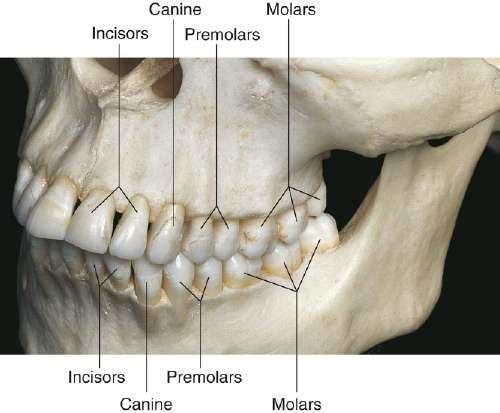
Classes of Human Teeth: Form and Function
Human teeth are divided into classes on the basis of form and function. The primary and permanent dentitions include the incisor , canine and molar classes. The fourth class, the premolar , is found only in the permanent dentition (Fig. 1.1). Tooth form predicts the function of teeth; class traits are the characteristics that place teeth into functional categories. Because the diet of humans
consists of animal and plant foods, the human dentition is called omnivorous
Incisors
The incisors are located near the entrance of the oral cavity and function as cutting or shearing instruments for food (Fig. 1.1). From a proximal view, the crowns of these teeth have a relatively triangular shape, with a narrow incisal surface and a broad cervical base. During mastication, incisors are used to shear (cut through) food.
Clinical Notes
Incisors are essential for the proper esthetics of the smile, facial soft tissue contours (e.g. lip support) and speech (phonetics).
Canines
Canines possess the longest roots of all teeth and are located at the corners of the dental arch. They function in the seizing, piercing, tearing and cutting of food. From a proximal view, the crown also has a triangular shape, with a thick incisal ridge. The anatomic form of the crown and the length of the root make these teeth strong, stable abutment teeth for a fixed or removable prosthesis.
Clinical Notes
Canines not only serve as important guides in occlusion because of their anchorage and position in the dental arches but also play a crucial role (along with the incisors) in the esthetics of smile and lip support (Fig. 1.1).
Figure 1.1 Maxillary and mandibular teeth in maximum inter-cuspal position. The classes of teeth are incisors, canines, premolars and molars. Cusps of mandibular teeth are one-half cusp anterior of corresponding cusps of teeth in the maxillary arch.
(From Logan BM, Reynolds P, Hutchings RT: McMinn’s color atlas of head and neck anatomy, ed 4, Edinburgh, 2010, Mosby.)
Premolars
Premolars serve a dual role:
I. They are similar to canines in the tearing of food. II. They are similar to molars in the grinding of food.
The occlusal surfaces of the premolars present a series of curves in the form of concavities and convexities that should be maintained throughout life for correct occlusal contacts and function.
Clinical Notes
Although less visible than incisors and canines, premolars still can play an important role in esthetics.
Molars
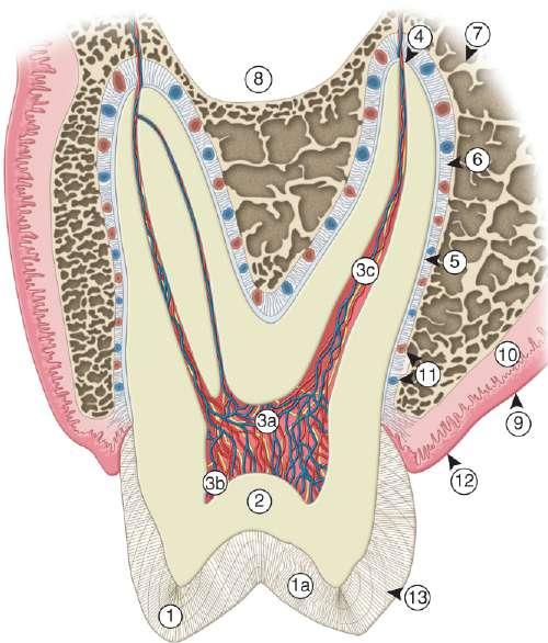
Molars are large, multi-cusped, strongly anchored teeth located nearest to the temporomandibular joint (TMJ), which serves as the fulcrum during function. These teeth have a major role in the crushing, grinding and chewing of food to the smallest dimensions suitable for swallowing. They are well suited for this task because they have broad occlusal surfaces and multi-rooted anchorage (Fig. 1.2).
Clinical Notes
Premolars and molars are important in maintaining the vertical dimension of the face (Fig. 1.1).
Figure 1.2 Cross-section of the maxillary molar and its supporting structures. 1, Enamel; 1a, gnarled enamel; 2, dentin; 3a, pulp chamber; 3b, pulp horn; 3c, pulp canal; 4, apical foramen; 5, cementum; 6, periodontal fibres in periodontal ligament; 7, alveolar bone; 8, maxillary sinus; 9, mucosa; 10, submucosa; 11, blood vessels; 12, gingival and 13, striae of Retzius.
Structures of Teeth
Teeth are composed of enamel, the pulp–dentin complex and cementum (Fig. 1.2). Each of these structures is discussed individually.
Enamel
Enamel formation, amelogenesis , is accomplished by cells called ameloblasts . These cells originate from the embryonic germ layer known as ectoderm . Enamel covers the anatomic crown of the tooth and varies in thickness in different areas (Fig. 1.2). It is thicker at the incisal and occlusal areas of a tooth and becomes progressively thinner until it terminates at the cementoenamel junction (CEJ). The thickness also varies from one class of tooth to another:
• Averaging 2 mm at the incisal ridges of incisors.
• 2.3–2.5 mm at the cusps of premolars.
• 2.5–3 mm at the cusps of molars.
The cusps of posterior teeth begin as separate ossification centres, which form lobes that coalesce. Enamel usually decreases in thickness towards the junction of these developmental features and can approach zero where the junction is fissured (non-coalesced).
Chemical composition
Enamel is a highly mineralized crystalline structure.
• Hydroxyapatite, in the form of a crystalline lattice, is the largest mineral constituent (90%–92% by volume).
• Organic matrix proteins (1%–2% by volume).
• Water (4%–12% by volume).
Structure of enamel
Enamel is composed of millions of enamel rods or prisms, which are the largest structural components, rod sheaths and a cementing inter-rod substance in some areas.
Human enamel is composed of rods that, in transverse section, have a rounded head or body section and a tail section, forming a repetitive series of interlocking prisms. The rounded head portion of each prism (5 µm wide) lies between the narrow tail portions (5 µm long) of two adjacent prisms (Fig. 1.3). Generally, the rounded head portion is oriented in the incisal or occlusal direction; the tail section is oriented cervically.
The rods vary in number from approximately 5 million for a mandibular incisor to about 12 million for a maxillary molar. The rods are densely packed and intertwined in a wavy course, and each extends from the dentinoenamel junction (DEJ) to the external surface of the tooth. Enamel rod diameter near the dentinal borders is about 4 µm and about 8 µm near the surface. This difference accommodates the larger outer surface of the enamel crown compared with the dentinal surface at the DEJ.
Clinical Notes
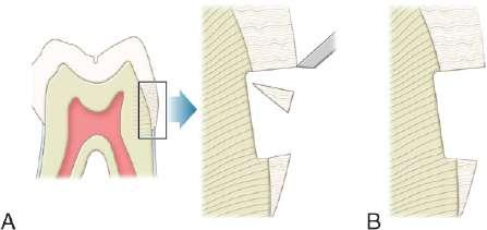
• Enamel is the hardest substance of the human body.
• Enamel rods that lack dentin support because of caries or improper preparation design are easily fractured away from neighbouring rods. For optimal strength in tooth preparation, all enamel rods should be supported by dentin (Fig. 1.4).
Apatite crystallites
The structural components of the enamel prism are millions of small, elongated apatite crystallites that vary in size and shape. The crystallites are tightly packed in a distinct pattern of orientation that gives strength and structural identity to the enamel prisms. The crystallites are irregular in shape, with an average length of 160 nm and an average width of 20–40 nm. Each apatite crystallite is composed of thousands of unit cells that have a highly ordered arrangement of atoms. A crystallite may be 300 unit cells long, 40 cells wide and 20 cells thick in a hexagonal configuration. An organic matrix or prism sheath also surrounds individual crystals and appears to be an organically rich interspace rather than a structural entity.
Gnarled enamel
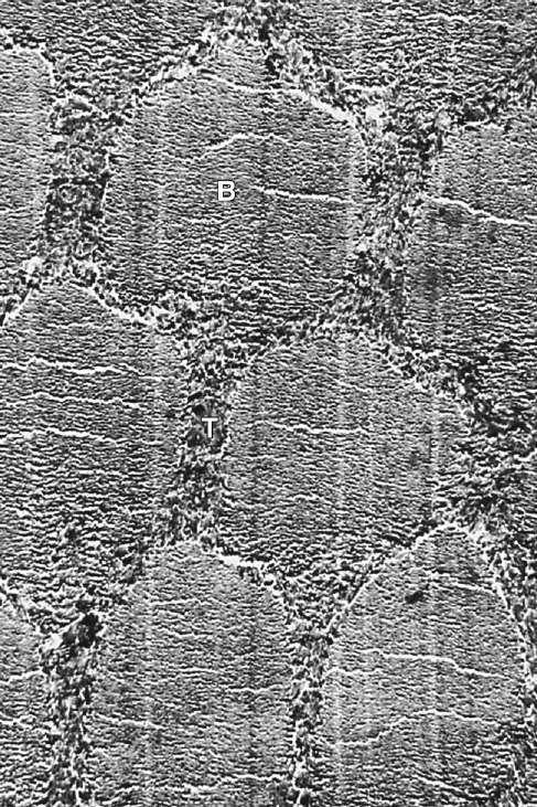
Enamel rods follow a wavy, spiralling course, producing an alternating arrangement for each group or layer of rods
Figure 1.3 Electron micrograph of cross-section of rods in mature human enamel. Crystal orientation is different in ‘bodies’ (B) than in ‘tails’ (T). Approximate level of magnification 5000× (From Meckel AH, Griebstein WJ, Neal RJ: Structure of mature human dental enamel as observed by electron microscopy, Arch Oral Biol 10(5):775–783, 1965.)
Figure 1.4 (A) Enamel rods unsupported by dentin are fractured away readily by pressure from hand instrument. (B) Cervical preparation showing enamel rods supported by dentin.
as they change direction in progressing from the dentin to the enamel surface, where they end a few micrometres short of the tooth surface. They initially follow a curving path through one-third of the enamel next to the DEJ. After that, the rods usually follow a more direct path through the remaining two-thirds of the enamel to the enamel surface. Groups of enamel rods may entwine with adjacent
(From Berkovitz BKB, Holland GR, Moxham BJ: Oral anatomy, histology and embryology, ed 4, Edinburgh, 2009, Mosby.)
groups of rods, and they follow a curving irregular path towards the tooth surface. These constitute gnarled enamel, which occurs near the cervical regions and the incisal and occlusal areas (Fig. 1.5). Gnarled enamel is not subject to fracture as much as a regular enamel. This type of enamel formation does not yield readily to the pressure of bladed, hand-cutting instruments in tooth preparation.
Clinical Notes
The orientation of the enamel rod heads and tails and the gnarling of enamel rods provide strength by resisting, distributing and dissipating impact forces.
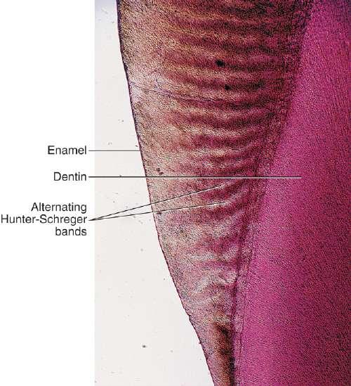
Hunter–Schreger bands
The changes in the direction of enamel prisms that minimize fracture in the axial direction produce an optical appearance called Hunter–Schreger bands (Fig. 1.6). These bands appear to be composed of alternate light and dark zones of varying widths that have slightly different permeability and organic content. These bands are found in different areas of each class of teeth. Because the enamel rod orientation varies in each tooth, Hunter–Schreger bands also have a variation in the number present in each tooth. In anterior teeth, they are located near the incisal surfaces. In molars, the bands occur from near the cervical region to the cusp tips.
Enamel tufts
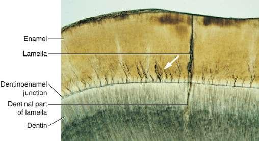
Enamel tufts are hypomineralized structures of the enamel rods and the inter-rod substance that project between adjacent groups of enamel rods from the DEJ (Fig. 1.7). These projections arise in dentin, extend into enamel in the direction of the long axis of the crown and may play a role in the spread of dental caries.
Figure 1.6 Photomicrograph of enamel photographed by reflected light of Hunter–Schreger bands.
(From Avery JK, Chiego DJ: Essentials of oral histology and embryology: a clinical approach, ed 3, St Louis, MO, 2006, Mosby.)
1.7 Microscopic view through lamella that goes from enamel surface into dentin. Note the
(From Bath Balogh M, Fehrenbach MJ: Illustrated dental embryology, histology, and anatomy, ed 3, St Louis, MO, 2011, Saunders. Courtesy: James McIntosh, PhD, Assistant Professor Emeritus, Department of Biomedical Sciences, Baylor College of Dentistry, Dallas, TX.)
Enamel lamellae
Enamel lamellae are thin, leaf-like faults between enamel rod groups that extend from the enamel surface to the DEJ, sometimes extending into dentin (Fig. 1.7). They contain mostly organic material, which is a weak area predisposing a tooth to the entry of bacteria and dental caries.
Striae of Retzius
Enamel rods are formed linearly by successive apposition of enamel in discrete increments. The resulting variations in structure and mineralization are called incremental
Figure
enamel tufts (arrow)
Figure 1.5 Gnarled enamel.
striae of Retzius and can be considered growth rings (Fig. 1.2). In horizontal sections of a tooth, the striae of Retzius appear as concentric circles. In vertical sections, the lines traverse the cuspal and incisal areas in a symmetric arc pattern, descending obliquely to the cervical region and terminating at the DEJ. When these circles are incomplete at the enamel surface, a series of alternating grooves, called imbrication lines of Pickerill, are formed. The elevations between the grooves are called perikymata; these are continuous around a tooth and usually lie parallel to the CEJ and each other.
Nasmyth’s membrane or primary enamel cuticle
Once damaged, enamel is incapable of repairing itself because the ameloblast cell degenerates after the formation of the enamel rod. The final act of the ameloblast is secretion of a membrane covering the end of the enamel rod. This layer is referred to as Nasmyth’s membrane or primary enamel cuticle. This membrane covers the newly erupted tooth and is worn away by mastication and cleaning. This membrane is replaced by an organic deposit called the pellicle, which is a precipitate of salivary proteins. Microorganisms may attach to the pellicle to form bacterial plaque, which, if acidogenic in nature, can be a potential precursor to dental disease.
Prismless enamel
A structureless outer layer of enamel about 30 µm thick is found most commonly towards the cervical area and less often on cusp tips. No prism outlines are visible, and all of the apatite crystals are parallel to one another and perpendicular to the striae of Retzius. This layer, referred to as prismless enamel, may be more heavily mineralized. Microscopically, the enamel surface initially has circular depressions indicating where the enamel rods end. These concavities vary in depth and shape, and they may contribute to the adherence of plaque material, with a resultant caries attack, especially in young individuals. The dimpled surface anatomy of the enamel, however, gradually wears smooth with age.
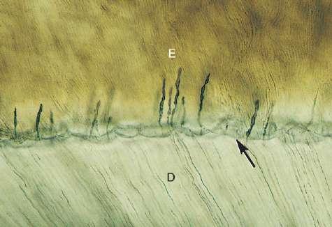
Dentinoenamel junction
The interface of enamel and dentin is known as dentinoenamel junction or DEJ. It is scalloped or wavy in outline, with the crest of the waves penetrating towards enamel (Fig. 1.8). The rounded projections of enamel fit into the shallow depressions of dentin. This interdigitation may contribute to the firm attachment between dentin and enamel.
Enamel is the hardest substance of the human body. Hardness may vary over the external tooth surface according to the location; also, it decreases inward, with hardness lowest at the DEJ. The density of enamel also decreases from the surface to the DEJ. Enamel is a rigid structure that is both strong and brittle (high elastic modulus, high compressive strength and low tensile strength). The ability of the enamel to withstand masticatory forces
Figure 1.8 Microscopic view of scalloped dentoenamel junction (DEJ) (arrow) E, Enamel; D, dentin.
(From Bath Balogh M, Fehrenbach MJ: Illustrated dental embryology, histology, and anatomy, ed 3, St. Louis, MO, 2011, Saunders. Courtesy: James McIntosh, PhD, Assistant Professor Emeritus, Department of Biomedical Sciences, Baylor College of Dentistry, Dallas, TX.)
depends on a stable attachment to the dentin by means of the DEJ. Dentin is a more flexible substance that is strong and resilient (low elastic modulus, high compressive strength and high tensile strength), which essentially increases the fracture toughness of the more superficial enamel.
The DEJ is approximately 2 µm wide and comprises a mineralized complex of interwoven dentin and enamel matrix proteins. In addition to the physical, scalloped relationship between the enamel and dentin, an interphase matrix layer (made primarily of a fibrillary collagen network) extends 100–400 µm from the DEJ to the enamel. This matrix-modified interphase layer is considered to provide fracture propagation limiting properties to the interface between the enamel and the DEJ and thus overall structural stability of the enamel attachment to dentin.
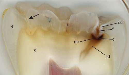
Pit and fissure
The occlusal surfaces of premolars and molars have grooves and fossae that form at the junction of the developmental lobes of enamel. Failure of the enamel of the developmental lobes to coalesce results in a deep invagination of the enamel surface and is termed fissure. Non-coalesced enamel at the deepest point of a fossa is termed as pit. These pit and fissures act as food and bacterial traps that predispose the tooth to dental caries (Fig. 1.9).
Enamel maturation
Although enamel is a hard, dense structure, it is permeable to certain ions and molecules. The route of passage may be through structural units that are hypomineralized and rich in organic content, such as rod sheaths, enamel cracks and other defects. Water plays an important role as a transporting medium through small inter-crystalline spaces. Enamel permeability decreases with age because of changes in the enamel matrix, a decrease referred to as enamel maturation
Figure 1.9 Fissure (f) at junction of lobes allows accumulation of food and bacteria predisposing the tooth to dental caries (c); enamel (e); dentin (d); enamel caries lesion (ec); dentin caries lesion (dc); transparent dentin (td); early enamel demineralization (arrow).
Clinical Notes
Effect of fluoride on enamel: Enamel is soluble when exposed to acidic conditions, but the dissolution is not uniform. Solubility of enamel increases from the enamel surface to the DEJ. When fluoride ions are present during enamel formation or are topically applied to the enamel surface, the solubility of surface enamel is decreased. Fluoride can affect the chemical and physical properties of the apatite mineral and influence the hardness, chemical reactivity and stability of enamel while preserving the apatite structures. Trace amounts of fluoride stabilize enamel by • lowering acid solubility;
• decreasing the rate of demineralization and
• enhancing the rate of remineralization.
Translucency of enamel
Because enamel is semitranslucent, the colour of a tooth depends on
• colour of the underlying dentin;
• thickness of the enamel and
• amount of stain in the enamel.
The amount of translucency of enamel is related to variations in the degree of calcification and homogeneity. Abnormal conditions of enamel usually result in aberrant colour. Enamel becomes temporarily whiter within minutes when a tooth is isolated from the moist oral environment by a rubber dam or cotton rolls. This change in colour is explained by the temporary loss of loosely bound (or exchangeable) water (<1% by weight).
Clinical Notes
The shade of a tooth must be determined before isolation and preparation of a tooth for a tooth-coloured restoration.
Pulp–Dentin Complex
Dentin and pulp tissues are specialized connective tissues of mesodermal origin, formed from the dental papilla of the tooth bud. Many investigators consider these two tissues as a single tissue, which form the pulp–dentin complex, with mineralized dentin constituting the mature end product of cell differentiation and maturation.
Pulp
The pulp is circumscribed by the dentin and is lined peripherally by a cellular layer of odontoblasts adjacent to dentin. Anatomically, the pulp is divided into the following:
I. Coronal pulp located in the pulp chamber in the crown portion of the tooth, including the pulp horns that are directed towards the incisal ridges and cusp tips.
II. Radicular pulp located in the pulp canals in the root portion of the tooth. The radicular pulp is continuous with the periapical tissues by connecting through the apical foramen or foramina of the root. Accessory canals may extend from the pulp canals laterally through the root dentin to the periodontal tissues. The shape of each pulp conforms generally to the shape of each tooth (Fig. 1.2).
The pulp contains nerves, arterioles, venules, capillaries, lymph channels, connective tissue cells, intercellular substance, odontoblasts, fibroblasts, macrophages, collagen and fine fibres. The pulp is circumscribed peripherally by a specialized odontogenic area composed of the odontoblasts, the cell-free zone and the cell-rich zone.
Functions of pulp
The dental pulp occupies the pulp cavity in the tooth and is a unique, specialized organ of the human body that serves four functions:
I. Formative or developmental. The formative function is the production of primary and secondary dentin by odontoblasts.
II. Nutritive. The nutritive function supplies nutrients and moisture to dentin through the blood vascular supply to the odontoblasts and their processes.
III. Sensory or protective. The sensory function provides nerve fibres within the pulp to mediate the sensation of pain. Dentin receptors are unique because various stimuli elicit only pain as a response. The pulp usually does not differentiate between heat, touch, pressure and chemicals. Motor fibres initiate reflexes in the muscles of the blood vessel walls for the control of circulation in the pulp.
IV. Defensive or reparative. The defensive function of the pulp is related primarily to its response to irritation by mechanical, thermal, chemical or bacterial stimuli. The deposition of reparative dentin acts as a protective barrier against caries and various other irritating factors. In cases of severe irritation, the pulp responds by an
inflammatory reaction similar to that for any other soft tissue injury. The inflammation may become irreversible, however, and can result in the death of the pulp because the confined, rigid structure of the dentin limits the inflammatory response and the ability of the pulp to recover.
Clinical Notes
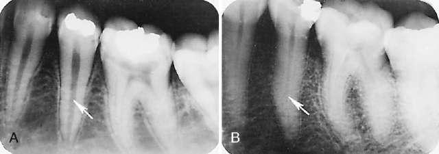
Knowledge of the contour and size of the pulp cavity is essential during tooth preparation. In general, the pulp cavity is a miniature contour of the external surface of the tooth. Pulp cavity size varies with tooth size among individuals and even within a single person. With advancing age, the pulp cavity usually decreases in size. Radiographs are an invaluable aid in determining the size of the pulp cavity and any existing pathologic condition (Fig. 1.10). A primary objective during operative procedures must be the preservation of the health of the pulp.
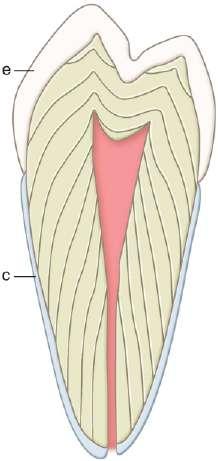
Dentin
Dentin forms the largest portion of the tooth structure, extending almost the full length of the tooth. Externally, dentin is covered by enamel on the anatomic crown and cementum on the anatomic root. Internally, dentin forms the walls of the pulp cavity (pulp chamber and pulp canals) (Fig. 1.11).
Dentinogenesis
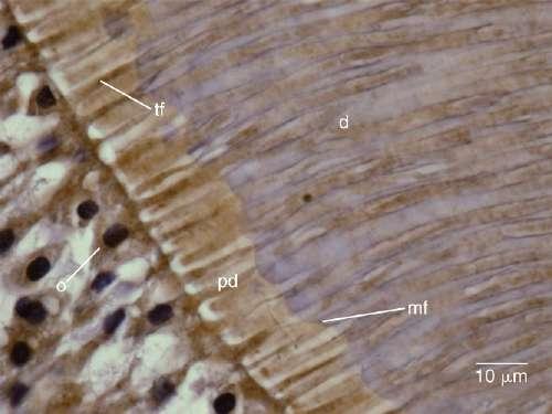
Dentin formation or dentinogenesis is accomplished by cells called odontoblasts. Odontoblasts are considered as a part of pulp and dentin tissues because their cell bodies are in the pulp cavity, but their long, slender cytoplasmic cell processes (Tomes’ fibres) extend well (100–200 µm) into the tubules in the mineralized dentin (Fig. 1.12). Because of these odontoblastic cell processes, dentin is considered a living tissue, with the capability of reacting to physiologic and pathologic stimuli. Dentin formation begins at areas subjacent to the cusp tip or incisal ridge and gradually spreads to the apex of the root (Fig. 1.11). In contrast to enamel formation, dentin formation continues after tooth eruption and throughout the life of the pulp.
the anatomic root.
Figure 1.10 Pulp cavity size. (A) Premolar radiograph of young person. (B) Premolar radiograph of older person. Note the difference in the size of the pulp cavity (arrows)
Figure 1.11 Pattern of formation of primary dentin. This figure also shows enamel (e) covering the anatomic crown of the tooth and cementum (c) covering
Figure 1.12 Odontoblasts (o) have cell processes (Tomes’ fibres [tf]) that extend through the predentin (pd) into dentin (d). mf, Mineralization front.