Bruce E. Katz MD and Neil S. Sadick MD, FAAD, FAACS, FACP, FACPh
ISBN 978-1-4377-0739-7
Non-Surgical Skin Tightening and Lifting
Murad Alam MD, MSCI and Jeffrey S. Dover MD, FRCPC, FRCP
ISBN 978-1-4160-5960-8
Botulinum Toxin
Fourth edition
Alastair Carruthers MA, BM, BCh, FRCPC, FRCP(Lon) and Jean Carruthers MD, FRCSC, FRC(Ophth), FASOPRS
ISBN 978-0-323-47659-1
Soft Tissue Augmentation
Fourth edition
Jean Carruthers MD, FRCSC, FRC(Ophth), FASOPRS and Alastair Carruthers MA, BM, BCh, FRCPC, FRCP(Lon)
ISBN 978-0-323-47658-4
Body Shaping: Skin Fat Cellulite
Jeffrey Orringer MD, Jeffrey S. Dover MD, FRCPC, FRCP and Murad Alam MD, MSCI
ISBN 978-0323321976
For Elsevier
Content Strategist: Belinda Kuhn
Content Development Specialist: Humayra Rahman Khan
Project Manager: Srividhya Vidhyashankar
Design: Miles Hitchen
Illustration Manager: Nichole Beard
Cosmeceuticals
Third edition
Zoe Diana Draelos MD, Murad Alam MD, MSCI and Jeffrey S. Dover MD, FRCPC, FRCP
ISBN 978-0-323-29869-8
Lasers and Lights
Fourth edition
George Hruza MD and Elizabeth Tanzi MD
ISBN 978-0-323-48006-2
Photodynamic Therapy
Second edition
Mitchel P. Goldman MD
ISBN 978-1-4160-4211-2
Liposuction
C. William Hanke MD, MPH, FACP and Gerhard Sattler MD
ISBN 978-1-4160-2208-4
Scar Revision
Kenneth A. Arndt MD
ISBN 978-1-4160-3131-4
Hair Transplantation
Robert S. Haber MD and Dowling B. Stough MD
ISBN 978-1-4160-3104-8
Blepharoplasty
Ronald L. Moy MD and Edgar F. Fincher MD
ISBN 978-1-4160-2996-0
PROCEDURES IN COSMETIC DERMATOLOGY
Soft Tissue Augmentation
Fourth Edition
Edited by Jean Carruthers MD, FRCSC, FRC (OPHTH), FASOPRS
Clinical Professor, Department of Ophthalmology and Visual Science, University of British Columbia, Vancouver, BC, Canada
Alastair Carruthers MA, BM, BCh, FRCPC, FRCP(Lon)
Clinical Professor, Department of Dermatology and Skin Science, University of British Columbia, Vancouver, BC, Canada
Series Editor
Jeffrey S. Dover MD, FRCPC, FRCP
Director, SkinCare Physicians, Chestnut Hill, MA; Associate Clinical Professor of Dermatology, Yale University School of Medicine; Adjunct Associate Professor of Dermatology, Brown Medical School, Providence, RI, USA
Associate Editor Murad Alam MD, MSCI
Professor of Dermatology, Otolaryngology, and Surgery; Chief, Section of Cutaneous and Aesthetic Surgery; Vice-Chair, Department of Dermatology, Northwestern University, Chicago, IL, USA
No part of this publication may be reproduced or transmitted in any form or by any means, electronic or mechanical, including photocopying, recording, or any information storage and retrieval system, without permission in writing from the publisher. Details on how to seek permission, further information about the Publisher’s permissions policies and our arrangements with organizations such as the Copyright Clearance Center and the Copyright Licensing Agency, can be found at our website: www.elsevier.com/permissions.
This book and the individual contributions contained in it are protected under copyright by the Publisher (other than as may be noted herein).
Notices
Knowledge and best practice in this field are constantly changing. As new research and experience broaden our understanding, changes in research methods, professional practices, or medical treatment may become necessary.
Practitioners and researchers must always rely on their own experience and knowledge in evaluating and using any information, methods, compounds, or experiments described herein. In using such information or methods they should be mindful of their own safety and the safety of others, including parties for whom they have a professional responsibility.
With respect to any drug or pharmaceutical products identified, readers are advised to check the most current information provided (i) on procedures featured or (ii) by the manufacturer of each product to be administered, to verify the recommended dose or formula, the method and duration of administration, and contraindications. It is the responsibility of practitioners, relying on their own experience and knowledge of their patients, to make diagnoses, to determine dosages and the best treatment for each individual patient, and to take all appropriate safety precautions.
To the fullest extent of the law, neither the Publisher nor the authors, contributors, or editors, assume any liability for any injury and/or damage to persons or property as a matter of products liability, negligence or otherwise, or from any use or operation of any methods, products, instructions, or ideas contained in the material herein.
ISBN: 978-0-323-47658-4
E-ISBN: 978-0-323-48008-6
The publisher’s policy is to use paper manufactured from sustainable forests
Video contents
Video 5.1 Juvederm Family in Nasolabial Folds
Patrick Coleman, IV, MD
Video 5.2 Treatment of the Lips with Volbella
Shannon Humphrey, MD, FRCPC, FAAD
Video 5.3 The MD Codes ™: Cheek Reshape
Mauricio de Maio, MD, ScM, PhD
Video 5.4 The MD Codes ™: Chin Reshape
Mauricio de Maio, MD, ScM, PhD
Video 5.5 Deep Fat Pads of the Mid Face
Rebecca Fitzgerald, MD
Video 5.6 Treatment of the Forehead with Vycross Products
Jean Carruthers, MD
Video 7 Radiesse Injection in the Nasolabial Folds, Cheeks and Marionette Lines
David M. Ozog, MD
Video 8.1 Poly-L-Lactic Acid: Pan Facial Injection
Stephen H. Mandy, MD
Video 8.2 Poly-L-Lactic Acid
Rebecca Fitzgerald, MD
Video 12.1 Silicone Oil
Alastair Carruthers, MD
Video 12.2 Liquid Injectable Silicone
Derek H. Jones, MD
Video 14 Forehead
Mariano Busso, MD
Video 16.1 Glabella/Central brow (1)
Hema Sundaram, MD
Video 16.2 Glabella/Central brow (2)
Hema Sundaram, MD
Video 16.3 Glabella/Central brow (3)
Hema Sundaram, MD
Video 16.4 Valley in Mid Forehead
Jean Carruthers, MD
Video 19 Cheek Rejuvenation: Hyaluronic Acid Augmentation
Davi de Lacerda, MD
Video 20 Nose
Kyle Koo-Il Seo, MD
Video 21 Perioral Filling
Jean Carruthers, MD, Alastair Carruthers, MD
Video 22.1 Lip Augmentation
Frederick C. Beddingfield III, MD
Video 22.2 Perioral Botulinum Toxin Lip Enhancement
Alastair Carruthers, MD
Video 22.3 Botox Lip Eversion
Jean Carruthers, MD
Video 23.1 Hands (1)
Heidi A. Waldorf, MD
Video 23.2 Hands (2)
Heidi A. Waldorf, MD
Video 23.3 Hands (3)
Heidi A. Waldorf, MD
Video 23.4 Hands (4)
Heidi A. Waldorf, MD
Video 25.1 Correction of Gluteal Depressions with Hyaluronic Acid Gel
Ada R. Trinidade de Almeida, MD
Video 25.2 Lower Gluteus
Raúl Banegas, MD
Video 25.3 Bocouture Injection
Raúl Banegas, MD
Video 27.1 Belotero Injection
Gerhard Sattler, MD
Video 27.2 Vertical Injection Technique
Gerhard Sattler, MD
Video 31.1 Reversers
Rebecca Fitzgerald, MD
Video 31.2 Positive Blood Reflux Test during Filler Injection in the Temporal Area
Gabriela Casabona, MD
Video 31.3 Case of Overcorrection after Filler Injection Lateral Brow Area Treated with Low Dose of Hialuronidase
Gabriela Casabona, MD
Video 34 Camera Usage
Kevin C. Smith, MD
Series preface
Much has changed since the first edition of this series. Non-invasive and minimally invasive cosmetic procedures, as pioneered by dermatologists, have become increasingly adopted by physicians and well-accepted by patients. Cosmetic dermatologic surgery procedures have been refined and improved. Interventions have become more effective, and also safer and more tolerable with increasing benefit:risk ratios. Combination cosmetic regimens that include multiple procedure types have been shown to achieve results comparable to those with more invasive procedures. And new devices and technologies continue to be introduced.
And how best to keep up with these advances and to ensure your offerings are state of the art and at the cutting edge? The newest edition of the Procedures in Cosmetic Dermatology series keeps you there, and for those starting out in the field these texts quickly introduce you and bring you to the state of the art. Each book in this series is designed to quickly impart basic skills as well as advanced concepts in an easy-to-understand manner. We focus not on theory but on how-to. Our expert book editors and chapter authors will guide you through the
learning process efficiently, so you can soon get back to treating patients.
The authors are leading dermatologists in the field. Dermatologists’ role in cosmetic medicine has continued to expand. Research has revealed that primary care physicians and the general public view dermatologists as the experts in less invasive cosmetic procedures. A nationwide advanced fellowship program in cosmetic dermatologic surgery has been initiated to train the next generation of dermatologists to the highest standards.
What has not changed is physicians’ need for clear, concise, and current direction on procedure techniques. Physicians need to be proficient in the latest methods for enhancing appearance and concealing the visible signs of aging.
To that end, we hope that you, our reader, find the books enjoyable and educational.
We thank our many contributors and wish you well on your journey of discovery.
Jeffrey S. Dover MD, FRCPC, FRCP and Murad Alam MD, MSCI
Series preface first edition
Although dermatologists have been procedurally inclined since the beginning of the specialty, particularly rapid change has occurred in the past quarter century. The advent of frozen section technique and the golden age of Mohs skin cancer surgery has led to the formal incorporation of surgery within the dermatology curriculum. More recently technological breakthroughs in minimally invasive procedural dermatology have offered an aging population new options for improving the appearance of damaged skin.
Procedures for rejuvenating the skin and adjacent regions are actively sought by our patients. Significantly, dermatologists have pioneered devices, technologies, and medications, which have continued to evolve at a startling pace. Numerous major advances including virtually all cutaneous lasers and light-source-based procedures, botulinum exotoxin, soft tissue augmentation, dilute anesthesia liposuction, leg vein treatments, chemical peels, and hair transplants have been invented or developed and enhanced by dermatologists. Dermatologists understand procedures, and we have special insight into the structure, function, and working of skin. Cosmetic dermatologists have made rejuvenation accessible to risk-averse patients by emphasizing safety and reducing operative trauma. No specialty is better positioned than dermatology to lead the field of cutaneous surgery while meeting patient needs.
As dermatology grows as a specialty, an ever-increasing proportion of dermatologists will become proficient in the delivery of different procedures. Not all dermatologists will perform all procedures, and some will perform very few, but even the less procedurally directed among us must be well versed in the details to be able to guide and educate our patients. Whether you are a skilled dermatologic surgeon interested in further expanding your surgical repertoire, a complete surgical novice wishing to learn a few simple procedures, or somewhere in between, this book and this series are for you.
The volume you are holding is one of a series entitled “Procedures in Cosmetic Dermatology.” The purpose of each book is to serve as a practical primer on a major topic area in procedural dermatology.
If you want to make sure you find the right book for your needs, you may wish to know what this book is and what it is not. It is not a comprehensive text grounded in theoretical underpinnings. It is not exhaustively referenced. It is not designed to be a completely unbiased review of the world’s literature on the subject. At the same time, it is not an overview of cosmetic procedures that describes these in generalities without providing enough specific information to actually permit someone to perform the procedures. And importantly, it is not so heavy that it can serve as a doorstop or a shelf filler.
What this book and this series offer is a step-by-step, practical guide to performing cutaneous surgical procedures. Each volume in the series has been edited by a known authority in that subfield. Each editor has recruited other equally practical-minded, technically skilled, handson clinicians to write the constituent chapters. Most chapters have two authors to ensure that different approaches and a broad range of opinions are incorporated. On the other hand, the two authors and the editors also collectively provide a consistency of tone. A uniform template has been used within each chapter so that the reader will be easily able to navigate all the books in the series. Within every chapter, the authors succinctly tell it like they do it. The emphasis is on therapeutic technique; treatment methods are discussed with an eye to appropriate indications, adverse events, and unusual cases. Finally, this book is short and can be read in its entirety on a long plane ride. We believe that brevity paradoxically results in greater information transfer because cover-to-cover mastery is practicable.
We hope you enjoy this book and the rest of the books in the series and that you benefit from the many hours of clinical wisdom that have been distilled to produce it. Please keep it nearby, where you can reach for it when you need it.
Jeffrey S. Dover MD, FRCPC, FRCP and Murad Alam MD, MSCI
Preface
“To improve is to change; to be perfect is to change often”.
Winston
Churchill
Since our third edition in the Procedures in Cosmetic Dermatology Series there has been an unprecedented amount of change in the field of soft tissue augmentation!
The concept of the three-dimensional filler approach has rapidly expanded with the development of new fillers, new methods of administering fillers, new anatomical concepts, new appreciation of the three-dimensional subcutaneous anatomy and consequent safety. A fresh appreciation of the best modalities to use in combination including fillers, neuromodulators and energy-based devices has been thoroughly explored as has the relationship of these approaches to traditional surgery.
The use of CT and MRI scans has given a pictorial improvement in our understanding of the various causes of facial deflation. We always saw the loss of facial fat associated with aging, but improved understanding of specific facial fat compartmentalization with age-related atrophy and descent has enabled us to more accurately reflate these collapsed compartments for a more natural result.
Loss and inward rotation of facial bone in the upper, mid and lower face begins earlier in women than in men, but appreciating the trends in age-related bony atrophy are as important as perceiving the changes in facial fat.
The improvement in our understanding of facial skin descent and expansion (dermatochalasis) due to collagen and elastin loss, its treatment and prevention have iced the facial cake. An appreciation of the similarities and differences in treating facial skin in all Fitzpatrick and Glogau skin types is so important.
After all, it is important to put the replacement volume where it will be seen to improve the facial aesthetic in our patient’s specific cultural milieu.
The concept of filler erasability has further elevated the popularity of the HA fillers and hyaluronidase is in every office. Moreover, when other fillers come to market we can hope that an “eraser” will be available at the same time.
Longer-lasting semi-permanent and permanent fillers also have their place in our therapeutic armamentarium. The calcium hydroxylapatite fillers, poly-L-lactic acid, liquid injectable silicone and PMMA in bovine collagen suspension are all approved for facial augmentation, and we expect more classes of fillers to become available.
Finally, we turn to the most important feature of all: our patients! No-one likes pain, and the introduction of the admixture of local anesthetic with fillers spearheaded by Marianno Busso, MD, has indeed allowed many more patients to avail themselves of this successful treatment. His work has stimulated us all to blend fillers so as to enhance patient comfort.
Finally, we feel the world of fillers will change yet again with their perception as drugs not devices. In other words, using the presence of a filler to cause collagen deposition and long-term natural correction is a very important concept.
The filler world has changed dramatically in the past few years. We hope that you enjoy this changed world as much as we do.
Jean Carruthers MD, FRCSC, FRC (OPHTH), FASOPRS Vancouver, BC, Canada
List of contributors
The editor(s) would like to acknowledge and offer grateful thanks for the input of all previous editions’ contributors, without whom this new edition would not have been possible.
Mathew M. Avram MD, JD
Director, MGH Dermatology Laser & Cosmetic Center, Massachusetts General Hospital, Harvard Medical School, Boston, MA, USA
Raul Alberto Banegas MD
Plastic Surgeon, Centro Arenales, Buenos Aires, Argentina
Katie Beleznay MD, FRCPC, FAAD
Clinical Instructor, Department of Dermatology, University of British Columbia, Vancouver, BC, Canada
Jeanette M. Black MD
Dermatologist, Skin Care and Laser Physicians of Beverly Hills, Los Angeles, CA, USA
Joanna G. Bolton MD, FAAD
ASDS Cosmetic Dermatologic Surgery Fellow, Cosmetic Laser Dermatology, San Diego, CA, USA
André Vieira Braz MD
Dermatologist, Clinical, Surgical and Cosmetic, Dermatology Clinic, Rio de Janeiro, RJ, Brazil
Harold J. Brody MD
Clinical Professor of Dermatology, Emory University School of Medicine, Atlanta, GA, USA
Alastair Carruthers MA, BM, BCh, FRCPC, FRCP(Lon)
Clinical Professor, Department of Dermatology and Skin Science, University of British Columbia, Vancouver, BC, Canada
Jean Carruthers MD, FRCSC, FRC (OPHTH), FASOPRS
Clinical Professor, Department of Ophthalmology and Visual Science, University of British Columbia, Vancouver, BC, Canada
Gabriela Casabona MD
Director at Clinica Vida Cosmetic, Laser and Mohs Surgery Center, New York, NY, USA
Kyle M. Coleman MD
Co-Owner, Etre, Cosmetic Dermatology and Laser Center, New Orleans, LA, USA
William P. Coleman III MD
Clinical Professor of Dermatology; Adjunct Professor of Surgery (Plastic Surgery), Tulane University Health Sciences Center, New Orleans, LA, USA
Steven Dayan MD
Clinical Assistant Professor, University of Illinois, Chicago, IL, USA
Jeffrey S. Dover MD, FRCPC, FRCP
Director, SkinCare Physicians, Chestnut Hill, MA; Associate Clinical Professor of Dermatology, Yale University School of Medicine; Adjunct Associate Professor of Dermatology, Brown Medical School, Providence, RI, USA
Jason J. Emer MD
Private Practice, The Roxbury Institute, Beverly Hills, CA, USA
Sabrina Guillen Fabi MD, FAAD, FAACS
Dermatologist, Cosmetic Laser Dermatology, Volunteer Assistant Clinical Professor, Department of Medicine/ Dermatology, University of California, San Diego, CA, USA
Dermatologist, Private Practice, Los Angeles, CA, USA
Laurel Naversen Geraghty MD
Dermatologist, Dermatology and Laser Associates of Medford, Medford, OR, USA
Marguerite Germain MD
Dermatologist, Private Practice, Mt Pleasant, SC, USA
Richard G. Glogau MD
Clinical Professor of Dermatology, University of California, San Francisco, CA, USA
Greg J. Goodman MD, FACD, GradDipClinEpi
Associate Professor, Monash University, Clayton, Victoria, and Chief of Surgery, Skin and Cancer Foundation Inc., Carlton, Victoria, Australia
Adele Haimovic MD
Dermatologist, SkinCare Physicians, Chestnut Hill, MA, USA
Bhushan Hardas MD, MBA
Chief Scientific Officer (Devices), Therapeutic area head of Dermatology and Aesthetics, Allergan, Irvine, CA, USA
Shannon Humphrey MD, FRCPC, FAAD
Clinical Assistant Professor, Director of CME, Department of Dermatology and Skin Science, University of British Columbia, Vancouver, BC, Canada
Omer Ibrahim MD
Dermatologist, SkinCare Physicians, Chestnut Hill, MA, USA
Derek H. Jones, MD
Founder and Director, Skin Care and Laser Physicians of Beverly Hills; Clinical Associate Professor, Dermatology, University of California, Los Angeles, CA, USA
Isabela Tollini Jones MD
ASDS Cosmetic Dermatologic Surgery Fellow, Cosmetic Laser Dermatology, San Diego, CA, USA
Shilpi Khetarpal MD
Dermatologist, Cleveland Clinic Foundation, Cleveland, OH, USA
Val Lambros MD, FACS
Assistant Clinical Professor, Plastic Surgery, University of California at Irvine, Irvine, CA, USA
Bassel H. Mahmoud MD, PhD
Dermatologist, Lahey Hospital and Medical Centre, Burlington, MA, USA
Paula Marchese MD
Assistant at Clinica Vida Cosmetic, Laser and Mohs Surgery Center, New York, NY, USA
Kavita Mariwalla MD
Dermatologist, Mariwalla Dermatology, West Islip, NY, USA
Gary D. Monheit MD
Dermatologist, Total Skin and Beauty, Birmingham, AL, USA
Jose R. Montes MD
Associate Professor and Director, José Raúl Montes Eyes & Facial Rejuvenation, San Juan, Puerto Rico
Diane K. Murphy MBA Consultant, Allergan, Irvine, CA, USA
Rhoda S. Narins MD, FAAD
Medical Director, Dermatology Surgery and Laser Center, New York, NY, USA
David M. Ozog MD, FAAD
Director of Cosmetic Dermatology, Division of Mohs and Dermatological Surgery, Vice-Chair Department of Dermatology, Henry Ford Hospital, Detroit, MI, USA
Berthold J. Rzany MD, ScM Professor, Rzany & Hund, Private Practice for Dermatology and Aesthetic Medicine, Berlin, Germany
Neil S. Sadick MD, FAAD, FAACS, FACP, FACPh
Clinical Professor, Weill Cornell Medical College, Cornell University, New York, NY, USA
Gerhard Sattler, MD
Founder, Medical Director, Rosenparkklinik, Darmstadt, Germany
Clinical Associate Professor, Department of Dermatology, Seoul National University College of Medicine, Seoul, South Korea
Renee Sheinin MD
Dermatology Resident, Henry Ford Health System, Detroit, MI, USA
Kevin C. Smith MD, FRCPC
Private Practice, Niagara Falls Dermatology and Skin Care Centre Ltd, Niagara Falls, ON, Canada
Ada R. Trindade de Almeida MD
Dermatologist, Dermatologic Clinic, Hospital do Servidor Público Municipal de São Paulo; Private Practice, São Paulo, Brazil
Heidi A. Waldorf MD
Waldorf Dermatology Aesthetics; Director, Laser & Cosmetic Dermatology; Mount Sinai Hospital; Associate Clinical Professor, Department of Dermatology, Icahn School of Medicine of Mount Sinai, New York, NY, USA
Monique Vanaman Wilson MD
ASDS Cosmetic Dermatologic Surgery Fellow, Cosmetic Laser Dermatology, San Diego, CA, USA
Dr Stuart Maddin is the godfather of Canadian dermatology. When we first returned to Vancouver from our postgraduate training in 1977, Alastair was enticed to write part of Stuart’s latest textbook. This led to San Francisco fellowships in 1982: in dermatologic surgery for Alastair and in the ophthalmological use of botulinum toxin A for Jean. Stuart’s vision of the future of procedural dermatology has since been proven indelibly and we are both so grateful to him for his charm, intellect, continuous energy and belief in our work. Fortunately for this new world of fillers, the demographics were perfect. The baby boom generation was just starting to deflate as bovine collagen was FDA-approved in the early 1980s! In these very early days we were also surrounded by dermatologists who were equally fascinated by the revolutionary new ability to treat the aging process: Hal Brody, Bill Coleman, Rick Glogau, Arnie Klein, Nick Lowe, Steve Mandy, Gary Monheit, Rhoda Narins, Sam Stegman, Ted Tromovitch, and Luitgard Wiest, amongst others. We have learned so much from you all: you have made this path the best!
A look at the authors of this book will show that the magic continues: new ideas, superb chapter contributions and videos have come from the Coleman family, from Steve Fagien, Shannon Humphrey, Derek Jones, Bert Rzany, Kyle Seo, Ada Trindade de Almeida, and Susan Weinkle, amongst others. We thank you.
Our office clinical and research teams are responsible for huge amounts of data collection, photography and patient followup. Without their dedication and their joy in this work we would never have the data to write about and teach with. We humbly thank you all.
Our three sons, their partners and our grandsons have given us so much love and encouragement over the years.
We love you all and we cherish all the times we have shared together. Now that this book is finished, there will be even more time!
Jean Carruthers MD, FRCSC, FRC (OPHTH), FASOPRS and Alastair Carruthers MA, BM, BCh, FRCPC, FRCP(Lon)
Dedication
We dedicate this volume to our children and their families. Our sons were young when the botulinum toxin story began and they have regarded the efforts of their parents to cope with this accidental discovery with tolerance and increasing pride over the years. We have appreciated the support and encouragement they have given us (rather than the other way round). The love they have given us means we are indeed fortunate!
Alastair Carruthers MA, BM, BCh, FRCPC, FRCP(Lon) and Jean Carruthers MD, FRCSC, FRC (OPHTH), FASOPRS
To the women in my life: my grandmothers, Bertha and Lillian, my mother, Nina, my daughters, Sophie and Isabel, and especially to my wife, Tania. For their never-ending encouragement, patience, support, love, and friendship. To my father, Mark – a great teacher and role model; to my mentor, Kenneth A. Arndt for his generosity, kindness, sense of humor, joie de vivre, and above all else curiosity and enthusiasm.
Jeffrey S. Dover MD, FRCPC, FRCP
Elsevier’s dedicated editorial staff has made possible the continuing success of this ambitious project. The team led by Belinda Kuhn, Humayra Khan and the production staff have refined the concept for this new edition while maintaining the series’ reputation for quality and cuttingedge relevance. In this, they have been ably supported by the graphics shop, which has created the signature high-quality illustrations and layouts that are the backbone of each book. We are also deeply grateful to the volume editors, who have generously found time in their schedules, cheerfully accepted our guidelines, and recruited the most knowledgeable chapter authors. And we especially thank the chapter contributors, without whose work there would be no books at all. Finally, I would also like to convey my debt to my teachers, Kenneth Arndt, Jeffrey Dover, Michael Kaminer, Leonard Goldberg, and David Bickers, and my parents, Rahat and Rehana Alam.
Murad Alam MD, MSCI
Introduction
Jean Carruthers, Alastair Carruthers
Summary and Key Features
• Knowledge of the subcutaneous neurovascular, bony, and muscular anatomy of each region of the face is increasingly important. Understanding of the aesthetic desires of each subject and careful targeted volumization is the key to aesthetic success with facial fillers.
• Understanding different regional concepts of beauty and the use of validated assessment scales have aided in achieving optimal outcomes.
• Reversers and better pain management have enhanced augmentation procedures. Better understanding of anatomy and improved injection techniques have reduced vascular occlusion complications.
• Three-dimensional fillers are now seen as stimulators of neocollagenesis, thus improving the texture of facial skin from the “inside.”
In 1885 two extremely powerful European figures, Kaiser Wilhelm and Chancellor Bismarck, decided to announce the new age of retirement, age 65. At that time the median age in the population was 16. Nowadays it is 41.7.1
In addition, adults are currently working longer and living better and are much more educated about the ways to live a healthy lifestyle. They are also very pressed for time because they are not only looking after their children and grandchildren but also are taking care of their aging parents.
There are also significant stresses from the workplace, in that older workers are competing for work and promotions with younger colleagues.
The new world of noninvasive rejuvenation is perfectly timed to assist them to recover and maintain their youthful and empowered appearance.
In the ASDS Consumer Survey on Cosmetic Dermatologic Procedures2 of 7315 individuals surveyed, 5 in 10 were considering a cosmetic procedure for aesthetic indications not only in the face but also over the entire body. They wished to look as young as they felt, to appear more attractive, and to feel more confident.1
In 2014 nearly half of all cosmetic patients in the United States requesting noninvasive or minimally invasive
interventions received multiple cosmetic procedures at the same time.2 They are also most interested in procedures that give little or no downtime.
Synthetic fillers, such as hyaluronans, calcium hydroxylapatite, polyLlactic acid, and silicone, allow threedimensional volumization without a prior harvesting procedure. Local tumescent anesthesia liposuction allows autologous product to be used for volumization.
The anatomic structures of facial aging of bone, fat, and skin have been further studied by computed tomography (CT), magnetic resonance imaging (MRI) scans, and detailed anatomic cadaver dissection which have allowed us to visualize accurately the underlying agerelated changes. A new descriptive language of agerelated facial changes using facial scales has been published, demonstrating their value in improving communication not only with our patients in our clinics, but also with each other as we work together toward better treatments.3
Patientreported outcomes (PROs) have become the standard for assessing treatment outcomes from the patient’s point of view. Published validated questionnaires, such as the Facial Line Outcomes (FLO) and SelfPerception of Age (SPA), can be used as easily in the clinic as in the research setting. Several validated PROs are used both by patients and by the treating and evaluating physician—such as the Lip Fullness Scale (LFS) and Look and Feel of the Lips (LAF) scale, as well as the severity scales for Perioral Lines at Rest (POL), Perioral Lines at Maximum Contraction (POLM), and Oral Commissure Severity (OCS), and the FaceQ scales.4–9 The recognition that the patient’s opinion is all important and must thus be recorded, studied, and understood is a gift of the field of aesthetic medicine.
Patients prefer their treatments to be as painfree as possible. In the past decade, we have learned that educating our subjects dramatically reduces their anxiety, as does topical chilling with ice and topical anesthesia and “talkesthesia.” Pain control for facial injections has largely evolved away from trigeminal nerve blocks, with the common addition of lidocaine to injected fillers. The addition of saline to dilute the filler decreases cohesiveness, allowing for smooth delivery and the ability to distribute the product evenly by gentle massage.
Reversibility has also become a cornerstone of facial filler injections. Hyaluronidase is an enzyme that will catabolize any hyaluronic acid (HA) filler, sometimes within 24 hours. New classes of fillers may be produced
with custommade “erasers” in the future. Posttreatment bruising can now be treated immediately using intense pulsed light at moderate settings, which allows the bruise to be absorbed within 24 to 48 hours instead of 7 to 10 days.
All subjects are aware that they will see an immediate filler effect with the desired new contour. They may not be aware of the neocollagenesis that occurs with the filler apparently stimulating the development of new collagen in the dermis,3 which will give a more reflectant glowing facial skin.
The past century has seen an explosion of development of new fillers and their global acceptance by a patient population that would rather look restored and younger without the trauma and downtime of surgery. Indeed, the introduction of noninvasive or minimally invasive injectable procedures represents a significant shift in the approach to facial rejuvenation. According to the website of the American Society of Aesthetic Plastic Surgeons 2016, injectables overall saw a 21% increase in 2015.10
References
1. Median Age of the world’s population Wikipedia.org. Accessed Oct 22, 2016.
2. ASDS Consumer survey on cosmetic Dermatologic Procedures 2016.
3. Carruthers J, Burgess C, Day D, Fabi SG, Goldie K, Kerscher M, Nikolis A, Pavicic T, Rho NK, Rzany B, Sattler G, Sattler S, Seo K, Werschler WP, Carruthers A. Consensus Recommendations for Combined Aesthetic Interventions in the Face Using Botulinum Toxin, Fillers, and EnergyBased Devices. Dermatol Surg. 2016;0:1–12.
4. Carruthers A, Carruthers J, Hardas B, et al. A validated lip fullness rating scale. Dermatol Surg. 2008a;34(suppl 2):S161–S166.
5. Carruthers A, Carruthers J, Hardas B, et al. A validated marionette lines rating scale. Dermatol Surg. 2008b; 34(suppl 2):S167–S172.
6. Carruthers A, Carruthers J, Hardas B, et al. A validated crow’s feet rating scale. Dermatol Surg. 2008c;34(suppl 2):S173–S178.
7. Carruthers A, Carruthers J, Hardas B, et al. A validated hand grading rating scale. Dermatol Surg. 2008d;34(suppl 2):S179–S183.
8. Carruthers J, Carruthers A, Monheit GD, Davis PG, Tardie G. Multicenter, randomized, parallelgroup study of onabotulinumtoxinA and hyaluronic acid dermal fillers (24mg/mL smooth, cohesive gel) alone and in combination for lower facial rejuvenation: satisfaction and patientreported outcomes. Dermatol Surg. 2011;36(suppl 4):2135–2145.
9. Carruthers J, Flynn TC, Geister TL, et al. Validated assessment scales for the mid face. Dermatol Surg. 2012;38:320–332.
10. American Society for Aesthetic Plastic Surgery website Accessed 22 Oct 2016.
Further reading
Carruthers A, Carruthers J, Hardas B, et al. A validated lip fullness rating scale. Dermatol Surg. 2008;34(suppl 2): S161–S166.
Carruthers A, Carruthers J, Hardas B, et al. A validated marionette lines rating scale. Dermatol Surg. 2008; 34(suppl 2):S167–S172.
Carruthers A, Carruthers J, Hardas B, et al. A validated crow’s feet rating scale. Dermatol Surg. 2008c;34(suppl 2):S173–S178.
Carruthers A, Carruthers J, Hardas B, et al. A validated hand grading rating scale. Dermatol Surg. 2008d;34(suppl 2): S179–S183.
Carruthers J, Carruthers A, Monheit GD, Davis PG, Tardie G. Multicenter, randomized, parallelgroup study of onabotulinumtoxinA and hyaluronic acid dermal fillers (24mg/mL smooth, cohesive gel) alone and in combination for lower facial rejuvenation: satisfaction and patientreported outcomes. Dermatol Surg. 2011;36(suppl 4):2135–2145.
Carruthers J, Flynn TC, Geister TL, et al. Validated assessment scales for the mid face. Dermatol Surg. 2012;38:320–332.
Carruthers JDA, Glogau R, Blizter A, Facial Consensus Group Faculty. Advances in facial rejuvenation: botulinum toxin type A, hyaluronic acid dermal fillers, and combination therapies: consensus recommendations. Plast Reconstr Surg. 2008;121(suppl 5):S5–S30.
Fagien S, Carruthers J. A comprehensive review of patient reported satisfaction with botulinum toxin type A for aesthetic procedures. Plast Reconstr Surg. 2008;122:1915–1925.
Fillers: paradigm shifts produce new challenges
Richard G. Glogau
Summary and Key Features
• A shift in treatment concept from two-dimensional to three-dimensional.
• Volume replacement rather than static wrinkle correction.
• Twenty years of collagen injectables replaced with hyaluronic acid (HA) gels.
• More robust—denser, harder, greater lift—HA gels introduced.
• Newer products are niche variations of existing HA technology.
• More robust fillers require deeper application.
• Deeper placement involves exposure to vascular communications with retinal artery and branches of carotid artery.
• Treatment of the temple area involves potential middle temporal vein injection, which can cause pulmonary or cerebral embolism.
• Intravascular injection or vascular compression from robust agents can cause catastrophic consequences.
• Risk factors include filler particle size, pressure generated, and speed of injection.
• Volume replacement gives satisfying aesthetic improvement unachievable by other means.
• Location of injection is key to avoiding risk and providing cosmetic benefit.
1926 real estate classified ad in the Chicago Tribune: “Attention salesmen, sales managers: location, location, location, close to Rogers Park.”
William Safire, June 26, 2009, New York Times Magazine
When this topic was reviewed in the 2012 edition of “Soft Tissue Augmentation,” we pointed out the shift in aesthetics that was occurring. Fillers were moving from treatment of static wrinkles in the two-dimensional plane to correcting the three-dimensional volume changes in the aging
face. What started out as very small and arbitrary amounts of material (0.5 and 1.0 mL syringes of bovine collagen or occasional microdroplets of silicone) in the 1970s and an unblinking focus on the single line or small dermal acne scar, suddenly blossomed into greater volumes of filler, especially with the introduction of hyaluronic acid (HA) gels. With the blockbuster aesthetic impact of botulinum toxin on the muscles of facial expression, the tools were at hand to address a second significant component of facial aging: the dramatic loss of subcutaneous and deep tissue volume associated with the aging face.
The single syringe of collagen commercially available for 25 years was completely inadequate to address the tasks at hand. The markets began to respond with a virtual flood of new filler products—HA gels, polylactic acid (PLA), calcium hydroxylapatite (CaHA), polymethylmethacrylate (PMMA)—and a renewed interest in two filler agents that had been in use before: silicone and fat. After 20 years of three fillers—collagen, silicone, and fat—the aesthetic market expanded in 5 years to more than 300 commercially available forms of injectable filler worldwide, with more coming every year.
The HA gels are now available in a variety of densities, with added lidocaine, and they constitute the lion’s share of the filler market in North America at the present time. Although Restylane was the first HA filler product US Food and Drug Administration (FDA)-approved to enter the US market, it was quickly followed by the Juvéderm family of fillers and has been joined by other injectable products of different compositions: Sculptra (Dermik Laboratories, Sanofi-Aventis, Bridgewater, New Jersey), a PLA filler, Radiesse (BioForm Medical, San Mateo, California), a CaHA filler, Bellafill (Suneva, San Diego, California), a PMMA filler, and Belotero (Merz, Frankfurt a.M., Germany), an HA gel filler for fine lines.
In 2012 we pointed out that many of the products were coming from European manufacturers and described two interesting agents, Aquamid (Contura, Soeborg, Denmark), a polyacrylamide gel, and Ellansé (AQTIS, Utrecht, Netherlands), polycaprolactone microspheres in carboxymethyl cellulose gel. Neither product currently has made it through the FDA approval process. Other HA gel fillers, such as the Emervel family of fillers (Galderma, S.A., Lausanne, Switzerland), are in trial or are awaiting FDA approval. What changes in professional market demand and perception are now driving the process forward? What have we learned since 2012?
As a result of the influx of new fillers, together with a heightened appreciation of the three-dimensional nature of volume loss in the aging face, there was a rapid increase in volume of material being injected into the deeper compartments of the face, frequently giving more significant and natural improvements to the aging face. Convex contours of the cheek in particular, but also the temples, lips, chin, eyes, were subtly restored to younger volumes in ways that traditional incisional surgery could not address. Whole new areas of application were suddenly available to the injector.
Homologous fat, which was a readily available by-product of liposuction techniques introduced in the early 1980s, was the only true volume injectable. Many liposuction surgeons tried various harvesting and processing techniques as they recycled the unwanted fat from abdomens and hips to various places on the face and body. However, for the most part the results were inconsistently dependable, not long-lasting for the majority of patients, and occasionally producing unwanted asymmetric outcomes. But the use of fat did increase further understanding of the nature and distribution of the subcutaneous fat compartments in the aging face. For example, noninvasive magnetic resonance imaging (MRI) and imaging techniques estimate the volume of the midface subcutaneous fat at somewhere between 13 and 17 mL per side1—not a deficit likely to be repaired with a single syringe of any material!
HA fillers have become the dominant filler in the United States and global markets. They are stable at room temperature, available “off the shelf,” and were manufactured in single preloaded syringes, very reminiscent of the old collagen injections. They provide good reversibility with hyaluronidase, an important safety consideration. They provide more immediate “lift” than the collagen fillers that preceded them, have longer duration of action, and require no allergy skin testing prior to treatment. They are easily injected through small-gauge needles, 30 gauge and smaller. Because they are derived from bacteria, they do not share the problems of animal-derived proteins like the collagen products.
The evolution of the HA gel products has made some technologic improvements in the products. One approach has been to vary the concentration of the HA gel. Another is to vary the degree of cross-linking between the polymer chains, and another significant improvement was the addition of lidocaine for increased comfort of injection. Changes in density and cross-linking variously affect the rheology of the materials and can improve duration, lift, and ease of injection through small-gauge needles.
Some of these changes have produced “niche” products for specific anatomic areas or applications: Restylane Silk (Galderma), and Volbella (Allergan) for lip augmentation, Volift (Allergan) for nasolabial folds, and Voluma (Allergan) and Restylane Lift (Galderma, formerly Perlane-L) for midface augmentation. However, the shared characteristic of all these “niche” fillers is that they are not “dermal” fillers but are placed in the deep subcutaneous,
submucosal, or submuscular space, often just above the periosteum. Each of these products offers a nuanced variation of the earlier HA gel fillers to appeal to physicians and patients alike.
However, the hallmark of the popular HA fillers is their adaptability, tolerability, and reversibility. Injectors can easily pick two or three of the available HA fillers and by diluting them slightly, they can often make one of the standard HA fillers perform satisfactorily in different anatomic depths. Although such activity must be recognized as “off-label” use of approved medical devices, the practice appears widespread as physicians and patients seek treatment of a variety of anatomic locations and aesthetic indications in a single office visit.
The challenge for the treating physician is to select the suitable agent and place it appropriately in the given anatomic location to produce the desired aesthetic result. In the 1970s and early 1980s when bovine collagen injections were in use, wrinkles were the aesthetic target, the placement was in the dermis, and the postinjection side effects were predictably local bruising at the site of injection. As the aesthetic range gradually included defects with some depth, like expression lines, vascular occlusions began to appear, particularly ischemic accidents in the supratrochlear vessels (from treatment of the glabellar lines), the angular branch of the facial artery (treatment of nasolabial folds), or the occlusion of the labial artery (lip augmentation).
These vascular accidents appeared to increase in number when the shift occurred from the original Zyderm collagen to Zyplast, a collagen product in which the collagen polymers were cross-linked to produce longer duration of effect. Zyplast was much more slippery than Zyderm, and injectors could inadvertently inject the entire syringe with one push on the plunger, potentially contributing to vascular accidents. Although debate swirled about the underlying cause of these occlusions (intravascular vs. extravascular compression,2 hemostatic effect of the collagen vs. mass effect of the bolus in the vessel), the nature of the phenomenon was that (1) it was irreversible—nothing existed to dissolve the collagen, and (2) it required the injection to occur below the dermis in the subcutaneous space, where the smaller arteries came close to the surface anatomy.
The evolution of use of the HA gel fillers followed the same process, although the dermal placement of these materials was certainly less common. Restylane, because of the particulate nature of the HA, produced a color shift known as the Tyndall effect when it was injected too superficially. However, it certainly did not take much time for the occlusive accidents to be seen with HA treatment of the glabellar creases, nasolabial folds at the base of the nose, and labial arteries, among others. The next evolutionary step was to use the HA fillers to go even deeper in the soft tissue to address true volume deficits: loss of premalar and malar fat, perioral volume loss, atrophy along the mandibular ridge, and loss of fat in the temples, forehead, and nose.
The filler materials themselves became robust, providing greater lift, duration, and volume. They required larger-caliber needles to inject the material that can allow greater aliquots of material to be injected with greater speed and higher pressures than possible with smaller needles. But the catastrophic vascular complications of retinal artery embolization in the literature reflect the deeper anatomy being targeted, along with many of the facial arteries sharing circulation with the retinal artery.
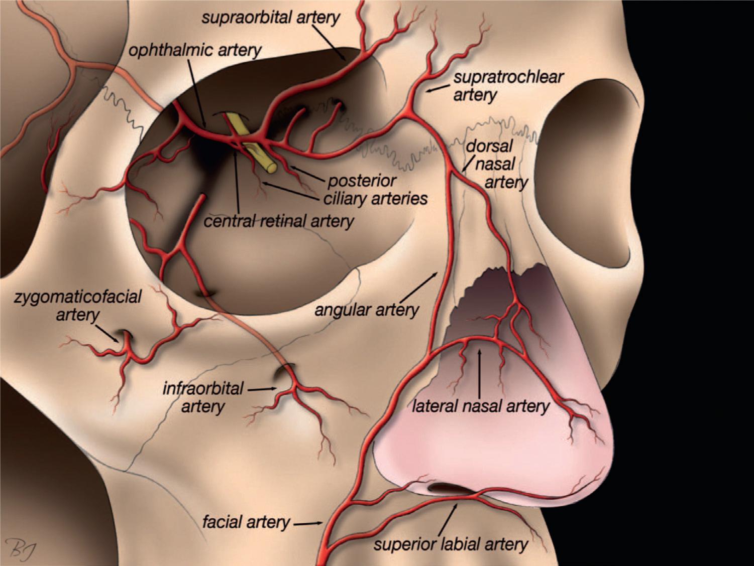
Several superficial arteries of the facial vasculature are distal branches of the ophthalmic artery (supraorbital, supratrochlear, dorsal nasal, and angular artery of the nose) (Fig. 2.1). The danger is not limited to the arterial side of the circulation. In the temple area, a frequent site of unwanted atrophy and an aesthetic target for deep filler injections, the middle temporal vein (MTV) communicates with the cavernous sinus through the periorbital veins and the pulmonary artery via the internal jugular vein,3 requiring judicious placement of filler in the immediate periosteal space to avoid intravascular injection of the MTV and potential vision loss or pulmonary and cerebral embolism.
Unfortunately, only small amounts of filler are required to produce retrograde injection from the distal branches to the retinal artery, reportedly as little as 0.5 mL of material. Although fortunately these deep vascular
complications with retrograde embolism are quite rare, they warrant a careful understanding of both the potentially problematic vasculature and available techniques to lessen the risk of the injections.4 The inextricable link between speed of injection and risk of side effect has been demonstrated in clinical trials.5 In addition, suggestions have been made to use smaller syringes (reducing pressure), blunt cannulae (less chance of direct vessel cannulation), multiple small aliquots rather than large single amounts, aspiration before injection to confirm location of the needle tip, use of vasoconstrictors in the area before injecting filler to reduce vessel caliber, and moving the needle or cannula slightly while injecting to avoid intravascular puncture. Consider injecting across the vessel’s path rather than parallel to it to minimize intravascular injection.
Although vascular accidents have been reported with all manner of filler, fat clearly seems to run a higher risk, probably secondary to the higher pressures and larger particle sizes injected. Along with other permanent or semipermanent fillers, such as CaHA, PMMA, PLA, and silicone, there is no chance of removing or dissolving the offending filler in case of vascular accident. HA gel fillers offer a chance to ameliorate some cases of vascular occlusion if the occlusion is in the superficial vasculature (e.g., glabellar ischemia, angular artery ischemia, or labial artery
Figure 2.1
The potential for retrograde communication and embolism to the central retinal artery can be traced to the facial artery, lateral nasal artery, dorsal nasal artery, infraorbital artery, zygomaticofacial artery, supratrochlear artery, supraorbital artery, and even the superior labial artery. Reprinted from Lazzeri D, Agostini T, Figus M, Nardi M, Pantaloni M, Lazzeri S. Blindness following cosmetic injections of the face, Plast Reconstr Surg. 2012;129(4):995–1012. p 1006.
occlusion).6 In these cases flooding the area with hyaluronidase (Vitrase, Bausch + Lomb, Bridgewater, New Jersey), using topical nitropaste 2% (Nitro-Bid, Fougera, Melville, New York),7 and supportive care with aspirin, emollient, and dressings may help minimize the damage.8
However, if deeper vascular occlusion has occurred (vision changes, symptoms of stroke, etc.), the time pressure is acute. Permanent retinal blindness occurs after 60 to 90 minutes. Emergency consultation with ophthalmology and neurology is required. Recovery of function is usually marginal at best. Use of hyaluronidase in the retroorbital space may have theoretical benefit but the use is not established.9–11
In addition to the vascular complications that occur at the time of treatment, the robust HA fillers for deeper placement have been reported to cause a late-onset inflammatory nodular reaction, which can be persistent and requires corticosteroids, hyaluronidase, and repeat treatments to manage.12 While they appear relatively infrequently, they are another potential problem to be managed as fillers moved from superficial to deep placement.
The upside to the transition to deeper placement of more robust fillers has been achievement of greater duration of effect and in most cases a more natural-appearing reversal of the loss of facial volumes that are lost in an aging face. In particular, the replacement of midface volumes has an effect on appearance that cannot be readily achieved by other means, including cold steel surgery. Following the adoption curve of many new, innovative therapies, there can be a tendency to overtreat in the initial transition to the newer materials and techniques. We see far too many patients with overfilled mid-faces and malar eminences, usually with overfilled lips to go along with it. However, the aesthetically intelligent injector can address superficial, medium, and deep defects using either a variety of niche filler products or various dilutions of a single product to produce natural results.
Although the manufacturers obviously prefer the targeted niche approach to differentiate their products from the competition, many patients and physicians are sensitive to the costs incurred by using multiple syringes of different products. The use of more than one syringe of a single product can address multiple anatomic sites and various depths and seems to be a trend. In addition, the use of combination therapy to address skin texture, repetitive movement of muscles of expression, and smaller volume changes (above the brow, above the upper lip, along the jaw line, earlobes, etc.) separate the experienced
injectors from the ordinary, but there can be no doubts that the progression to deeper fillers can make all of us look better. Remember: location, location, location.
References
1. Barrera JE, Most SP. Volumetric imaging of the malar fat pad and implications for facial plastic surgery. Arch Facial Plast Surg. 2008;10(2):140–142.
2. Chang SH, Yousefi S, Qin J, et al. External compression versus intravascular injection: a mechanistic animal model of filler-induced tissue ischemia. Ophthal Plast Reconstr Surg. 2016;32:261–266.
3. Tansatit T, Apinuntrum P, Phetudom T. An anatomical study of the middle temporal vein and the drainage vascular networks to assess the potential complications and the preventive maneuver during temporal augmentation using both anterograde and retrograde injections. Aesthetic Plast Surg. 2015;39(5):791–799.
4. Beleznay K, Carruthers JD, Humphrey S, Jones D. Avoiding and treating blindness from fillers: a review of the world literature. Dermatol Surg. 2015;41(10):1097–1117.
5. Glogau RG, Kane MA. Effect of injection techniques on the rate of local adverse events in patients implanted with nonanimal hyaluronic acid gel dermal fillers. Dermatol Surg. 2008;34(suppl 1):S105–S109.
6. DeLorenzi C. Complications of injectable fillers, part 2: vascular complications. Aesthet Surg J. 2014;34(4):584–600.
7. Kleydman K, Cohen JL, Marmur E. Nitroglycerin: a review of its use in the treatment of vascular occlusion after soft tissue augmentation. Dermatol Surg. 2012;38(12):1889–1897.
8. Cohen JL, Biesman BS, Dayan SH, et al. Treatment of hyaluronic acid filler-induced impending necrosis with hyaluronidase: consensus recommendations. Aesthet Surg J. 2015;35(7):844–849.
9. Goodman GJ, Clague MD. A rethink on hyaluronidase injection, intraarterial injection, and blindness: is there another option for treatment of retinal artery embolism caused by intraarterial injection of hyaluronic acid? Dermatol Surg. 2016;42(4):547–549.
10. Fagien S. Commentary on a rethink on hyaluronidase injection, intra-arterial injection and blindness. Dermatol Surg. 2016;42(4):549–552.
12. Beleznay K, Carruthers JD, Carruthers A, Mummert ME, Humphrey S. Delayed-onset nodules secondary to a smooth cohesive 20 mg/mL hyaluronic acid filler: cause and management. Dermatol Surg. 2015;41(8):929–939.
Further reading
Lazzeri D, Agostini T, Figus M, Nardi M, Pantaloni M, Lazzeri S. Blindness following cosmetic injections of the face. Plast Reconstr Surg. 2012;129(4):995–1012.
Facial attractiveness and the central role of volume
Greg J. Goodman
Introduction
The issue practitioners are facing is that we are now able to positively impact on most of our patient’s appearance in ways we never could before. To take on that responsibility we need to understand what is our desired result. Central to this is an understanding of what is beauty, as there is little doubt that it is beauty that we are ultimately trying to achieve. To successfully manage patients we start by understanding why beauty exists, why it is important to our patients, and how to enhance this. In addition, we need to tackle the concept of universality of beauty across ethnicities.
The very existence of beauty
Arguably, beauty is innate and is about survival itself.1 By recognizing what is good and bad around us in terms of beauty or ugliness we are likely to choose good (or beautiful) and avoid bad or harmful (ugly).2,3
Many different professions have studied attractiveness and beauty—psychologists,4–9 neuroscientists,10 biologists, human behaviorists,11 anthropologists, dentists and orthodontists,12 dermatologists and surgeons,13–15 as well as other specialties16—all approaching this topic from their own unique perspectives.
Attractiveness may seem a frivolous topic but with the prospect for a more fulfilling life enhanced purely by its presence or absence,17,18 it clearly has an important function.
role in our visual clues when assessing facial beauty and handsomeness.
The appraisal of beauty and recognition of another’s face take only a fraction of a second.19,20 Brain responses to facial beauty have been widely studied.21–23 Magnetic resonance imaging (MRI) has shown that beauty results in widespread brainwave activity that directly correlates to the degree of facial attractiveness.24 In another study, performing a task was found to take longer if one was distracted by an attractive facial image, even if it was outside direct vision. In other words, facial beauty automatically competes with any other task we are doing.25
Given that our visual sense is the strongest, it is no surprise that our appreciation of facial appearance is rapid, efficient, and even in a subliminal way a very important part of mate selection. Therefore maximizing facial attractiveness is uppermost in all our patients’ hearts and minds. Although movement and surface-related issues are undoubtedly important aspects in a beautiful face, arguably volume is the most important (Fig. 3.1).
Volume is central to rejuvenation of the aging face or facial improvement at any age and plays a pivotal
Question: What are the principles that underpin our understanding of beauty?
The total attractiveness of an individual is not quite the same as facial beauty. Attractiveness is a total package and may relate to how a person moves,26 appearance of fitness,27 and how one expresses himself or herself,28 sounds,29 or thinks.30 It may relate to their power or success, to the reputation they have, how they relate to others, and many other facets that go in to making an individual who they are.
Attractiveness in humans is certainly not limited to the face. A determinant of female attractiveness and beauty is termed the ogee curve. This curve is simply a convexity followed by a concavity, which is best appreciated by the example of the classic 1950s pinup, in which we see a convexity on either side of a concavity at the waistline. The waist–hip ratio (WHR) is one of the most alluring aspects of body attractiveness.31 However, the thrust of this chapter is more specifically about female facial beauty and the role of volume in that beauty, and here we see volume-expanded ogee curves (Fig. 3.2) as clues to sexual maturity in the curve of the high cheekbones, eyelid–cheek junction, lips, and eyebrow32–34
The
role of volume in the appreciation of facial beauty
Facial volume gives the face its characteristic shape and is a prominent aspect of beauty. It is the concept that
Pearl 1
Attractiveness appreciation is an innate human skill, with all senses participating but the visual sense dominating.
Pearl 2
Although we very quickly assess beauty, the exact mechanism of how remains elusive.
underpins much of what we perceive as the symmetry and geometry of facial attractiveness. This shape fluctuates with weight gain and loss and with age as tissues lose volume in some areas and appear to add volume in others, thus contributing to the rounding of a more youthful face (Fig. 3.3) or squaring of an aging face.
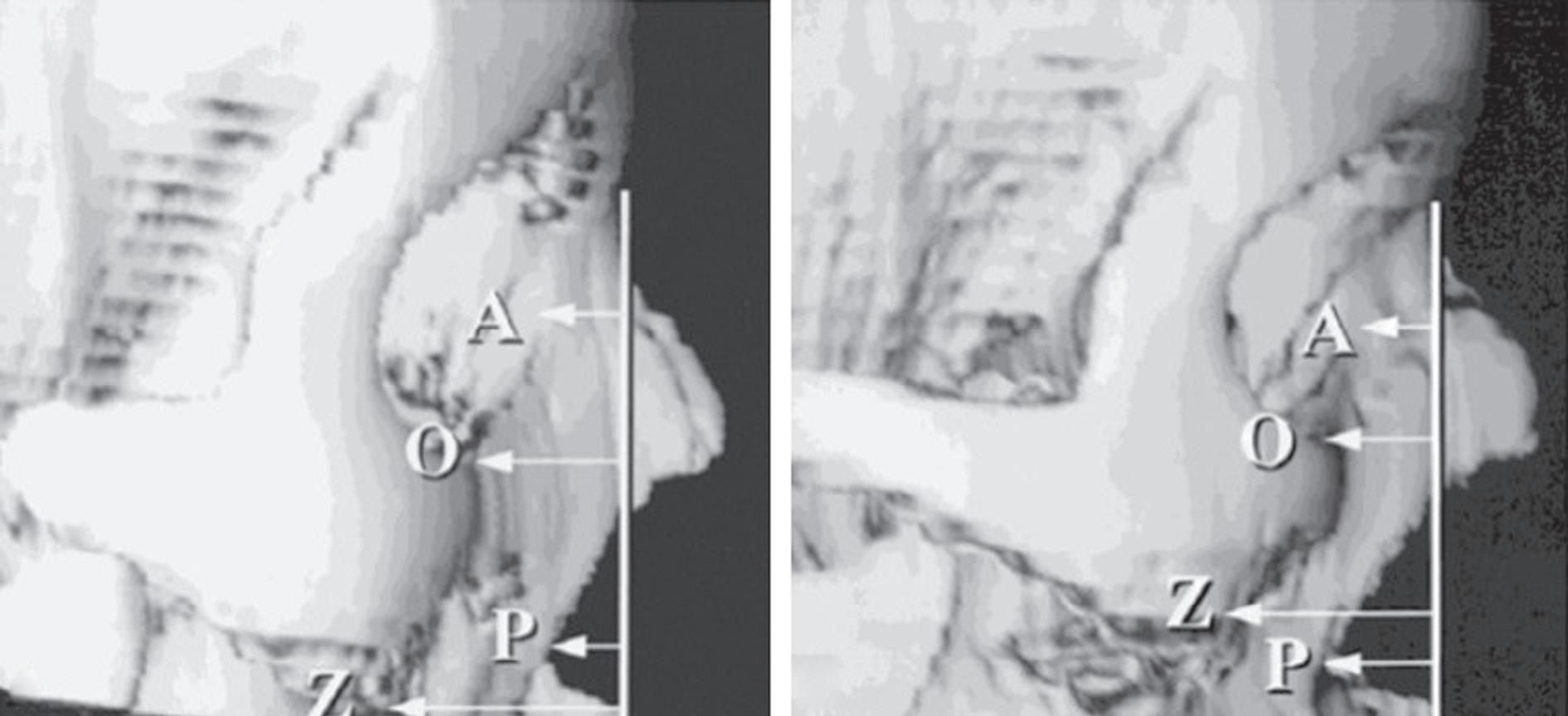
Volume is important in all facial layers from the base presented by the bone and teeth through to muscle, fat, and the superficial layers of the skin. Most volume loss occurs deeply with resorption around and within major facial orifices. The orbits expand, and the piriform aperture (Fig. 3.4)35 widens, allowing posterior displacement of the nasal base with drooping of the nose and deepening of the nasolabial fold.
Dentition volume decreases and the mandible and maxilla volume diminishes. Anterior projection of the periorbital zone, midface, nose, perioral area, and chin all suffer as a result. The decrease in volume of the large muscles of mastication (especially the masseter muscle) and changes in the mandible, particularly shortening in the posterior ramus height and the body length, also contribute to an increasingly obtuse mandibular angle.36 This has significant ramifications for the fading of beauty in older age with the facial shape changing and the support for the lower face waning.
The
of mastication
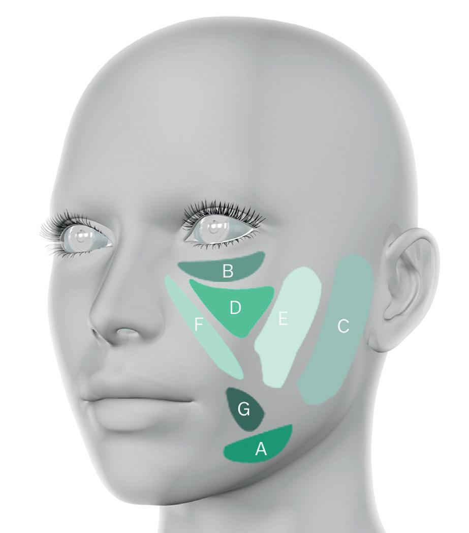
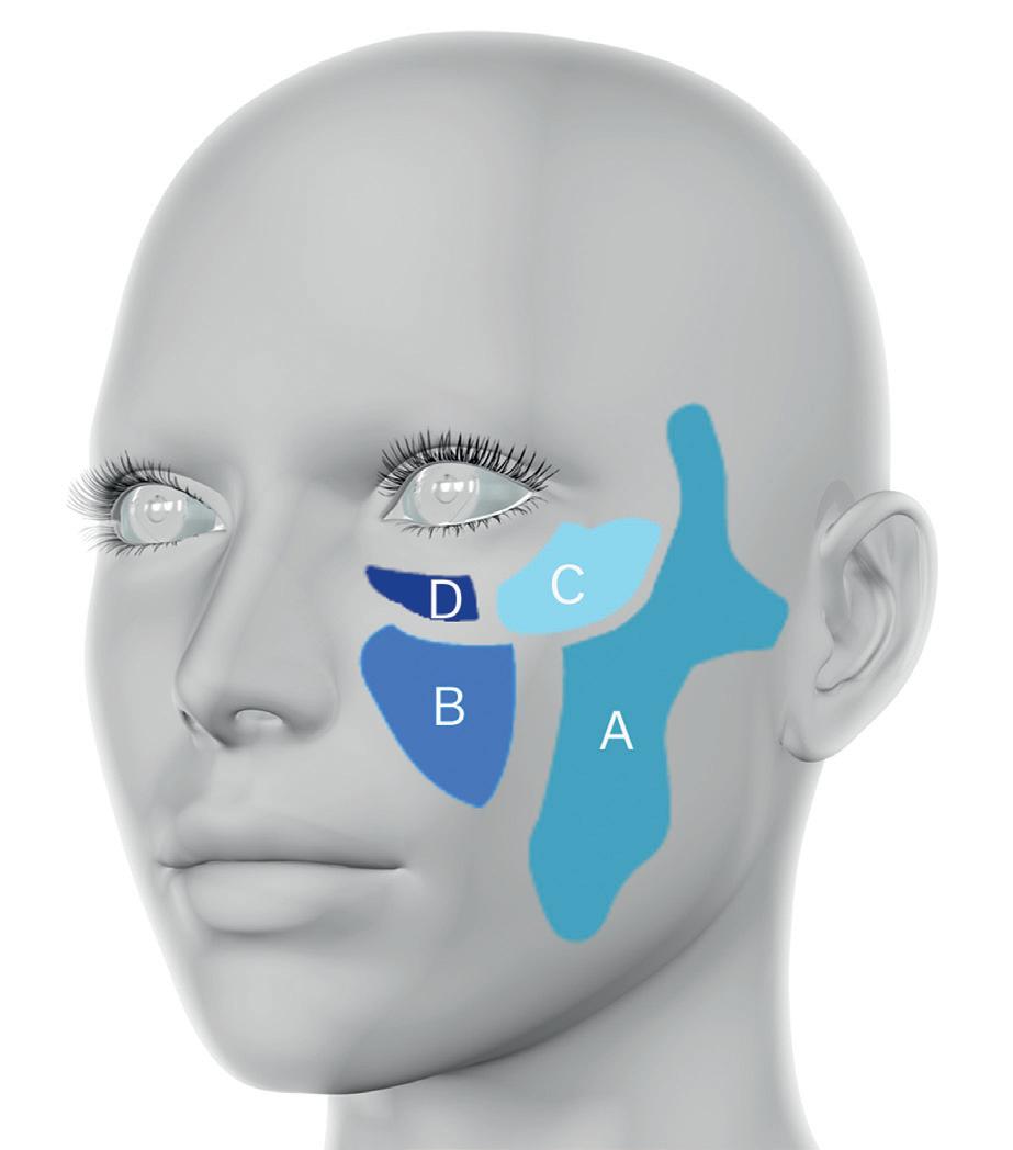
There have been separate facial fat compartments (Fig. 3.5) described in both the superficial and deep layers of the face, each with their defined pockets of fat separated by retaining ligaments.37,38 The major retaining ligaments, such as the mandibular and zygomatic ligaments, do not
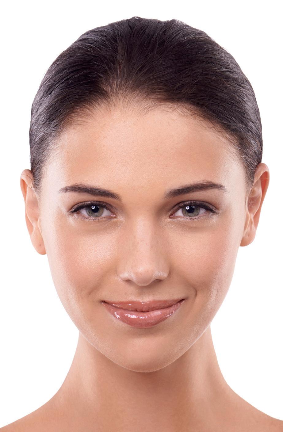
Figure 3.1 Beautiful face.
Figure 3.2 Diagram of ogee curve taking in the eyelid–cheek junction, high cheek bone and the concavity inferior to this.
Pearl 3
Volume is important in many of the indicators of youth, maturity, attractiveness, symmetry, and gender differentiation.
Pearl 4
Bone and dentition support the facial form and structure of overlying muscles and resorb with age periorificially and in the bones supporting mastication.
Pearl 5
muscles
(masseter and temporalis particularly) lose volume in the aging process.
3.4 The areas of the facial skeleton that selectively resorb with aging with the size of the arrows indicating relative tendency for bone loss. From Mendelson B, Wong C. Changes in the facial skeleton with aging: implications and clinical applications in facial rejuvenation. Aesth Plast Surg. 2012;36:4:753–760.
Figure 3.3 Facial shape before (A) and after (B) weight loss, showing a change from round to oval.
Figure
Table 3.1 Superficial and corresponding deep fat compartments of the face. Deep compartments may be more prone to atrophy with age whereas superficial ones tend to hypertrophy
Superficial fat compartments Deep fat compartments
Nasolabial
Deep medial cheek (medial component)
Superficial medial cheek Deep medial cheek (lateral component)
significantly change in length over time. The zones of the face and its proportions are an effortless balance in youth and when optimal features are at work. With age or poorly developed features comes a burden on retaining ligaments as volume shifts or sags, exaggerating the deep folds of the face.
Pearl 6
Most major retaining ligaments induce deep facial grooves as the face cascades forwards and inwards with age.
The facial fat compartments appear to be bilayered, with a superficial and deeper set of compartments (Table 3.1).39 The deeper fat pads allow sliding of mimetic muscles and muscles of mastication, but with aging it appears that the midfacial fat compartments migrate medially with an inferior shift within the individual compartments.40 This has led to the popularization of a deeper injection augmenting the deep system and reinflating the superficial fat pads, allowing better support and projection.41
7
Facial fat compartments appear to have deep and superficial elements with the deeper ones particularly prone to atrophy with aging.
Replenishing atrophic compartments may aid facial rejuvenation.42,43 For example, deflation of the deep periorbital fat pad with age induces a V-shaped concavity between the medial eyelid and cheek area, opening up the inferior orbital hollows, and a more obvious transition
Figure 3.5 Diagram of facial fat compartments. (A) Superficial fat compartments: A, Inferior jowl fat. B, Infraorbital fat. C, Lateral cheek fat. D, Medial cheek fat. E, Middle cheek fat. F, Nasolabial fat. G, Superior jowl fat. (B) Deep fat compartments: A, Buccal fat. B, Deep medial cheek fat. C, Lateral suborbicularis oculi fat. D, Medial suborbicularis oculi fat.
Pearl
between the lid and cheek. Hence deep augmentation of the medial suborbicularis fat pads or deep medial cheek fat pad using a deep supraperiosteal injection may improve this tear trough deformity.
Pearl 8
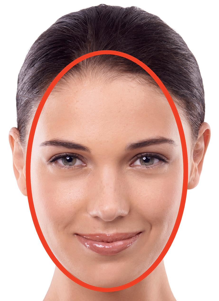
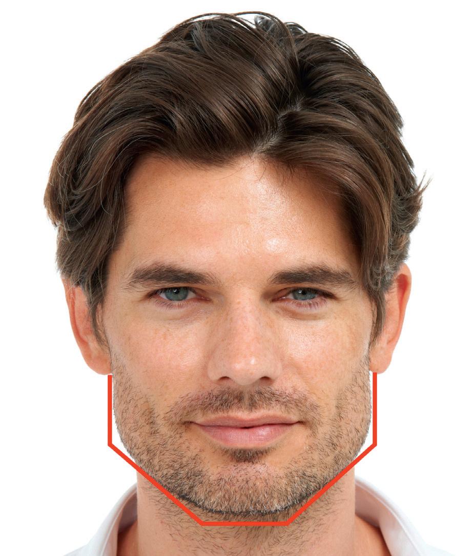
The facial shape seen from the front should be an oval in youth, independent of ethnicity in females, and a sharp angular shape, more base-heavy in males.
Facial shapes
There are many facial shapes which should be pleasing, even at times arresting from any angle; for example, the sweep of the cheek may be best appreciated from an oblique pose and the projected aspects from a lateral one.
We will start from the anteroposterior (AP) view because this is the view we see mostly in the mirror and when in conversation. It should not be forgotten that photographic capture and posing is not usually front-on but various shades of a side-on pose. Looking at the face front-on, volume is important for the outline, shape, symmetry, and width of the features.
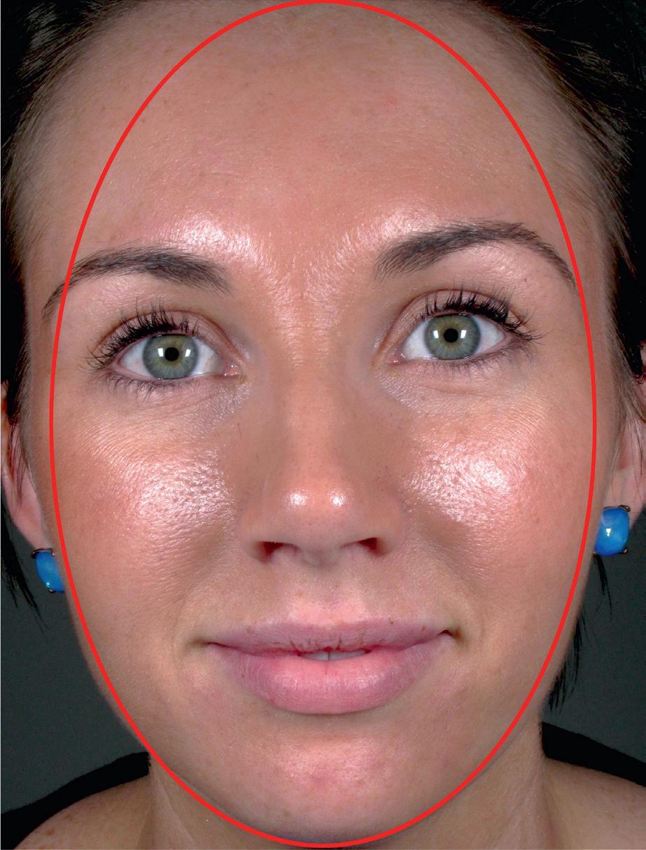
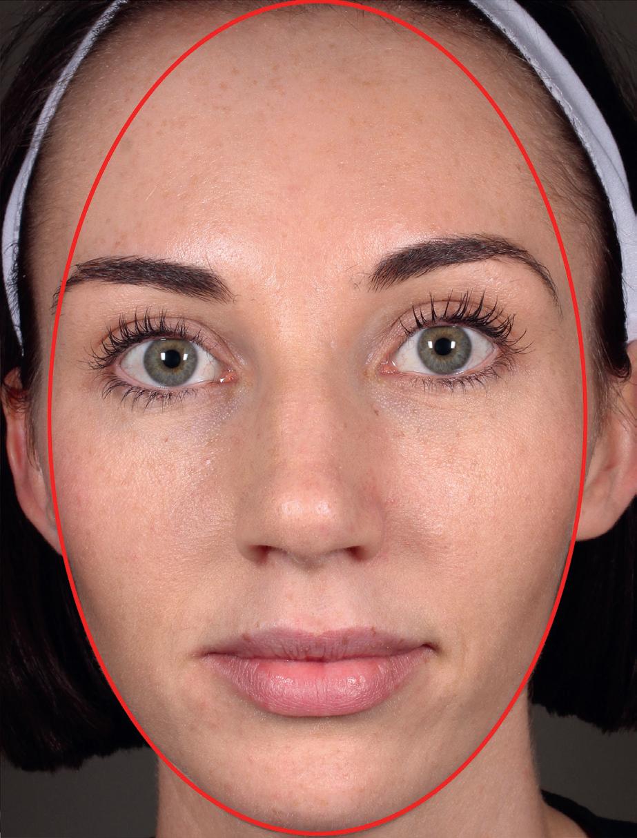
Facial shape is an essential aspect underpinning attractiveness, the appearance of youth, and gender identification. In the female, looking at the face front-on, the facial outline should approximate a smooth oval. This oval is a combination of bony skeleton overlain by soft tissues. The oval line should begin at the forehead and curve rather seamlessly around the outside of the face through the temples, outer cheeks, preauricular area, angle of the jaw, jawline, and all the way to the chin.44 The edge of the features it passes on its way around the face from forehead to chin should sit on the line, neither falling far short of nor projecting past the oval’s edge (Fig. 3.6).
retaining ligaments. However, further weight gain may affect these compartments prone to age-related hypertrophy resulting in facial squaring and plentiful folds, which are not aesthetically ideal.
A square male face is acceptable, even desirable, but this squareness must have certain characteristics.47–50 In the upper face, adequate bi-zigonial distance is required, with the male facial shape most defined in the lower half, requiring strong masseteric volume and jaw angle (Fig. 3.7). What is not desirable is jowls or excess volume posterior to the masseters overlying the parotids.
This facial shape is a very similar beauty consideration across all ethnicities and is a strong determinant of age perception. Variations have been termed heart-shaped or betel leaf, but these may be variations in the upper half of the oval rather than different to the intrinsic oval face. However, not all agree with this stance,45 but all would agree the female facial shape should be wider in the upper half, tapering to a point at the chin, with no angular sharpness. Squaring of the face begins soon after a female’s peak of maturation, around the age of 25, with a more rapid progression into middle age and older years.46 This masculinizes the face, making it less attractive.
In youth a fatter face is acceptable with an evening out of the fat compartments and support provided by
A strong, defined jawline and strong square chin are the masculinizing facial shape and attractiveness requirements.
Symmetry
Symmetry appears to be our visual clue to the outward show of ideal genetics.
Figure 3.6 Facial features sitting on oval outline.
Pearl 9
The oval outline of the attractive female face should pass seamlessly touching on the temples, cheeks, jaw angle, and jawline through to the chin.
Pearl 10
Attractive female faces are much wider in the mid and upper face than the lower face, whereas attractive males tend to possess greater lower face volume.
Although symmetry may refer to any aspect of beauty, such as surface irregularities or facial movement peculiarities, it is the differential volume effects that detract most from the beauty of an individual’s face.
In one study, only 6 of the 21 subjects were symmetrical across their upper face (bi-zigonial distance) and only 4 symmetrical across the lower face (angle of the jaws). Only 3 out of 21 appeared symmetrical across both the upper and lower face. These subjects were taken from some of the objectively most beautiful females in current and past history—film stars, models, and pageant winners.45
Ideal proportions of beauty
We now understand that to have a well-proportioned face is a major step towards beauty. The ancient Greeks believed that all beauty was mathematical, and one of the more robust concepts is the golden ratio. This ratio is a mathematical construct fixed at 1.618 : 1.
In geometry it is a linear relation in which the smaller length is to the larger length as the larger length is to the complete line. It defines ratios we find appealing in nature, architecture, the human body, and faces. This ratio has
been applied to the many aspects making up the attractive face and has helped us to understand what defines facial beauty.
Swift and Remington explored these Phi concepts to illustrate how best to rejuvenate the face in a concept they term “beautiphication.”51
It is odd that a two-dimensional line has been so extensively used to assess facial beauty and correct proportionality, as the proportions really represent a three dimensionality. The vertical height of the upper and lower lips, the relationship of eye width to the width across the malar ridges, and the proportions of the nose and the teeth are measured by this two-dimensional line but represent volume, a three-dimensional concept.
Divisions of the face into thirds horizontally and fifths vertically are other useful constructs for facial assessment and treatment planning. It is most likely that an inbuilt sense of mathematical analysis and concepts is at work in all of us when assessing beauty.
Maturity indicators
Gender selection is a hardwired, innate attraction to a particular gender. It is based on sexual dimorphism, or what makes a person obviously one gender or the other.
The soft sloping ogee curves in females and the strong angles of males are volume indicators of the attainment of sexual maturity associated with estrogen and testosterone, respectively.
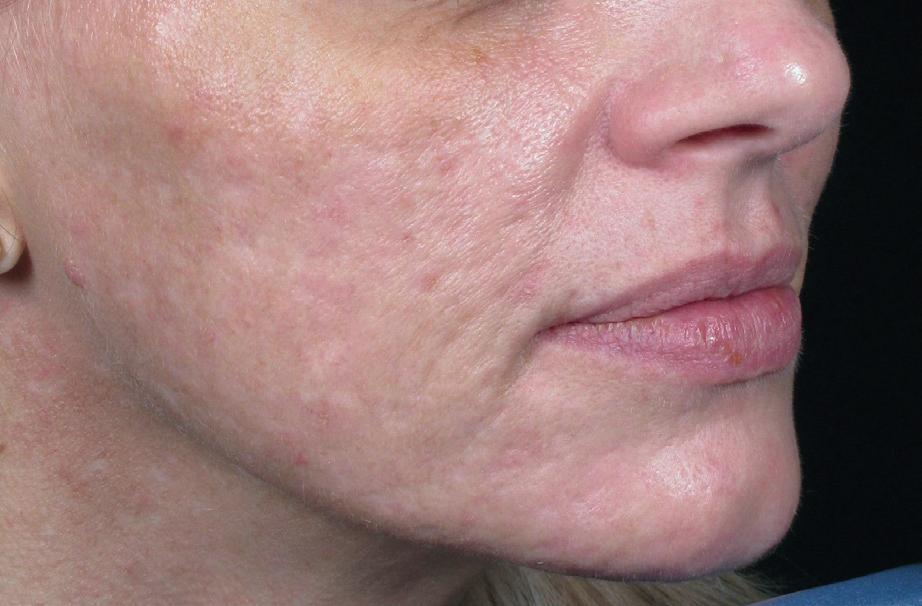
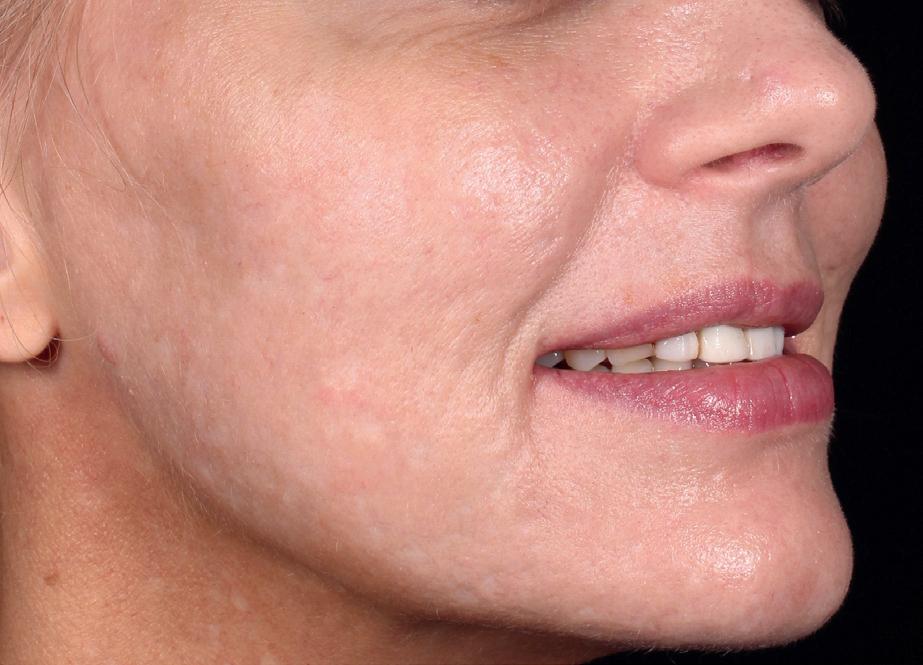
On a more localized level the human face is meant to be predictable, devoid of volume imperfections. One of the most common local volume abnormalities dermatologists and their patients face is atrophic scars. This is so disfiguring to patients that it is a source of depression and suicidal ideation.52 It deeply offends the patient cosmetically and is a common reason for seeking treatment. Volume correction in this instance of atrophic scarring has been a neglected aspect in treatment (Fig. 3.8).53,54
Facial volume, beauty, and ethnicity
Initially researchers believed that different cultures adhere to different styles of beauty, but this may not be correct. Darwin espoused that there is no universal standard of beauty. However, much of the research since the 1970s contradicts this and suggests a more universal standard of beauty.55–57
Does our ideal face shape vary with ethnicity? Is an oval facial shape something females of any ethnicity can and should aspire to? It is probable that a smoothly contoured oval-shaped perimeter to a woman’s face is the cornerstone of universal beauty and that an angular face
Figure 3.7 Strong masseteric volume and jaw angle in a male face.
Pearl 11
Very few people, even beautiful people are symmetrical.
Pearl 12
The oval facial shape transcends ethnicity and is the cornerstone of universal beauty, with differences lying more in an individual’s internal features.
with lower facial prominence, defined jawline, square strong chin, large nose, and heavy horizontal brows are similar cornerstones in men.
So where do the differences lie?
The strength and weaknesses of the ethnicities are more obvious in the internal features of the face—the nose, lips, cheeks, eyes, brows, and chin—and how they project from the face.
These structures are emphasized in Caucasians and Indians but poorly projected in East Asians and Africans. Lip projection varies between ethnicities. Although the upper lip is often quite full in East Asian females, the lower lip tends to be a little less developed. Lips of Caucasians may be more volume-deficient, whereas in West Asians, Africans, and Hispanics the proportions and size more often conform to optimal ratios and size.
A consensus group issued a number of consensus statements with regard to East Asian versus Caucasian aesthetics.58 Quoting from that paper:
• “Beautiful people of all races show similarity in facial characteristics while retaining distinct ethnic features.
• Asians are not a homogeneous group but rather comprise many varied ethnic origins, with each group having its own unique facial characteristics.
• Treatment to achieve esthetic changes in Asians should not be viewed as an attempt at Westernization, but rather the optimization of Asian ethnic features, in the same way that Westerners who receive lip enhancement, lateral malar enhancement, or skin tanning are not trying to ‘Easternize’ their appearance as they attempt to make up for their intrinsic ethnicity-associated structural weaknesses.”
These statements may be extrapolated to describe the universality of beauty, with each racial or ethnic group exhibiting greater strength in certain volume-related characteristics than other groups. Caucasians may have more anterior projection of facial features, the midface, and lower face, whereas East Asian and African populations may have more facial width intrinsically. In each group, benefit can be sought by maximizing good structural characteristics, while improving common structural deficiencies typical of their ethnicity. Truly beautiful people of any ethnicity show the strong points of the group but may be outliers with regard to that group’s deficiencies. So in terms of volume, an oval facial shape in females and a square, angular face in men are truly youthful and attractive in any race.
Conclusion
Returning to the questions posed at the beginning of this chapter, namely:
1. The very existence of beauty.
Why does beauty exist in nature and in human beings? Why do we strive to achieve a goal of increased beauty? Why do people wish to attain it? What is in it for them?
The answers appear to be that beauty is a basic appreciative skill we all have, which may have survival value. The survival value is to keep us safe and our current gene pool safe.
2. The principles upon which we judge facial beauty.
A major principle is the selective placement of volume. It allows recognition of each other and to distinguish age and gender for mate selection. The different distribution between males and females and the volume shifts occurring from childhood to maturity are our indicators of sexual maturity.
Beauty in the guise of optimal facial shape, symmetry, sweeping curves in a young adult female, angles and sharpness in a young male, proportions of the face, and the relationship between the features all rely on volume distribution. Aging distorts the flow of this volume, creating compartmentalization cascading volume shifts caught up by the retaining ligaments of the face. Volume is utterly instrumental in every aspect of our understanding of facial beauty.
Figure 3.8 Acne scarring before (A) and after (B) volume treatment.
References
1. Baig MA. Surgical enhancement of facial beauty and its psychological significance. Ann R Australas Coll Dent Surg. 2004;17:64–67.
2. Smith CU. Evolutionary neurobiology and aesthetics. Perspect Biol Med. 2005;48:17–30.
3. Dutton D. TED talk <http://www.ted.com/talks/denis_ dutton_a_darwinian_theory_of_beauty.html>; 2010. Accessed 29.07.16.
4. Rhodes G. The evolution of facial attractiveness. Annu Rev Psychol. 2006;57:199–226.
5. Little AC, Jones BC, DeBruine LM. Facial attractiveness: evolutionary based research. Philos Trans Roy Soc B. 2011;366:1638–1659.
6. Little AC, Perrett DI. Facial attractiveness. In: Adams RA Jr, Ambady N, Nakayama K, Shimojo S, eds. The Science of Social Vision. Oxford: Oxford University Press; 2011:164–185.
7. Whitehead RD, Ozakinci G, Perrett DI. Attractive skin coloration: harnessing sexual selection to improve diet and health. Evol Psychol. 2012;10:842–854.
8. Fink B, Neave N. The biology of facial beauty. Int J Cosmet Sci. 2005;27:317–325.
9. Penton-Voak IS, Morrison ER. Structure, expression and motion in facial attractiveness. In: Calder AJ, Rhodes G, Johnson MH, Haxby JV, eds. The Oxford Handbook of Face Perception. New York, NY: Oxford University Press; 2011:653–672.
10. Hogan PC. Literary aesthetics: beauty, the brain, and Mrs. Dalloway. Prog Brain Res. 2013;205:319–337.
11. Samson N, Fink B, Matts PJ. Visible skin condition and perception of human facial appearance. Int J Cosmet Sci. 2010;32:167–184.
12. Baig MA. Surgical enhancement of facial beauty and its psychological significance. Ann R Australas Coll Dent Surg. 2004;17:64–67.
13. Swift A, Remington K. BeautiPHIcation™: a global approach to facial beauty. Clin Plast Surg. 2011;38:347–377.
14. Borelli C, Berneburg M. “Beauty lies in the eye of the beholder”? Aspects of beauty and attractiveness. J Dtsch Dermatol Ges. 2010;8:326–330.
15. Patel U, Fitzgerald R. Facial shaping: beyond lines and folds with fillers. Drugs Dermatol. 2010;9(8 suppl ODAC Conf Pt 2):s129–s137.
16. Buggio L, Vercellini P, Somigliana E, Viganò P, Frattaruolo MP, Fedele L. “You are so beautiful”: behind women’s attractiveness towards the biology of reproduction: a narrative review. Gynecol Endocrinol. 2012;28:753–757.
17. Eagly AH, Ashmore RD, Makhijani MG, Longo LC. What is beautiful is good, but…: a meta-analytic review of research on the physical attractiveness stereotype. Psychol Bull. 1991;110:109–128.
18. Dion K, Berscheid E, Walster E. What is beautiful is good. J Pers Soc Psychol. 1972;24:285–290.
19. Yokoyama T, Noguchi Y, Tachibana R, Mukaida S, Kita S. A critical role of holistic processing in face gender perception. Front Hum Neurosci. 2014;8:477(1–10).
20. Zhao M, Hayward WG. Holistic processing underlies gender judgments of faces. Atten Percept Psychophys. 2010;72:591–596.
21. Werheid K, Schacht A, Sommer W. Facial attractiveness modulates early and late event-related brain potentials. Biol Psychol. 2007;76:100–108. Epub 2007 Jul 4.
22. Schacht A, Werheid K, Sommer W. The appraisal of facial beauty is rapid but not mandatory. Cogn Affect Behav Neurosci. 2008;8(2):132–142.
23. Lu Y, Wang J, Wang L, Wang J, Qin J. Neural responses to cartoon facial attractiveness: An event-related potential study. Neurosci Bull. 2014;30:441–450.
24. Chatterjee A, Thomas A, Smith SE, Aguirre GK. The neural response to facial attractiveness. Neuropsychology. 2009;23:135–143.
25. Sui J, Liu CH. Can beauty be ignored? Effects of facial attractiveness on covert attention. Psychon Bull Rev. 2009;16:276–281.
26. Cappelle T, Fink B. Changes in women’s attractiveness perception of masculine men’s dances across the ovulatory cycle: preliminary data. Evol Psychol. 2013;11:965–972.
27. Postma E. A relationship between attractiveness and performance in professional cyclists. Biol Lett. 2014;10:20130966.
28. Golle J, Mast FW, Lobmaier JS. Something to smile about: the interrelationship between attractiveness and emotional expression. Cogn Emot. 2014;28:298–310.
29. Xu Y, Lee A, Wu WL, Liu X, Birkholz P. Human vocal attractiveness as signaled by body size projection. PLoS One. 2013;8:e62397.
30. Zhang H, Teng F, Chan DK, Zhang D. Physical attractiveness, attitudes toward career, and mate preferences among young Chinese women. Evol Psychol. 2014;12:97–114.
31. Fisher ML, Voracek M. The shape of beauty: determinants of female physical attractiveness. J Cosmet Dermatol. 2006;5:190–194.
32. Shetty R. Outer Circle Versus Inner Circle: Special Considerations While Rejuvenating an Indian Face Using Fillers. J Cutan Aesthet Surg. 2015;8:169–172.
33. Little JW. Volumetric perceptions in midfacial aging with altered priorities for rejuvenation. Plast Reconstr Surg. 2000;105:252–266; discussion 286–289.
34. Shetty R. Under eye infraorbital injection technique: the best value in facial rejuvenation. J Cosmet Dermatol. 2014;13:79–84.
35. Mendelson B. Wong C. Changes in the facial skeleton with aging: implications and clinical applications in facial rejuvenation. Aesth Plast Surg. 2012;36:4:753–760.
36. Wong CH, Mendelson B. Newer understanding of specific anatomic targets in the aging face as applied to injectables: aging changes in the craniofacial skeleton and facial ligaments. Plast Reconstr Surg. 2015;136(5 suppl):44S–48S. doi:10.1097/ PRS.0000000000001752.
37. Wan D, Amirlak B, Rohrich R, Davis K. The clinical importance of the fat compartments in midfacial aging. Plast Reconstr Surg Glob Open. 2014;1(9):e92. doi:10.1097/ GOX.0000000000000035 [eCollection 2013].
38. Alghoul M, Codner MA. Retaining ligaments of the face: review of anatomy and clinical applications. Aesthet Surg J. 2013;33(6):769–782.
39. Ramanadham SR, Rohrich RJ. Newer understanding of specific anatomic targets in the aging face as applied to injectables: superficial and deep facial fat compartments—an evolving target for site-specific facial augmentation. Plast Reconstr Surg. 2015;136(5 suppl):49S–55S. doi:10.1097/ PRS.0000000000001730.
40. Gierloff M, Stöhring C, Buder T, Gassling V, Açil Y, Wiltfang J. Aging changes of the midfacial fat compartments: a computed tomographic study. Plast Reconstr Surg. 2012;129:263–273.
41. Wan D, Amirlak B, Rohrich R, Davis K. The clinical importance of the fat compartments in midfacial aging. Plast Reconstr Surg Glob Open. 2013;1(9):e92. doi:10.1097/ GOX.0000000000000035.
42. Rohrich RJ, Ghavami A, Constantine FC, Unger J, Mojallal A. Lift-and-fill face lift: integrating the fat compartments. Plast
43. Fitzgerald R, Rubin AG. Filler placement and the fat compartments. Dermatol Clin. 2014;32(1):37–50. doi:10.1016/j.det.2013.09.007.
44. Goodman GJ. The oval female facial shape—a study in beauty. Dermatol Surg. 2015;41:1374–1382.
45. Kane M. Commentary on the Oval Female Facial Shape—A Study in Beauty. Dermatol Surg. 2015;41:1384–1388.
46. Goodman GJ, Halstead MB, Rogers JD, et al. A software program designed to educate patients on age-related skin changes of facial and exposed extrafacial regions: the results of a validation study. Clin Cosmet Investig Dermatol 2012;5:23–31.
47. Conway CA, Jones BC, DeBruine LM, Little AC. Sexual dimorphism of male face shape, partnership status and the temporal context of relationship sought modulate women’s preferences for direct gaze. Br J Psychol. 2010;101:109–121.
48. Claes P, Walters M, Shriver MD, et al. Sexual dimorphism in multiple aspects of 3D facial symmetry and asymmetry defined by spatially dense geometric morphometrics. J Anat. 2012;221:97–114.
49. Thayer ZM, Dobson SD. Sexual dimorphism in chin shape: implications for adaptive hypotheses. Am J Phys Anthropol. 2010;143:417–425.
50. Glassenberg AN, Feinberg DR, Jones BC, Little AC, Debruine LM. Sex-dimorphic face shape preference in heterosexual and
homosexual men and women. Arch Sex Behav. 2010;39:1289–1296.
51. Swift A, Remington K. BeautiPHIcation™: a global approach to facial beauty. Clin Plast Surg. 2011;38:347–377.
52. Goodman GJ. Acne and acne scarring—the case for active and early intervention. Aust Fam Phys. 2006;35:503–504.
53. Goodman GJ. Treating scars: addressing surface, volume and movement to optimize results: Part 1. Mild grades of scarring. Dermatol Surg. 2012. doi:10.1111/j.1524-4725.2012.02434.x.
54. Goodman GJ. Treating scars: addressing surface, volume and movement to optimize results: part 2. More severe grades of scarring. Dermatol Surg. 2012;doi:10.1111/j.1524-4725 .2012.02439.x.
55. Rhodes G, Yoshikawa S, Clark A, Lee K, McKay R, Akamatsu S. Attractiveness of facial averageness and symmetry in non-western cultures: in search of biologically based standards of beauty. Perception. 2001;3:611–625.
56. Magro AM. Evolutionary-derived anatomical characteristics and universal attractiveness. Percept Mot Skills. 1999;88(1):147–166.
57. Jefferson Y. Facial beauty—establishing a universal standard. Int J Orthod Milwaukee. 2004;15:9–22.
58. Liew S, Wu WT, Chan HH, et al. Consensus on changing trends, attitudes, and concepts of Asian beauty. Aesthetic Plast Surg. 2015, Sep 25 [Epub ahead of print].