All rights reserved. No part of this publication may be reproduced or transmitted in any form or by any means, electronic or mechanical, including photocopying, recording, or any information storage and retrieval system, without permission in writing from the publisher. Details on how to seek permission, further information about the Publisher’s permissions policies, and our arrangements with organizations such as the Copyright Clearance Center and the Copyright Licensing Agency can be found at our website: www.elsevier. com/permissions
This book and the individual contributions contained in it are protected under copyright by the Publisher (other than as may be noted herein).
Notices
Knowledge and best practice in this field are constantly changing. As new research and experience broaden our understanding, changes in research methods, professional practices, or medical treatment may become necessary.
Practitioners and researchers must always rely on their own experience and knowledge in evaluating and using any information, methods, compounds, or experiments described herein. In using such information or methods they should be mindful of their own safety and the safety of others, including parties for whom they have a professional responsibility.
With respect to any drug or pharmaceutical products identified, readers are advised to check the most current information provided (i) on procedures featured or (ii) by the manufacturer of each product to be administered, to verify the recommended dose or formula, the method and duration of administration, and contraindications. It is the responsibility of practitioners, relying on their own experience and knowledge of their patients, to make diagnoses, to determine dosages and the best treatment for each individual patient, and to take all appropriate safety precautions.
To the fullest extent of the law, neither the Publisher nor the authors, contributors, or editors assume any liability for any injury and/or damage to persons or property as a matter of products liability, negligence or otherwise, or from any use or operation of any methods, products, instructions, or ideas contained in the material herein.
Library of Congress Cataloging-in-Publication Data
Names: Davis, Peter J. (Anesthesiologist), editor. | Cladis, Franklyn P., editor.
Title: Smith’s anesthesia for infants and children / [edited by] Peter J. Davis, Franklyn P. Cladis.
Other titles: Anesthesia for infants and children
Description: Edition 9. | St. Louis, Missouri : Elsevier, [2017] | Includes bibliographical references and index.
Identifiers: LCCN 2016032263 | ISBN 9780323341257 (hardcover : alk. paper)
Subjects: | MESH: Anesthesia | Infant | Child
Classification: LCC RD139 | NLM WO 440 | DDC 617.9/6083–dc23 LC record available at https://lccn.loc.gov/2016032263
Executive Content Strategist: Dolores Meloni
Senior Content Development Specialist: Margaret Nelson
Publishing Services Manager: Julie Eddy
Book Production Specialist: Clay S. Broeker
Design Direction: Patrick Ferguson
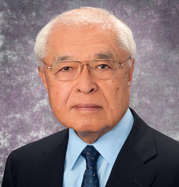
This ninth edition of Smith’s Anesthesia for Infants and Children is dedicated to Dr. Etsuro K. Motoyama, an academician, researcher, clinician, and mentor. Dr. Motoyama graduated from Chiba University School of Medicine in Japan and completed his anesthesia residency at the Graduate Hospital of the University of Pennsylvania. Dr. Motoyama was a fellow in pediatric anesthesia at the Boston Children’s Hospital where he was mentored by Dr. Smith and eventually became his protégé. In addition to clinical training, Dr. Motoyama was also a research fellow in respiratory physiology, studying with Dr. Charles D. Cook at the Harvard Medical School.
Dr. Motoyama’s success and academic advancements have been at the Yale School of Medicine and the University of Pittsburgh School of Medicine. He has also received adjunct professor appointments in Japan at the Keio University School of Medicine, Kobe University School of Medicine, and the National Center for Child Health and Development in Tokyo. Among his accomplishments, Dr. Motoyama has helped to pioneer the fields of both pediatric anesthesia and pediatric pulmonology. His basic science research, supported by multiple National Institutes of Health research grants, has been on pulmonary surfactant and bronchopulmonary dysplasia. His clinical research areas have involved (1) the effects of medical preoperative stabilization on outcomes of neonates with congenital diaphragmatic hernia, (2) the differential sensitivity of halothane on airway and thoracic respiratory muscles as a cause of airway obstruction in infants, (3) the effects of anesthesia on FRC, atelectasis, and PEEP, and (4) the longitudinal pulmonary function changes in patients with bronchopulmonary dysplasia and congenital diaphragmatic hernia and in children with early onset scoliosis undergoing repetitive VEPTR thoracoplasties. The ability to perform these pulmonary function studies was made possible by Dr. Motoyama’s creation of a specialized device that allowed the patients to be studied while under general anesthesia. His basic science and clinical research contributions have shaped the landscape of pediatric anesthesia, pediatric medicine, and pediatric surgery.
Though currently a Professor Emeritus at the University of Pittsburgh, Dr. Motoyama is still actively engaged in improving the lives of children by providing anesthesia services to pediatric patients in underdeveloped countries. The legacy and spirit of Smith’s Anesthesia for Infants and Children was maintained when Dr. Smith passed on the responsibilities of the book’s editorship to Dr. Motoyama, who edited and co-edited the book’s fifth through eighth editions. Though Dr. Motoyama has stepped down from the editorial role in the present edition, the legacies of Drs. Smith and Motoyama continue in the current edition of the book. Through his scientific contributions to the field of pediatric anesthesia as well as through his continued clinical involvement, Dr. Motoyama continues to positively impact the lives of children throughout the world.
CONTRIBUTORS
Ann G. Bailey, MD
Professor of Anesthesiology and Pediatrics University of North Carolina Chapel Hill, North Carolina
Jeffrey R. Balzer, PhD
Associate Professor of Neurological Surgery, Neuroscience, and Acute and Tertiary Care Nursing Director, Clinical Operations
Center for Clinical Neurophysiology Director
Cerebral Blood Flow Laboratory University of Pittsburgh Medical Center Pittsburgh, Pennsylvania
Victor C. Baum, MD
United States Food and Drug Administration
Silver Spring, Maryland
Adjunct Professor of Anesthesiology and Critical Care Medicine and of Pediatrics
George Washington University Washington, DC
David S. Beebe, MD
Professor Department of Anesthesiology University of Minnesota Minneapolis, Minnesota
Sue R. Beers, PhD
Professor Department of Psychiatry University of Pittsburgh School of Medicine Pittsburgh, Pennsylvania
Kumar G. Belani, MBBS, MS
Professor and Division Chief, Pediatric Anesthesiology Department of Anesthesiology University of Minnesota Minneapolis, Minnesota
Bruno Bissonette, MD, FRCPC Professor Emeritus of Anesthesia University of Toronto Founder and President
Children of the World Anesthesia Foundation Rimouski, Quebec, Canada
Brian Blasiole, MD, PhD
Assistant Professor Department of Anesthesiology
Children’s Hospital of Pittsburgh of UPMC Pittsburgh, Pennsylvania
Adrian T. Bosenberg, MBChB FFA(SA) Professor
Department of Anesthesiology and Pain Management University of Washington
Pediatric Anesthesiologist Department of Anesthesiology and Pain Management
Seattle Children’s Hospital Seattle, Washington
Barbara W. Brandom, MD
Professor of Anesthesiology (Retired) University of Pittsburgh Co-Director
North American Malignant Hyperthermia Registry
Malignant Hyperthermia Association of the United States Pittsburgh, Pennsylvania
Claire M. Brett, MD, FAAP Professor
Departments of Anesthesia and Perioperative Care and of Pediatrics University of California, San Francisco San Francisco, California
James G. Cain, MBA, MD
Director of Perioperative Medicine, Transplant, and Trauma Anesthesiology
Department of Pediatric Anesthesiology
Children’s Hospital of Pittsburgh of UPMC Pittsburgh, Pennsylvania
Thomas M. Chalifoux, MD
Assistant Professor of Anesthesiology University of Pittsburgh School of Medicine Department of Anesthesiology
Children’s Hospital of Pittsburgh of UPMC Magee-Women’s Hospital of UPMC Pittsburgh, Pennsylvania
Franklyn P. Cladis, MD
Associate Professor
Department of Anesthesiology
University of Pittsburgh School of Medicine
Children’s Hospital of Pittsburgh of UPMC Pittsburgh, Pennsylvania
David E. Cohen, MD
Associate Professor, Anesthesiology and Critical Care Medicine and Pediatrics
The Children’s Hospital of Philadelphia Perelman School of Medicine at the University of Pennsylvania
Perioperative Medical Director
The Children’s Hospital of Philadelphia Philadelphia, Pennsylvania
Ira T. Cohen, MD
Professor, Anesthesiology
Children’s National Medical Center Washington, DC
Joseph P. Cravero, MD
Senior Associate in Anesthesiology and Pain Medicine
Associate Professor of Anesthesiology Department of Anesthesiology, Perioperative, and Pain Medicine
Boston Children’s Hospital Boston, Massachusetts
Nicholas M. Dalesio, MD
Assistant Professor
Departments of Anesthesiology and Critical Care Medicine and of Otolaryngology and Head and Neck Surgery
Johns Hopkins School of Medicine
Baltimore, Maryland
Andrew Davidson, MBBS, MD, FANZCA Staff Anaesthetist
Department of Anaesthesia and Pain Management Director of Clinical Research
Royal Children’s Hospital
Associate Professor Department of Paediatrics
University of Melbourne Melbourne, Victoria, Australia
Jessica Davis, BA, JD, LLM
Senior Professional Responsibility Attorney Pepper Hamilton, LLP Philadelphia, Pennsylvania
Peter J. Davis, MD, FAAP Professor
Departments of Anesthesiology and Pediatrics
Dr Joseph H. Marcy Endowed Chair in Pediatric Anesthesia
University of Pittsburgh School of Medicine
Anesthesiologist-in-Chief
Children’s Hospital of Pittsburgh of UPMC Pittsburgh, Pennsylvania
Duncan G. de Souza, MD, FRCPC
Clinical Assistant Professor
Department of Anesthesiology
University of British Columbia
Vancouver, British Columbia, Canada
Director, Cardiac Anesthesia
Kelowna General Hospital
Kelowna, British Columbia, Canada
Nina Deutsch, MD
Associate Professor, Anesthesiology and Pediatrics
Department of Anesthesiology, Pain, and Perioperative Medicine
Children’s National Medical Center Washington, DC
Laura K. Diaz, MD
Assistant Professor of Clinical Anesthesiology and Critical Care Medical Director, General Operating Room Cardiac Resources
The Children’s Hospital of Philadelphia Department of Anesthesiology and Critical Care Medicine Philadelphia, Pennsylvania
James A. DiNardo, MD, FAAP
Professor of Anaesthesia
Harvard Medical School Chief
Division of Cardiac Anesthesia
Francis X. McGowan, Jr. MD Chair in Cardiac Anesthesia
Boston Children’s Hospital Boston, Massachusetts
Peter F. Ehrlich, MD, MSC
Associate Professor of Pediatric Surgery
Department of Surgery
University of Michigan CS Mott Children’s Hospital
Ann Arbor, Michigan
Demetrius Ellis, MD
Professor, Nephrology and Pediatrics University of Pittsburgh School of Medicine
Children’s Hospital of Pittsburgh of UPMC Pittsburgh, Pennsylvania
James J. Fehr, MD
Professor of Anesthesiology and Pediatrics
Washington University St. Louis, Missouri
Jeffrey M. Feldman, MD, MSE
Division Chief, General Anesthesia
The Children’s Hospital of Philadelphia Professor, Clinical Anesthesiology and Critical Care
Perelman School of Medicine at the University of Pennsylvania Philadelphia, Pennsylvania
Kathryn Felmet, MD
Assistant Professor, Critical Care Medicine and Pediatrics
Children’s Hospital of Pittsburgh of UPMC Pittsburgh, Pennsylvania
Jonathan D. Finder, MD
Professor of Pediatrics
University of Pittsburgh School of Medicine
Division of Pulmonology, Department of Pediatrics
Children’s Hospital of Pittsburgh of UPMC Pittsburgh, Pennsylvania
Sean Flack, MBChB DA FCA
Associate Professor, Anesthesiology and Pain Medicine
Director, Regional Anesthesia Division University of Washington
Seattle Children’s Hospital Seattle, Washington
Randall P. Flick, MD Consultant
Department of Anesthesiology
Associate Professor of Anesthesiology College of Medicine
Mayo Clinic Rochester, Minnesota
Michelle A. Fortier, PhD Assistant Professor
Department of Anesthesiology and Perioperative Care
School of Medicine
University of California, Irvine Orange, California
Geoff Frawley, MBBS,FANZCA
Anaesthetist
Department of Paediatric Anaesthesia
Royal Children’s Hospital
Clinical Associate Professor
Department of Paediatrics
Melbourne University Melbourne, Victoria, Australia
Samir K. Gadepalli, MD, MBA Clinical Lecturer
Co-Director of Pediatric Surgical Critical Care
University of Michigan CS Mott Children’s Hospital Ann Arbor, Michigan
Jeffrey L. Galinkin, MD
Professor of Anesthesiology and Pediatrics University of Colorado, Anschutz Medical Campus
Director of Scientific and Medical Affairs
CPC Clinical Research Aurora, Colorado
Nancy Glass, MD
Director, Chronic and Palliative Pain Service
Texas Children’s Hospital
Professor, Pediatrics and Anesthesiology
Baylor College of Medicine
Houston, Texas
Salvatore R. Goodwin, MD
Offiice of VP-Quality and Safety
Chair Professional Performance and Quality Committee-Nemours
Associate Professor, Anesthesiology
Mayo Medical School
Jacksonville, Florida
George A. Gregory, MD
Professor Emeritus of Anesthesia and Perioperative Care and of Pediatrics
University of California, San Francisco
San Francisco, California
Lorelei Grunwaldt, MD Director
Vascular Anomalies Center and Brachial Plexus Clinic
Associate Professor of Surgery
Children’s Hospital of Pittsburgh of UPMC Pittsburgh, Pennsylvania
Padma Gulur, MD
Director, Pain Management Services
Department of Anesthesiology and Perioperative Care
University of California, Irvine
Division of Pain Management Irvine, California
Nina A. Guzzetta, MD, FAAP
Associate Professor of Anesthesiology and Pediatrics
Emory University School of Medicine
Children’s Healthcare of Atlanta Atlanta, Georgia
Dawit T. Haile, MD Consultant
Department of Anesthesiology
Assistant Professor in Anesthesiology College of Medicine
Mayo Clinic
Rochester, Minnesota
Denise M. Hall-Burton, MD, FAAP
Assistant Professor of Anesthesiology
Department of Anesthesia
Children’s Hospital of Pittsburgh of UPMC Pittsburgh, Pennsylvania
Gregory B. Hammer, MD
Professor of Anesthesia and Pediatrics
Stanford University School of Medicine
Stanford, California
Director of Research
Department of Anesthesia Lucile Packard Children’s Hospital Palo Alto, California
Jennifer L. Hamrick, MD
Assistant Professor
Division of Pediatric Anesthesia and Pain Medicine
University of Arkansas for Medical Sciences Little Rock, Arkansas
Justin T. Hamrick, MD
Assistant Professor
Division of Pediatric Anesthesia and Pain Medicine
University of Arkansas for Medical Sciences Little Rock, Arkansas
Daniel M. Hayward, MD
Department of Anesthesiology and Critical Care Medicine
Johns Hopkins School of Medicine Baltimore, Maryland
Eugenie S. Heitmiller, MD
Professor
Department of Anesthesiology and Critical Care Medicine
Johns Hopkins University School of Medicine
Baltimore, Maryland
Andrew Herlich, DMD, MD, FAAP
Professor and Special Assistant to the Chair for Academic and Faculty Affairs
Department of Anesthesiology
University of Pittsburgh School of Medicine
Attending Physician
Department of Anesthesiology
UPMC Mercy Pittsburgh, Pennsylvania
Robert S. Holzman, MD, MA (Hon), FAAP
Senior Associate in Perioperative Anesthesia Boston Children’s Hospital Professor of Anaesthesia
Harvard Medical School Boston, Massachusetts
Vincent C. Hsieh, MD, MS
Assistant Professor of Anesthesiology and Pain Medicine
University of Washington and Seattle Children’s Hospital Seattle, Washington
Elizabeth A. Hunt, MPH, PhD, MD
Assistant Professor
Department of Anesthesiology and Critical Care Medicine
The Johns Hopkins University School of Medicine
Drs. David S. and Marilyn M. Zamierowski Director
The Johns Hopkins Medicine Simulation Center Baltimore, Maryland
James W. Ibinson, MD, PhD
Assistant Professor Center for Pain Research
Department of Anesthesiology University of Pittsburgh Pittsburgh, Pennsylvania
Lori T. Justice, MD, FAAP
Clinical Staff Pediatric Anesthesiologist Children’s Anesthesiologists, PC East Tennessee Children’s Hospital Knoxville, Tennessee
Zeev N. Kain, MD, MBA
Professor, Anesthesiology and Pediatrics and Psychiatry and Human Behavior Chair
Department of Anesthesiology and Perioperative Care
Associate Dean of Clinical Operations School of Medicine
University of California, Irvine Orange, California
Evan Kharasch, MD, PhD
Vice Chancellor for Research
Russell D. and Mary B. Shelden Professor of Anesthesiology
Director, Division of Clinical and Translational Research
Department of Anesthesiology
Professor of Biochemistry and Molecular Biophysics
Washington University in St. Louis St. Louis, Missouri
Rahul Koka, MD, MPH
Assistant Professor
Department of Anesthesiology and Critical Care Medicine
Johns Hopkins University School of Medicine
Baltimore, Maryland
Sabine Kost-Byerly, MD
Associate Professor and Director of Pediatric Pain Management
Department of Anesthesiology and Critical Care Medicine
The Johns Hopkins University School of Medicine
Baltimore, Maryland
Elliot J. Krane, MD
Professor of Anesthesiology, Perioperative, and Pain Medicine (Pediatric Anesthesia)
Stanford University School of Medicine
Stanford, California
Professor of Pediatrics
Lucile Salter Packard Children’s Hospital at Stanford Palo Alto, California
Barry D. Kussman, MBBCh, FFA (SA), FAAP
Associate Professor of Anaesthesia
Harvard Medical School
Senior Associate in Cardiac Anesthesia
Department of Anesthesiology
Perioperative, and Pain Medicine
Boston Children’s Hospital Boston, Massachusetts
Robert Scott Lang, MD
Clinical Assistant Professor
Department of Anesthesiology
Children’s Hospital of Pittsburgh of UPMC Pittsburgh, Pennsylvania
Helen Victoria Lauro, MD, MPH, MSEd, FAAP
Clinical Associate Professor of Anesthesiology
State University of New York (SUNY) Downstate Medical Center Brooklyn, New York
Jennifer K. Lee, MD
Associate Professor of Anesthesiology and Critical Care Medicine
Associate Professor of Pediatrics
Johns Hopkins School of Medicine Baltimore, Maryland
Joseph Losee, MD
Ross H. Musgrave Professor of Pediatric Plastic Surgery
Department of Plastic Surgery
University of Pittsburgh Medical Center Chief
Division of Pediatric Plastic Surgery
Children’s Hospital of Pittsburgh Pittsburgh, Pennsylvania
Igor Luginbuehl, MD
Associate Professor
University of Toronto
Department of Anesthesia and Pain Medicine
The Hospital for Sick Children Toronto, Ontario, Canada
Mohamed Mahmoud, MD
Associate Professor
Department of Anesthesia Cincinnati Children Medical Center University of Cincinnati Cincinnati, Ohio
Brian Martin, DMD, MHCDS
Medical Director—Clinical Excellence Department of Medical Affairs
Division Chief, Pediatric Dentistry
Children’s Hospital of Pittsburgh of UPMC Pittsburgh, Pennsylvania
Keira P. Mason, MD
Senior Associate in Perioperative Anesthesia Department of Anesthesia
Boston Children’s Hospital
Associate Professor of Anaesthesia (Radiology)
Department of Anaesthesia Harvard Medical School Boston, Massachusetts
William J. Mauermann, MD
Consultant
Department of Anesthesiology
Associate Professor of Anesthesiology College of Medicine
Mayo Clinic Rochester, Minnesota
Lynne G. Maxwell, MD
Senior Anesthesiologist
Department of Anesthesiology and Critical Care Medicine
Children’s Hospital of Philadelphia Associate Professor
Department of Anesthesiology and Critical Care
Perelman School of Medicine at University of Pennsylvania Philadelphia, Pennsylvania
Francis X. McGowan Jr., MD, FAAP Professor of Anesthesiology and Critical Care
The Children’s Hospital of Philadelphia Philadelphia, Pennsylvania
Bruce E. Miller, MD
Associate Professor of Anesthesiology and Pediatrics
Emory University School of Medicine
Children’s Healthcare of Atlanta Atlanta, Georgia
Constance L. Monitto, MD
Assistant Professor
Department of Anesthesiology and Critical Care Medicine
The Johns Hopkins University School of Medicine Baltimore, Maryland
Philip G. Morgan, MD
Professor of Anesthesiology and Pain Medicine
University of Washington and Seattle Children’s Hospital Seattle, Washington
Michael L. Moritz, MD
Clinical Director, Pediatric Nephrology
Medical Director, Pediatric Dialysis
Professor of Pediatrics
University of Pittsburgh School of Medicine
Division of Pediatric Nephrology Pittsburgh, Pennsylvania
Etsuro K. Motoyama, MD, FAAP Professor Emeritus
Departments of Anesthesiology and Pediatrics (Pulmonology)
University of Pittsburgh School of Medicine
Former Director, Pediatric Pulmonology Laboratory
Children’s Hospital of Pittsburgh of UPMC Pittsburgh, Pennsylvania
Michael E. Nemergut, MD, PhD Consultant
Department of Anesthesiology
Assistant Professor of Anesthesiology College of Medicine
Professor Department of Anesthesiology Duke University Durham, North Carolina
Ari Y. Weintraub, MD
Attending Anesthesiologist
Department of Anesthesiology and Critical Care Medicine
Children’s Hospital of Philadelphia Assistant Professor of Clinical Anesthesiology and Critical Care Department of Anesthesiology and Critical Care
Perelman School of Medicine at the University of Pennsylvania Philadelphia, Pennsylvania
Timothy P. Welch, MD, MSPH
Assistant Professor of Anesthesiology and Pediatrics
Washington University St. Louis, Missouri
Robert K. Williams, MD
Professor of Anesthesia and Pediatrics
The University of Vermont Burlington, Vermont
Eric P. Wittkugel, MD, FAAP
Associate Professor of Anesthesia and Pediatrics University of Cincinnati College of Medicine
Cincinnati Children’s Hospital Medical Center Cincinnati, Ohio
Susan Woelfel, MD
Associate Professor of Anesthesiology University of Pittsburgh School of Medicine
Children’s Hospital of Pittsburgh of UPMC Pittsburgh, Pennsylvania
Myron Yaster, MD
Richard J. Traystman Professor Departments of Anesthesiology and Critical Care Medicine, Pediatrics, and Neurosurgery
The Johns Hopkins University School of Medicine
Baltimore, Maryland
Koichi Yuki, MD
Assistant Professor of Anaesthesia Department of Anesthesiology, Perioperative, and Pain Medicine
Harvard Medical School
Boston, Massachusetts
Division of Cardiac Anesthesia
Boston Children’s Hospital Boston, Massachusetts
Steven Zgleszewski, MD, FAAP
Assistant Professor of Anaesthesia
Harvard Medical School
Boston, Massachusetts
Basil J. Zitelli, MD
Edmund R. McCluskey Professor of Pediatric Medical Education
University of Pittsbugh School of Medicine
Department of Pediatrics
Children’s Hospital of Pittsburgh of UPMC Pittsburgh, Pennsylvania
Aaron L. Zuckerberg, MD Director
Children’s Diagnostic Center
North American Partners of Anesthesia Departments of Anesthesiology and Pediatrics
Sinai Hospital of Baltimore Baltimore, Maryland
PREFACE
Dr. Robert Smith, a distinguished pioneer in pediatric anesthesia and a great teacher and clinician, wrote the first edition of this book in 1959, a book subsequently referred to as “the bible” of pediatric anesthesia. The forward to the first edition was written by the famous pediatric surgeon Robert E Gross, the William E. Ladd professor of Children’s Surgery at the Harvard Medical School. Though his words in the forward were written over 50 years ago, at a time when the specialty of pediatric anesthesia and surgery was in its infant stages, his words and ideas are still poignant and insightful today.
During the past decade surgery has made important strides in providing safer and improved methods for handling various problems in infancy and childhood, indeed now making it possible to correct some conditions which were previously thought to be entirely hopeless. Many factors have contributed to these dramatic advances in pediatric surgery. Outstanding among them is the work of anesthesiologists who have focused on the field and have provided well standardized procedures for carrying small and critically ill patients through operations on literally all portions and every system of the body. The surgeon realizes that his chances for success or failure are determined in great measure by the capabilities of the person at the head of the table who is administering the anesthetic.
In some medical circles, there seems to be an attitude that the surgical operator is managing the show; in others, the anesthetist has an overly possessive feeling toward the patient. Neither approach is proper. It is best for each to be cognizant of his own problems and also to know of the other’s difficulties; both must work together for total care of the patient. Certainly this is the most pleasant way to work, and surely it is the most effective way to conduct a child through a surgical ordeal.
Since the initial printing of this textbook in 1959, the book has been markedly transformed in its content and in its appearance. The book has gone from mainly a single- to a multi-author book and from a 400-page 7” by 10” book to a 1400-page 11.5” by 8.5” text. As learning styles have changed, so has the format of this book. The book uses multimedia presentations to supplement, emphasize, and reinforce concepts of pediatric anesthesia. However, even with the increases in page number, new information, and media platforms, the basic tenets of anesthesia care and patient compassion, the legacy and tradition of the of the eight previous editions, have been retained.
The ninth edition has been prepared with the same considerations as the previous editions: to give anesthesiology care providers comprehensive coverage of physiology, pharmacology, and clinical anesthetic management of infants and children of all ages. The ninth edition has been reorganized into six main sections. Part I, Basic Physiology and Principles, contains updated chapters on behavioral development and respiratory, cardiovascular, renal, and thermal physiology. The pharmacology chapter in the previous editions has been expanded into its own section. This new Part II, Pharmacology, now has additional authors and specific chapters in developmental pharmacology, intravenous anesthetic agents, inhaled anesthetic agents, opioids, local anesthetic agents, neuromuscular blocking agents, and anesthetic adjuncts. Part III, General Approach, addresses the basic concepts of caring for children as well as the principles involved in the administration of anesthetics to children. The chapters have all been updated. The chapter on regional anesthesia has new authorship and, with the advent of ultrasound guidance and increased popularity in the use of regional anesthesia in infants and children, the reader will be able to access video demonstrations of specific regional anesthetic techniques in children. Part IV, Clinical Management of Specialized Surgical Problems, contains new material written by new authors. The previous edition chapter of Anesthesia for General Abdominal, Thoracic, Urological, and Bariatric Surgery has now been divided into separate chapters to better organize the material. New chapters on sedation and anesthesia for surgical missions have been added. The use of video has been maintained to further supplement the clinical material. The chapter on Neonatology for the Anesthesiologist has been revised into a comprehensive work that updates the anesthesia provider with perinatal outcome data as well as serves as a primer for pediatric anesthesiologists to better understand the pathophysiology of prematurity and the developmental physiology that occur with neonatal growth. This chapter also serves as a rich resource for the chapters on Anesthesia for Fetal Surgery and Anesthesia for General Surgery in Neonates.
In view of the significant number of disorders that pediatric anesthesiologists are confronted with in the everyday care of their patients, Part V, Systemic Disorders and Associated Problems, was created to better organize and provide information for both unusual patient diseases and to address everyday common perioperative anesthetic concerns. Three chapters on obesity, uncommon diseases, and dermatology for the anesthesiologist are new additions to the book. The chapter on dermatology has an extensive number of figures (both in the book and online) of lesions and rashes that anesthesiologists frequently encounter. Part VI, Critical Care in Pediatric Anesthesia, contains revised chapters on critical care medicine, cardiac intensive care, medicolegal and ethical issues, history
of pediatric anesthesia, and cardiopulmonary resuscitation. The CPR chapter contains the latest (2015) recommendations from the American Heart Association. Part VI also includes new chapters on statistics, safety and patient outcomes, and cardiac intensive care.
In keeping with advancements in technology, this edition is in color, and text material is further supplemented by a website. Videos of airway techniques, single-lung isolation, regional anesthesia, the use of ultrasound, and anatomic dissections of congenital heart lesions are accessible with just a click of the mouse. In addition, supplemental materials on organ transplantation, airway lesions, and pediatric syndromes remain available.
The appendices, which can be found online at ExpertConsult.com, include an updated list of drugs and their dosages, normal growth curves, normal values for pulmonary function tests in children, and an expanded list of common and uncommon syndromes of clinical importance for pediatric anesthesiologists.
Finally, this edition, like the last edition, also includes online multiple-choice questions with answers and explanations. As with any learning process, it is important for the reader to have some method to affirm that they understood the salient features and to reinforce the learning process. Most chapters have associated questions to aid the reader in understanding of the material
In summary, considerable developments and progress in the practice of pediatric anesthesia are reflected in this new edition. The emphasis on the safety and wellbeing of young patients during the perianesthetic period remains unchanged—just as Dr Smith would have wanted.
Peter J. Davis, MD, FAAP
Franklyn P. Cladis, MD
ACKNOWLEDGMENTS
The project of revising a classic medical textbook presents many opportunities and challenges. The chance to review the many new developments that have emerged in pediatric anesthesia since the publication of the last edition of Smith’s Anesthesia for Infants and Children in 2011 and to evaluate their effects on clinical practice has been rewarding. As always, we are deeply indebted to the extraordinary work done and commitment made by Dr. Robert M. Smith in the first four editions that made Anesthesia for Infants and Children a classic textbook in pediatric anesthesia.
Our ability to maintain this book’s standard of excellence is not just a reflection of the many gifted contributors but also a result of the level of support that we have received at work and at home. We wish to thank the staff members of the Department of Anesthesiology at Children’s Hospital of Pittsburgh of UPMC for their support and tolerance.
Our special thanks go to Joy Holden and Patty Klein, administrative assistants, of the Department of Anesthesiology, Children’s Hospital of Pittsburgh of UPMC, for their many hours of diligent work on the book. We are also appreciative of Dr. Basil Zitelli, Professor of Pediatrics, University of Pittsburgh at Children’s Hospital of Pittsburgh of UPMC, for his generosity in allowing us to use many of the photographs published in his own book, Atlas of Pediatric Physical Diagnosis.
Our special thanks also go to Elsevier’s Kellie Heap and William Schmitt, Content Strategists; Margaret Nelson, Senior Content Development Specialist; and Clay Broeker, Book Production Specialist, for their editorial assistance.
As with the previous editions, we are deeply indebted to our family members Katie, Evan, Zara, Will, and Hunter Davis; Julie, Andy, and Mugsy Peet Potash; and Joseph Losee and Hudson Cladis Losee for remaining loyal, for being understanding, and for providing moral support throughout the lengthy and, at times, seemingly endless project. Finally, we are indebted to our patients, who grant us the privilege to care and learn from them as well as keep us humble.
29-6: Anterior mediastinal mass: visualization with the lung deflated
29-7: Posterior mediastinal mass resection
29-8: Thoracoscopic aortopexy for tracheomalacia
29-9: Thoracoscopic excision of esophageal duplication cyst
30-1: Laparoscopic reduction of gastric volvulus and repair of giant hiatal hernia in a neonate
30-2: Ventral wall repair
30-3: Laparoscopic pyloromyotomy
30-4: Laparoscopic Meckel’s diverticulectomy and appendectomy
30-5: Nissen fundoplication
31-1: Airway evaluation during sleep endoscopy
31-2: Fiberoptic pharyngoscopy
31-3: Demonstration of airway endoscopy
31-4: Laryngomalacia
35-1: Placement of donor aortic conduit
35-2: Completion of hepatectomy
35-3: Implantation of donor organs
35-4: Reperfusion of abdominal organs
37-1: Pediatric trauma
37-2: Cervical spine precautions
Special Characteristics of Pediatric Anesthesia
Peter J. Davis, Etsuro K. Motoyama, and Franklyn P. Cladis
OUTLINE
Introduction, 2
Perioperative Monitoring, 2
Anesthetic Agents, 3
Airway Devices and Adjuncts, 4
Intraoperative and Postoperative Analgesia in Neonates, 4
Regional Analgesia in Infants and Children, 5
INTRODUCTION
In the past few decades, new scientific knowledge of physiology and pharmacology in developing humans, as well as technologic advancements in equipment and monitoring, has markedly changed the practice of pediatric anesthesia. In addition, fur ther emphasis on patient safety (e.g., correct side-site surgery, correct patient identification, correct procedure, appropriate prophylactic antibiotics) coupled w ith advances in minimally invasive pediatric surgery, have created a need for better pharmacologic approaches to infants and children, as well as improved skills in pediatric anesthetic management.
As a result of the advancements and emphasis on pediatric subspecialty training and practice, the American Board of Anesthesiology has now come to recognize the subspecialty of pediatric anesthesiology in its certification process.
PERIOPERATIVE MONITORING
In the 1940s and 1950s, the techniques of pediatric anesthesia, as well as the skills of those using and teaching them, evolved more as an ar t than as a science, as Dr. Robert Smith† vividly and eloquently recollects through his firsthand experiences in his chapter on the history of pediatric anesthesia (see Chapter 58, “History of Pediatric Anesthesia,” updated by Mark A. Rockoff). The anesthetic agents and methods available were limited, as was the scientific knowledge of developmental differences in organ-system function and anesthetic effect in infants and children. Monitoring pediatric patients was limited to inspection of chest movement and occasional palpation of the pulse until the late 1940s, when Smith introduced the first physiologic monitoring to pediatric anesthesia by using the precordial stethoscope for continuous auscultation of hear tbeat and breath sounds (Smith 1953, 1968). Until the mid-1960s, many anesthesiologists monitored only the hear t rate in infants and small children during anesthesia and surgery. Electrocardiographic and blood pressure measurements were either too difficult or too extravagant and were thought to provide little or no useful
†Deceased.
Fundamental Differences in Infants and Children, 6
Psychological Differences, 6
Differences in Response to Pharmacologic Agents, 6
Anatomic and Physiologic Differences, 6
Summary, 9
information. Measurements of central venous pressure were thought to be inaccurate and too invasive, even in major surgical procedures. The insertion of an indwelling urinary (Foley) catheter in infants was considered invasive, and surgeons resisted its use.
Smith also added an additional physiologic monitoring: soft, latex blood pressure cuffs suitable for newborn and older infants, which encouraged the use of blood pressure monitoring in children (Smith 1968). The Smith cuff (see Chapter 58, “History of Pediatric Anesthesia” and Fig 58-4) remained the standard monitoring device for infants and children until the late 1970s, when automated blood pressure devices began to replace them.
The introduction of pulse oximetry for routine clinical use in the early 1990s has been the single most important development in monitoring and patient safety, especially related to pediatric anesthesia, since the advent of the precordial stethoscope in the 1950s (see Chapter 16, “Equipment,” Chapter 17, “Pediatric Anesthesia Monitoring,” and Chapter 57, “Safety and Outcome in Pediatric Anesthesia”) (Smith 1956). Pulse oximetry is superior to clinical observation and other means of monitoring, such as capnography, for the detection of intraoperative hypoxemia (Coté et al. 1988, 1991). In addition, Spears and colleagues (1991) have indicated that experienced pediatric anesthesiologists may not have an “educated hand” or a “feel” adequate to detect changes in pulmonary compliance in infants. Pulse oximetry has revealed that postoperative hypoxemia occurs commonly among otherwise healthy infants and children undergoing simple surgical procedures, presumably as a result of significant reductions in functional residual capacity (FRC) and resultant airway closure and atelectasis (Motoyama and Glazener 1986). Consequently, the use of supplemental oxygen in the postanesthesia care unit (PACU) has become a par t of routine postanesthetic care (see Chapter 3, “Respiratory Physiology”). Although pulse oximetry greatly improved patient monitoring, there were some limitations, namely, motion ar tifact and inaccuracy in low-flow states, and in children w ith levels of low oxygen saturation (e.g., cyanotic congenital hear t disease). Advances have been made in the new generation of pulse oximetry, most notably through the use of Masimo Signal Extraction Technology (SET). This device minimizes the effect of motion ar tifact, improves accuracy, and has been shown to have advantages over the existing system in low-flow states, mild
hypothermia, and moving patients (Malviya et al. 2000, Hay et al. 2002, Irita et al. 2003).
Trending of hemoglobin (Hgb) can also be performed w ith oximetry. Noninvasive pulse co-oximetry (SpHb) has been used in both children and neonates to measure SpHb Pulse co-oximetry uses pulse oximeter technology that involves sensors w ith light-emitting diodes of many wavelengths. Patino and colleagues (2014) demonstrated in children undergoing major surgical procedures w ith anticipated substantial blood loss that SpHb followed the trend in invasively measured Hgb w ith respect to bias and precision and that the trend accuracy was better than the absolute accuracy. In both term and preterm neonates who weighed less than 3000 grams at birth, Nicholas and colleagues (2015) noted a good agreement between the noninvasive SpHb and the invasive Hgb
Monitoring of cerebral function and blood flow, as well as infrared brain oximetry, has advanced the anesthetic care and perioperative management of infants and children w ith congenital hear t disease and traumatic brain injuries. Depth of anesthesia can be difficult to assess in children, and anesthetic overdose was a major cause of anesthesia-associated cardiac arrest and mortality. Depth-of-anesthesia monitors (bisectral index monitor [BIS], Patient State Index, Narcotrend) have been used in children and have been associated w ith the administration of less anesthetic agent and faster recovery from anesthesia. However, because these monitors use electroencephalography and a sophisticated algorithm to predict consciousness, the reliability of these monitors in children younger than 1 year of age is limited.
More recently, interest has developed in the use of noninvasive monitors to assess fluid responsiveness. Static variables (central venous pressure, pulmonary ar tery wedge pressure, and left ventricle area) are not reliable predictors of fluid responsiveness. Dynamic indicators that are based on cardiopulmonary interactions in mechanically ventilated patients, such as aortic peak velocity, systolic blood pressure variation (SPV), pulse pressure variation (PPV), and pleth variability index (PVI), have been shown to be predictive in adults. In children, the results of studies involving dynamic variables have been mixed, but it appears that aortic peak velocity is a reliable indicator of fluid responsiveness (Marik et al. 2009, Feldman et al. 2012, Byon et al. 2013, Gan et al. 2013, Pinsky 2014, Nicholas et al. 2015).
In addition to advances in monitors for individual patients, hospital, patient, and outside-agency initiatives have focused on more global issues. Issues of patient safety, side-site markings, time outs, and proper patient identification, together w ith appropriate administration of prophylactic antibiotics, have now become major priorities for health care systems. World Health Organization ( WHO) checklists are positive initiatives that have ensured that the correct procedure is performed on the correct patient, as well as fostered better communication among health care workers. In anesthesia, patient safety continues to be a mantra for the specialty. Improved monitoring, better use of anesthetic agents, and the development of improved airway devices, coupled w ith advancements in minimally invasive surgery, continue to advance the frontiers of pediatric anesthesia as a specialty medicine, as well as improve patient outcome and patient safety.
ANESTHETIC AGENTS
More than 1 decade after the release of isoflurane for clinical use, two volatile anesthetics, desflurane and sevoflurane, became available in the 1990s in most industrialized countries. Although these two agents are dissimilar in many ways, they share common physiochemical and pharmacologic characteristics: very low blood-gas par tition coefficients (0.4 and 0.6, respectively), which are close to those of nitrous
oxide and are only fractions of those of halothane and isoflurane; rapid induction of and emergence from surgical anesthesia; and hemodynamic stability (see Part II, “Pharmacology”; Chapter 19, “Induction, Maintenance, and Recovery”; and Chapter 42, “Anesthesia for SameDay Surgery”). In animal models, the use of inhaled anesthetic agents has been shown to attenuate the adverse effects of ischemia in the brain, hear t, and kidneys.
Although these newer, less-soluble, inhaled agents allow for faster emergence from anesthesia, emergence excitation or delirium associated w ith their use has become a major concern to pediatric anesthesiologists (Davis et al. 1994, Sarner et al. 1995, Lerman et al. 1996, Welborn et al. 1996, Cravero et al. 2000, Kuratani and Oi 2008). Adjuncts, such as opioids, analgesics, serotonin antagonists, and α1adrenergic agonists, have been found to decrease the incidence of emergence agitation (Aono et al. 1999, Davis et al. 1999a, Galinkin et al. 2000, Cohen et al. 2001, Ko et al. 2001, Kulka et al. 2001, VoepelLewis et al. 2003, Lankinen et al. 2006, Aouad et al. 2007, Tazeroualti et al. 2007, Erdil et al. 2009, Bryan et al. 2009, Kim et al. 2009).
Propofol has increasingly been used in pediatric anesthesia as an induction agent, for intravenous sedation, or as the primary agent of a total intravenous anesthetic technique (Martin et al. 1992). Propofol has the advantage of aiding rapid emergence and causes less nausea and vomiting during the postoperative period, par ticularly in children with a high risk for vomiting. When administered as a single dose (1 mg/kg) at the end of surgery, propofol has also been shown to decrease the incidence of sevoflurane-associated emergence agitation (Aouad et al. 2007).
Dexmedetomidine is an α1-adrenergic agonist approved for use as a sedation agent for adult ICU patients (Mason and Lerman 2011). In pediatrics, off-label use of dexmedetomidine is common and has been used in the settings of procedural sedation and ICU sedation. It also has been administered as an adjunct to general anesthesia in order to decrease both opioid and inhalational anesthetic requirements. It has been used to treat junctional ectopic tachycardia in pediatric cardiac patients and has been used successfully for both prophylaxis and treatment of emergence agitation in postoperative surgical patients (Erdil et al. 2009, Jooste et al. 2010, Gupta et al. 2013, Sun et al. 2014). In order to attenuate the biphasic hemodynamic response of dexmedetomidine, the package insert recommends infusing the drug over 10 minutes. However, studies involving rapid bolus administration (less than 3 seconds) of dexmedetomidine in both healthy children and children who had received a hear t transplant had minimal clinical significance ( Jooste et al. 2010, Hauber et al. 2015).
Remifentanil, a µ-receptor agonist, is metabolized by nonspecific plasma and tissue esterases. The organ-independent elimination of remifentanil, coupled w ith its clearance rate (highest in neonates and infants compared w ith older children), makes its kinetic profile different from that of any other opioid (Davis et al. 1999b, Ross et al. 2001). In addition, its ability to provide hemodynamic stability, coupled w ith its kinetic profile of rapid elimination and nonaccumulation, makes it an attractive anesthetic option for infants and children. Numerous clinical studies have described its use for pediatric anesthesia ( Wee et al. 1999, Chiaretti et al. 2000, Davis et al. 2000, 2001, German et al. 2000, Dönmez et al. 2001, Galinkin et al. 2001, Keidan et al. 2001, Chambers et al. 2002, Friesen et al. 2003). When combined, intravenous hypnotic agents (remifentanil and propofol) have been shown to be as effective and of similar duration as propofol and succinylcholine for tracheal intubation.
The development of more predictable, shorter-acting anesthetic agents (see Part II, “Pharmacology”) has increased the opportunities for pediatric anesthesiologists to provide safe and stable anesthesia with less dependence on the use of neuromuscular blocking agents.
AIRWAY DEVICES AND ADJUNCTS
Significant changes in pediatric airway management that have patientsafety implications have emerged over the past few years. The lar yngeal mask airway (LMA), in addition to other supraglottic airway devices (e.g., the King LT-D, the Cobra pharyngeal airway), has become an integral par t of pediatric airway management. Although the LMA is not a substitute for the endotracheal tube, it can be safely used for routine anesthesia in both spontaneously ventilated patients and patients requiring pressure-controlled support. The LMA can also be used in the patient w ith a difficult airway to aid in ventilation and to act as a conduit to endotracheal intubation both w ith and w ithout a fiber-optic bronchoscope.
In addition to supraglottic devices, advances in technology for visualizing the airway have also improved patient safety. Since the lar ynx could be visualized, at least 50 devices intended for lar yngoscopy have been invented. The newer airway-visualization devices have combined better visualizations, video capabilities, and high resolution.
The development and refinement of airway visualization equipment such as the Glidescope, Shikani Seeing Stylet, and the Bullard laryngoscope have added more options to the management of the pediatric airway and literally give the lar yngoscopist the ability to see around corners (see Chapter 16, “Equipment” and Chapter 18, “Airway Management”).
The variety of pediatric endotracheal tubes (ET Ts) has focused on improved materials and designs. ET Ts are sized according to the internal diameter; however, the outer diameter (the parameter most likely involved w ith airway complications) varies according to the manufacturer ( Table 1-1). Tube tips are both flat and beveled, and a Murphy eye may or may not be present. The position of the cuff varies w ith the manufacturer. The use of cuffed endotracheal tubes in pediatrics continues to be controversial. In a multicenter, randomized prospective study of 2,246 children from birth to 5 years of age undergoing general anesthesia, Weiss and colleagues (2009) noted that cuffed ET Ts compared w ith uncuffed ET Ts did not increase the risk for postextubation stridor (4.4% vs. 4.7%) but did reduce the need for ET T exchanges (2.1% vs. 30.8%), thereby reducing the possibility of additional trauma from multiple intubation attempts.
There has been a gradual but steady trend over the last decade toward the routine and exclusive use of cuffed ET Ts in pediatric anesthesia including infants (Dullenkopf et al. 2005, Weiss et al. 2009, Litman and Maxwell, 2013, Tobias 2015). Murat (2001) was first to
propose the use of cuffed ET Ts exclusively for children of all ages w ith the record of no complications w ithout using uncuffed ET Ts for a three-year span in a major children’s hospital in Paris. The change in practice of not using uncuffed ET T is due to the recognition that the shape of the glottic opening at the cricoid ring, the narrowest fixed diameter in the upper airways, is more elliptic in shape than circular with a larger anteroposterior (AP) diameter and a narrower transverse diameter (Litman and Maxwell 2013, Dalal et al. 2009). These findings mean that the most appropriate-sized uncuffed ET T (<20 cm H2O leak pressure) would compress the lateral wall mucosa of the cricoid, causing ischemia even while there are enough anteroposterior spaces left for air leaks (Motoyama, 2009). A recently developed thin-walled (with smaller outer diameter), cuffed endotracheal tube specifically designed for pediatric anesthesia (Microcuff by Kimberly-Clark) has two major modifications: the cuff is made of ultrathin polyurethane, allowing a more effective tracheal seal at a much lower pressure than the pressure known to cause tracheal mucosal necrosis, and the short cuff is located more distally near the tip of the endotracheal tube shaft, allowing more reliable placement of the cuff below the nondistensible cricoid ring, as well as reducing the chance of endobronchial intubation (Dullenkopf et al. 2005, Litman and Maxwell 2013). Whether the new, more costly endotracheal tube actually reduces the incidence of intubation-related airway injury is being investigated.
A main concern w ith cuffed endotracheal tubes relates to excessive pressure in the cuff. The exact pressure a cuff needs to exert against the wall of the tracheal mucosae to induce ischemia is not known; recommendations range from 20 to 30 cm H2O. In an observation trial of 200 pediatric patients, Tobias and colleagues (2012) noted that when cuff pressures were measured, 23.5% of the patients had pressures greater than 30. Various devices have been prepared to monitor intracuff pressure (Ramesh et al. 2014, Krishna et al. 2014, Tobias 2015, Kako et al. 2015). The role of cuffed ET Ts in neonates and infants who require prolonged ventilation has yet to be determined (Sathyamoorthy et al. 2015).
INTRAOPERATIVE AND POSTOPERATIVE ANALGESIA IN NEONATES
It has long been thought that newborn infants do not feel pain the way older children and adults do and therefore do not require anesthetic or analgesic agents (Lippmann et al. 1976). Thus, in the past, neonates undergoing surgery were often not afforded the benefits of anesthesia.
1-1 Measured Outer Diameters of Pediatric Cuffed Tracheal Tubes According to the Internal Diameter of Tracheal Tubes Supplied by Different Manufacturers
ID, Inner diameter; OD, outer diameter.
Modified from Weiss M, Dullenkopf A, Gysini C, et al: Shortcomings of cuffed paediatric tracheal tubes, Br J Anaesth 2004;92:78–88.
TABLE
Later studies, however, indicated that pain experienced by neonates can affect behavioral development (Dixon et al. 1984, Taddio et al. 1995, 2005). Rats exposed to chronic pain w ithout the benefit of anesthesia or analgesia showed varying degrees of neuroapoptosis (Anand et al. 2007). However, to add fur ther controversy to the issue of adequate anesthesia for infants, concerns have been raised regarding the neurotoxic effects of both intravenous and inhalational anesthetic agents (GABAminergic and NMDA antagonists). Postoperative cognitive dysfunction (POCD) has been noted in adult surgical patients ( Johnson et al. 2002, Monk et al. 2008). In adults, POCD may also be a marker for 1-year survival after surgery.
Although POCD is an adult phenomenon, animal studies by multiple investigators have raised concerns about anesthetic agents being toxic to the developing brains of infants and small children ( JevtovicTodorovic et al. 2003, 2008, Mellon et al. 2007, Wang and Slikker 2008, Rappaport et al. 2015). Early work by Uemura and coworkers (1985) noted that synaptic density was decreased in rats exposed to halothane in utero. Further work w ith rodents, by multiple investigators, has shown evidence of apoptosis in multiple areas of the central nervous system during the rapid synaptogenesis period. This w indow of vulnerability appears to be a function of time, dose, and duration of anesthetic exposure. In addition to the histochemical changes of apoptosis, the exposed animals also demonstrated learning and behavioral deficits later in life.
Neuroapoptotic changes in nonhuman primates (rhesus monkeys) exposed to ketamine (an NMDA antagonist) also occur. As w ith the rodents, ketamine exposure in monkeys resulted in long-lasting deficits in brain function (Paule et al. 2011). How these animal studies relate to human findings is unclear. However, a number of clinical studies have been reported, and all are retrospective. Wilder et al. (2009) studied a cohort group of children from Rochester, Minnesota, and noted that children exposed to two or more anesthetics in the first 4 years of life were more likely to have learning disabilities, compared with children exposed to one anesthetic or none at all. Kalkman and coworkers (2009) studied a group of children undergoing urologic surgery before 6 years of age and reported that there was a tendency for parents to report more behavioral disturbances than those operated on at a later age. In a group of children anesthetized before 3 years of age, Ing and colleagues (2012) noted an association of anesthesia and neuropsychological outcome and that the deficits in language and abstract reasoning were also present at 10 years of age (Ing et al. 2012). However, not all studies have demonstrated an association of anesthesia w ith neurocognitive deficits. In a tw in cohort study from the Netherlands, Bartels and coworkers (2009) reported no causal relationship between anesthesia and learning deficits in 1,143 monozygotic tw in pairs. In a cohort of children anesthetized after 3 years of age, Ing and associates (2014) noted that language and cognitive function testing were not affected, compared w ith a control population of children not exposed to anesthetic. Ing and colleagues have noted variations in results that have also been shown to be a function of the outcome measure that was studied (Ing et al. 2014). In an effort to determine the impact of anesthetic agents on neurocognitive development, a collaborative par tnership between the U.S. Food and Drug Administration (FDA) and the International Anesthesia Research Society created SmartTots, a program designed to fund and promote research in this area. A recent publication from this collaboration has been the randomized, prospective study that compared neurodevelopmental outcome of infants undergoing either general anesthesia or spinal anesthesia. In infants operated on before 1 year of age and evaluated at 2 years of age, Davidson and colleagues (2016) reported no difference in adverse neurodevelopmental outcomes between the two groups.
REGIONAL ANALGESIA IN INFANTS AND CHILDREN
Although conduction analgesia has been used in infants and children since the beginning of the twentieth century, the controversy about whether anesthetic agents can be neurotoxic has caused a resurgence of interest in regional anesthesia (Abajian et al. 1984, Williams et al. 2006).
As newer local anesthetic agents w ith less systemic toxicity become available, their role in the anesthetic/analgesic management of children is increasing. Studies of levobupivacaine and ropivacaine have demonstrated safety and efficacy in children that are greater than that of bupivacaine, the standard regional anesthetic used in the 1990s (Ivani et al. 1998, 2002, 2003, Hansen et al. 2000, 2001, Lönnqvist et al. 2000, McCann et al. 2001, Karmakar et al. 2002). A single dose of local anesthetics through the caudal and epidural spaces is most often used for a variety of surgical procedures as par t of general anesthesia and for postoperative analgesia. Insertion of an epidural catheter for continuous or repeated bolus injections of local anesthetics (often w ith opioids and other adjunct drugs) for postoperative analgesia has become a common practice in pediatric anesthesia. The addition of adjunct drugs, such as midazolam, neostigmine, tramadol, ketamine, and clonidine, to prolong the neuroaxial blockade from local anesthetic agents has become more popular, even though the safety of these agents on the neuroaxis has not been determined (see Chapter 21, “Pain Management,” and Chapter 22, “Regional Anesthesia”) (Ansermino et al. 2003, de Beer and Thomas 2003, Walker and Yaksh 2012).
In addition to neuroaxial blockade, specific nerve blocks that are performed w ith or w ithout ultrasound guidance have become an integral par t of pediatric anesthesia (see Chapter 22, “Regional Anesthesia”) (Boretsky et al. 2013, Visoiu et al. 2014, Hall-Burton and Boretsky 2014, Suresh et al. 2015, Long et al. 2014). The use of ultrasound has allowed for the administration of smaller volumes of local anesthetic and for more accurate placement of the local anesthetic ( Willschke et al. 2006, Gurnaney et al. 2007, Ganesh et al. 2009). The use of catheters in peripheral nerve blocks has also changed the perioperative management for a number of pediatric surgical patients. Continuous peripheral nerve catheters w ith infusions are being used by pediatric patients at home after they have been discharged from the hospital (Ganesh et al. 2007, Gurnaney et al. 2014, Visoiu et al. 2014). The use of these at-home catheters has allowed for shorter hospital stays.
As pediatric regional anesthesia becomes more prevalent, the ability to collect data, audit practice patterns, and report on complications in infants and children undergoing regional anesthesia becomes essential to improving care for children. In this context, the Pediatric Regional Anesthesia Network (PRAN) was formed (Polaner et al. 2012, Long et al. 2014, Taenzer et al. 2014, Suresh et al. 2015).
In addition to advances in anesthetic pharmacology and equipment, advances in the area of pediatric minimal invasive surgery have improved patient morbidity, shortened the length of hospital stays, and improved surgical outcomes (Fujimoto et al. 1999).
Although minimally invasive surgery (MIS) imposes physiologic challenges in the neonate and small infant, numerous neonatal surgical procedures can nevertheless be successfully approached w ith such methods, even in infants w ith single ventricle physiology (Georgeson 2003, Ponsky and Rothenberg 2008). The success of MIS has allowed for the evolution of robotic techniques, stealth surgery (scarless surgery), and Natural Orifice Transluminal Endoscopic Surgery (NOTES) (Dutta and Albanese 2008, Dutta et al. 2008, Isaza et al. 2008).
FUNDAMENTAL DIFFERENCES IN INFANTS AND CHILDREN
Regardless of all the advances in equipment, monitoring, and patient safety initiatives, pediatric anesthesia still requires a special understanding of anatomic, psychological, and physiologic development. The reason for undertaking a special study of pediatric anesthesia is that children, especially infants younger than a few months of age, differ markedly from adolescents and adults. Many of the important differences, however, are not the most obvious. Although the most apparent difference is size, it is the physiologic differences related to general metabolism and immature function of the various organ systems (including the hear t, lungs, kidneys, liver, blood, muscles, and central nervous system) that are of major importance to the anesthesiologist.
Psychological Differences
For a child’s normal psychological development, continuous support of a nurturing family is indispensable at all stages of development; serious social and emotional deprivation (including separation from parents during hospitalization), especially during the first 2 years of development, may cause temporary or even lasting damage to psychosocial development (Forman et al. 1987). A young child who is hospitalized for surgery is forced to cope w ith separation from parents, to adapt to a new environment and strange people, and to experience the pain and discomfort associated w ith anesthesia and surgery (see Chapter 2, “Behavioral Development” and Chapter 14, “Psychological Aspects of Pediatric Anesthesia”).
The most intense fear of an infant or a young child is created by separation from the parents, and it is often conceived as loss of love or abandonment. The sequence of reactions observed is often as follows: angry protest w ith panicky anxiety, depression, and despair, and eventually apathy and detachment (Bowlby 1973). Older children may be more concerned w ith painful procedures and the loss of self-control that is implicit w ith general anesthesia (Forman et al. 1987). Repeated hospitalizations for anesthesia and surgery may be associated w ith psychosocial disturbances in later childhood (Dombro 1970). In children who are old enough to experience fear and apprehension during anesthesia and surgery, the emotional factor may be of greater concern than the physical condition; in fact, it may represent the greatest problem of the perioperative course (see Chapter 14, “Psychological Aspects of Pediatric Anesthesia”) (Smith 1980).
All of these responses can and should be reduced or abolished through preventive measures to ease the child’s adaptation to the hospitalization, anesthesia, and surgery. The anesthesiologist’s role in this process, as well as having a basic understanding of neurobehavioral development, is important ( Table 1-2). Anesthesiologists must also be open to new ideas regarding the role of family-centered care, specifically in regard to pediatric patients w ith psychiatric diagnoses or special needs who may benefit from the presence of service animals. Ambardekar and colleagues (2013) reported on the use of a service animal to help w ith the induction of anesthesia.
Differences in Response to Pharmacologic Agents
The extent of the differences among infants, children, and adults in response to the administration of drugs is not just a size conversion. During the first several months after birth, rapid development and growth of organ systems take place, altering the factors involved in uptake, distribution, metabolism, and elimination of anesthetics and related drugs. Interindividual variability of a response to a given drug may be determined by a variety of genetic factors. Genetic influences in biotransformation, metabolism, transport, and receptor site all affect an individual’s response to a drug. These changes appear to be
TABLE 1-2 Aspects of Developmental Assessment and Common Developmental Milestones
Follows dangling object from midline through a range of 90 degrees 1 month
Follows dangling object from midline through a range of 180 degrees
Consistent conjugate gaze (binocular vision)
Alerts or quiets to sound
Head up 45 degrees
Head up 90 degrees
Weight on forearms
Weight on hands with arms extended
Complete head lag, back uniformly rounded
Slight head lag
Rolls front to back
back to front
Sits with no support
Hands predominantly closed
Hands predominantly open
play
Transfers objects from hand to hand 6 months
Index finger approach to small objects and finger-thumb opposition
pat-a-cake
to stand
with one hand held
well
smile
months
Smiles at image in mirror 5 months
Separation anxiety/stranger awareness 6–12 months
Interactive games: peek-a-boo and pat-a-cake 9–12 months
Waves “bye-bye” 10 months
Cooing 2–4 months
Babbles with labial consonants (“ba,” “ma,” “ga”) 5–8 months
Imitates sounds made by others 9–12 months
First words (approximately four to six, including “mama,” “dada”) 9–12 months
Modified from Illingworth RS: The development of the infant and young child: normal and abnormal, New York, 1987, Churchill Livingstone; ages are averages based primarily on data from Arnold Gesell.
responsible for developmental differences in drug response and can be further modified by age-related and environmental-related factors. The pharmacology of anesthetics and adjuvant drugs and their different effects in neonates, infants, and children are discussed in detail in Part II, “Pharmacology.”
Anatomic and Physiologic Differences
Body Size
As stated, the most striking difference between children and adults is size, but the degree of difference and the variation even w ithin the pediatric age group are hard to appreciate. The contrast between an infant weighing 1 kg and an overgrown and obese adolescent weighing more than 100 kg who appear in succession in the same operating room is overwhelming. It makes considerable difference whether body weight, height, or body surface area (BSA) is used as the basis for size comparison. As pointed out by Harris (1957), a normal newborn infant who weighs 3 kg is one-third the size of an adult in length but the adult size in BSA and of adult size in weight (Fig. 1-1). Of these body measurements, BSA is probably the most important, because it
FIG 1-1 Proportions of Newborn to Adult with Respect to Weight, Surface Area, and Length. (Data from Crawford JD, Terry ME, Rourke GM: Simplification of drug dosage calculation by application of the surface area principle, Pediatrics 1950;5:785.)
TABLE 1-3 Relation of Age, Height, and Weight to Body Surface Area (BSA)*
*Based on standard growth chart and the formula of DuBois and DuBois (1916): BSA (m2) = 0.007184 × Height0.725 × Weight0.425
closely parallels variations in basal metabolic rate measured in kilocalories per hour per square meter. For this reason, BSA is believed to be a better criterion than age or weight in judging basal fluid and nutritional requirements. For clinical use, however, BSA proves somewhat difficult to determine, although a nomogram such as that of Talbot and associates (1952) facilitates the procedure considerably (Fig. 1-2). For the anesthesiologist who carries a pocket calculator, the following formulas may be useful to calculate BSA:
Formula of DuBois and DuBois()1916
BSAm HeightWeight (). 2 0725 0425 0007184
Gehan and George()1970
FIG 1-2 Body Surface Area Nomogram for Infants and Young Children. (From Talbot NB, Sobel EH, McArthur JW, Crawford JD: Functional endocrinology from birth through adolescence, Cambridge: Harvard University Press, 1952.)
TABLE 1-4 Approximation of Body Surface Area (BSA) Based on Weight
Modified from Vaughan VC III, Litt IF: Assessment of growth and development. In: Behrman RE, Vaughn VC III, eds: Nelson’s textbook of pediatrics, ed 13, Philadelphia: Saunders, 1987.
At full-term birth, BSA averages 0.2 m2, whereas in the adult it averages 1.75 m2 Table 1-3 shows the relation of age, height, and weight to BSA. A simpler, crude estimate of BSA for children of average height and weight is given in Table 1-4 The formula is also reasonably accurate in children of normal physique weighing 21 to 40 kg ( Vaughan and Litt 1987):
BSAmkg()(.). 2 002040=×+
The caloric need in relation to BSA of a full-term infant is about 30 kcal/m2 per hour. It increases to about 50 kcal/m2 per hour by
2 years of age and then decreases gradually to the adult level of 35 to 40 kcal/m2 per hour.
Relative Size or Proportion
Less obvious than the difference in overall size is the difference in relative size of body structure in infants and children. This is par ticularly true w ith the head, which is large at birth (35 cm in circumference)— in fact, larger than chest circumference. Head circumference increases by 10 cm during the first year and an additional 2 to 3 cm during the second year, when it reaches three-fourths of the adult size (Box 1-1).
At full-term birth, the infant has a short neck and a chin that often meets the chest at the level of the second rib; these infants are prone to upper airway obstruction during sleep. In infants w ith tracheostomy, the orifice is often buried under the chin unless the head is extended w ith a roll under the neck. The chest is relatively small in relation to the abdomen, which is protuberant w ith weak abdominal muscles (Fig. 1-3). Furthermore, the rib cage is car tilaginous, and the thorax is too compliant to resist inward recoil of the lungs. In the awake state, the chest wall is maintained relatively rigid w ith sustained
BOX 1-1 Typical Patterns of Physical Growth
Weight
Birth weight is regained by the tenth to fourteenth day.
Average weight gain per day: 0–6 months = 20 g; 6–12 months = 15 g.
Birth weight doubles at ≈4 months, triples at ≈12 months, and quadruples at
≈24 months.
During the second year, average weight gain per month: ≈0.25 kg.
After 2 years of age, average annual weight gain until adolescence: ≈2.3 kg.
Length/Height
By the end of the first year, birth length increases by 50%.
Birth length doubles by 4 years of age and triples by 13 years of age.
Average height gain during the second year: ≈12 cm.
After 2 years of age, average annual growth until adolescence: ≈5 cm.
Head Circumference
Average head growth per week: 0–2 months = ≈0.5 cm; 2–6 months = ≈0.25 cm.
Average total head growth: 0–3 months = ≈5 cm; 3–6 months = ≈4 cm; 6–9 months = ≈2 cm; 9–12 months = ≈1 cm.
1-3 A
inspiratory muscle tension, which maintains the end-expiratory lung volume (i.e., functional residual capacity [FRC]). Under general anesthesia, however, the muscle tension is abolished and FRC collapses, resulting in airway closure, atelectasis, and venous admixture unless continuous positive airway pressure (CPAP) or positive end-expiratory pressure (PEEP) is maintained.
Central and Autonomic Nervous Systems
The brain of a neonate is relatively large, weighing about 1/10 of the body weight compared w ith about 1/50 of the body weight in an adult. The brain grows rapidly; its weight doubles by 6 months of age and triples by 1 year of age. By the third week of gestation, the neural plate appears, and by 5 weeks’ gestation, the three main subdivisions of the forebrain, midbrain, and hindbrain are evident. By the eighth week of gestation, neurons migrate to form the cortical layers, and migration is complete by the sixth month. Cell differentiation continues as neurons, astrocytes, oligodendrocytes, and glial cells form. Axons and synaptic connections continually form and remodel. Fig. 1-4 plots gestational brain growth as a percentage of brain weight at term (Kinney 2006). At birth, about one fourth of the neuronal cells are present. The development of cells in the cortex and brain stem is nearly complete by 1 year of age. Myelinization and elaboration of dendritic processes continue well into the third year. Incomplete myelinization is associated with primitive reflexes, such as the Moro and grasp reflexes in the neonate; these are valuable in the assessment of neural development.
At birth, the spinal cord extends to the third lumbar vertebra. By the time the infant is 1 year of age, the cord has assumed its permanent position, ending at the first lumbar vertebra (Gray 1973).
In contrast to the central nervous system, the autonomic nervous system is relatively well developed in the newborn. The parasympathetic components of the cardiovascular system are fully functional at birth. The sympathetic components, however, are not fully developed until 4 to 6 months of age (Friedman 1973). Baroreflexes to maintain blood pressure and hear t rate, which involve medullary vasomotor centers (pressor and depressor areas), are functional at birth in awake newborn infants (Moss et al. 1968, Gootman 1983). In anesthetized newborn animals, however, both pressor and depressor reflexes are diminished ( Wear et al. 1982, Gallagher et al. 1987).
FIG 1-4 Normal Brain Growth from 20 to 40 Weeks’ Gestation. Brain weight is expressed as a percentage of term brain weight. (From Kinney HC: The near-term (late preterm) human brain and risk for periventricular leukomalacia: a review, Semin Perinatol 2006;30:81-88. Data from Guihard-Costa AM, Larroche JC: Differential growth between the fetal brain and its infratentorial part. Early Hum Dev. 1990;23[1]:27-40.)
FIG
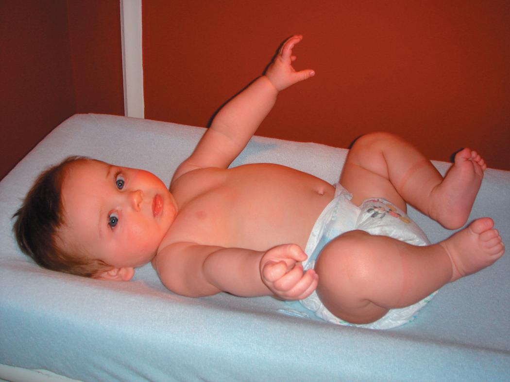
Normal Infant Has a Large Head, Narrow Shoulders and Chest, and a Large Abdomen.
The lar yngeal reflex is activated by the stimulation of receptors on the face, nose, and upper airways of the newborn. Reflex apnea, bradycardia, or lar yngospasm may occur. Various mechanical and chemical stimuli, including water, foreign bodies, and noxious gases, can trigger this response. This protective response is so potent that it can cause death in the newborn (see Chapter 3, “Respiratory Physiology” and Chapter 4, “Cardiovascular Physiology”).
Respiratory System
At full-term birth, the lungs are still in the stage of active development. The formation of adult-type alveoli begins at 36 weeks postconception but represents only a fraction of the terminal air sacs w ith thick septa at full-term birth. It takes more than several years for functional and morphologic development to be completed, w ith a 10-fold increase in the number of terminal air sacs to 400 to 500 million by 18 months of age, along w ith the development of rich capillary networks surrounding the alveoli. Similarly, control of breathing during the first several weeks of extrauterine life differs notably from control in older children and adults. Of par ticular importance is the fact that hypoxemia depresses, rather than stimulates, respiration. Anatomic differences in the airway occur w ith growth and development. Recently, the age-old concept of the child having a funnel-shaped lar ynx w ith the cricoid as the narrowest portion of the airway has been challenged. Findings by Litman and colleagues (2003) using MRI and video-bronchoscopic images by Dalal and colleagues (2009) both revealed that the shape of the infant lar ynx was more cylindrical (as for adults) than funnel shaped and did not change much w ith growth.
They also suggest for infants and children that the glottis, not the cricoid, may be the narrowest portion in the paralyzed or cadaveric position (which can be gently w idened w ith an ET T); the cricoid remains to be the solid narrowest segment of the upper airway system. The development of the respiratory system and its anatomy and physiology are detailed in Chapter 3, “Respiratory Physiology.”
Cardiovascular System
During the first minutes after birth, the newborn infant must change his or her circulatory pattern dramatically from fetal to adult types of circulation to survive in the extrauterine environment. Even for several months after initial adaptation, the pulmonary vascular bed remains exceptionally reactive to hypoxia and acidosis. The hear t remains extremely sensitive to volatile anesthetics during early infancy, whereas the central nervous system is relatively insensitive to these anesthetics. Cardiovascular physiology in infants and children is discussed in Chapter 4, “Cardiovascular Physiology.”
Fluid and Electrolyte Metabolism
Like the lungs, the kidneys are not fully mature at birth, although the formation of nephrons is complete by 36 weeks’ gestation. Maturation continues for about 6 months after full-term birth. The glomerular filtration rate (GFR) is lower in the neonate because of the high renal vascular resistance associated w ith the relatively small surface area for filtration. Despite a low GFR and limited tubular function, the fullterm newborn can conserve sodium. Premature infants, however, experience prolonged glomerulotubular imbalance, resulting in sodium wastage and hyponatremia (Spitzer 1982). On the other hand, both full-term and premature infants are limited in their ability to handle excessive sodium loads. Even after water deprivation, concentrating ability is limited at birth, especially in premature infants. After several days, neonates can produce diluted urine; however, diluting capacity does not mature fully until after 3 to 5 weeks of life (Spitzer 1978). The premature infant is prone to hyponatremia when sodium supplementation is inadequate or w ith overhydration. Furthermore, dehydration
is detrimental to the neonate regardless of gestational age. The physiology of fluid and electrolyte balance is detailed in Chapter 5, “Regulation of Fluids and Electrolytes.”
Temperature Regulation
Temperature regulation is of par ticular interest and importance in pediatric anesthesia. There is a better understanding of the physiology of temperature regulation and the effect of anesthesia on the control mechanisms. General anesthesia is associated w ith mild to moderate hypothermia, resulting from environmental exposure, anesthesiainduced central thermoregulatory inhibition, redistribution of body heat, and up to 30% reduction in metabolic heat production (Bissonette 1991). Small infants have a disproportionately large BSA, and heat loss is exaggerated during anesthesia, par ticularly during the induction of anesthesia, unless the heat loss is actively prevented. General anesthesia decreases but does not completely abolish thermoregulatory threshold temperature to hypothermia. Mild hypothermia can sometimes be beneficial intraoperatively, and profound hypothermia is effectively used during open-heart surgery in infants to reduce oxygen consumption. Postoperative hypothermia, however, is detrimental because of marked increases in oxygen consumption, oxygen debt (dysoxia), and resultant metabolic acidosis (Bissonette 1991). Regulation of body temperature is discussed in detail in Chapter 6, “Thermoregulation.”
SUMMARY
Pediatric anesthesia as a subspecialty has evolved because the needs of infants and young children are fundamentally different from those of adults. The pediatric anesthesiologist should be aware of the child’s cardiovascular, respiratory, renal, neuromuscular, and central nervous system responses to various drugs, as well as to physical and chemical stimuli, such as changes in blood oxygen and carbon dioxide tensions, pH, and body temperature. Their responses are different both qualitatively and quantitatively from those of adults and among different pediatric age groups. More importantly, the pediatric anesthesiologist should always consider the child’s emotional needs and create an environment that minimizes or abolishes fear and distress.
There have been many advances in the practice of anesthesia to improve the comfort of young patients over the last decade. These advances include a relaxation of preoperative fluid restriction, more focused attention to the child’s psychological needs w ith more extensive use of preoperative sedation via the transmucosal route, the w ide use of topical analgesia w ith a eutectic mixture of local anesthetic cream before intravenous catheterization, expanded use of regional anesthesia w ith improved accuracy and safety by means of ultrasound devices, and more general acceptance of parental presence during anesthetic induction and in the recovery room. Furthermore, a more diverse anesthetic approach has evolved through the combined use of regional analgesia, together w ith the advent of newer and less-soluble volatile anesthetics, intravenous anesthetics, sedatives, and shorteracting synthetic opioids and muscle relaxants. Finally, the scope of pediatric anesthesia has significantly expanded w ith the recent development of organized pain services in most pediatric institutions. As a result, pediatric anesthesiologists have assumed the leading role as pain management specialists, thus fur ther extending anesthesia services and influence beyond the boundary of the operating room.
REFERENCES
Complete references used in this text can be found online at ExpertConsult.com.
REFERENCES
Abajian JC, Mellish RW, Browne AF, et al. Spinal anesthesia for surgery in the high-risk infant. Anesth Analg 1984;63:359-362.
Ambardekar AP, Litman RS, Schwartz AJ. “Stay, give me your paw.” The benefits of family-centered care. Anesth Analg 2013;116(6):1314-1316.
Anand KJS, Garg S, Rovnaghi CR, et al. Ketamine reduces the cell death following inflammatory pain in newborn rat brain. Pediatr Res. 2007;62:283-290.
Ansermino M, Basu R, Vandebeek C, Montgomery C. Nonopioid additives to local anaesthetics for caudal blockade in children: a systematic review. Paediatr Anaesth. 2003;13:561-573.
Aono J, Mimiya K, Manube M. Preoperative anxiety is associated with high incidence of problematic behavior on emergence after halothane anesthesia in boys. Acta Anaesthesiol Scand 1999;43:542-544.
Aouad MT, Yazbeck-Karam VG, Nasr VG, et al. A single dose of propofol at the end of surgery for the prevention of emergence agitation in children undergoing strabismus surgery during sevoflurane anesthesia. Anesthesiology 2007;107:733-738.
Bartels M, Althoff RR, Boomsma DI. Anesthesia and cognitive performance in children: No evidence for a causal relationship. Twin Res Hum Genet. 2009;12:246-253.
Bissonette B. Body temperature and anesthesia. Anesth Clin N Am 1991;9:849.
Boretsky K, Visoiu M, Bigeleisen P. Ultrasound-guided approach to the paravertebral space for catheter insertion in infants and children. Paediatr Anaesth 2013;23:1193-1198.
Bowlby J. Attachment and loss New York: Basic Books; 1973.
Bryan YF, Hoke LK, Taghon TA, et al. A randomized trial comparing sevoflurane and propofol in children undergoing MRI scans. Paediatr Anaesth 2009;19:672-681.
Byon HJ, Lim CW, Lee JH, et al. Prediction of fluid responsiveness in mechanically ventilated children undergoing neurosurgery. Br J Anaesth 2013;110:586-591.
Chambers N, Lopez T, Thomas J, James MFM. Remifentanil and the tunneling phase of paediatric ventriculoperitoneal shunt insertion: a double-blind, randomized, prospective study. Anaesthesia 2002;57:133-139.
Chiaretti A, Pietrini D, Piastra M, et al. Safety and efficacy of remifentanil in craniosynostosis repair in children less than 1 year old. Pediatr Neurosurg 2000;33:83-88.
Cohen IT, Hannallah RS, Hummer KA. The incidence of emergence agitation associated with desflurane anesthesia in children is reduced by fentanyl. Anesth Analg 2001;93:88-91.
Coté CJ, Goldstein EA, Coté MA, et al. A single-blind study of pulse oximetry in children. Anesthesiology 1988;68:184.
Coté CJ, Rolf N, Liu LMP, Gousouzian NG. A single-blind study of pulse oximetry and capnography in children. Anesthesiology 1991;74:984.
Cravero J, Surgenor S, Whalen K. Emergence agitation in paediatric patients after sevoflurane anaesthesia and no surgery: a comparison with halothane. Paediatr Anaesth. 2000;10:419-424.
Dalal PG, Murray D, Messner H, et al. Pediatric laryngeal dimensions: an age-based analysis. Anesth Analg 2009;108:1475-1479.
Davidson AJ, Disma N, de Graaff JC, et al. Neurodevelopmental outcome at 2 years of age after general anaesthesia and awake-regional anaesthesia in infancy (GAS): an international multicentre, randomised controlled trial. Lancet 2016;387(10015):239-250.
Davis PJ, Cohen IT, McGowan FX Jr, Latta K. Recovery characteristics of desflurane vs. halothane for maintenance of anesthesia in pediatric ambulatory patients. Anesthesiology. 1994;80:293-302.
Davis PJ, Finkel J, Orr R, et al. A randomized, double-blinded study of remifentanil versus fentanyl for tonsillectomy and adenoidectomy surgery in pediatric ambulatory surgical patients. Anesth Analg 2000;90:863-871.
Davis PJ, Galinkin J, McGowan FX, et al. A randomized multicenter study of remifentanil compared with halothane in neonates and infants undergoing pyloromyotomy. Anesth Analg 2001;93:1387-1392.
Davis PJ, Greenberg JA, Gendelman M, Fertal K. Recovery characteristics of sevoflurane and halothane in preschool aged children undergoing bilateral
myringotomy and pressure equalization tube insertion. Anesth Analg 1999a;88:34-38.
Davis PJ, Wilson AS, Siewers RD, et al. The effects of cardiopulmonary bypass on remifentanil kinetics in children undergoing atrial septal defect repair. Anesth Analg 1999b;89:904-908.
Dawes J, Myers D, Görges M, et al. Identifying a rapid bolus dose of dexmedetomidine (ED50) with acceptable hemodynamic outcomes in children. Paediatr Anaesth 2014;24:1260-1267.
de Beer DA, Thomas ML. Caudal additives in children: solutions or problems? Br J Anaesth. 2003;90:487-498.
Dixon S, Snyder J, Holve R, Bromberger P. Behavioral effects of circumcision with and without anesthesia. J Dev Behav Pediatr 1984;5:246.
Dombro RH. The surgically ill child and his family. Surg Clin North Am 1970;50:759.
Dönmez A, Kizilkan A, Berksun H, et al. One center’s experience with remifentanil infusions for pediatric cardiac catheterization. J Cardiothoracic Vasc Anaesth 2001;15:736-739.
DuBois D, DuBois EF. A height-weight formula to estimate the surface area of man. Proc Soc Exp Biol Med. 1916;3:77.
Dullenkopf A, Gerber AC, Weiss M. Fit and seal characteristics of a new paediatric tracheal tube with high volume-low pressure polyurethane cuff. Acta Anaesthesiol Scand 2005;9:232-237.
Dutta S, Albanese CT. Transaxillary subcutaneous endoscopic release of the sternocleidomastoid muscle for treatment of persistent torticollis. J Pediatr Surg 2008;43:447-450.
Eichhorn SM, Cooper JB, Cullen DJ, et al. Standards for patient monitoring during anesthesia at Harvard Medical School. JAMA 1986;56: 1017.
Erdil F, Demirbilek S, Begec Z, et al. The effects of dexmedetomidine and fentanyl on emergence characteristics after adenoidectomy in children. Anaesth Intensive Care 2009;37:571-576.
Feldman JM, Sussman E, Singh D, et al. Is the pleth variability a surrogate for pulse pressure variation in a pediatric population undergoing spine fusion? Paediatr Anaesth. 2012;22:250-255.
Forman MC, Kerschbaum WE, Hetznecker WH, Dunn JM. Psychosocial dimensions. In: Behrman RE, Vaughan VC III, eds. Nelson’s textbook of pediatrics Philadelphia: Saunders; 1987.
Friedman WF. The intrinsic physiologic properties of the developing heart. In: Friedman WF, Lesch M, Sonnenblick EH, eds. Neonatal heart disease New York: Grune & Stratton; 1973.
Friesen RH, Veit AS, Archibald DJ, Campanini RS. A comparison of remifentanil and fentanyl for fast track paediatric cardiac anaesthesia. Paediatr Anaesth 2003;13:122-125.
Fujimoto T, Segawa O, Lane GJ, et al. Laparoscopic surgery for newborn infants. Surg Endosc 1999;13:773-777.
Galinkin JL, Davis PJ, McGowan FX, et al. A randomized multicenter study of remifentanil compared with halothane in neonates and infants undergoing pyloromyotomy: II Perioperative breathing patterns in neonates and infants with pyloric stenosis. Anesth Analg 2001;93:1387-1392.
Galinkin JL, Fazi LM, Cuy RM, et al. Use of intranasal fentanyl in children undergoing myringotomy and tube placement during halothane and sevoflurane anesthesia. Anesthesiology 2000;93:1378-1383.
Gallagher T, Lerman J, Volgyesi GA, et al. Effects of halothane and isoflurane on the baroreceptor response in newborn swine. Anesth Analg. 1987;66:564.
Gan H, Cannesson M, Chandler J, et al. Pediatric fluid responsiveness in children: a systematic review. Anesth Analg 2013;17:1380-1392.
Ganesh A, Gurnaney HG. Ultrasound guidance for pediatric peripheral nerve blockade. Anesthesiol Clin. 2009;27:197-212.
Ganesh A, Rose JB, Wells L, et al. Continuous peripheral nerve blockade for inpatient and outpatient postoperative analgesia in children. Anesth Analg 2007;105:1234-1242.
Gehan EA, George SL. Estimation of human body surface area from height and weight. Cancer Chemother Rep 1970;54(Pt 1):225.
Georgeson K. Minimally invasive surgery in neonates. Semin Neonatol 2003;8:243-248.
German JW, Aneja R, Heard C, Dias M. Continuous remifentanil for pediatric neurosurgery patients. Pediatr Neurosurg 2000;33:227-229.
Gootman PM. Neural regulation of cardiovascular function in the perinatal period. In: Gootman N, Gootman PM, eds. Perinatal cardiovascular function New York: Marcel Dekker; 1983.
Gray H. Anatomy of the human body 29th ed. Philadelphia: Lea & Febiger; 1973.
Gupta N, Rath GP, Prabhakar H, Dash HH. Effect of intraoperative dexmedetomidine on postoperative recovery profile of children undergoing surgery for spinal dysraphism. J Neurosurg Anesthesiol 2013;25:271-278.
Gurnaney HI, Ganesh A, Cucchiaro G. The relationship between current intensity for nerve stimulation and success of peripheral nerve blocks performed in pediatric patients under general anesthesia. Anesth Analg 2007;105:1605-1609.
Gurnaney H1, Kraemer FW, Maxwell L, et al. Ambulatory continuous peripheral nerve blocks in children and adolescents: a longitudinal 8-year single center study. Anesth Analg 2014;118:621-627.
Hall-Burton DM, Boretsky KR. A comparison of paravertebral nerve block catheters and thoracic epidural catheters for postoperative analgesia following the Nuss procedure for pectus excavatum repair. Paediatr Anaesth 2014;24:516-520.
Hansen TG, Ilett KF, Lim SI, et al. Pharmacokinetics and clinical efficacy of long-term epidural ropivacaine infusion in children. Br J Anaesth 2000;85:347-353.
Hansen TG, Ilett KF, Reid C, et al. Caudal ropivacaine in infants. Anesthesiology 2001;94:579-584.
Harris JS. Special pediatric problems in fluid and electrolyte therapy in surgery. Ann N Y Acad Sci 1957;66:966.
Hauber JA, Davis PJ, Bendel LP, et al. Dexmedetomidine as a rapid bolus for treatment and prophylactic prevention of emergence agitation in anesthetized children. Anesth Analg 2015;121:1308-1315.
Hay WW Jr, Rodden DJ, Collins SM, et al. Reliability of conventional and new pulse oximetry in neonatal patients. J Perinatol 2002;22:360-366.
Ibacache ME, Muñoz HR, Brandes V, et al. Single-dose dexmedetomidine reduces agitation after sevoflurane anesthesia in children. Anesth Analg 2004;98:60-63.
Ing CH, DiMaggio CJ, Malacova E, et al. Comparative analysis of outcome measures used in examining neurodevelopmental effects of early childhood anesthesia exposure. Anesthesiology 2014;120:1319-1392.
Ing CH, DiMaggio CJ, Whitehouse AJ, et al. Long-term differences in language and cognitive function after childhood exposure to anesthesia. Pediatrics. 2012;130:e476-e485.
Ing CH, DiMaggio CJ, Whitehouse AJ, et al. Neurodevelopmental outcomes after initial childhood anesthetic exposure between ages 3 and 10 years. J Neurosurg Anesthesiol 2014;26:377-386.
Irita K, Kai Y, Akiyoshi K, et al. Performance evaluation of a new pulse oximeter during mild hypothermic cardiopulmonary bypass. Anesth Analg 2003;96:11-14.
Isaza N, Garcia P, Dutta S. Advances in pediatric minimal access therapy: a cautious journey from therapeutic endoscopy to transluminal surgery based on the adult experience. J Pediatr Gastroenterol Nutr. 2008;46:359-369.
Ivani G, DeNegri P, Conio A, et al. Comparison of racemic bupivacaine, ropivacaine, and levobupivacaine for pediatric caudal anesthesia: effects on postoperative analgesia and motor block. Reg Anesth Pain Med. 2002;27:157-161.
Ivani G, DeNegri P, Lönnqvist P, et al. A comparison of three different concentrations of levobupivacaine for caudal block in children. Anesth Analg 2003;97:368-371.
Ivani G, Lampugnani E, Torre M, et al. Comparison of ropivacaine with bupivacaine for paediatric caudal block. Br J Anaesth 1998;81: 247-248.
Jevtovic-Todorovic V, Hartman RE, Izumi Y, et al. Early exposure to common anesthetic agents causes widespread neurodegeneration in the developing rat brain and persistent learning deficits. J Neurosci 2003;23:876-882.
Jevtovic-Todorovic V, Olney JW. PRO: Anesthesia-induced developmental neuroapoptosis: status of the evidence. Anesth Analg. 2008;106(6):1659-1663.
Johnson T, Monk T, Rasmussen LS, et al. Postoperative cognitive dysfunction in middle-aged patients. Anesthesiology 2002;96:1351-1357.
Jooste EH, Muhly WT, Ibinson JW, et al. Acute hemodynamic changes after rapid intravenous bolus dosing of dexmedetomidine in pediatric heart transplant patients undergoing routine cardiac catheterization. Anesth Analg 2010;111:1490-1496.
Kako H, Alkhatib O, Krishna SG, et al. Changes in intracuff pressure of a cuffed endotracheal tube during surgery for congenital heart disease using cardiopulmonary bypass. Paediatr Anaesth 2015;25(7):705-710.
Kalkman CJ, Peelen L, Moons KG, et al. Behavior and development in children and age at the time of first anesthetic exposure. Anesthesiology 2009;110:805-812.
Karmakar MK, Aun CST, Wong ELY, et al. Ropivacaine undergoes slower systemic absorption from the caudal epidural space in children than bupivacaine. Anesth Analg 2002;94:259-265.
Keidan I, Berkenstadt H, Segal E, Perel A. Pressure versus volume-controlled ventilation with a laryngeal mask airway in paediatric patients. Paediatr Anaesth 2001;11:691-694.
Keidan I, Berkenstadt H, Sidi A, Perel A. Propofol/remifentanil versus propofol alone for bone marrow aspiration in paediatric haematooncological patients. Paediatr Anaesth 2001;11:297-301.
Kim JY, Chang YJ, Lee JY, et al. Post-induction alfentanil reduces sevofluraneassociated emergence agitation in children undergoing an adenotonsillectomy. Acta Anaesthesiol Scand 2009;53:678-681.
Kinney HC. The near-term (late preterm) human brain and risk for periventricular leukomalacia: a review. Semin Perinatol 2006;30:81-88.
Ko Y-P, Huang C-J, Hung Y-C, et al. Premedication with low-dose oral midazolam reduces the incidence and severity of emergence agitation in pediatric patients following sevoflurane anesthesia. Acta Anaesthesiol Sin. 2001;36:169-177.
Krishna SG, Ramesh AS, Jatana KR, et al. A technique to measure the intracuff pressure continuously: an in vivo demonstration of its accuracy. Paediatr Anaesth 2014;24(9):999-1004.
Kulka PJ, Bressem M, Tryba M. Clonidine prevents sevoflurane-induced agitation in children. Anesth Analg 2001;93:335-338.
Kuratani N, Oi Y. Greater incidence of emergence agitation in children after sevoflurane anesthesia as compared with halothane. Anesthesiology 2008;109:225-232.
Lankinen U, Avela R, Tarkkila P. The prevention of emergence agitation with tropisetron or clonidine after sevoflurane anesthesia in small children undergoing adenoidectomy. Anesth Analg 2006;102:1383-1386.
Lerman J, Davis PJ, Welborn LG, et al. Induction, recovery, and safety characteristics of sevoflurane in children undergoing ambulatory surgery. A comparison with halothane. Anesthesiology 1996;84:1332-1340.
Lippmann M, Nelson J, Emmanouilides GC, et al. Ligation of patent ductus arteriosus in premature infants. Br J Anaesth 1976;48:365.
Litman RS, Maxwell LG. Cuffed versus uncuffed endotracheal tubes in pediatric anesthesia: the debate should finally end. Anesthesiology 2013;118:500-501.
Long JB, Birmingham PK, De Oliveira GS Jr, et al. Transversus abdominis plane block in children: a multicenter safety analysis of 1994 cases from the PRAN (Pediatric Regional Anesthesia) database. Anesth Analg 2014;119:395-399.
Lönnqvist PA, Westrin P, Laarsson BA, et al. Ropivacaine pharmacokinetics after caudal block in 1- to 8-year-old children. Br J Anaesth. 2000;85:506-511.
Lorenzo AJ, Lynch J, Matava C, El-Beheiry H, Hayes J. Ultrasound guided transversus abdominis plane vs surgeon administered intraoperative regional field infiltration with bupivacaine for early postoperative pain control in children undergoing open pyeloplasty. J Urol. 2014;192:207-213.
Malviya S, Reynolds PI, Voepel-Lewis T, et al. False alarms and sensitivity of conventional pulse oximetry versus the Masimo SET technology in the pediatric postanesthesia care unit. Anesth Analg. 2000;90: 1336-1340.