Preface
The third edition differs considerably from the earlier editions in that the book is no longer separated in two parts, the first providing the nuts and bolts of what might be termed classical signal transduction. In fact, this classification does not really apply for two reasons. First, for students everything is as new as the latest scientific article is for a teacher and dividing between classical or nonclassical is not really making the subject clearer and might even pretend that cells employ classical (important) and not-so classical (less important) mechanisms that bring about changes in their metabolism, gene expression, secretion rate, contraction state, and so on. Second, with the recent structural revelations of G protein-coupled receptor and the action of biased agonists, classical signal transduction has suddenly lost its classical touch and has become very modern. Instead, an introductory chapter has been added in which a number of principles are outlined common to many signal transduction events, and these are placed in the context of the most “Nobel”, the most classic, of all pathways: adrenaline to glycogen phosphorylase. While the previous editions were written by three authors and rewritten by Bastien Gomperts, so that it appeared as written by one mind, one hand, this edition also differs in that it has been written by one hand only (or, less poetically, typed by two hands on a keyboard). The two “greybeards” have pulled out after publication of the second edition and, very sadly, Bastien Gomperts has passed away in October 2013. If, from the previous editions, you appreciate the writing style, the wit, and the anecdotes, bearing from some unusual sources, much of the credit goes to him. He was an inspiring mentor indeed. With a few exceptions, for instance the chapter on protein phosphatases, each chapter now has a theme of its own, around which specific aspects of signal transduction pathways are developed. For teaching purposes, Table 1-1 attempts to give a short overview of each chapter’s subjects and highlights, so that, depending on the pathway to be explored, a relevant (suitable) context can be selected. Although there still is a gradual buildup of the subject, where later chapters make reference to earlier chapters, each chapter could stand on its own. Naturally, the signaling aspects highlighted are not necessarily unique to the context in which they are developed, but it allows teachers (and students) to tell a story rather than just listing a sequence of signaling events (Kramer and Thomas, 2006) As a consequence, this edition contains more on cell biology, physiology, pathology, and immunology. By providing precise examples, embedded in precise contexts, the book offers the possibility to integrate signaling
knowledge into the above-mentioned disciplines and, therefore, facilitates a constructive approach to teaching (for more information, see http://www. cellbiol.net/docs/Constructive_teachingKramer.pdf).
Again, there has been no attempt to be comprehensive and certain important topics that well qualify for inclusion in this book, such as signals initiated by damaged DNA or unfolded proteins, are conspicuous only by their absence. Although the book touches the leading edges of the subject, it also endeavors to provide an elementary basis with some historical background to all the topics covered. The “prologue” has been extended considerably with new information about the first observations of “irritability,” a phenomenon that qualifies as “an essential element of the living” (besides template replication and metabolism). Historical background not only provides a broader insight into the subject and pays tribute to the wisdom of our forebears, whose freedom of thought and sometimes serendipitous discoveries in the nineteenth and early twentieth centuries led to the creation of the modern sciences, for certain people it may also provide meaning to the numerous odd names, abbreviations, and acronyms (collectively named symbols) that riddle the book. For unexplained symbols the reader is encouraged to consult UniProt (paragraphs “function” and “names & taxonomy”) or relevant Wikipedia articles. Besides the chapter about intracellular calcium, the book does not reveal a good deal of experimental techniques. A lack of time and a growing complexity of technology are to blame. Moreover, experience tells that explaining technology is revealing for learners who already master the cellular context and signaling events but tend to mystify matters, because of a substantial increase in cognitive burden, when learners are still struggling with the molecular mechanisms that drive the pathways.
In preparing the book, I have had the benefit of advice and opinions from many friends and colleagues. These include (in order of their first name) Alan Hall (New York), Alasdair Gibb (London), Bob Weinberg (Cambridge, USA), Bob Lefkowitz (Durham, USA), Bruno Klaholz (Illkirch), Carsten Hoege (Dresden), Chris de Graaf (Amsterdam), Christopher Glass (San Diego), David Armstrong (Durham, USA), David Strutt (Sheffield), Filip van Petegem (Vancouver), Geerten Vuister (Leicester), George Mosialos (Thessaloniki), Graham Dunn (London), Jean Dessolin (Bordeaux), Jeff Saucerman (Charlottesville), Jennifer LippincottSchwartz (Bethesda), Jürgen Knoblich (Vienna), Karin Rittinger (London), Karl Matter (London), Maria Schumacher (Durham, USA), Marian Joëls (Utrecht), Marina Gloukova (Paris), Mark Dell’Acqua (Aurora), Matthew Gold (London), Michel Laguerre (Bordeaux), Miho Lijima (Baltimore), Mingjie Zhang (Hong Kong), Peter van Haastert (Groningen), Purna Joshi (Toronto), Roel Sterckx (Cambridge, UK), Romuald Nargeot (Bordeaux), Sander van den Heuvel (Utrecht), Shiva Malek (South San Francisco), Stuart Firestein (New York), Yohanns Bellaiche (Paris), Vadim Asharvsky (Durham, USA), Wai Leong (Singapore), and Wei-Min Shen Los Angeles. Special gratitude also goes to all authors, curators, Website developers, and
technicians who contributed and continue to contribute to an increasing number of outstanding annotated databases (UniProt, PubMed, OMIM, UniGene, GenomeNet, HGNC, PhosphoSite, and so on) and Wikis. These databases have become key sources of information for research as well as education. I encourage students to go out on the web!
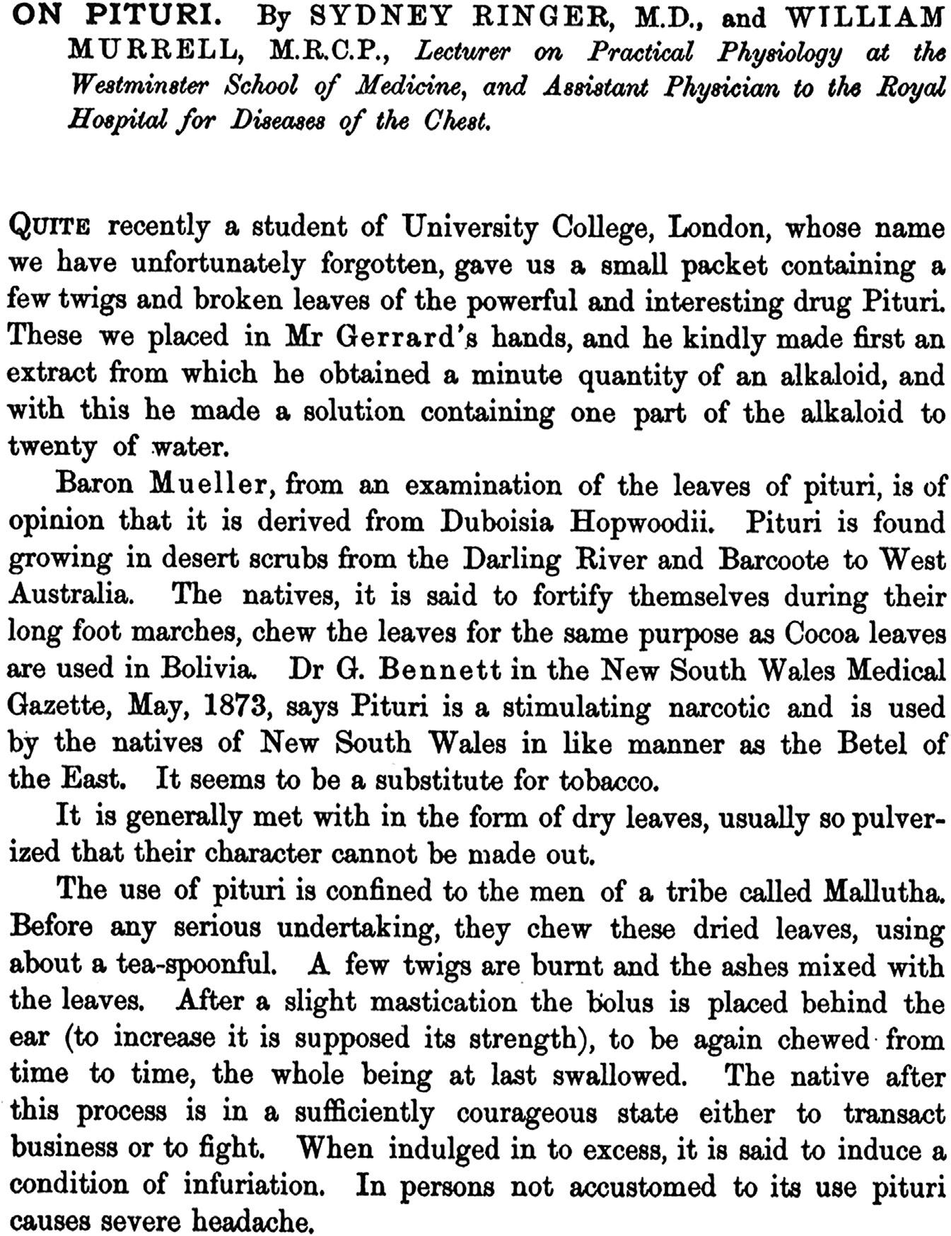
In acknowledgment of their contribution I offer the following quotation by one of the pioneers of signal transduction (Figure 1-1; Ringer and Murrell, 1878).
FIGURE 1-1
Of course, the authors of this paper would themselves never have recognized the expression signal transduction, and it would be a further 100 years before it made its appearance in the biological literature. The sensations brought about by pituri, an alkaloid that Ringer and Murrell described as sharing some of the pharmacological properties of atropine (courage, infuriation, frustration, and headaches), are not dissimilar to those experienced in the writing of this book. Indeed, they will be familiar to many students and investigators in this and other fields of research. However, we should not take this too far. When Ringer (1879) tested the effects of the application of pituri on four men, he noted that it also causes drowsiness, faintness, pallor, giddiness, hurried and superficial breathing, dilates the pupil, produces general weakness with convulsive twitchings, and antagonizes the action of muscarin on the heart. Unlike atropine, it produces sickness and increases the salivary secretion in large doses copiously, the breathing becomes quick and shallow, and general weakness ensues. Reading all this, it leads one to wonder who, among their students, colleagues, and servants, may have offered themselves up as willing, or less than willing, guinea pigs in the furtherance of scientific research. Ringer and his friends apparently preferred to eschew membership of the very honorable brotherhood of selfexperimenters, of which the more famous members include Sir, Humphry Davey, who breathed nitrous oxide as well as other more noxious gases, John Scott Haldane, who too inhaled lethal gases; and more recently Barry Marshall, who has swallowed a culture of Helicobacter pylori to show that it caused stomach ulcers and who with Robin Warren was awarded the Nobel Prize in Physiology or Medicine in 2005. Another member of this fraternity, Charles Eduard Brown-Sequard, figures prominently in “prologue” chapter.
NOTES
For web-support of this book
See the companion website: http://booksite.elsevier.com/9780123948038
For protein structural data we have made use of
The Protein Data Bank: Berman et al. (2000).
Protein structures have been generated using PyMol (education version), a molecular visualization system on open source foundation, maintained and distributed by Schrodinger.
References
We have tried to provide original text sources to nearly all the statements, experiments, and discoveries discussed. The main reason for this
is that we ourselves have necessarily had to extend the treatment of nearly all the topics presented far beyond the areas of our own experience or expertise. Thus, comprehensive lists are there to provide us with some sort of reassurance that what we have written has not simply been conjured out of the air. Also, because we have made a particular feature of presenting original historical source material by quotation, which necessarily required referencing, it seemed logical also to include literature references to modern sources as well. Thus we hope that this book may serve as a valuable resource, in the manner of a basic literature review, for anyone wanting to explore further.
Protein symbols (gene products)
We have named proteins according to the symbols agreed upon in the HUGO gene nomenclature database (HGNC) (www.genenames.org). Similar symbols have been adopted by protein databases such as UniProt. Some of the new symbols are simply awkward, so different from conventional names that they are even not recognized by scientists who made major contributions to the field, and when they really are uncommon alternative more conventional names are provided, but from a pedagogical point of view it is vital that students can search the web with unique (unambiguous) symbols and find out about the relevant proteins (and genes) themselves. And remember, certain symbols may be more familiar to experts in the field, for students they are all the same: new and often gruesome.
References
Kramer, I.M., Thomas, G., Spring 2006. Meeting report: teaching signal transduction. CBE Life Sci. Educ. 5, 19–26. Ringer, S., Murrell, W., 1878. On pituri. J. Physiol. 377–383. Ringer, S., 1879. On the action of pituri on man. Lancet 290–291. Berman, H.M., Westbrook, J., Feng, Z., Gilliland, G., Bhat, T.N., Weissig, H., Shindyalov, I.N., Bourne, P.E., 2000. The protein data bank. Nucleic Acids Res. 28, 235–242. http://www. rcsb.org/pdb/
TABLE 1-1 Contexts, pathways, subjects, and proteins/molecules elaborated in different chapters
Title/context/main pathway Subjects
1. Prologue: Signal transduction from an historical perspective
An account of how the term “signal transduction” entered biomedical research and how stimulus–response coupling, hormones, neurotransmitters, growth factors, and their receptors were brought to light
Highlights on molecules, proteins, and personalities
• Alfred Gilman
• Martin Rodbell
• Thomas Henry Huxley
• Steve Grand
• Charles Edouard Brown-Séquard
• Henry Hallett Dale
• Otto Loewi
• George Oliver
• Edward Scharpey-Schäfer
• Ernest Henry Starling
• Willam Maddock Baylis
• CONRO, a self-reconfigurable robot
• Paul Ehrlich
• John Newport Langley
• Francis Peyton Rous
• Rita Levi-Montalcini
• Stanley Cohen
• Alexis Carrel
• Howard Temin
• Renato Dulbecco
2. An introduction to signal transduction
Context: Adrenalin-mediated activation of glycogen phosphorylase in striated muscle
• Signaling pathways serve to create symbolic representations of the cellular environment
• First messengers, description of hormones, growth and differentiation factors, cytokines, inflammatory mediators, vasoactive agents, neurotransmitters
• Aph(2′)-Ib, aminoglycoside-2 phosphotransferase, Escherichia coli (ancient) protein kinase fold, structural composition, conserved residues, αC helix
• Docking site, substrate-binding motifs in MAPKinase, D motif, and DEF motif, examples of substrates, sequence of the transcription factor ELK1
• Signaling in context, the signal itself is ambiguous but context, cell type, and cell condition, determine the outcome
• Receptor–ligand concept, classic pharmacology, different receptor types
• Signaling mechanisms, the sequence of receptor–transducer–effector–second messenger
• Adrenaline to glycogen phosphorylase, pioneer studies in signal transduction, detailed transcription of the pathway as operated in muscle
• Nobel laureates associated with the adrenaline to glycogen phosphorylase pathway
• Wired allostery, signal integration, and thoughtful decisions
• Posttranslational modifications, a broad overview
• Feedback mechanisms, modeling of EGFRmediated activation of MAP-kinase
• Edmond Fischer and Edwin Krebs, Nobel Laureates 1992 (protein phosphorylation, glycogen phosphorylase)
• Eicosanoids, molecular composition, prostaglandin, prostacyclin, thromboxane, leukotriene, anandamide
• GNAI1, Gαi subunit of heterotrimeric G-protein, structural composition, conserved residues, GTP-binding pocket, GαRAS and alpha-helical segment
• H3F3A, histone-3.3, posttranslational modifications, acetylation, crotonylation, methylation, and phosphorylation
• HRAS, monomeric G-protein, molecular structure, position of conserved residues, mechanism of nucleotide exchange and GTP hydrolysis, GTPase cycle, switch regions, farnesylation
• Cellular communication, modes of, endocrine, paracrine, juxtacrine, synaptic, and autocrine
• Pharmacology, agonist, antagonist, inverse agonist, receptor–ligand interaction, affinity, dose–response curve, receptor number, and signal sensitivity
• Phosphoenolpyruvate-dependent phosphotransferase system in bacteria
• Phosphorylation, consequences for activity and subcellular localization of proteins, potential phosphate donors, ATP favorite but not exclusive Continued
Title/context/main pathway Subjects
Highlights on molecules, proteins, and personalities
• Phospho-amino acids, most occurring phosphate acceptors in proteins, stability of phospho-ester, phospho-ramidate, and phospho-anhydride bond
• Phosphoryl transferase, phosphorylation, protein kinase, catalytic mechanism, role of conserved residues
• PRKACA, protein kinase A, catalytic subunit, serine/threonine kinase, structural composition, conserved residues, N-lobe, C-lobe, signature sequences, αC-helix, catalytic mechanism
• Protein kinases, classification, serine/threonine, tyrosine and dual-specificity protein kinases, their mode of regulation
• Protein phosphatases, classification, serine/ threonine, tyrosine and dual-specificity phosphatases, structural composition, catalytic mechanisms
• PYGM, glycogen phosphorylase muscle form, structural composition, dimer, hydrolysis of glycogen and phosphorylation of glucose (glucose1-phosphate), allosteric regulation of enzyme, phosphorylation by glycogen phosphorylase kinase on serine-14, change in configuration, tense to relaxed state
• RASA1, RAS GTPase-activating protein, RAS–GAP domain, molecular structure, catalyzes hydrolysis of GTP
• Receptors, classification, ligands, overview of different types
• SOS1, RAS-nucleotide exchange factor, GEF, Cdc25 domain, molecular structure, αH helix, mechanism of removal of GDP from nucleotide pocket
• Two-component signaling and environmental sensors in bacteria
3. Regulation of muscle contraction by adrenoceptors
Contexts:
a. Cardiac muscle contraction
b. Smooth muscle contraction
Pathways:
a. Action potential, voltage sensitive Ca2+ channels, Ca2+induced-Ca2+ release (ryanodine receptor), intracellular free Ca2+ release, troponin, myosin–actin cross-bridge cycle
b. Adrenoceptor β1, heterotrimeric G-proteins (Gαs), adenylyl cyclase, cAMP, and protein kinase A
c. Heterotrimeric G-proteins (Gαs), beta-adrenergic receptor kinase, arrestin, switch to MAP-kinase pathway, non-receptor tyrosine kinases, and protein kinase B (AKT)
• Catecholamines (adrenaline, noradrenaline, dopamine), molecular composition
• Central and autonomic–peripheral nervous system, anatomy, and neurotransmitters involved
• Adrenoceptors, adrenergic receptors, history, classification
• Agonist, inverse agonist, biased agonist, and antagonist, mode of action, examples
• Muscle contraction, cardiac and smooth muscle, anatomy, myosin–actin cross-bridge cycle
• G-protein effectors, adenylyl cyclase and phospholipase C, second messengers cAMP, and diacyglycerol/IP3
• Noradrenaline-mediated control of cardiacmuscle contraction: ADRB1 (β1AR), GNAS (Gαs), ACDY5, cAMP, PRKACA (PKA), CACNA1C (voltage-sensitive Ca2+ channel), RYR2, TNNC1, TTNI3 (troponin), myosin–actin cross-bridge cycle, PLN (phospholamban), ATP2A2 (SERCA2, Ca2+ pump), KCNQ1 (K+ channel), ATP2B2 (Ca2+ pump)
• G protein-receptor kinases (GRK), among which ADRBK1 and GRK6
• Arrestins and arrestin-dependent signaling (among others, SRC, RAF–MAPK, AKT1, NFKB)
• ADCY, adenylyl cyclase, structure, catalytic mechanism, production of cAMP, family members, mode of activation, and inhibition
• ADRA, ADRB, adrenoceptor, adrenergic receptor, ligand-binding site, conformational changes, energy landscape, coupling to heterotrimeric G-proteins or G protein-receptor kinases (GRK)
• adrenalin, composition, its agonists, inverse agonists, biased agonist, and neutral antagonists
• ARRB, arrestin proteins, structure, family members
• GRK6, G protein-receptor kinase, structure, function, family members, mechanism of attachment to GPCR
• Heterotrimeric G-proteins, mechanism of nucleotide exchange, guanine-exchange function (GEF) of seven-membrane-spanning receptors (GPCR)
• ITPR1, IP3 receptor, cryo-electromicroscopydetermined structure, molecular detail of the IP3-binding site
• PLC, phospholipase C, domain architecture, structural composition, TIM barrel, X/Ylinker, catalytic mechanism, production of diacylglycerol and inositol-1,4,5-trisphosphate (IP3), family members, family tree, control of PLCB3 by GNAQ (Gαq)
• Feedback mechanisms, modeling of noradrenaline-mediated molecular events
Continued
Title/context/main pathway
4. Cholinergic signaling and muscle contraction
Contexts:
a. Skeletal muscle contraction
b. Cardiac muscle contraction and relaxation
c. Smooth muscle contraction and relaxation
Pathways:
a. Nicotinic acetylcholine receptor, voltage sensitive Na+-channel, membrane depolarization, voltage sensitive Ca2+channel, Ca2+-induced-Ca2+release (ryanodine receptor), intracellular free Ca2+, troponin, myosin-actin cross-bridge cycle
b. Muscarinic acetylcholine receptor-3, G protein-coupled receptors, heterotrimeric G-proteins (Gαi, Gαq, Gα12, and Gβγ),
- Pacemaker, cardiomyocyte, Gαi, adenylyl cyclase down, Gβγ-gated K+-channel conductivity, cAMP-gated Na+ channel
Subjects
• Acetylcholine, history, composition, synthesis, and breakdown
• Synapse, active zone, vesicle docking, membrane fusion machinery
• Muscarinic and nicotinic receptors
• Nicotinic receptor (type IV), neuromuscular junction, membrane depolarization and skeletal muscle contraction
• Muscarinic receptor (M2) and vagal (parasympathetic) control of cardiac force and rhythmicity
• Cyclic nucleotide phosphodiesterase (PDE) and pathway control
• Muscarinic receptor (M3), bronchial smooth muscle contraction, mucus production, salbutamol, and asthma
• Nitric oxide, history, NO synthase, smooth muscle relaxation
• Guanylyl cyclases, cGMP, protein kinase G (PRKG)
• PDE5A, cGMP-specific 3′,5’-cyclic phosphodiesterase-5A, in corpus cavernosum, inhibition by sildenafil (Viagra) and penile erection
• Examples of ionotropic and metabotropic receptors for neurotransmitters
Highlights on molecules, proteins, and personalities
• ACh, acetylcholine, composition, synthesis, breakdown, agonists, and antagonists
• CHRN, nicotinic acetylcholine receptors, channel topology, structural composition, subunits, control of pore permeability (gating), detail ligand binding pocket, aromatic cage, comparison with AChBP, snail protein
• CHRM, muscarinic acetylcholine receptors, structural composition, bitopic ligand-binding site
• GUCY, guanylyl cyclase (soluble), family tree of guanylyl cyclases, structural composition, α- and β-subunit, catalytic mechanism
• Inward rectifier K+ channel (Kcnj6) in complex with Gβγ
• NOS (nitric oxide synthase), structural composition, prosthetic groups, catalytic mechanism
• PDE, cyclic nucleotide phosphodiesterase, structure, cyclic-nucleotide binding (cAMP versus cGMP), regulation of activity
• PDE5A, cGMP-specific 3 ′ ,5 ′ -cyclic phosphodiesterase-5A, target of sildenafil (Viagra), molecular structure with inhibitor
• PRKG1, protein kinase G, molecular structure, regulation by cGMP
- bronchial Smooth muscle, Gαq, phospholipase Cβ, phosphatidylinositol-4,5bisphosphate (PIP2), inositol1,4,5-trisphosphate (IP3), diacylglycerol, IP3 receptor, intracellular-free Ca2+, smooth muscle myosin light chain kinase, actin–myosin cross-bridge cycle, protein kinase C, guanine nucleotide exchange factor (ARHGEF1), monomeric GTPase (RHOA), ROCK1, inhibitory subunit of serine/threonine phosphatases
c. Muscarinic acetylcholine receptor-3 endothelium, G protein-coupled receptors, phospholipase Cβ, phosphatidyl4,5-bisphosphate, diacylglycerol, inositol-1,4,5-trisphosphate, IP3receptor, intracellular free Ca2+, NO synthase, nitric oxide
d. Nitric oxide, smooth muscle cell, soluble guanylyl cyclase, cGMP, protein kinase G, effect on regulatory subunit of serine/ threonine phosphatase and dephosphorylation of smoothmuscle myosin-regulatory light chain, myosin–actin cross-bridge cycle Continued
Title/context/main pathway
5. Sensory signal processing: visual transduction and olfaction
Contexts:
a. Vision and retinal photoreceptors
b. smell and olfactory epithelium
Pathways:
a. G-protein coupled receptors (rhodopsin), heterotrimeric G-protein (Gαt or transducin), guanylyl cyclase, removal cyclic cGMP, nucleotide-gated Na+/ Ca2+ ion channel, voltage-gated Ca2+ channel, neurotransmitter release (glutamate), G proteincoupled kinase, arrestin, transducin GAP-complex
b. G protein-coupled odorant receptors, Gαolf, adenylyl cyclase, production cAMP, cAMP-sensitive Na+/Ca2+ channel, Ca2+/calmodulingated Cl channel, membrane depolarization
Subjects
• Eye, development, anatomy, retina, cell types
• Rods and cones, disks, rhodopsin, absorption spectra, color vision, 11-cis-retinal, all-transretinal, vitamin A
• Retinal metabolism, retinal-pigment epithelium
• Rhodopsin (RHO), metarhodopsin, structure, conformational change of helical bundle, binding of Gαt (transducin) or GRK1
• Effectors, inhibition of guanylyl cyclase GUCY2D, activation phosphodiesterase PDE6, cGMPsensitive cation channel (CNGA1)
• Signal attenuation, GRK1, arrestin (SAG), and transducin GAP complex (PDE6G, GBB5, RGS9)
• Light, darkness, adaptation
• Drosophila compound eye, norpA (PLC), InaC (PRKC), InaD (scaffold) and Trp-channels
• Chemosensory organs mouse, fly and human, olfactory bulb, olfactory epithelium
Highlights on molecules, proteins, and personalities
• G protein-coupled receptor, GPCR, 7TMreceptor, excursion: classification, topology of transmembrane helical bundle, Ballesteros–Weinstein generic numbering of highly conserved residues, contact network of ligand and of Gα, ligand-binding pockets across the category A (rhodopsin-type) receptors, structure comparison between category A and B (secretin type)
• Metarhodopsin II, molecular structure, interaction with effectors, Gαt (transducin) or GRK1
• Retinal, chromophore, 11-cis-retinal lysine, all-trans-retinal, photoisomerization, vitamin A, metabolic pathway, retinal-pigment epithelium
• Rhodopsin, opsin +11-cis-retinal, GPCR, different members (OPN1SW, OPN1MW, OPN1LW, RHO), molecular structure of RHO, residues that determine differences in absorption spectra
• Transducin GAP complex, molecular structure of GNAT1, PDE6G, RGS9, domain architecture, membrane recruitment via RGS9BP, acceleration of GTP hydrolysis of transducin
• Odorant receptors, GPCR, Gαolf (GNAL), adenylyl cyclase (ACDY3), cyclic nucleotidegated channel (CNG), Ca2+/calmodulin-gated chloride channel (ANO2)
• Excursion on G protein-coupled receptors
6. Intracellular calcium
Contexts:
a. Muscle contraction
b. Neurotransmitter release
c. Cell migration
Pathway:
a. Phospholipase C, inositol-1,4,5trisphosphate, diacylglycerol, IP3-receptor, Ca2+-induced Ca2+-release (ryanodine receptor), intracellular free Ca2+
- Ca2+/calmodulin sensitive smooth muscle myosin light chain kinase, myosin–actin cross-bridge cycle smooth muscle
- Synaptotagmin, membrane fusion, voltage-gated Ca2+ channel neurotransmitter release
b. Protein kinase C, phosphorylation of Rhodissociation inhibitor
- Guanine nucleotide exchange (ARHGEF1), monomeric GTPase (RHOA), Rhokinase, phosphatase inhibitor (MYPT), smooth muscle myosin activation, myosin–actin cross-bridge cycle
- Guanine Nucleotide exchange (TIAM1), monomeric GTPase (RAC1), activation of actin nucleation (WASP/ARP), membrane protrusion
• Calcium-storing organelles and Ca2+ transporter
• Ca2+-coordination geometry, Ca2+-binding proteins and Ca2+ chelators (EDTA, EGTA)
• Ca2+-binding domains (EF-hand, C2-domain, Pand C-domain, Calx-β-motif, gelsolin-repeat)
• Calmodulin-binding proteins as effectors
• Ca2+ indicators (from aquorin to genetically encoded indicators)
• Ca2+-permeable channels (ITPR, RYR, TPCN, KNCA, TRP, THEM16A, CACNA1, ORAI1)
• Store replenishment through ORAI1 (calcium release-operated calcium channel), the Ca2+sensor STIM1 and the Ca2+-ATPase (ATP2A1)
• Ca2+ blips, puffs, spikes and waves
• Vesicle fusion with membrane, SNARE complex, synaptotagmin, voltage-gated Ca2+ channel (CACNA1C)
• Cell migration, protrusion, retraction, actin cytoskeleton (arcs and stress fibers)
• Chemokine receptor-mediated localized Ca2+ oscillations, activation of smooth-muscle myosin light-chain kinase (MYLK) and activation of WAVE/SCAR/WASP-mediated actin-filament nucleation
• Michel Abercrombie, a pioneer in cell migration
• Ca2+-binding domains or motifs, molecular structure of EF-hand, C2-domain, Calx-β motif and gelsolin repeat
• Ca2+-permeable channels, members, membrane topology
• CALM, calmodulin, structural composition, change in conformation upon binding of four Ca2+ ions, interaction with Camk2a, Mylk2, and Kcnn2
• CAMK, Ca2+/calmodulin protein kinase, molecular structure of hub, linker and kinase domain, schematic representation of the assembly into a multiprotein complex, control of kinase activity by Ca2+ oscillations
• GCaMP3, genetically encoded Ca2+ indicator, EGFP bound to Ca2+/calmodulin, molecular structure
• ITPR, IP3-receptor, cryo-microscopy-derived structure, control of Ca2+ conductivity, molecular structure of IP3-binding site
• Metal coordination geometry, Ca2+-binding proteins and metal chelators (EDTA, EGTA)
• Michel Abercrombie, a pioneer in cell migration
• RYR, ryanodine receptor, cryo-microscopyderived structure, molecular composition of cytoplasmic vestibule, and control of Ca2+ conductivity
Continued
Title/context/main pathway
7. Bringing the signal into the nucleus: Regulation of gene expression
Context:
a. Gluconeogenesis
Pathway:
a. G protein-coupled receptors, heterotrimeric G-protein, Gαs, adenylyl cyclase, cAMP, protein kinase A, cyclic AMP-response element-binding protein (CREB), induction of the gluconeogenic program
Subjects
• Estimated number of human genes and the central dogma of molecular biology
• Starvation and the processes that control gluconeogenesis, role of glucagon and glucocorticoid
• Signaling by the glucagon receptor, adenylyl cyclase, cAMP, and regulation of activity of protein kinase A
• AKAP, anchoring of protein kinase A to subcellular compartments and scaffolding of signaling complexes, example of AMPA receptor, DLG1, and AKAP79
• CREB, transcription factor, is a nuclear target of protein kinase A
• Gene transcription and transcription factors, from Jacob and Monod until today, histone acetylation and methylation, pre-initiation transcription complex
• CREB recruits co-activators, CREBBP, PE300 and CRTC2
• CREB1, FOXO1, PPAR and the glucocorticoid receptor NR4A1 drive the gluconeogenic program
• Insulin disables the gluconeogenic program (cytoplasmic sequestration of CRTC2 and FOXO1, phosphorylation and dissociation of CREBBP)
Highlights on molecules, proteins, and personalities
• AKAP79, schematic representation of scaffolding role in signaling complex assembly (ADCY8, PPP3CB, PPP3CA, PRKACA, PRKARIIA, DLG1), role in control of phosphorylation of the AMPAtype glutamate receptor (GRIA1)
• bZIP, basic leucine-zipper protein, list of members of the protein family
• CREB1, cAMP response-element binding protein, structural composition of bZIP domain bound to cAMP-response element (CRE), phosphorylation sites in KID domain and interaction with CREBBP
• GCGR, glucagon receptor, bound to glucagon, predicted structure
• Histone-3, methylation and acetylation signatures of a repressed and activated enhancer, promotor and coding region
• PIC, transcription pre-initiation complex, schematic representation of proteins
• PRKACA, catalytic subunit of protein kinase
A, domain architecture, structural composition, conserved residues, position of N-tail with myristate and of C-tail, co- and posttranslational phosphorylation and phosphosite sequence logo of substrates
• PRKARIA, regulatory subunit of protein kinase
A, domain architecture, molecular structure with or without cAMP, function of pseudo-substrate (RRGAI), dimerization through dimerization domain and CNBA interactions
8. Nuclear receptors
Contexts:
a. Sperm motility and capacitation
b. Mammary gland development
c. Consolidating memory (and dealing with pregnancy)
Pathways: Nuclear receptor-mediated regulation of gene transcription
• Steroid hormones, everything from domestic animals, the Chinese Pharmacopoeia of 725 AD, to nineteenth and twentieth century personalities in the discovery of steroids
• Steroids accumulate in the nucleus, nuclear receptors
• Regulation of transcription discovered in giant chromosomes of insect salivary glands
• Superfamily of nuclear receptors and recognition of specific promoter enhancer sites (inverted or direct repeat DNA sequences)
• Ligand-mediated activation or repression of gene transcription, histone acetylation or de-acetylation
• Chaperone (or heat-shock) proteins and the loading of receptors with their ligand
• Cooperation with other transcription factors (transrepression or transactivation)
• Non-genomic action of nuclear receptors (activation of SRC, interfering with integrin binding, gating of the AMPA-type glutamate receptor)
• Paracrine signaling between estrogen and progesterone receptor-bearing epithelial cells and mammary-gland stem cells
• Sperm capacitance and motility induced by progesterone in a non-genomic fashion (effect on the CATSPER Ca2+ channel)
• Glucocorticoid-mediated synapse strengthening
• Endocrine disruption in a plastic world (bisphenol A)
• ESR1, estrogen receptor, molecular structure of ligand binding with agonist (DES), detail of coordinating amino acids of the ligandbinding pocket, position of NCOA2 co-activator, molecular structure of dimer of DNA-binding domain, comprising two C4-type Zn2+ fingers
• Ludwig Fraenkel and the search for progesterone
• Nuclear receptors, domain architecture, classification, ligands, molecular composition of ligands
• RARA and ESR1, conformational changes of the ligand-binding domains and interaction with NCOR1 or NCOA1, in the presence of an inverse agonist (BMS493), agonists (AM580, DES) or antagonists (BMS614, tamoxifen)
• VDR/RXRA, molecular structure of dimer bound to DNA, cryo-electron microscopyderived structure, crystal structure modeled to cryo-EM, associated ligands 1,25-dihyroxy vitamin D3 and 9-cis retinoic acid Continued
Title/context/main pathway Subjects
9. Protein kinase C in oncogenic transformation and cell polarity
Contexts:
a. Oncogenic transformation
b. Cell polarity (spindle orientation in development, migration of astrocytes, and axonal outgrowth)
Pathways:
a. Classical protein kinase C: - RAF kinase inhibitor, RAF, MAP-kinase pathway, FOS transcription factor, precancer stem-cell program - Jun-kinase-1, JUN transcription factor, precancer stem-cell program
b. Atypical protein kinase C: Monomeric GTPase (CDC42), polarity complex (PARD3, PARD6), cell polarity substrates NUMB, LGL, CRB3
• Phorbol ester and the characteristics of inflammation
• Protein kinase C family, member of AGC kinases, domain architecture
• Classical protein kinase C, role of Ca2+ and diacyglycerol (or phorbol ester) in activation of the kinase, displacement of pseudo-substrate sequence by C1 and C2 domains
• Atypical protein kinase C, role of AC1 and PB1 domain plus substrate in rendering atypical protein kinases catalytically competent
• Protein kinase C anchoring proteins, RACKs, STICKs, and PICKs
• Protein kinase C as a potential oncogene, history, phorbol ester and signaling to AP-1 transcription complexes
• Role of classical protein kinase C (PRKCA) in facilitating the cancer stem-cell program, role of JNK1 and JUN, and RAF and FOS
• Atypical protein kinase C, different types of polarity cues and the localization of PARDpolarity complexes
• Discovery of PAR proteins in Caenorhabditis elegans (zygote)
• Polarity complexes in flies and mice (CDC42, PRKCI or PRKCZ, PARD3, PARD6, GNAI3, GPSM2, INSC, NUMA, and DLG1)
Highlights on molecules, proteins, and personalities
AP-1, activator protein-1, structure composition of JUN and FOS bound to DNA and linked through leucine zipper, different protein combinations (JUN/JUNB, JUN/ATF7, FOS/JUN, FOS/ATP7, and so on), different response elements (TRE, CRE, MARE1, MAREII and ARE)
• BP1 domain, molecular structure of type-I, type-II, and type I/II domains, OPCA motif, lysine, heterodimer assembly, and formation of homotypic array
• C1A, C1B, and C2 domains, Ca2+ binding, membrane binding, role in protein kinase activation
• Polarity complexes, composition, membrane anchors (GNAI or CDC42), signaling complex (PARD3, PARD6, atypical PKC), adaptors (GPMS2, NUMA or INSC,GPMS2, DLG1), motor proteins (dynein complex or kinesin complex), and microtubules
• Phorbol ester, phorbol-12-myristate-13-acetate, PMA, TPA, molecular composition
• PRKCI, atypical protein kinase iota, molecular structure of kinase domain and PB1 domain, potion of AGC tail, and model of activation mechanism (removal of AC1, pseudo-substrate, and PB1, role of substrate in rendering kinase fully competent
• Protein kinase C, PKC, classical and novel, family members, molecular structure of domains, priming of the protein kinase through phosphorylation of the AGC tail by mTORC2 (co-translational), and through phosphorylation of the activation segment by PDK1, comparison of conserved phosphorylation sites and phosphosite sequence logo of PRKCA substrates
10. Regulation of cell proliferation by receptor tyrosine protein kinases
Context:
EGF-mediated activation of gene expression, cell division
Pathway
EGFR, recruitment of nucleotide exchange factor (SOS/GRB2), activation of monomeric GTPase (RAS), activation of kinase cascade RAF, MEK, MAPK, translocation into the nucleus, regulation of transcription factors
• Receptor tyrosine protein kinases, classification, family
• EGF/ERBB receptor, family members, ligands, dimer combinations, intracellular adaptors and effectors
• Adaptors and effector proteins of receptor tyrosine kinases, their discovery, SH2 and PTB domains
• SH2 and PTB-containing proteins, enzymes, transcription factors, adaptors, docking proteins
• Drosophila compound eye, C. elegans vulval induction, and the elucidation of the Ras–MAPkinase pathway
• EGFR, SOS, RAS, BRAF, MEK1, ERK1 pathway, detail of BRAF dimer
• RAS and RAF oncogenes, detail of multiple regulation mechanisms that control RAF
• MAP-kinase (ERK) docking sites
• MAP-kinase-activated kinases, family tree (MNK, RPS6K, MAPKAPK)
• MAPK-mediated phosphorylation of transcription factors, example ELK4
• MNK1-mediated regulation of protein synthesis, phosphorylation of components of the ribosome translation initiation complex
• Scaffold for the RAS–MAPK pathway, yeast STE5, mammalian KSR2
• Why are signaling pathways so complicated?
• MAP-kinase-related proteins, subfamilies, ERK, JNK, and p38 pathways, family tree
• Other branches of EGFR signaling pathways, Ca2+/calmodulin, PI-3-kinase, STAT proteins
• Transactivation, from GPCR to EGFR
AKT1, serine/threonine protein kinase, structural composition, conserved residues, substrate-binding site (penetrates into catalytic cleft, comparison with INSR)
• BRAF, domains, structural composition, conserved residues, activation mechanism, sideto-side dimerization, oncogenic mutants
• EGFR, extracellular and transmembrane segment, domain architecture, structural composition, ligand-mediated conformational changes, CR domain-mediated dimerization
• EGFR, intracellular segment, role of LLRRL helix in juxta-membrane segment in membrane binding of receptor monomer and in stabilization of kinase domain in receptor dimer, allosteric regulation through asymmetric dimer formation of two kinase domains
• EGFR, kinase domain, detail of regulation of kinase activity through removal of leucine wedge (L858, L861), illustration of how oncogenic L858R mutation removes the inhibitory constraint (increase in kinase activity without need of receptor dimerization)
• ELK4 and SRF, DNA-binding domain, SRE enhancer element
• INSR, insulin receptor kinase domain, tyrosine protein kinase, structural composition, conserved residues and substrate-binding site (surface-oriented, long tyrosine required to reach catalytic residue, comparison with AKT1)
• MAPK1 (ERK2), structural composition, conserved residues, activation mechanism, activation segment phosphorylation
Continued
Title/context/main pathway
Subjects
11. Signal transduction to and from adhesion molecules
Context:
a. Integrins, cell survival and cell proliferation
Pathways: Integrin dimier, paxillin, recruitment of focal adhesion kinase (PTK2), activation by nonreceptor tyrosine kinase (SRC), phosphorylation of scaffold protein (BCAR1)
- Activation of protein kinase B (AKT) survival pathway
• Three modes of communication (endocrine, paracrine, juxtacrine)
• Overview of adhesion molecules, members of the immunoglobulin superfamily (VCAM, ICAM, SIGLEC), junctional adhesion molecule, occludin, claudin, integrin, cadherin, selectin, and cartilage link protein
• Highlight of integrin activation, role of talin and its F3 FERM domain
• Receptor-mediated integrin activation, RHOA, RAP1, their guanine–nucleotide exchange factors, PI 5-kinase, RASSF5 (Rap-ligand), SKAP1, APBB1IP (Riam), TLN1 (talin)
• Focal adhesion complex formation, RHOA, ARHGEF18, PI 5-kinase, PI-4,5-P2, VCL (vinculin), TLN1 (talin), and ACTN1 (α-actinin)
Highlights on molecules, proteins, and personalities
• MAP-kinase-related proteins, scheme of threetier cascades of ERK, p38, and JNK pathways, family tree of MAPK members, activation segment signature
• Receptor tyrosine protein kinases, RTK, classification, domain architecture
• SH2 domain, selectivity, contextual peptide, and selectivity of phosphotyrosine recognition
• SOS1, domain architecture, structural composition, function, tandem-binding sites for RAS.GTP
• CD22, Siglec-2, V-set immunoglobulin domain bound to sialic acid, structural composition, intracellular domain ITIM motif
• CD44, cartilage link proteins, domain architecture, family members, link domainbound hyaluronan, molecular structure
• CDH, cadherin protein, domain architecture, family members, molecular structure of EC1 and EC2 domains, role of tryptophan, different types of homophilic interactions (cis/trans, involving EC1 and/or EC3)
• ITGA, ITGB, integrin proteins, domain architecture, family members, molecular structure (ITGAV, ITGB3), dimer combinations of α- and β-integrin, activation mechanism (switch blade), role of F3 domain of TLN1 in changing the position of the transmembrane segment of β-integrin binding to TLN1
- Recruitment of guanine nucleotide exchange factor (RAPGEF1), monomeric GTPases (RAP1), protein kinase-cassette RAF-MAPkinase, pathway to cell division cycle
- Recruitment of adaptor protein (CRK), recruitment of guanine exchange protein (DOCK1), monomeric GTPase (RAC1), protein kinase (PAK1), protein kinase (JNK1), pathway to cell division cycle
12. WNT signaling and regulation of cell adhesion and differentiation
Contexts:
a. Epithelial–mesenchymal transition
b. Stem cells
Pathways:
a. Canonical Wnt
b. Noncanonical Wnt
• Focal adhesion signaling complex, PTK2 (focal adhesion kinase), PXN (paxillin), SRC, BCAR1 (Cas)
• Focal adhesion signaling complex, PTK2, TLN1 (talin), SOS1, PI 3-kinase, proliferation (RAS–MAPK and STAT pathway) and survival (AKT1) through control of apoptosis
• Control of cell division-cycle inhibitor CDKN1B (p27kip) through ubiquitinylation by the SCFSKP2 E3–ubiquitin ligase complex (SKP1, SKP2, CUL1 and E2–ubiquitin conjugation enzyme)
• Cadherin-mediated activation of PI 3-kinase (survival), microtubule recruitment (vesicle transport, provision of mRNA and proteins) and development of the zonula adherens (actin contractile filaments).
• Dissipation of cell polarity and de-differentiation
• Markers of epithelial mesenchymal transition
• Wnt family of cytokines, history of discovery (int and Wg), epistatic analysis of Drosophila mutants
• β-catenin switches TCF from a gene transcription repressor to an activator
• Different partners of β-catenin (CDH1, TCF) and the role of AXIN/APC (destruction complex) in elimination of unbound protein
• SCFBTRC E3–ubiquitin ligase complex and ubiquitinylation of β-catenin
• PTK2, focal adhesion kinase, interaction with BCAR (Cas), PXN (paxillin), domain architecture and phosphorylation sites, other interacting proteins
• SCFskp2, E3–ubiquitin ligase complex, schematic representation of structural composition
• SEL, selectin proteins, domain architecture, family members, lectin-like domain bound to fucose of SELPLG, structural composition
• CTNNB1, β-catenin armadillo repeats, interaction with fragment of CDH1 (E-cadherin),
• CTNNB1, β-catenin armadillo repeats, interaction with fragment of APC (adenomatous polyposis coli),
• CTNNB1, β-catenin armadillo repeats, interaction with fragment of AXIN (Xenopus axis inhibitory protein)
• CTNNB1, β-catenin armadillo repeats, interaction with LEF1 (lymphoid enhancerbinding factor)
• CTNNB1, β-catenin N-terminal segment phosphorylation and ubiquitinylation sites
Continued
Title/context/main pathway
Subjects
• Wnt signaling disables the AXIN/APC destruction complex (role of FZD, LRP, DVL, CSNK1G1, casein kinase)
• Wnt signaling, induction of expression of SNAI1, SNAI2 (slug), TWIST1, ZEP1 and suppression of expression of CDH1 (E-cadherin)
• Wnt signaling (WNT3 and FDZ7/LPR6) and stem cell self-renewal, stem cell niche of crypts in small intestine, role of R-spondins and LGR5 receptor in boosting Wnt response
• Wnt expression and ephrin-B receptors (EphB)
• Wnt and planar cell polarity in Drosophila and mammalian cells (RHO, ROCK, myosin light chain, actin filament nucleation and organization)
• Adenomatous polyposis coli, colon cancer, mutations in CTNNB1, AXIN, and APC
Highlights on molecules, proteins, and personalities
• Domain architecture and domain interactions of components of Wnt pathway (LRP6, DVL1, AXIN, APC, GSK3B, CSNK2A2 (casein kinase), CTNN1B, CDH1)
• Domain architecture and domain interactions of TCF7 and TCF7L1 and their association with transcriptional repressor and activator complexes
• Epithelium small intestine, villus, crypt of Lieberkühn, enterocyte, Paneth cell, goblet cells, crypt base columnar cell (Lgr5+ stem cell)
• Fzd8, Frizzled-8, receptor extracellular segment bound to wnt-8 (xWnt8), structural composition
• LEF1, lymphoid enhancer factor, C-terminal segment (HMG-BT) bound to DNA
• TCF family members, gene structure and domain architecture
13. Activation of innate immune system: Toll-like receptor-4 and signaling through ubiquitinylation
Context:
a. Innate immunity (activation of dendritic cell)
Pathway:
a. TLR4, dimerization by bacterial lipopolysaccharide (LPS), assembly of ubiquitin-mediated signaling complexes
• Sensing microbial universe by pattern-recognition receptors of dendritic cells
• Toll and toll-like receptors (TLR), discovery in Drosophila mutants, types and ligands
• Composition of bacterial cell walls, Gram positive, negative and mycobacteria, lipopolysaccharides acting as a shield and pathogen-associated molecular pattern, which acts as ligand for TLR4 receptor on dendritic cells
• LPS-mediated receptor dimerization and assembly of large signaling complexes through assembly of DD domain carrying proteins (MYD88, IRAK4 and IRAK1)
• CBL, E3-ligase, UBE2L3, E2-conjugating enzyme, ubiquitin, substrate complex, structural composition
• E3–ubiquitin ligase classes, RING single component (TRAF6, CBL), RING multicomponent (SCF), HECT (HECT, SMURF) and RING-between-RING (RNF31)
• Enhanceosome of interferon-β gene (IFNB1), atomic model of IRF3, IRF7, NFKB, JUN and ATF2 bound to their enhancer elements
• NKκB/Rel and iκB (ankyrin repeat proteins), family members, domain architecture and proteolytic processing
- Pathway via MYD88, IRAK1, IRAK4, E3–ubiquitin ligase TRAF6, recruitment of TAB2/ MAP3K7 and IKBKG/ IKBKB/CHUK, activation of NFKB and of JUN/ATF2
- Pathway via TICAM1, TICAM2, E3–ubiquitin ligase TRAF3, recruitment of TANK/IKBKE/TBK1, activation of IRF3
• Signaling complex formation through K63connected ubiquitin chains, role of TRAF6 E3-ubiquitin ligase complex, binding of TAB2 associated with MAP3K7 (Tak1), and of IKBKG (Nemo) associated with IKBKB (IKKβ) and CHUK (IKKα)
• Phosphorylation of NFKBIA (inhibitor of κB), recognition by SCFBTRC E3-ubiquitin ligase complex, K48-ubiquitinylation, destruction by proteasome, nuclear localization of NFKB and induction of gene transcription
• MAPK14 (p38a) and MAPK8 (JNK1) activation through MAP3K7 (TAK1), activation of JUN/ ATF2 (AP-1 complex) and induction of gene transcription
• TLR4-mediated signaling complex assembly comprising TICAM2 (Tram), TICAM1 (Trif) and E3–ubiquitin ligase TRAF3. Ubiquitin-chain formation, binding of TANK, activation of IKBKE and TBK1, leading to phosphorylation of IRF3 followed by nuclear translocation and induction of gene transcription
• Feedback mechanisms, holding inflammatory response in check.
• Excursion on ubiquitinylation and sumoylation
• TBK1 activation, molecular structure of protein kinase (kinase- + ULD- + SDD domain), conserved residues and phosphorylation of the activation segment, role of TANK binding to the K63–ubiquitin chain in bringing inactive protein kinases together and phosphorylation of the activation segment in trans.
• TLR4, toll-like receptor-4, structural composition, leucine-rich repeats, toll-interleukin receptor domain (TIR), death-domain (DD) and interaction with cellular adaptors and effectors
• TLR4, receptor dimer-mediated assembly of large signaling complexes through sequential reenforcing DD domain interactions of MYD88 (adaptor), IRAK4 and IRAK1 (kinases), molecular structure and assembly mechanism
• Proteasome, model of capture of phosphorylated CTNNB1 by PSMD4 and RAD23A, molecular structure of RAD23A, XPC, ubiquitin-binding domain (UBA) and ubiquitin-like domain (UBL)
• Ubiquitinylation process, E1–ubiquitin activation, E2–ubiquitin conjugation, and E3–ubiquitin ligation, isopeptide bond, peptide bond (linear linkage), different linkages, M1, K11, K48, K63
14. Chemokines and traffic of white blood cells
Context: Immunity and recruitment of leukocytes to sites of inflammation
• First evidence of extravasation in the tongue of a frog, the account of Augustus Waller (1864)
• Bacterial infection (S erysipelas) in treatment from “new growth” and the discovery of tumor necrosis factors
• Inflammatory mediators and their sources
• Chemokine receptors, downstream enzymes, their products and biological effects
• Chemokines, classification, topology of cysteine bonds
• CXCL8, interleukin-8, bound to fragment of CXCR1, receptor, structural composition Continued
Title/context/main pathway
Pathways:
a. TNF, TNF-receptor, ubiquitinylation-mediated signaling-complex assembly (TRADD, TRAF2, RIPK1 and the E3–ubiquitin ligase BIRC2), recruitment of TAB2/ MAP3K7 (TAK1) and IKBKG/ IKBKB/CHUK (IKKα/β), activation of transcription factor NFKB, transcription of inflammatory mediators and enhanced expression of adhesion molecules (among which ICAM1 and VCAM1)
b. Chemokine CXCL1 (Gro), CXCR2 receptor, phospholipaseCβ, phosphatidylinositol-4,5bisphosphate, inositol-1,4,5trisphosphate, diacylglycerol, guanine exchange factor RASGRP2, monomeric GTPase RAP1A, activation of intergrins, arresting leukocytes
Subjects
• TNF receptor (TNFRSF1A), trimerization, assembly of signaling complex through death domains (DD), binding of TRADD, TRAF2, RIPK1
• K63–ubiquitin chain attachment to RIPK1 by the E3–ubiquitin ligase complex BIRC2/UB2D3, binding of IKBKG (Nemo) and TAB2 associated with MAP3K7 (TAK1), IKBKB (IKKβ) and CHUK (IKKα)
• Phosphorylation of NFKBIA (inhibitor κB), recognition by CSFBTRC followed by its destruction, translocation of NFKB to the nucleus
• LUBAC, linear ubiquitin-chain assembly, by E3–ubiquitin ligase RNF31, reenforces signaling complex
• Role of E3–ubiquitin ligase PELI3 in preventing apoptotic pathway
• Chemokine receptor-mediated activation of phospholipase A2 (PLA2G6), phospholipase C (PLCB2 or PLCG1), phospholipase C (PKD1) and PI3-kinase (PIK3CA), enzyme products and their biological effects
• CXCL1 (Gro), CXCR2 and activation of PLCB2, formation of diacylglycerol, activation of RASGRP2 (guanine nucleotide exchange factor), loading of GTP on RAP1A, leading to membrane recruitment of RASSF5, APBB1IP, and talinmediated activation of integrins, binding to ICAM1, and arresting the cell on the surface of vascular endothelium
• Chemokine-mediated migration of leukocytes, role of RHOA and RAC1
Highlights on molecules, proteins, and personalities
• Extravasation, a three-step process, circulation, attachment/rolling and arrest/diapedesis
• LUBAC, role of linear ubiquitin chains, M1, in stabilization of signaling complexes, complex of RNF31 (HOIP), RBCK1 (HOIL), SHARPIN
• Protrusion and retraction, mode of cell migration, proteins involved, domain architecture, and domain interactions (PI 3-kinase-γ, TIAM, RAC1, GNA13, ARHGEF1, RHOA, ROCK1)
• TAB2, adaptor, binding to K63–ubiquitin dimer, structural aspect
• RNF31 (HOIP), domain architecture, linear ubiquitin chain formation (M1–G76), RINGbetween-RING segment of E3–ubiquitin ligase, schematic representation of association with SHARPIN, RBCK1 (Hoil), OTULIN, CYLD, and ubiquitin chains
• TNFRSF, members of the tumor necrosis factor receptor superfamily, domain architecture and ligands
• TNFRSF1A, TNF-receptor-1, domain architecture, structural composition, schematic representation of receptor associated adaptors and effectors (TRADD, TRAF2, BIRC2, RIPK1) or the inhibitor (BAG4)
15. Activating the adaptive immune system: role of non-receptor tyrosine kinases
Context: T-cell activation, T-cell receptor engaged by antigen bound to MHCII
Pathways:
a. Phospholipase Cγ, phosphatidylinositol4,5,bisphosphate, diacylglycerol and inositol-1,4,5-trisphosphate, IP3, intracellular free Ca2+, serine/threonine phosphatases (calcineurin), dephosphorylation of transcription factor (NFAT), nuclear translocation
b. Protein kinase Cθ, CARD11, adaptor protein BCL10, recruitment of E3–ubiquitin ligase TRAF6, caspase-like protease (MALT1), ubiquitin chain, recruitment TAB2/ MAP3K7, IKBKG (Nemo)/ IKBKB (IKKβ)/CHUK(IKKα), phosphorylation of inhibitor kappa B, nuclear translocation of transcription factor (NFKB)
• Overview of members of non-receptor protein tyrosine kinases
• T-cell receptor, antigen-presenting dendritic cell, MHC class II, CD4, CD28, CD80
• Activation of ZAP70 by LCK, phosphorylation of LAT, recruitment of various effectors, among which phospholipase Cγ (PLCG1)
• Production of diacylglycerol and IP3, liberation of Ca2+ from intracellular stores, activation of Ca2+/ calmodulin-sensitive calcineurin (phosphatase), dephosphorylation of NFAT, nuclear translocation, DNA binding, and gene expression
• Activation of atypical protein kinase-θ (PRKCQ), phosphorylation of CARD11 (Carma1), filamentous assembly of BCL10 (CARD domains), binding of MALT1 and TRAF6 leading to ubiquitin-mediated assembly of a large signaling complex, comprising TAB2/MAP3K7, IKBKG (Nemo)/IKBKB (IKKβ)/CHUK(IKKα), phosphorylation of inhibitor kappa B, nuclear translocation of transcription factor NFKB/RELA, DNA binding and gene expression
• Interferon-mediated dimerization of IFNAR1 and -2, activation of the non-receptor protein tyrosine kinases TYK2 and JAK1, JAK1-mediated phosphorylation of STAT1 and -2, dimerization, binding of IRF9, nuclear translocation, DNA binding, gene expression
• Down-regulation of interferon pathway
• BCL10, adaptor protein with CARD domain, cryo-electromicroscopy-derived evidence of filamentous assembly, formation of a large adaptor structure that recruits numerous MALT1 proteins (caspase-like protease) and these, in turn, recruit TRAF6 proteins (E3–ubiquitin ligase), assembly of a big signaling complex
• CARD11 (CARM1), schematic representation of protein unfolding, nucleation site of BCL10 assembly through CARD–CARD interaction
• CSK, non-receptor tyrosine kinase, activator kinase of SRC, domain architecture, structural composition, membrane recruitment. and activation through phosphorylated PAG1 (CBP)
• IFN, interferons, classification
• IFNAR1, IFNAR2, interferon receptors bound to interferon-α (IFNA), molecular structure combined with schematic representation
• MHCII, major histocompatibility complex classII, schematic representation, binding of CD4
• Non-receptor tyrosine protein kinases (NRTK), classification, domain architecture, members acting as oncogenes
• SRC, non-receptor tyrosine kinase, domain architecture, structural composition, activation mechanisms, role of the membrane recruitment protein PAG1 (CBP), the upstream protein tyrosine kinase CSK and the tyrosine phosphatase PTPRC (CD45)
Continued