No part of this publication may be reproduced or transmitted in any form or by any means, electronic or mechanical, including photocopying, recording, or any information storage and retrieval system, without permission in writing from the Publisher. Details on how to seek permission, further information about the Publisher’s permissions policies, and our arrangements with organizations such as the Copyright Clearance Center and the Copyright Licensing Agency can be found at our website: www.elsevier.com/permissions.
This book and the individual contributions contained in it are protected under copyright by the Publisher (other than as may be noted herein).
Notice
Practitioners and researchers must always rely on their own experience and knowledge in evaluating and using any information, methods, compounds, or experiments described herein. Because of rapid advances in the medical sciences, in particular, independent verification of diagnoses and drug dosages should be made. To the fullest extent of the law, no responsibility is assumed by Elsevier, authors, editors, or contributors for any injury and/or damage to persons or property as a matter of products liability, negligence or otherwise, or from any use or operation of any methods, products, instructions, or ideas contained in the material herein.
Cover photos of goats and sheep courtesy of Kim Benson
ISBN: 978-0-323-62463-3
Content Strategist: Jennifer Catando
Content Development Specialist: Kim Benson
Project Manager: Manchu Mohan
Design: Brian Salisbury
Marketing Manager: Bergen Farthing Printed in China Last digit is the print number: 9 8 7 6 5 4 3 2 1
For my parents, Terry and the late Jack Pugh, who taught Helen, Joel, and me to love the Lord, work hard, and try our best.
For my wife, partner, and best friend, Ms. Jayne Moore Pugh, who taught our three children the same ideas for life.
For our three wonderful children, Rebekah, Natalie, and Dylan, their spouses, Brent, Aaron, and Chasity, and our four grandchildren, Ella, Eli, Layne, and Leah, all of whom we are so very proud.
For the Lord, who has given me a multitude of blessings. Keep the faith.
David G. Pugh
To the memory of my parents Aubrey and Arline, who taught me to always give my best and that with opportunity comes responsibility. I hope they would be proud.
To Debra, my love and my life, who graciously agreed to sacrifice time while I tackled another book.
To our children, Taylor (Purdue DVM 2021), Tanner (Casper College 2017, Fightin’ Texas Aggie 2019), and Kaycee (TBD 2024), who have given us many great memories and the hope of many more to come.
Thank you to my great friend of over 30 years, David Pugh, for inviting me to participate in this project and his never-ending work to make this book the best it could be.
And thanks to God, through which all this is possible.
A. N. Baird
To my parents, Barbara Fechner and the late Hans Passler, for their love and emotional, moral, and financial support. They instilled in me the work ethic, perseverance, and desire to succeed in all tasks large or small.
To my wife Nicole and son William. It is their love and smiles that I look forward to when I come home every day. I would be lost without you and appreciate your support of the extracurricular activities in my career!
And to my host family, Roger, Peggy, Nathan, Nick, and Nora Borgmeyer and their respective spouses and children, who have always treated me like their own and have fostered my desire to become a veterinarian. They successfully turned a city slicker into a country boy!
Thomas Passler
To my parents, John and Priscilla Abrams, who taught me, John, and Kristy the importance of family, the value of hard work, and to always do our best in everything.
To my husband, Jason, for always pushing me to follow my dreams and for giving me mine.
To our two amazing children, Wyatt and Laken, who make everything worthwhile. We are so proud of you both and thank God for you every day.
To my teacher, mentor and friend, David Pugh, for all of the wonderful veterinary and life lessons and for all of the fun along the way. Thank you for all that you have done to help me. I will be forever grateful and will always Keep the Faith.
Misty A. Edmondson
List of Contributors
The editors would like to acknowledge and offer grateful thanks for the input of all previous edition contributors, without whom this new edition would not have been possible.
A. N. Baird, DVM, MS, DACVS
Professor of Surgery, Large Animal Chief-of-Staff
Department of Veterinary Clinical Sciences
Purdue University, West Lafayette Indiana United States
Jenna E. Bayne, DVM, PhD, DACVIM (LA)
Clinical Assistant Professor
Veterinary Diagnostic and Production Animal Medicine
Iowa State University College of Veterinary Medicine, Ames Iowa United States
Kenneth R. Brown, JD, DVM
Program Director
Veterinary Services
Rural Veterinary Outreach, Bell
Florida
United States
Chris Cebra, VMD, MA, MS, DACVIM
Department Chair, Clinical Sciences
Carlson College of Veterinary Medicine, Oregon State University, Corvallis
Oregon United States
Margaret Cebra, VMD, DACVIM Corvallis
Oregon United States
Manuel F. Chamorro, DVM, MS, PhD, DACVIM
Assistant Professor
Department of Clinical Sciences
Auburn University, College of Veterinary Medicine, Auburn Alabama
United States
Lionel J. Dawson, BVSc, MS, DACT
Professor
Department of Veterinary Clinical Sciences
Oklahoma State University, Stillwater
Oklahoma
United States
Professor
American Institute for Goat Research
Langston University, Langston
Oklahoma
United States
Misty A. Edmondson, DVM, MS, DACT
Associate State Veterinarian
Alabama Department of Agriculture and Industries, Montgomery Alabama United States
Virginia R. Fajt, DVM, PhD
Clinical Professor
Veterinary Physiology and Pharmacology
Texas A&M University College of Veterinary Medicine and Biomedical Sciences, College Station
Texas
United States
Eric J. Fish, DVM, PhD, DACVP
Clinical Pathologist
IDEXX Laboratories
Westbrook Maine United States
Nar Kaji Gurung, PhD, PAS, DACAN
Associate Professor
Agricultural and Environmental Sciences Tuskegee University, Tuskegee Alabama
United States
Affiliate Professor
Department of Animal Sciences
Auburn University, Auburn Alabama
United States
Meredyth Jones, DVM, MS, DACVIM
Associate Professor, Food Animal Medicine and Surgery
Large Animal Clinical Sciences
Oklahoma State University, Stillwater
Oklahoma United States
President
Large Animal Consulting & Education, Perkins
Oklahoma
United States
Richard J. McMullen Jr., DVM, DACVO, DECVO
Associate Professor
Department of Clinical Sciences
Auburn University, College of Veterinary Medicine, Auburn Alabama
United States
Matt Miesner, DVM, MS, DACVIM (LAIM)
Associate Clinical Professor
Veterinary Clinical Sciences
Kansas State University College of Veterinary Medicine, Manhattan Kansas United States
Roger Merkel, BS, MS, PhD
American Institute for Goat Research
Langston University, Langston Oklahoma
United States
Benjamin W. Newcomer, DVM, PhD, DACVPM (Epidemiology), DACVIM, DABT
Associate Professor
Department of Clinical Sciences
Auburn University, College of Veterinary Medicine, Auburn Alabama
United States
Daniel K. Newhard†, DVM
Cardiology Resident
Department of Clinical Sciences
Auburn University, College of Veterinary Medicine, Auburn Alabama
United States
Thomas Passler, DVM, PhD, DACVIM
Associate Professor
Department of Clinical Sciences
Auburn University, College of Veterinary Medicine, Auburn Alabama
United States
Paul J. Plummer, DVM, PhD, DACVIM, DECSRHM
Executive Director
National Institute for Antimicrobial Resistance Research and Education, Ames
Iowa
United States
Jenny Pope, DVM, DACVP
Veterinary Pathologist
Thompson-Bishop-Sparks State Diagnostic Laboratory
Alabama Department of Agriculture and Industries, Auburn Alabama
United States
David G. Pugh, BSA, DVM, MS, MAG, DACT, DACVN, DACVM
Kansas State University College of Veterinary Medicine, Manhattan
Kansas
United States
Jessica Rush, DVM, MS, DACT
Assistant Professor
Department of Clinical Sciences
Food & Fiber Animal Services, J.T. Vaughan Large Animal Teaching Hospital, College of Veterinary Medicine
Auburn University, Auburn Alabama
United States
Clifford F. Shipley, DVM, DACT
Associate Clinical Professor Emeritus Veterinary Clinical Medicine University of Illinois, Urbana
Illinois
United States
Lindsay A. Starkey, DVM, PhD, DACVM (Parasit)
Assistant Professor
Pathobiology
Auburn University, Auburn Alabama
United States
Kelly M. Still Brooks, DVM, MPH, DACVPM, DABVP-Food Animal
Assistant Professor
Dairy and Livestock Production Medicine
Colorado State University, Fort Collins
Colorado
United States
Jenna Workman Stockler, BS, DVM
Graduate Teaching Assistant Clinical Sciences
Auburn University, Auburn Alabama
United States
Ricardo M. Stockler, DVM, MS, DABVP Dairy Practice
Assistant Clinical Professor of Farm Animal Ambulatory Clinical Sciences
Auburn University College of Veterinary Medicine, Auburn Alabama
United States
Sandra D. Taylor, DVM, PhD, DACVIM
Associate Professor
Department of Veterinary Clinical Sciences
Purdue University, West Lafayette Indiana United States
Heather Walz, DVM, PhD, DACVP
Veterinary Pathologist
Thompson-Bishop-Sparks State Diagnostic Laboratory
Alabama Department of Agriculture and Industries, Auburn Alabama United States
Paul H. Walz, DVM, MS, PhD, DACVIM
Professor
Department of Pathobiology
Auburn University, College of Veterinary Medicine, Auburn Alabama United States
Ann B. Weil, MS, DVM, DACVAA
Clinical Professor of Veterinary Anesthesiology
Department of Veterinary Clinical Sciences
Purdue University, West Lafayette Indiana United States
Preface
The first edition of Sheep and Goat Medicine was published in 2002, with me as the only editor and primary author. The second edition, published in 2012, was improved over the first edition by asking Dr. Baird to also be an editor. His work helped the content tremendously. In this, the third edition of the book Sheep and Goat Medicine, we have added Cervid and changed the name to Sheep, Goat, and Cervid Medicine. This new rendition, with the addition of cervids (deer, elk, etc.), came about by way of a phone conversation in 2015 with Dr. Baird, when I asked him, “If Elsevier asks us to re-edit/write ‘The Book’ again, how would you change it?” He said, “Add deer and other cervids, as farm raising those critters is becoming a big industry here in the Midwest (USA).” Great recommendation! I had an interest in cervids, as part of my MS degree dealt with mercury toxicity in WTD (White Tailed Deer), but I remained very limited in my cervid knowledge base. I also had a difficult time finding readily accessible information that could help me with cervid medicine for our university practice. In late 2016, Ms. Jennifer Flynn-Briggs, of Elsevier, contacted me about editing/writing a new edition of the book. We discussed the addition of cervids and settled on the term “Cervid Medicine,” as the book Nutrient Requirements of Small Ruminants: Sheep, Goats, Cervids, and New World Camelids (2007), NAS/NRC had set the precedence to use that term to encompass many of the “cervidae” family that may be encountered by practicing veterinarians. Once they agreed to that change, I told the folks at Elsevier I would only take on the task if Dr. Baird would reprise his role from the second edition, and if we could add two other exceptional large-animal veterinarians as editors, Drs. Misty Edmondson (Professor of Large Animal Medicine, Auburn University) and Thomas Passler (Associate Professor and Food Animal Medicine Section Chief). Like Dr. Baird, both Drs. Edmondson and Passler had superbly written chapters in the second edition of this book. Both these clinicians, like Dr. Baird, had a wealth of small ruminant knowledge and experience. Dr. Passler’s PhD was in virology of white-tailed deer, and Dr. Edmondson had been a food animal clinician at Auburn University’s CVM (College of Veterinary Medicine) from 2004 to 2018 and she had assumed the role as the predominant small ruminant veterinarian much of that time. Thankfully, the folks from Elsevier agreed. We formatted the editing process, where each editor would plan, pick new authors where needed, oversee the writing, and edit the final version of certain chapters. Dr. Baird oversaw Chapters 3, 4, 10, 11, and 18 (Fluid Therapy and Parenteral Nutrition, Oral-Esophageal Diseases, Diseases of the Integumentary System, Diseases of the Musculoskeletal System, and Anesthetic and Pain Management, respectively) and contributed to the surgery sections for each chapter; Dr. Edmondson oversaw Chapters 5, 8, 12, 15, and 16 (Diseases of the Gastrointestinal System, Theriogenology of Sheep, Goats, and Cervids, Diseases of the Urinary System, Diseases of the Mammary Gland, Diseases of the Hematologic, Immunologic, and Lymphatic Systems [Multisystem Diseases], respectively); Dr. Passler oversaw
Chapters 7, 9, 13, 14, and 17 (Diseases of the Respiratory System, Diseases of the Endocrine System, Diseases of the Neurologic System, Diseases of the Eye, and Diseases of the Cardiovascular System, respectively); and I oversaw Chapters 1, 2, 6, 19, 20, Appendix 1 and Appendix 2 (Physical Examination, Handling, and Restraint of Sheet, Goats, and Cervids, Goats, and Cervids, Feeding and Nutrition, Internal Parasites of Sheep, Goats, and Cervids, Herd and Flock Health, Field Necropsy and Diagnostic Tests, Commonly Used Drugs and Vetrinary Feed Directive in Sheep, Goats, and Cervids, and Reference Intervals and Conversions). The authors were charged with re-writing where needed, updating all information, and adding cervids to each chapter and the appendices. In addition to the editors, we asked Dr. Cliff Shipley (2017 ACT (American College of Theriogenologists) Theriogenologist of the Year, Professor of the University of Illinois, and noted small ruminant veterinarian) to help us add some of the deer and other cervid information to Chapters 1, 4, 8, 10, 11, and 19, and he did a great job. Dr. Kelley Steury, a diagnostic specialist at the Al State Diagnostic Laboratory, found many of the figures used in multiple chapters, mainly of white-tailed deer. We have added new authors and/or co-authors to all chapters except Chapters 3 and 13. Chapter 20 (Necropsy) was added to the second edition, written by Dr. John Roberts (Auburn University), and was an excellent tool for use on necropsy in sheep and goats. In this third edition, Drs. Heather Walz and Jenny Pope covered necropsy on sheep and goats very well and aimed much of the new material and many of the figures toward cervid and field necropsy. In a very farsighted and novel move, Drs. Walz and Pope wrote their chapter with the potential use of all the editions. We added many very knowledgeable and experienced clinicians. I have always had a (bad?) habit of wanting to learn from new folks, and the authors.
I am very blessed to have been able to work with three awesome and very talented editors. The book would not have happened if it were not for Drs. Baird, Edmondson, and Passler. They all are such a credit to the veterinary profession, and it was a joy to be able to watch, read, and learn from them. I was able to read the entire book after all authors, then editors, had finished each chapter. I learned so much, and hope all the readers/users of this book do, as well.
Finally, I would be remiss if I didn’t mention several clinicians who, either directly or indirectly, contributed to this undertaking. Dr. Christine Navarre (chapter author in the first two editions); the late Drs. Bob Carson, Alan Heath, and Tom Powe; and Drs. Dwight Wolfe, Darrel Rankins, Jim Wenzel, Gatz Riddel, Debra Taylor, Julie Gard, and Hui-Chu Lin all had a great and positive influence on the all three editions of this book.
Keep the Faith David G. Pugh, BSA, DVM, MS, MAG, DACT, DACVN, DACVM Southern Traxx Veterinary Services Waverly, AL 36879
1 Physical Examination, Handling, and Restraint of Sheep, Goats, and Cervids, 1
Ricardo M. Stockler, Jenna Workman Stockler, Clifford F. Shipley, and David G. Pugh
2 Feeding and Nutrition, 15
Nar Kaji Gurung, Jessica Rush, and David G. Pugh
3 Fluid Therapy and Parenteral Nutrition, 45
Sandra D. Taylor and A. N. Baird
4 Oral-Esophageal Diseases, 51
A. N. Baird and Clifford F. Shipley
5 Diseases of the Gastrointestinal System, 63
Jenna E. Bayne and Misty A. Edmondson
6 Internal Parasites of Sheep, Goats, and Cervids, 97
Lindsay A. Starkey and David G. Pugh
7 Diseases of the Respiratory System, 119
Paul J. Plummer, Kelly M. Still Brooks, and Jenna E. Bayne
8 Theriogenology of Sheep, Goats, and Cervids, 141
Misty A. Edmondson and Clifford F. Shipley
9 Diseases of the Endocrine System, 209
Benjamin W. Newcomer and Manuel F. Chamorro
10 Diseases of the Integumentary System, 221
A. N. Baird and Clifford F. Shipley
11 Diseases of the Musculoskeletal System, 251 A. N. Baird and Clifford F. Shipley
12 Diseases of the Urinary System, 281
Meredyth Jones, Matt Miesner, and Misty A. Edmondson
13 Diseases of the Neurologic System, 311
Thomas Passler and Paul H. Walz
14 Diseases of the Eye, 349
Richard J. McMullen Jr. and Thomas Passler
15 Diseases of the Mammary Gland, 385
Paul J. Plummer, Kelly M. Still Brooks, and Misty A. Edmondson
16 Diseases of the Hematologic, Immunologic, and Lymphatic Systems (Multisystem Diseases), 405
Benjamin W. Newcomer, Chris Cebra, Manuel F. Chamorro, Emily Reppert, Margaret Cebra, and Misty A. Edmondson
17 Diseases of the Cardiovascular System, 439
Daniel K. Newhard, Jenna E. Bayne, and Thomas Passler
18 Anesthetic and Pain Management, 461
Ann B. Weil and A. N. Baird
19 Herd and Flock Health, 479
Lionel J. Dawson, Clifford F. Shipley, Roger Merkel, and David G. Pugh
20 Field Necropsy and Diagnostic Tests, 499 Heather Walz, Jenny Pope, and David G. Pugh
Appendix 1 Commonly Used Drugs and Veterinary Feed Directive in Sheep, Goats, and Cervids, 517
Virginia R. Fajt, Kenneth R. Brown, and David G. Pugh
Appendix 2 Reference Intervals and Conversions, 539
Eric J. Fish and David G. Pugh
Index, 545
1 Physical Examination, Handling, and Restraint of Sheep, Goats, and Cervids
RICARDO M. STOCKLER, JENNA WORKMAN STOCKLER, CLIFFORD F. SHIPLEY, AND DAVID G. PUGH
Introduction
In general, there are three parts to any physical examination: history, distance examination, and the actual “hands-on” systematic examination. This three-part approach is true for individual patients and for a herd or flock evaluation.
A systematic method allows the practitioner to assess all body systems in an organized and comprehensive manner. The development of a problem list and their localization to a body system in conjunction with a thorough history and understanding of the patient`s husbandry and the farm management practices will undoubtedly offer enough evidence for the practitioner to create a list of acceptable differential diagnoses. All of the aforementioned (history, physical examination, and farm management practices) will lead to a diagnosis, the institution of a treatment plan, and improvement of the herd or flock management practices.
History
Questioning the owner or farmhand for basic information such as breed, age, sex, production level, and purpose (pet, wool, leather, meat, milk, antler size, etc.) is imperative. Medical history including when the perceived problem started, initial signs and symptoms, duration of the current disease, core vaccination history, as well as reproduction and production status are critical questions to ascertain the answers to. On-farm treatment details regarding antimicrobials administered, amount and route of administration, and response to therapy are important moving forward with the treatment of the disease. The authors realize owner experience and ability to provide specific details vary significantly from farm to farm, however, any information gathered is known to be relevant and should be taken into consideration. Questions regarding housing and detailed dietary routine are also important aspects to consider in any individual clinical case. When assessing a herd or flock problem, full understanding of overall husbandry (housing, feeding, animal movement, population density, etc.), as well as herd health information (see Chapter 19) are imperative and will further guide the veterinarian towards a solid diagnosis.
Visual Examination
Initial evaluation of the group or individual is critical; at this time the practitioner has the ability to appreciate individual animal or group behavior and interaction in their natural environment. Small ruminants (sheep, goats, and cervidae) are prey animals by nature and want to remain with the group even if they are sick. The veterinarian should be suspect of those animals that segregate themselves, do not interact as usual (pets), or are physically unable to ambulate. It should not be assumed that all animals within the herd or flock are well just because the individual patient appears to be keeping up with the rest of the group.
Visual examination allows the veterinarian to observe any of the following: abnormal respiratory pattern, the presence of ocular or nasal discharge, lethargy, active diarrhea or a stained perineal area, rumen tympany, rumination activity via cud chewing, lameness, swollen joints, and submandibular edema (“bottle jaw”). Further assessments can be made related to body condition score, conformation, and mental status. Mentation and neurological assessment/clinical signs not limited to depression, head pressing, opisthotonos, strabismus, and circling (see Chapter 13) are syndromes commonly noted in small ruminants with many disease processes.
Body condition scoring of both the herd or flock and the individual animal is a great tool to prompt more attention to on-farm problems, such as inappropriate dietary management and internal parasitism. Both distance examination and hands-on palpation are necessary for complete body condition scoring (BCS) of the herd or flock (Table 1.1; see Chapter 19). Cervids are often scored similar to sheep and goats but, to the authors’ knowledge, no official BCS system has been proposed or accepted for white-tailed deer or mule deer. A score from 1 to 5 is also used, taking into consideration overall appearance, fat deposits over the ribs, hook and pin bones and over-the-chest area (Table 1.1). Using a scoring system similar to sheep would be acceptable assuming that the parameters are clearly defined. A cervid’s hair coat will usually not hide body condition to a great extent, so a practiced eye should be able to determine with some accuracy a BCS for the individual
1.1 Body Condition Scoring of Sheep, Goats, and Cervids.
animal as well as for the herd. An attentive observer may identify additional sick animals that may have initially been overlooked by the owner/producer (Figure 1.1).
Farmed cervids may also have rough hair coats from “barbering.” This is a condition that may occur when one or more animals eat/chew/pick at others’ hair. It may manifest itself as one or all animals in a group. Careful observation of the pattern and frequency of the hair loss will lead to a proper diagnosis. Hair is broken or pulled out and there is no itch involved. The cervids also cannot pull hair where they can’t reach (head if self-inflicted) or where they resist such activity from others.
Once the thorough history is gathered and a focused observation performed, the clinician than proceeds to the medical systematic “hands-on” physical examination.
Systematic Physical Examination
Several approaches may be taken to accomplish this step. Consistency in execution of the examination makes it extremely doubtful the practitioner will overlook any system. Having knowledge of the normal physiological parameters is imperative. Table 1.2 shows a comprehensive summary of values the practitioners is expected to remember.
For biosecurity purposes, the veterinarian and assistant(s) must always wear gloves and protective clothing when handling animals. Human protection against zoonotic pathogens and the transmission of communicable diseases between cohorts of animals and between farms must be conveyed to assistants and instituted by the attending veterinarian.
Head and Neck Examination
Objectives:
1. Symmetry
2. Skin condition
3. Oral and ocular examination
Evaluation of symmetry of the head, neck, ears, eyes, and muzzle is important as potential abnormalities due to congenital defects, trauma, or neurological disease may be diagnosed. Swelling under the chin is frequently consistent with submandibular edema, often associated with hypoproteinemia associated with protein-losing enteropathy due to heavy parasitism. Masses that lead to abscessation and are adjacent to a peripheral lymph node, submandibular or pre-scapular may be linked to Corynebacterium pseudotuberculosis infection. Swelling at the level of the larynx may be an indication of goiter caused by an enlarged thyroid gland (see Chapter 9). Horns and wattles should also be evaluated as they are normal structures in many breeds. A central whorl of hair is usually found in polled goat breeds, whereas horned breeds may have palpable horn buds with overlying whorls of hair. Wattles can be present in both males and females.
Overall skin and hair or wool condition must be evaluated. Alopecia, the presence of ectoparasites (lice, mites, and ticks), dermatitis (see Chapter 10), and location of the lesions assist the veterinarian in making a diagnosis and guiding further diagnostic testing. Presence of crusting or vesicular lesions at the mucocutaneous junctions of the face are often a sign of contagious ecthyma, a zoonotic disease.
Oral examination is conducted with the help of a flashlight and speculum; in some cases sedation may be warranted. Evaluating for
• Fig. 1.1 Distance observation as an assessment of overall health of the flock/herd. Body condition scoring and disposition of the individuals are evaluated during this time.
TABLE
TABLE 1.2
Normal Physiological Parameters of Sheep and Goats.
aIn cervids, the physiological parameters vary greatly depending on environmental factors and the circumstances of those parameters, e.g., during sedation or anesthesia.
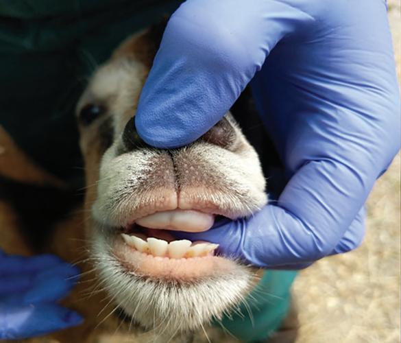
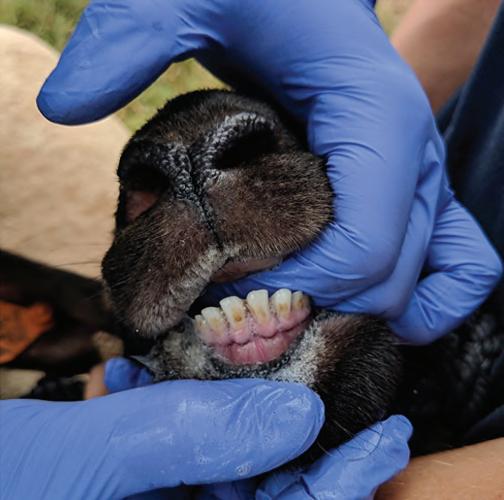
structural abnormalities (e.g., presence of cleft palate), teeth condition, presence of prognathism and brachygnathism, and mucosal lesions such as vesicles or ulcerations is easily achieved during the examination. The presence of a foul oral odor could be an indication of disease associated with the oral cavity, gastrointestinal system (specifically the rumen), or respiratory tract. Teeth eruption and wear patterns can be easily used to estimate the age of sheep and goats (Table 1.3, Figure 1.2). Conversely, cervidae are mostly aged via eruption and wear of the premolars and molars. Typically, eruption of premolars starts at 1.5 years wear and full eruption and wear of molars occurs by 3.5 years. Wear is then evaluated until all premolars and molars reach the gum line at approximately 10 years of age and various wear patterns of the cusp and dentine help to determine the age of the animal (Table 1.3).
Detailed aging information is available from most wildlife and hunting agencies. The most accurate way to determine age is to submit to a laboratory for examination of annular rings.
The use of this method to age the animal becomes less accurate once all of the permanent incisors have erupted and are in wear. Abnormal wear patterns or poor dentition (loose teeth, absence of teeth, and tooth root abscess) may be contributors to a chronic weight loss complaint, especially in situations of competition for food (see Chapter 4).
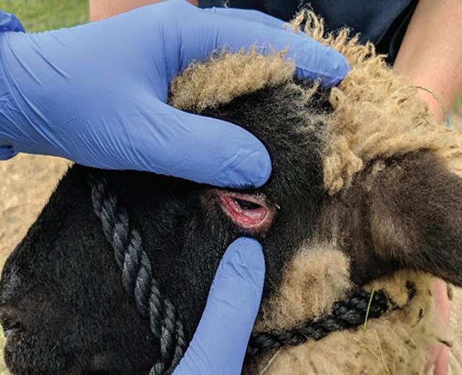
The assessment of hydration status and FAMACHA scoring is accomplished during the ocular examination. Eyeball recession and eyelid skin tenting are the two reliable methods to subjectively determine the hydration status of the animal. FAMACHA scoring (see Chapter 6, Box 6.2 & Figure 6.2 and Chapter 19) is recommended to be part of the routine care of any herd or flock as an important aspect of parasite management and control. The conjunctival membrane color is used to estimate systemic perfusion. (Figure 1.3). Oral mucous membranes should not be used for this assessment as many breeds have a pigmented oral cavity and the rough nature of the mouth may portray an erroneous estimation. As a general rule, pale membranes may indicate anemia, most likely due to intestinal parasitism (Haemonchus contortus infection) or coccidiosis. Jaundice or icteric mucous membranes may indicate a hemolytic crisis or liver disease, such as copper toxicity, and congested (red in color) membranes may be indicative of fever, septicemia, or toxemia.
TABLE 1.3
Estimating Age of Sheep, Goats, and Cervids by Teeth Eruption.
Estimating Age of Sheep and Goats Using the Incisors (I)
Estimating Age of Cervids Using the Premolar and Molar Eruptionsa
Deciduous Eruption Age Fawn–6 months old
I1 Birth–1 week Five or fewer teeth present and the third premolar (tooth 3) has three cusps
I2 1–2 weeks 1½ years of age
I3 2–3 weeks Tooth 3 (third premolar) has three cusps. Tooth 6 has erupted and is slightly visible just above the gum line
I4 3–4 weeks 2½ years of age
Permanent Lingual crest on all molars are sharp and pointed. Tooth 3 now has two cusps. Back cusp of tooth 6 is sharp and pointed
I1 1–1.5 years 4½ years of age
I2 1.5–2 years Lingual crest on tooth 4 rounded off, and in tooth 5 blunt. The dentine in tooth 4 is twice as wide as the enamel. The dentine in tooth 5 is wider than the enamel.
I3 2.5–3 years 6½ years of age
I4 3.5–4 years Tooth 4 is worn completely smooth; no enamel ridge should be visible in the center of tooth 4. Small enamel ridge will be present in center of tooth 5 and tooth 6. Lingual crest on tooth 5 is almost worn away and rounded in tooth 6
aCain and Wallace: A Guide to Age Determination of White-Tailed Deer Austin, TX: Texas Parks and Wildlife, 2003.
Cardiovascular Examination
Objectives:
1. Auscultate both left and right side skin conditions
2. Presence of jugular vein distention
3. Peripheral perfusion
4. Peripheral edema
Auscultation of both the left and right side of the thorax is imperative. Assessment of rate, rhythm, character, and intensity of the heart sounds should be performed. Auscultation of the heart is accomplished by moving the stethoscope over the location of the valves and determining the point of maximal intensity. The pulmonic valve (low third intercostal space, below the elbow), the aortic valve (high fourth intercostal space, above the elbow), and the left (mitral) atrioventricular (AV) valve (at the low fifth intercostal space, at the level of the elbow) are found on the left chest. The right AV valve or tricuspid valve (high fourth
intercostal space, above the elbow) should be auscultated on the right side.
As a general rule, normal heart rate should vary between 70 and 90 beats per minute in adults and between 80 and 130 beats per minute in neonates. There is physiological variation according to environmental conditions (i.e., ambient temperature) and situations that cause hyperexcitability (i.e., aggressive handling or movement). Anemia, murmurs, pain, heart failure, and infectious and inflammatory processes are certain conditions known to effect the heart rate.
Observing for jugular pulses and palpation of distal appendages, such as tip of the ears and limbs are indicators of appropriate peripheral perfusion when warm. Distention of the jugular veins and the presence of pulsations may indicate heart failure. Peripheral edema is known to be consistent with either hypoproteinemia or congestive heart failure and warrants further investigation (see Chapter 17).
• Fig. 1.2 The practitioner may insert the index finger inside of the sheep/goat mouth, laterally, and with the other hand lower the bottom lip allowing exposure of the incisors. The approximate age may be determined according to Table 1.3
• Fig. 1.3 To FAMACHA score sheep and goats, the practitioner gently pushes the upper eyelid medially and rolls the lower eyelid ventrally to access conjunctival membrane color.
Respiratory Evaluation
Objectives:
1. Observe and record rate at a distance first
2. Auscultate both left and right side
The clinician must be aware that the respiratory system should be examined in conjunction with the cardiovascular system and drawing major conclusions without examining both may impair the ability to accurately determine a diagnosis.
On average, the normal respiratory rate should vary between 10 and 30 breaths per minute in adults and between 20 and 40 breaths per minute in neonates. The rate can be obtained by observing the movement of the coastal arch and nostrils from a distance. In cervids, similar rates to sheep and goats can be expected, however, cervids are highly sensitive to excitement and may be hard to evaluate except at a distance. Neonates may “hold” their breath (mule deer fawns especially) when hiding as a reflex to avoid predators.
As noted for the cardiovascular system, environmental conditions and systemic illness are known to influence respiratory rate and must be taken into consideration when appropriate. Animals in apparent respiratory distress, either dyspneic or tachypneic, with open mouth breathing, flaring the nostrils, abducted elbow stance, and with excessive abdominal effort must be evaluated critically and efficiently. Air flow should be symmetric from both nostrils. Mild, clear, serous nasal discharge is a common finding, especially in sheep; however, excessive clear to mucoid to purulent exudate must be explored.
Bilateral auscultation of the lung fields should be performed in a systematic approach. The margins of the lung fields are as follows: the cranial border is deep to the triceps, the dorsal border extends from the point of the shoulder to the last rib, and the caudoventral border arches from the point of the elbow to the last rib. Bronchial sounds usually are loudest over the craniodorsal lung fields at the level of the tracheal bifurcation. Generally, tracheal sounds should be absent. When tracheal disease is present, wheezes can be auscultated, indicating tracheal collapse; obstructive lesions and crackling sounds are characteristic of tracheitis. Elicitation of a cough can be achieved with minimal compression of the trachea and pharyngeal region. The normal animal will cough one to two times, while the diseased patient coughs repeatedly and often with forced effort.
Crackles are auscultated when air moves through inflammatory fluid in the alveoli, whereas wheezes are reverberations of air moving through inflamed, narrowed airways. One must remember that significant lung pathology may be present and not necessarily appreciated on auscultation. Coughing, nasal discharge, dyspnea with a fever, and severe open mouth breathing may be the only indication of lung pathology.
Upper airway diseases, such as rhinitis, tracheitis, foreign body, and compressive lesions, are usually characterized by a loud, harsh, dry, nonproductive cough of acute onset. Lower airway diseases, such as pneumonia, pulmonary edema, lung abscessation, and lungworm infection, are characterized by a chronic, soft, productive cough. Animals with lower airway disease typically cough infrequently and will swallow after coughing, which is different from animals with upper airway diseases who typically do not swallow after coughing.
Cervids can be difficult to assess due to restraint in drop chutes (poor access) or because they are highly excited. Many that have respiratory disease may have advanced disease that has consolidated portions of the lung, leading to “dead” spots that show no
air movement/sounds. They are poor anesthetic as well, so evaluation is difficult at best. Ultrasound, transtracheal wash, radiographs, and other diagnostic tools can be used as well, but risk/benefit ratio and economics must be taken into consideration (see Chapter 7).
Gastrointestinal Examination
Objectives:
1. Examination extends from mouth to rectum
2. Auscultation, palpation, and observation
The gastrointestinal system is one of the largest and most important in the body. Evaluation should be performed in a systematic and stepwise fashion from the mouth to rectum. The mouth should be examined for the presence of vesicles, ulcerations, swellings, and ptyalism. Inspection of the teeth for wear and soundness is important, and the upper dental pad should also be evaluated for evidence of abnormal wear. Although not easy to examine, and sedation or anesthesia may be necessary for a complete examination, the molars should be sound and present as their role in mastication of forages before swallowing and in proper cud chewing is critical. The use of a mouth gag and a bright light source is helpful. It is important to take into consideration that wear patterns may present in different ways and are dependent on the environmental conditions and primary diet of the herd or flock. The practitioner can then make a judgment of whether the wear pattern is abnormal or normal for the living conditions of the animal. Often, culling of lambs or kids is needed due to poor dentition.
The neck area is examined via thorough palpation. Masses, enlarged lymph nodes, or swelling may be causing esophageal compression and subsequent obstruction/choke. Rumen tympany, ptyalism, bruxism, and pain are common clinical signs that may be associated with esophageal disease.
It is wise if the clinician examines one side of the animal first, as this will help to avoid missing any aspect of the examination. On the left side of the animal, the rumen constitutes the major forestomach. Due to its size, the rumen may give an asymmetrical appearance to the abdominal contour favoring a larger “bulge” to the left side, which is considered normal and is expected. Healthy rumen striation consists of a gas cap dorsally, fiber mat in the middle, and fluid (digested ingesta) ventrally. Using the stethoscope, the practitioner should auscultate and perform succussion (i.e., shaking) of the abdomen. Within the left paralumbar fossa, rumen contractions can be auscultated in the healthy animal (sheep, goats, and cervids) at a rate of one to two primary contractions (active rolling of the ingesta) and one secondary rumination (eructation) per minute. A solid understanding of the individual or herd/flock dietary management and medical history, along with a physical examination, helps the practitioner determine the primary cause of rumen fill. Ballottement of the paralumbar fossa while listening with the stethoscope is imperative to support abnormal findings related to the striation of the rumen, displaced abomasum, and ascites. Auscultation of the right paralumbar fossa will allow the practitioner to evaluate the cecum, spiral colon, and small intestines. Illness associated with of any of these structures will lead to fluid and gas accumulation and distension of the viscus in the upper right quadrant. Dilation lower on the right side of the animal may be related to abomasal impaction, late gestation, or a severe rumen impaction.
If bilateral abdominal distention is seen, one may suspect vagal indigestion syndrome (chronic indigestion, failure of the omasal
transport, or pyloric stenosis), ileus, or free fluid accumulation. This fluid accumulation could be caused by diffuse peritonitis, ascites due to protein losing enteropathy, liver failure, or severe congestive heart failure.
Body temperature should be taken rectally observing common biosecurity practices. Normal temperature typically varies from 100.5° F to 103.5° F. In general, sheep tend to have a higher body temperature than goats, and cervids typically fall in the same range (101.5° F–102.5° F) with variations due to age, activity level, and environmental temperature. The practitioner must distinguish between true hyperthermia and a febrile response. A febrile response is more likely to be associated with an inflammatory or infectious process, whereas hyperthermia is going to be associated with the location of the patient (e.g., barn, paddock, pasture, etc.), behavior (e.g., hyperexcitability does increase body temperature), and environmental conditions (e.g., high temperature and humidity).
The authors would like to stress that obtaining body temperature should be the first procedure to be performed when examining sheep and goats and the results interpreted in conjunction with other clinical signs.
Fecal consistency and staining of the perineum, tail, and back of legs is a good way to assess the history of diarrhea. A thorough history of dietary management and fecal examination (fecal float or fecal egg count) is the only way the practitioner will reliably make a diagnosis and then recommend a targeted treatment.
In young stock, the authors recommend full examination of the umbilical structures both externally and internally. The use of ultrasound imaging if pain or swelling is found is highly valuable. Any signs or history of diarrhea in lambs, kids, or fawns must be addressed quickly as it can be life threatening. Lastly, atresia ani and atresia coli have been reported in kids and lambs, so the practitioner needs to be sure there is a patent anus and fecal passage present (see Chapter 5).
Urogenital Examination
Objectives:
1. Examination from a distance
2. History
3. Ultrasound imaging
The examination commences at the external genitalia of both males and females. In males, the prepuce should be free of adhesions, swelling, or any signs of trauma. The preputial opening should be evaluated for the presence of crystals, blood, excessive dryness, scabs, or ulcerations since any of these may be indicative of urethral calculi, obstructive urolithiasis, or ulcerative posthitis.
Urine samples in both sheep and goats can often be obtained by briefly occluding the nostrils. Young cervids can be encouraged to urinate with gentle stimulation. Older cervids that are bottle raised may be able approached for a “free catch” urine sample.
The penis is difficult to examine without the use of sedation or anesthesia (cervids). The authors strongly recommend the use of acepromazine or a benzodiazepine (see Chapters 8, 12, and 18) for sedation and relaxation. Rams and bucks can be placed in lateral recumbency or sitting up on their rump (preferred method) by an assistant, then the practitioner pushes the prepuce caudally while pushing the sigmoid flexure cranially. Once exteriorized, the practitioner can hold the penis using gauze. The surface of the penis should be examined for color, scabs, and any traumatic lesions. Palpation of the penis may reveal the presence of uroliths, swelling, or a focal area of pain. The urethral process in sheep and
goats should be examined closely for the presence of a urolith or sandy grit, which may be indicative of urolithiasis or urethral blockage. Cervids do not have a urethral process.
Frequently, the presentation of a sheep or goat with suspected urogenital disease involves standing in a stretched out position, intermittent straining, vocalization, and wagging of the tail when attempting to urinate. This stance is often confused by owners and their perception is that the animal is constipated when in all actuality the animal has a urinary obstruction. History of inability to urinate followed by relaxation and acute abdominal distention may indicate rupture of the urinary bladder, whereas caudal ventral edema (often reported by the owner as “broken penis”) may indicate distal urethral rupture.
It is important to take into consideration, contrary to what is commonly done in small animal practice, catheterization of the urethra is difficult in does and ewes owing to the presence of the urethral diverticulum at the floor of the pelvis and close to impossible in bucks and rams. Multiple anatomic locations in male anatomy (urethral process, sigmoid flexure, urethral diverticulum) are difficult to traverse with a catheter. Attempts to pass a urinary catheter can actually cause more harm due to severe trauma caused by the procedure.
The testicles are gently palpated to ensure they are not adhered to the scrotum, and there are no signs of epididymitis, orchitis, and poor testicular tone, which are often associated with suboptimal sperm production. In breeding males, the phrase “big is beautiful, mobility meaningful, resilience respectable, softness suspicious” is helpful to remember when evaluating males for breeding soundness. In addition, the scrotum should be free of traumatic lesions with intact skin. Signs of dermatitis due to ectoparasites, frostbite, or asymmetry are undesirable findings (see Chapter 8).
In females, the labia of the vulva is examined for their color, size, and presence of discharge. Pale mucous membranes may be an indication of anemia, whereas hyperemia and swelling may indicate the onset of estrus or an impending parturition. If calculi or sandy grit is found attached to the hairs below the urethral orifice, urolithiasis is suspected and the practitioner must evaluate further. Reproductive history is important when it comes to evaluating a potential vaginal or uterine discharge. Color, consistency, and volume are a good start as they may characterize a late estrus discharge, a postpartum normal lochia, or an infection. Lochia is considered a normal finding between days 0 and 21 post parturition. The finding of large protruding vulvar labia or clitoris, or a short anogenital distance is suggestive of an intersex condition (see Chapter 8).
In both males and females with suspected obstructive urolithiasis, an enlarged bladder may be palpable extending from the pelvis to the abdomen; in this case, the authors recommend further examination using ultrasound imaging. Caution should be used when applying manual pressure to the abdomen because there is a risk of rupturing the bladder and causing more pain to the patient (see Chapter 12).
Musculoskeletal Examination
Objectives:
1. Examination from a distance
2. History
3. Knowledge of foot conditions
4. Imaging examination
First, posture and locomotion are evaluated at a distance for both sheep and goats, as well as cervids. The animals are then
observed as they walk away from and towards the practitioner. It is important to note that lameness issues often present in a variety of ways and because of the prey mentality of small ruminants, the lameness may be very subtle. The patient may prefer to not bear weight on the limb at rest and use it sparingly while in motion, or may bear weight at rest and hop on three legs while in motion.
All claws should be observed for appropriate wear, hoof-wall separation due to white line disease, and defects in the sole. The interdigital space should be checked for pain, exudate, or a foul odor. The coronary bands should be observed for pain, swelling, ulceration, or separation from the foot. Separation of the hoof wall from the hoof in cervids is a common sequelae to hemorrhagic disease survivors. All joints should be palpated and checked for appropriate range of motion. Older and/or heavier small ruminants may have “clicking” within their joints indicating chronic osteoarthritis or overuse of the joint(s). This may or may not be an abnormal finding but should be recorded in the medical record.
In young stock, septic joints are typically diagnosed before swelling is ever a problem. It is an extremely painful condition affecting one or more joints and likely a sequelae from failure of passive transfer. Many of these patients present with non–weight-bearing lameness rather than swelling being noticed at one or more of the joints.
In goats, hygromas and synovitis secondary to caprine arthritis encephalitis infection can be differentiated on clinical examination. Hygromas are nonpainful, whereas synovitis typically is a painful condition.
Fractures must be evaluated immediately. The age of the animal, location of the fracture, and intended purpose (pet or production animal) will allow for an appropriate treatment plan and prognosis. Prognosis is also easily determined by radiographic examination (see Chapter 11).
Nervous System Examination
Objectives:
1. History
2. Examination—localizing the lesion
It is imperative for the practitioner to always wear gloves when interacting with an animal showing neurological disease. In general, the neurological examination should start by obtaining a thorough history of the patient. The examiner should have a full understanding of the animal’s diet and behavior within the past 24 to 48 h, housing and environment, new additions to the herd, travel, and interaction with wildlife.
From a distance, gait, posture, and overall behavior when interacting with herd or flock mates and with humans must be noted. Known traumatic events must be taken into consideration.
Clinical signs will help the practitioner to localize the lesion to the peripheral or central nervous system.
In sheep, goats, and cervids, infectious peripheral nerve disorders are less common than traumatic events leading to peripheral nerve damage. The peripheral nerves and their most likely clinical presentation when traumatized are summarized in Table 1.4 Often, one or more lesions is appreciated on clinical examination and this is attributable to multiple nerve roots or pathways being affected (e.g., complicated dystocia followed by traumatic obstetric maneuvers).
Sciatic and obturator nerve paresis and paralysis are the most common peripheral pelvic limb disorders. Radial nerve paralysis is the most common nerve palsy affecting the thoracic limb in sheep, goats, and cervids.
TABLE 1.4
Typical Clinical Signs Associated With Peripheral Nerve Disease.
Peripheral Nerves Clinical Signs
Femoral nerve Inability to bear weight and advance the limb, absent patellar reflex
Sciatic nerve Knuckled fetlock with dropped hock and intact patellar reflex
Peroneal nerve
Hyperflexed fetlock, overextending the hock, and inability to extend digit
Obturator nerve Inability to adduct limbs
Tibial nerve Knuckling of fetlock but no dropped hock
Radial nerve Inability to advance the limb
TABLE 1.5
Typical Clinical Signs Associated With Central Nerve Disease.
Area Affected Clinical Signs
Cortical and cerebral
Cortical
Cerebellar and spinal cord
Cerebellar
Spinal cord
Brain stem
Changes in mentation with normal gait, posture, and spinal reflexes
Head pressing, propulsive walking, convulsions, and blindness
Altered gait and posture with normal mentation
Ataxia with normal strength and proprioception, truncal sway, hypermetria and head tremor
Increased extensor tone and exaggerated spinal reflexes or paresis to paralysis with decreased spinal reflexes
Change in mentation, gait, posture, and spinal reflexes may or may not be present. Cranial nerve deficits which may manifest as head tilt, flaccid tongue, facial paralysis, circling, or ptosis
The central nervous system is divided into four major anatomic sites to which clinical signs may be localized: cortical, cerebral, cerebellar, and spinal cord. Diseases at any of these locations may be characterized by alterations in mentation, gait, posture, and spinal reflexes. The common clinical signs associated with the location in the nervous system are summarized in Table 1.5
Chapter 13 discusses in detail differential diagnoses for each location, treatment, and prognosis associated with nervous system diseases.
Mammary Gland Examination
Objectives:
1. Production history
2. Reproduction history
3. Examination
If in lactation, both halves of the mammary gland, teats, and teat sphincter are observed and palpated for symmetry, size, conformation, temperature, and consistency. Infectious and noninfectious mastitis is detrimental to the production life of the female, and can be a life-threatening disease if not treated promptly and correctly.
It is recommended to first and foremost gather a complete history and observe the young stock. Problems associated with the udder can be appreciated first in the lambs or kids that are weak, show poor body condition, or are failing to gain weight. Malnourished neonates can be an indication of poor milk production or a painful udder in the dam that has resulted in the dam not allowing the neonate to nurse.
The presence of edema that extends symmetrically and cranial ventral to the udder is a common finding shortly after parturition, especially in first-time ewes or does. A diffusely hard or firm udder noted in the first few days after lambing may indicate ovine progressive pneumonia (OPP) infection in sheep or caprine arthritis encephalitis (CAE) in goats. Low milk production and no signs of clinical mastitis are a common occurrence in most cases of OPP and CAE (see Chapter 16).
Aside from palpation of the mammary gland, if the doe is in lactation, the practitioner must remove a few streams of milk from both sides to assess patency of the sphincter. Color, consistency, and presence of abnormal clots or flakes in the secretion should prompt the practitioner to investigate further. The California Mastitis Test (CMT) can be used to determine if a subclinical mastitis is present and if further culture of the secretion is necessary.
Prepartum mastitis, although uncommon in small ruminants, can be caused by a herd/flock mate suckling or by a pathogen. This condition must be evaluated and treated promptly as it can severely affect colostrogenesis and ability to lactate after the birth of the offspring (see Chapters 15 and 19).
Lymphatic Examination
Objectives:
1. History of Corynebacterium pseudotuberculosis (CL) in the herd 2. Examination
It is part of the physical examination to palpate all the peripheral lymph nodes. Submandibular, retropharyngeal, parotid, prescapular, prefemoral, and supramammary (in females) are the most common palpable lymph nodes. It is important to note that often the practitioner will be unable to physically feel them, either because they are too small, or in wool breeds of sheep, the thick wool will impair access to them.
The authors recommend that attention be paid to lymph nodes that are enlarged and draining purulent exudate. CL infection is known to be the most common disease associated with draining lymph nodes in small ruminants and is extremely contagious in nature and is considered a zoonotic pathogen. Cervids commonly have lymph node involvement with Fusobacterium infections (see Chapter 16).
Integumentary Examination
Objectives:
1. Examination
2. Environment
Lesions like abrasions, lacerations, papules, pustules, scabs, and hair or wool loss are clinical signs associated with and indicative
TABLE 1.6
Typical Clinical Signs and Their Associated Differential Diagnosis.
Clinical Signs Potential Common Etiologies
Pruritus Mange, allergy, and scrapie
Hair loss Ringworm, mange, and nutrition
Skin nodules Abscesses, pustules, and demodectic mange
Dandruff Dry environment and often poor or improper nutrition
Crustiness Chorioptic mange (under the dew claws), fungal or bacterial dermatitis
Sunburn Hairless parts of the body in white animals (often seen on the top line, tip of nose, ears). Must differentiate from photosensitization
Barbering Chewing, biting, pulling of hair or wool by self or others
of dermatological issues. Always take into consideration the season and type of environment where the animals are being housed. Haired breeds of sheep e.g., Barbados, Katahdin, St. Croix, etc.) and goats will shed winter coats in the spring. Wooly sheep (e.g., Dorset, Suffolk, Merino, Corriedale, etc.) need to be sheared at least once a year, during the summer months. Wool blindness is a term often used by producers in reference to sheep with excessive wool above their eyes leading to their sight being physically impaired. In these cases, shearing of the periorbital area must be performed to avoid further damage, such as severe dermatitis and eye damage.
In cases where the practitioner encounters a flock with more than one case of poor wool quality, nutrition issues should always to be discussed. Hairiness or abnormal wool pigmentation, such as presence of brown fibers over the nape of the neck in wool sheep, may indicate border disease infection. Table 1.6 summarizes the most common clinical signs associated with skin or coat diseases in sheep and goats (see Chapter 10).
Restraining and Handling
Handling Sheep, Goats, and Cervids
Biosecurity. The practitioner should always be aware of potential zoonotic diseases during routine handling of small ruminants. Protective clothing and gloves should be worn at all times when visiting a herd or flock, and while interacting with animals. Clothes must be changed and footwear thoroughly washed between farms as it can easily serve as a fomite for infectious and contagious pathogens. As mentioned earlier, part of the physical examination is to learn about the herd health status through the use of historical information. This information will help the practitioner to identify the potential risk for the presence of zoonotic disease within a flock or herd. Segregation of sick animals and dedicated areas for lambing or kidding are strongly recommended to avoid and prevent pathogen transmission.
To prevent the introduction of new diseases to an established herd or flock, a prepurchase examination performed by a veterinarian is strongly recommended. Although prepurchase examinations do not guarantee the future health of that individual at the
future farm, it serves as an assurance that at that point in time there is a healthy female or breeding male. Aside from obtaining historical data from the herd/flock as a whole, the veterinarian must also ask directed questions concerning vaccination history, dietary protocols, and if any previous health events have occurred. It is also imperative that the practitioner question whether any treatments have been performed and by whom. Also, the veterinarian may decide to perform diagnostic tests including serology (for caprine arthritis encephalitis, caseous lymphadenitis, paratuberculosis, etc.), serum biochemistry, complete blood count, and a fecal examination. A reproductive or breeding soundness examination in both males and females may be indicated. It is strongly recommended that the new owner quarantine new animal additions a minimum of 4 weeks with no physical or visual contact with existing animals. Thirty days is known to be sufficient for most of the diseases that are worrisome and for those animals to show clinical signs. The authors understand that quarantine may be logistically difficult for some herds/flocks. Quarantine allows new animals the chance to acclimate to the environment, diet, and behavior patterns, allowing a stress free and productive atmosphere (see Chapter 19).
Behavior and Facilities. The use of behavior patterns and handling principles like “flight zone” and “point of balance”, as well as providing appropriate facilities are the hallmark to successfully and safely working with sheep, goats, and cervids.
TABLE 1.7 Typical Behavior Characteristics of Sheep and Goats.
Once one enters the animal’s flight zone, to the point where they feel threatened, the animal will walk/run away and face the person to assess the situation. The way the farmer or the veterinarian handles this situation is exactly the way the animal will handle it. That is, if the practitioner enters the flight zone calmly, the animal will behave calmly; if the person aggressively and loudly runs or walks towards the animal, the normal behavior is to also run. It is imperative to remember sheep, goats, and cervids are typically small and fast, yet extremely strong. They can injure themselves and/or injure the people that are attempting to work with them. In tightly enclosed spaces, some cervids will choose to fight and have been known to cause injury or death, especially males with antlers.
The level of the shoulder is known to be the point of balance. When working a herd or flock, the location where the people managing the animals are standing or moving about makes a significant difference in how effectively and timely the job can be executed. Standing in front of the chute or alleyway intended for the animals to walk through is counterproductive. If the desire is to encourage the animals to walk forward, the practitioner must stay behind the point of balance (behind the level of the shoulder). If the goal is to encourage the animal to back up, then the individual may walk past the level of the shoulder; this will invariably make the animals walk backwards. Having knowledge of behavior patterns in sheep and goats is a fundamental part of successful handling (Table 1.7).
TYPICAL BEHAVIOR CHARACTERISTICS
Activity Sheep Goats
Food preference Grass and succulent herbage Browse (weeds, leaves, twigs)
Food variety Accept monotonous diet
Habitat selection Lowlands or hilly grasslands
Antagonistic behavior Butt head on
Require variety
Climb rocks and elevations
Sideways hooking motion
Fighting Butt Rear on hind legs
Sexual behavior Less herding
Newborn young behavior Remain by dam (“lying in”)
Herding of females
Freezing some distance from the dams (“lying out”)
Alarm signal Snort and stamp forefoot Frequent high pitched “sneeze”
Alarm Form compact bunch Form thin line
Hornless condition Fertile
Sterile (usually) in males
Tail Hangs down Stands up
Beard Absent
Wattles Absent
Hear a low flying plane Frightened and likely to run
Stress Results from isolation or subjugation to unfamiliar environment
Cervids
Browse broadleaf herbaceous weeds, leaves and tender twigs, and grass
Require variety
Hardwoods, croplands, brush lands, and pastures
Bite and push
Fallow deer tend to maintain sexual segregation and make lion-like vocalizations
First days, they hide in tall grass becoming more active the second week of life
Snort or whistle, groan, or bleat when predators are around
Run
N/A
Down when calm/up with alarm
Present in buck and some females Absent
May be present
Often stand and watch
More of a problem in young kids and doelings
Absent
Alarm/run
Segregation or confinement
Another crucial aspect of managing small ruminants is that the farmer must be able to gather, restrain, and handle animals with minimal stress. Injury prevention for both animals and personnel is crucial. Small ruminants will readily follow one another and will move away from things that frighten them. They move better around slight corners or curves and will not move toward an area that appears to be a dead end. Sheep and goats will move away from buildings and prefer to move uphill. Lit areas are preferred as the animals will resist movement into dark barns, alleys, and chutes. Handling areas should be well lit and free of objects that may project shadows into the visual path. Solid sides in alleyways will help maintain forward momentum and minimize attempts at escape.
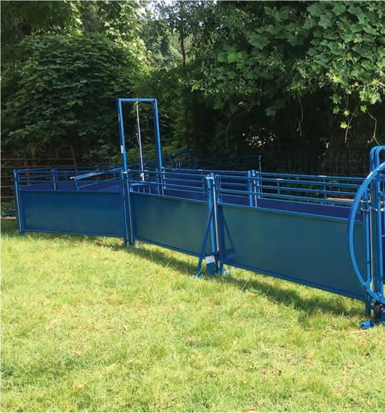
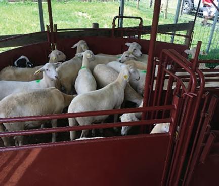
Well-planned working facilities to deal with a flock of sheep for example, are known to provide the desirable details mentioned above. Commercially available chute systems (Figure 1.4A) placed in an appropriate, well-lit location that is free of random loose
objects, allows the flock to ease their way into the holding area. This is considered one of the best-known ways to work a flock (Figure 1.4B).
If a chute system is not available, the farmer should consider moving the flock into a small paddock or stall as a group (Figure 1.5). It is wise to always try to work the flock together, even though you may not need all animals present in that group. Sheep have an extremely strong flocking instinct, therefore, if one animals is seen segregated and away from the flock, the practitioner and famer must investigate further. Once the group is in a small area, the practitioner carefully and calmly enters the area to attempt to catch an individual animal. Always remember they can be flighty animals and if necessary, they will give their full potential to escape by head butting or jumping.
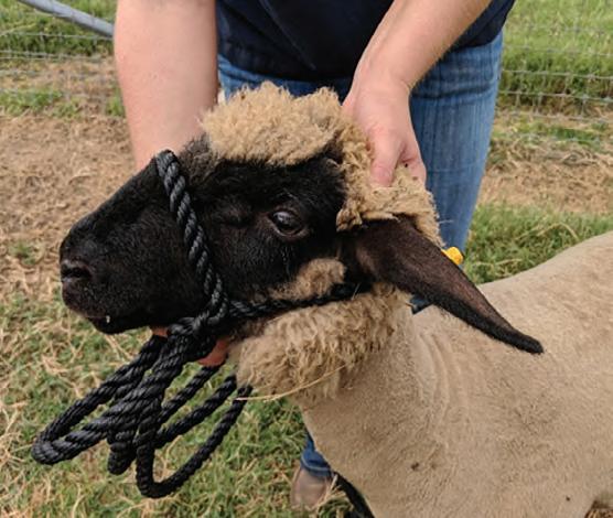
To catch a sheep, the handler can cup a hand under the animal’s jaw, grasping the bony part of the jaw—not the throat.
A B
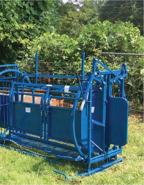
• Fig. 1.4 A. Commercially available chute restraint system to facilitate herd work. (Photo courtesy Sims Pond Farm.) B. Operating the chute system to safely restrain the animals allowing routine livestock procedures to be performed. (Photo courtesy Hunt Road Katahdin Sheep Farm.)
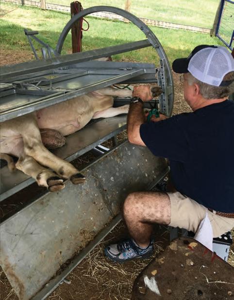
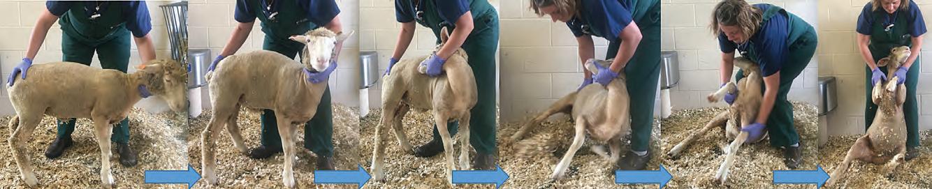
Once it has been caught, a second hand should be placed behind the head below the animal’s ears. It is important to note that for better control, the animal’s nose should be pointed upward to stop its forward motion, as sheep have a lot more power when the head is down. The handler should never grab the sheep by the wool or hair. A crook or lariat also is an acceptable catching device. A sheep can be handled using various handling points— for example, under the mandible, tail, and flank (Figure 1.6). After it has been caught, a sheep can be “tipped” on its rump for examination, shearing, foot trimming, and other routine procedures (Figure 1.7).
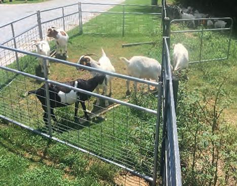
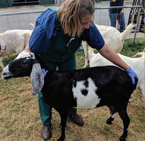
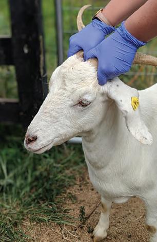
Goats are different in many respects when it comes to handling and to the facilities needed to work them. Goats are not as concerned about herding, but rather they develop close relationships with certain herd mates and can be seen playing and socializing. Goats typically spread out while browsing and ruminating. To catch a goat, the use of the horns as “handles” is an acceptable way to get a hold on them (Figure 1.8A, B); restraint by their ears is painful and considered abusive. Goats housed with a collar or halter can be caught using this, with the handler looping an arm around the animal’s neck. It is strongly recommended not to hold a goat by its hindlimbs as it may possibly dislocate a hip joint in an attempt to escape.
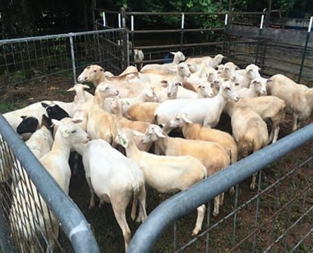
• Fig. 1.5 Depicted in this image is an example of a corral area. If a chute system is not available, small groups are moved into these smaller areas allowing herd work to be performed.
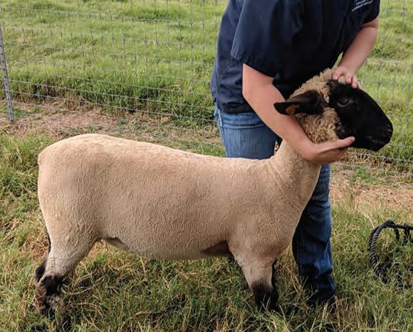
• Fig. 1.6 Proper method of individual animal restraint.
• Fig. 1.7 Series of images on how to tip a sheep and place it on its rump. This is a common method used to restrain adult sheep, allowing a multitude of livestock procedures to be performed (e.g., foot trimming, shearing).
• Fig. 1.8 A. This image shows how to properly
breeds, it is important to note that the horns must be
table with solid cushioned sides for cervids and manual restraint of the horns.
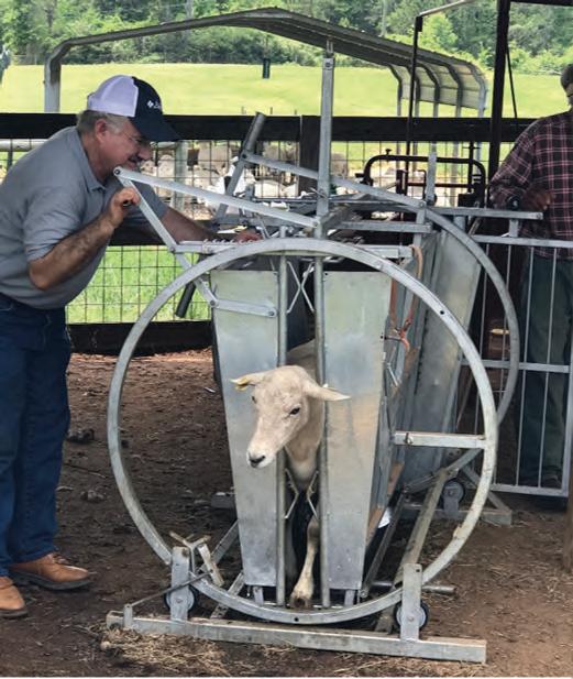
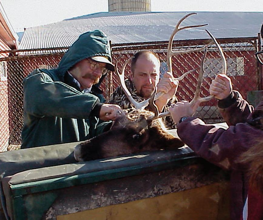
Special handling facilities for cervids will include a drop chute (Figure 1.9) and a box system leading to the chute, or box stalls with remote door opening to minimize animal-human contact (Figure 1.10). Animals may actually be calmer in the dark and work better through the facility. Training the animals to use the facility is very important and will result in less stress to the animals and handlers. Small fawns may be restrained manually, but larger fawns and adults may injure themselves or the handlers if not sedated or restrained in a chute. Cervids can strike with their front feet, or males in hard antler may charge and attempt to gore a handler. Shields may be used, but properly designed facilities usually do not require handlers to enter small confined areas. Extremely tame cervids may lie down and refuse to move in some cases and may have to be manually pushed or prodded into a chute or pen.
Fencing for cervids may be dictated by state statute. Most cervid fencing comes in 8- or 10-foot heights and is high-tensile
restrain a sheep and a goat by the head. In horned
held at the base. B. This image shows a restraint
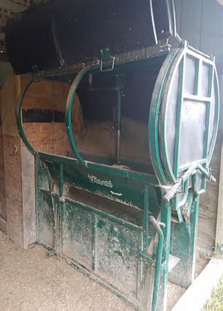
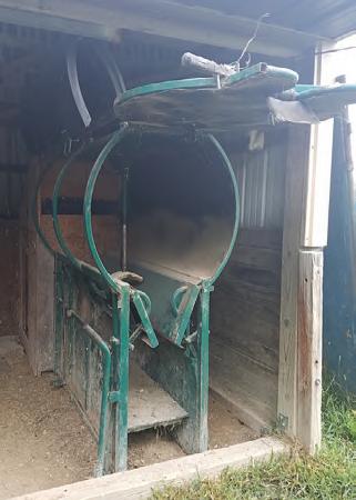
• Fig. 1.9 Commercially available chute restraint system to facilitate herd work of cervids.