LIST OF CONTRIBUTORS
Anthony P. Auger
University of Wisconsin–Madison, Neuroscience Training Program, Departments of Zoology and Psychology, Madison, WI, USA
Jill B. Becker
University of Michigan, Molecular and Behavioral Neuroscience Institute; University of Michigan, Department of Psychiatry; University of Michigan, Department of Psychology, Ann Arbor, MI, USA
Kristen M. Culbert
University of Nevada Las Vegas, Department of Psychology, Las Vegas, NV, USA
Kelly M. Dumais
Boston College, Neurobiology of Social Behavior Laboratory, Department of Psychology, Chestnut Hill, MA, USA
Yosefa Ehrlich
Brooklyn College of the City University of New York, Department of Psychology, Brooklyn, NY, USA
Liisa A.M. Galea
University of British Columbia, Department of Psychology; University of British Columbia, Centre for Brain Health, Vancouver, BC, Canada
Juan L. Gomez
Oregon Health & Sciences University, Department of Behavioral Neuroscience, Portland, OR, USA
Gian D. Greenberg
Oregon Health Sciences University, Department of Behavioral Neuroscience, Portland, OR, USA
Robert J. Handa
University of Arizona College of Medicine, Department Basic Medical Sciences, Phoenix, AZ, USA
Ashley A. Keiser
University of Michigan, Department of Psychology, Ann Arbor, MI, USA
Kelly L. Klump
Michigan State University, Department of Psychology, East Lansing, MI, USA
Victoria N. Luine
Hunter College of CUNY, Department of Psychology, New York, NY, USA
Lisa Y. Maeng
Harvard Medical School, Department of Psychiatry, Boston, MA, USA
Anna M. Malysz
Baylor College of Medicine, Department of Molecular and Cellular Biology, Houston, TX, USA
Shailaja K. Mani
Baylor College of Medicine, Department of Neuroscience, Houston; Baylor College of Medicine, Department of Molecular and Cellular Biology, Houston, TX, USA
Christian J. Merz
Ruhr-University Bochum, Institute of Cognitive Neuroscience, Department of Cognitive Psychology, Bochum, Germany
Mohammed R. Milad
Massachusetts General Hospital, Department of Psychiatry, Charlestown, MA, USA
Gretchen N. Neigh
Emory University, Department of Physiology, Department of Psychiatry & Behavioral Sciences, Atlanta, GA, USA
Christina L. Nemeth
Emory University, Department of Physiology, Department of Psychiatry & Behavioral Sciences, Atlanta, GA, USA
Mario G. Oyola
Baylor College of Medicine, Department of Neuroscience, Houston, TX, USA
Adam N. Perry
University of Texas at El Paso, Department of Biology, El Paso, TX, USA
Sarah E. Racine
Ohio University, Department of Psychology, Athens, OH, USA
Doodipala Samba Reddy
TAMHSC College of Medicine, Department of Neuroscience and Experimental Therapeutics, Bryan, TX, USA
Meighen Roes
University of British Columbia, Department of Psychology,Vancouver, BC, Canada
Sydney A. Rowson
Emory University, Department of Physiology, Department of Psychiatry & Behavioral Sciences, Atlanta, GA, USA
Jaclyn M. Schwarz
University of Delaware, Department of Psychological and Brain Sciences, Newark, DE, USA
Farida Sohrabji
TAMHSC College of Medicine, Department of Neuroscience and Experimental Therapeutics, Bryan, TX, USA
Brian C. Trainor
University of California, Department of Psychology, Davis, CA, USA
Natalie C. Tronson
University of Michigan, Department of Psychology, Ann Arbor, MI, USA
Alexa H. Veenema
Boston College, Neurobiology of Social Behavior Laboratory, Department of Psychology, Chestnut Hill, MA, USA
Deborah J. Walder
Brooklyn College of the City University of New York, Department of Psychology, Brooklyn, NY, USA
C. Jane Welsh
Texas A&M University, Department of Veterinary Integrative Biosciences, College Station, TX, USA
Christel Westenbroek
University of Michigan, Molecular and Behavioral Neuroscience Institute, Ann Arbor, MI, USA
Oliver T. Wolf
Ruhr-University Bochum, Institute of Cognitive Neuroscience, Department of Cognitive Psychology, Bochum, Germany
Beril Yaffe
Brooklyn College of the City University of New York, Department of Psychology, Brooklyn, NY, USA
Sex Differences in Immunity and Inflammation: Implications for Brain and Behavior
Gretchen N. Neigh, Christina L. Nemeth, Sydney A. Rowson
Emory University, Department of Physiology, Department of Psychiatry & Behavioral Sciences, Atlanta, GA, USA
1 INTRODUCTION
Are sex differences in the immune system evolutionary in nature? Sir Peter Medawar was the first to address the question of how a mother is able to immunologically tolerate her fetus (Trowsdale and Betz, 2006). This complex issue where a fetus, up to 50% immunologically foreign, is able to pass inert has been discussed since the 1950s (Abrams and Miller, 2011; Trowsdale and Betz, 2006). The necessity for a mother to have an immune system that can fluctuate in order to prevent the rejection of the foreign fetus is one explanation for the baseline differences we observe in the male and female immune systems (Van Lunzen and Altfeld, 2014; Abrams and Miller, 2011).
Sex differences in the immune response and susceptibility to immune-related diseases cannot be disputed.The exact nature of these differences and how these differences contribute to sickness and disease incidence is quite complex, depending on a multitude of factors including age, genetics, and environment. The evolutionary “need” brought on by these factors carries with it far reaching effects on both peripheral and central functions of the immune system. The sections within this chapter highlight the basic immune-related differences between men and women, citing studies of both human disease and model animal systems. Differences in disease incidence and baseline immune activity will be outlined, followed by potential mechanisms to explain these differences, and finally, sex-dependent immune effects on behavior and the manifestation of comorbid disease states will be discussed.
1.1 Immune mediators
A vast array of inflammatory cells mediates peripheral and central immune responses. The immune response is divided into two component systems, the innate and adaptive systems, which differ in their ability to recognize and remember specific pathogens and antigens. While the innate system mounts a generalized and nonspecific response, the adaptive system triggers a response that is both pathogen/antigen specific and based on an immunological memory of previous responses. Despite this major difference, both
Sex Differences in the Central Nervous System. http://dx.doi.org/10.1016/B978-0-12-802114-9.00001-9
Copyright © 2016 Elsevier Inc. All rights reserved.
the innate and adaptive immune responses are composed of cell-mediated and humoral components. It is important to note that while these immunological components may originate peripherally, the brain is not unaffected – or immune privileged. Macrophages and dendritic cells are located within the brain and respond to inflammatory stimuli (Dantzer et al., 2008), and furthermore, activation of microglial cells, the brain’s resident macrophages, occurs readily following infection. In addition to resident immune cells in the brain, several different routes of immune-to-brain access are possible including humoral access through circumventricular organs (Rivest, 2009), primary afferent nerve activation (vagal and trigeminal nerves; Goehler et al., 2000; Dantzer et al., 2008), cytokine trafficking through increased permeability of the blood–brain barrier, and activation of macrophage or endothelial cell interleukin-1 (IL-1) receptors, which cause a local increase of cytokine and prostaglandin release (Kubera et al., 2011; Rivest, 2009; Dantzer et al., 2008).
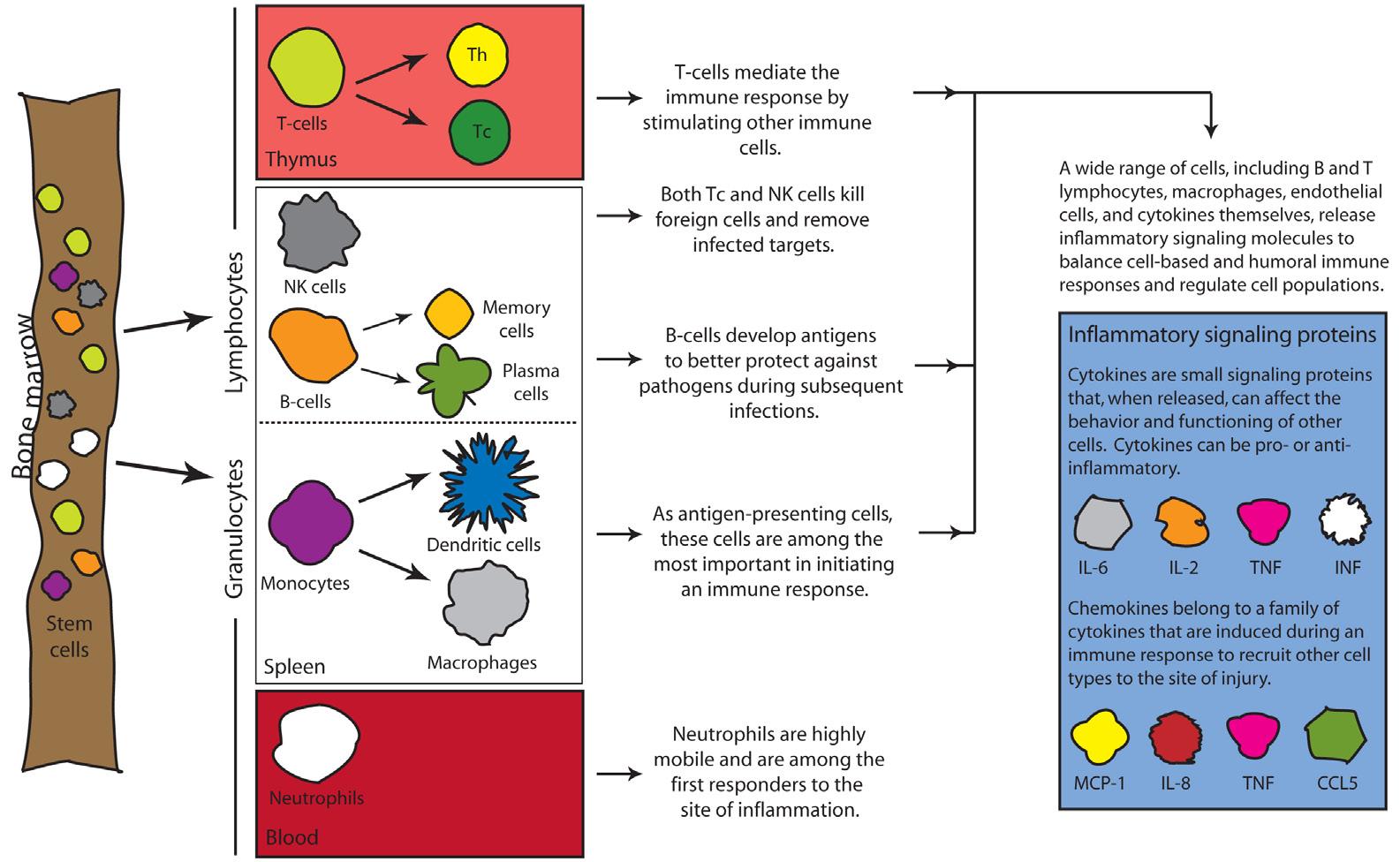
Within the body, lymphoid tissues and immune-relevant organs house the immunological system. Four major organs of the immunological system are bone marrow, thymus, spleen, and lymph nodes. All cells of the body are derived from the bone marrow. Here, stem cells develop into mature red blood cells, platelets, lymphocytes, and granulocytes while some migrate out of the bone marrow to mature. The following is a brief review of inflammatory mediators to aid in the understanding of how basic sex differences affect these cell populations and the immune response ( Figure 1.1 ).
T-cells: They derive from immature lymphocytes, mature in the thymus and are released into the blood stream. In action,T-cells have two very different functions. T helper (Th) cells coordinate the immune response and activate other necessary immune cells. There are two distinct types of Th cells, Th1 and Th2, which are differentiated by the inflammatory cytokines that they release. Th1 cells release inflammatory cytokines that promote phagocytosis while Th2 cells release cytokines that stimulate the production of antibodies. Cytotoxic T lymphocytes (Tc cells) are important for the downregulation or destruction of parasites, tumor cells, and virus-infected cells. Like many other immune cells, T-cell recruitment to the brain occurs following injury and in many immune-related disorders (Engelhardt and Ransohoff, 2012).
B-cells: They originate in the spleen and are important for the development of specific antigens against foreign bacteria, viruses, and tumor cells. B-cells respond to inflammation within the brain and are thought to be one of the main effector cells in multiple sclerosis pathogenesis, promoting inflammatory activity on both sides of the blood–brain barrier (Büdingen et al., 2012).
Natural killer (NK) cells: These cells, similar to T- and B-cells, derive from lymphocytes. NK cells are the most effective killer cells and are similar to Tc cells. NK cells destroy parasitic or infected foreign targets. Unlike Tc cells, NK cells do not
Figure 1.1 Immune cells originate from hematopoietic stem cells in the bone marrow. A number of these cells then mature within the bone marrow while other cells migrate to other tissues to mature and further differentiate (left). Immune cells are important for the recognition of pathogens and the initiation of inflammatory processes. Once activated, many cells are capable of releasing cytokines and chemokines to further regulate cellular activity and the immune response (right).
require target recognition prior to killing infected cells, and therefore work more efficiently than Tc cells. Because NK cells act in a general fashion, they are one of the quickest responders to cerebral injury and are recruited to sites of injury within the brain. NK cells have been shown to have detrimental effects following injury, such as stroke (Gan et al., 2014).
Neutrophils: These are the most abundant white blood cells, and are produced in the bone marrow. Neutrophils are one of the first responders to the site of injury where they promote secretion of anti-inflammatory molecules while reducing cell death and the release of toxic substances. In the brain, neutrophils migrate to the site of injury within hours and can contribute to neurotoxicity in response to injury (Allen et al., 2012).
Macrophages:These are essential to the immune response. Activation of the immune system is triggered when macrophages and/or dendritic cells present antigens to T- or B-cells within the spleen. Macrophages also participate in phagocytosis and release cytokines that modulate the immunological response. Macrophages stem from perivascular monocytes, which are derived from blood-borne monocytes, and reside just outside of the basement membrane of the brain. These cells cycle
in and out of blood vessels and play an important role in immune responses within the brain.
Microglia: They serve as the brain’s resident macrophages and are highly involved in the central immune response and the release of inflammatory signaling proteins. These cells are derived from hematopoietic stem cells and enter the brain early in gestation to mature. Though similar in nature, recent evidence suggests microglia to be very different from macrophages in both origin and function (Prinz and Priller, 2014). Microglial involvement is implicated in a variety of immune-related disorders as discussed throughout this chapter.
Dendritic cells: Similar to macrophages, dendritic cells originate in the bone marrow and are capable of presenting antigens. Dendritic cells, due to their vast presence, are more efficient antigen-presenting cells than macrophages and work to reduce cytokine release while increasing cell death pathways. Derived from monocytes, dendritic cells also infiltrate to the brain and participate in localized increases in inflammation (Prinz and Priller, 2014).
2 SEX-DEPENDENT BASELINE DIFFERENCES IN IMMUNE FUNCTIONING AND RESPONSE
Given that immune cells of both peripheral and central origin have the capacity to alter function of the central nervous system, we will first review the relationship between sex steroids and the immune system with a focus on peripheral organ systems before honing in on specific interactions and implications within the central nervous system. Sex hormones play a pivotal role in the differences and fluctuations in immune activation between men and women (see Table 1.1). Estrogen exerts a biphasic effect on the immune system: low levels stimulate the immune system, while high levels suppress it. Similarly, progesterone, a highly cyclical hormone, reduces immune activity (Abrams and Miller, 2011). While baseline immune strength is higher for women, immune activity, including the presence and activity of immune cells, fluctuates with the menstrual cycle as well as with reproductive stage (Fish, 2008). Spiking levels of estrogen and progesterone during pregnancy serves to inhibit immune function and reduce cell-mediated immune activity (Abrams and Miller, 2011) leaving pregnant women more susceptible to illness, such as influenza. Furthermore, higher levels of estrogen and progesterone during pregnancy actively shift the helper T-cell ratio toward Th2 thereby decreasing Th1 activity (Fish, 2008), a pattern consistent with observations of reduced symptoms of certain autoimmune disorders (those related to Th2 cells) during pregnancy (Fish, 2008). Therefore, a balance must exist between reducing immunity so as not to attack the maturing fetus and stabilizing immunity to prevent infection, also a serious threat to a healthy fetus.
Table 1.1 Quick reference guide of the sex hormone-induced differences and fluctuations in immune activation between males and females
Effects of sex hormones on immune activity
Estrogen Progesterone Testosterone
• Increases dendr itic cell production of IL-6 and IL-8
• Increases B lymphocyte production of IgG and IgM
• Increases number of regulatory T-cells
• Increases expression of Cd22, Ptpn6, Bcl2, and Vcam1
• Increases nitric oxide synthase
• Decreases monocyte secretion of IL-6 and IL-12
• Decreases natural killer cell activity
• Decreases monocyte levels
• Inhibits neutrophil chemotaxis
• Protects B-cells from apoptosis
• Favors Th1 over Th2 in helper T cell differentiation
• Favors Th2 over Th1 cell type in helper T cell differentiation
• Increases production of INF-a
• Decreases IgG and IgM
• Increases IL-12
• Increases IL-10
Full descriptions of these effects are provided in the text.
Regulatory T-cells are required to maintain immune tolerance of the body’s cells, and deficiencies in regulatory T-cells have been implicated in sex-dependent differences and the development of autoimmune disorders (Gratz and Campbell, 2014). Regulatory T-cells are particularly susceptible to hormonal fluctuations during a woman’s menstrual cycle. For example, when estrogen levels are lowered during the luteal phase, the number of regulatory T-cells decreases, potentially contributing to increased immune activity (Fish, 2008). Regulatory T-cell effects have been implicated in multiple sclerosis and rheumatoid arthritis and may contribute to the increased incidence rates in females in these two diseases (Fish, 2008). Estrogen effects on B-cell levels have also been proposed to contribute to the difference in rates of autoimmune diseases by increasing circulating levels of immunoglobulins G and M (IgG, IgM; Fish, 2008; Grimaldi et al., 2002; Lamason et al., 2006).
Later, in adulthood, expression of multiple signaling molecules differs between the sexes, which further influences immune functioning, and the susceptibility to disease. Gene expression of IL-10, its receptor (IL-10r), IL-16, IL1a, and toll-like receptor signaling protein as well as IL-1b protein expression are higher in females. Male rats, on the other hand, express higher levels of C–C motif chemokine 22 (CCL22) and C–C chemokine receptor type 4 (CCR4), its receptor. CCL22 and CCR4 are important players
in regulatory T-cell recruitment, and specifically during allograft tolerance (Schwarz et al., 2012; Lee et al., 2005;Yoshie and Matsushima, 2014).
The immune response triggered by viral exposure differs distinctly between males and females. For example, following viral exposure, the production of chemokines and cytokines is higher in females than males. More specifically, lymphocyte activation, the production of antibodies, as well as CD4+, T-cell, and CD8+ cell activity become elevated in the female response compared with males. Furthermore, females sustain a higher level of immune activation after viral clearance – an effect that can be both beneficial and damaging. Females have a higher likelihood of resisting persistence of virus, but the increased period of heightened immune activation can also be detrimental to the female central nervous system (Nau and Brück, 2002; Klein, 2012). Unlike females, males avoid this immunopathology by returning to baseline immune activity more quickly after viral clearance, but males then risk the re-emergence of viral infection (Klein, 2012). Moreover, sustained immune activity in females can also contribute to comorbidities associated with increased immune activation that will be discussed in later sections (Dantzer and Kelley, 2007; Schwarz and Bilbo, 2012).
2.1 Sex-dependent development and maturation of immune cells, immune responses, and immune signaling pathways
Sex hormones play an important role in the development of the immune system. Estrogen in particular has been thoroughly investigated and found to impact the production of many important immune signaling molecules. In addition to regulating immune signaling, estrogen has a wide impact on the functioning of immune cells (see Table 1.2). Estrogen increases the production of multiple immune molecules and expression of genes associated with the adaptive immune system (Oertelt-Prigione, 2012; Fish, 2008). Furthermore, hormonal regulation of immune processes is not limited to estrogen, as progesterone, testosterone, and allopregnanolone all play roles in several immune mediated pathways (Piccinni et al., 1995;Trinchieri, 1995; Oertelt-Prigione, 2012;
Table 1.2 Estrogen alone acts on a variety of immune markers
Effects of estrogen on immune markers
IL-6
IL-8
IL-12
IgG
IgM
Nitric oxide synthase
Increases production in dendritic cells
Decreases secretion from monocytes
Decreases production in dendritic cells
Increases secretion from monocytes
Increases production
Increases production
Increases production
Though generally anti-inflammatory in nature, estrogen induced effects may vary by host cell type.
Shiau et al., 2014). Differences in hormonal concentrations provide one explanation for differences in immune activity between males and females, but hormonal fluctuations over an individual’s lifetime also play an important role in altering immune function. Many genes affected by sex hormones (especially those associated with the adaptive immune system) change in expression after the onset of puberty in females (Lamason et al., 2006). Along these lines, the onset of menopause alters levels of monocytes to concentrations comparable to what is observed in males (Oertelt-Prigione, 2012). Further details on the effects of sex hormones on immune molecules are discussed in Section 4. Though hormones are important, they may not be solely responsible for sex differences in immune cell function. Without immune stimulation, girls as young as the age of 6 years still have higher levels of IgM than boys. It is therefore likely that additional sex differences, beyond the hormonal influence over immune activation, are at play in the larger picture of sex-dependent immune activity (Butterworth et al., 1967). Potential mechanisms that underlie these differences will be discussed later in the chapter.
2.2 Incidence of autoimmune disorders in men versus women
Autoimmune disorders are characterized by the body’s immune system attacking healthy tissue to generate disease and dysfunction. Overall, the incidence of autoimmune disorders is higher in females than males, though the incidence ratio between males and females differs greatly among diseases and sometimes among geographic location (Ngo et al., 2014). Diseases such as systemic lupus erythematosus, Sjogren’s syndrome, rheumatoid arthritis, Grave’s disease, and primary biliary cirrhosis favor females while ankylosing spondylitis (a form of arthritis) is more common in males. Other diseases, such as ulcerative colitis and Crohn’s disease have close to equal incidences in males and females (Ngo et al., 2014). One possible explanation for the increased incidence of many autoimmune diseases in women is due to the increased immune reactivity observed in women (Hewagama et al., 2009) though multiple potential mechanisms will be discussed later in this chapter.
2.3 Incidence of infectious diseases in men versus women
Due to differences in baseline and activated immune activity, the prevalence of infectious diseases varies between males and females. While behavioral differences that influence the likelihood of infection are partially responsible for differences in incidence rates of various infectious diseases, it is becoming increasingly clear that biological differences between the sexes are at least partially responsible for differing incidence rates. The mechanism of transmission of various infectious diseases differs between men and women, particularly in the case of sexually transmitted diseases; however, this does not account for the entirety of incidence rate differences.
The incidence of the human immunodeficiency virus (HIV) is significantly higher in females compared with males. The progression of HIV infection to acquired immune deficiency syndrome (AIDS) is faster in women, and females with HIV have higher immune activation and higher levels of helper T-cells. The increased activity of innate immunity in females has been proposed to be responsible for the differences in incidence rates (Addo and Altfeld, 2014); however, it is important to note that transmission mechanism may be partly responsible for differences in rates of various diseases, as is the case in HIV.
Males are more commonly affected with hepatitis C viral infection (HCV). Because HCV is transmitted both sexually and through blood, higher HCV incidence rates in males must be dependent on other sex-dependent mechanisms that, in this case, cause males to be more susceptible. In a study in the United States between 1999 and 2002, of those found to have anti-HCV antibodies, 89% of males developed a chronic HCV infection while only 63.4% of females experienced chronic infection. Furthermore, the progression of HCV is often more serious in males, and females are more likely to spontaneously clear the virus ( Armstrong et al., 2006 ; Baden et al., 2014 ). Similar to other conditions, female hormones have been cited as a major contributor to the decreased prevalence in females. Additionally, throughout the lifespan of the disease, females have been found to have varying progression rates of HCV, with the highest rate of progression observed after menopause, further implicating the influence of female hormones over HCV incidence ( Baden et al., 2014 ).
Viral diseases such as hepatitis B, dengue virus, West Nile virus, and hantavirus are all more prevalent in males. On the other hand, cytomegalovirus, herpes simplex virus type 2, influenza A, and measles are all more prevalent in females. Furthermore, while the prevalence of hantavirus is higher in males, mortality is greater in females. Because females have a tendency to exhibit a heightened innate and adaptive immune response, females may be better suited to fight off viral infection. In contrast, female elevations in the inflammatory response due to heightened immune activity may actually be detrimental in the consequences of disease exposure: enhanced inflammation may ultimately increase the severity of symptoms and subsequent damage, an effect commonly observed in influenza ( Klein, 2012 ). Influenza infection results in higher female mortality compared with males, but in theory, enhanced immune activation should be better suited to protect the body from the virus. This difference could be explained by the fact that mortality correlates with levels of proinflammatory immune activity in severe strains of influenza. Heightened female immune activity in response to influenza viral infection may mediate higher female mortality rates due to complications from enhanced and sustained immune activation ( Klein, 2012 ).
3 SEX-DEPENDENT DIFFERENCES WITHIN THE CENTRAL NERVOUS SYSTEM
As referenced in Section 1.1, microglia are the brain’s resident macrophages and make up 5–12% of the cells in the central immune system (Lenz and McCarthy, 2014). Microglia are the predominant immune cell of the central nervous system and are essential for detection and clearance of pathogens (Lehnardt, 2010). Microglia are antigen-presenting cells capable of releasing cytokines, characteristic of immune-competent cells (Schwarz and Bilbo, 2012). When microglia are activated by an immune challenge, they change morphology and adopt what is classified as an amoeboid form, and perform a multitude of actions to protect the central nervous system. Microglia release growth factors, remove debris and microorganisms, and release signaling molecules such as IL-1b, IL-6, and tumor necrosis factor-a (TNF-a; Kreutzberg, 1996; Lenz and McCarthy, 2014). Interestingly, though baseline differences exist, microglia also differ widely in males and females during development and throughout life (see Section 4.2).
Similarly, astrocytes have a sexually dimorphic nature in the brain. At birth, astrocytes in the preoptic area and arcuate nucleus in males have more complex projections. The processes of male astrocytes are longer, more frequent, and have a greater complexity of branching than females (McCarthy et al., 2003). In the medial amygdala, female rats have simpler and fewer astrocytes (Johnson et al., 2008). Differences in astrocyte makeup can be, at least partially, attributed to the presence of sex hormones though the activity and influence of other signaling molecules within the CNS likely drive differences between the sexes (Conejo et al., 2005).
While female hormones are commonly examined for their role in increasing susceptibility to autoimmune disorders, male hormones may also play a protective role after disease onset. For example, testosterone has been found to reduce brain atrophy and improve cognitive performance in men with multiple sclerosis (MS; Sicotte et al., 2007), an autoimmune disease of the central nervous system. MS is characterized by an immune attack on oligodendrocytes, cells that produce myelin. Females are disproportionately diagnosed with MS at a significantly younger age than males, causing investigation into the mechanism of this sex bias (Arnold and Chen, 2009). In a rodent model of multiple sclerosis, termed experimental autoimmune encephalomyelitis (EAE), sex differences in EAE severity are observed, reminiscent of the sex bias in human MS. In the specific sex bias EAE model, supplemental treatment with androgens exerts a protective effect from EAE symptoms and furthermore, castrated males experienced symptoms of increased severity (Palaszynski et al., 2004). Another study found that supplementation with dihydrotestosterone (an androgen hormone) caused a decreased severity of EAE (Dalal et al., 1997). Further implicating sex hormones in a role in MS severity, and a protective role of androgens, 24% of human males in an MS patient cohort had low levels of testosterone (Wei and Lightman, 1997). Additionally,
the microglial-produced immune molecules nitric oxide and TNF-a are toxic to oligodendrocytes, further implicating the neuroimmune system in the pathology of MS (Drew et al., 2003).
Although females are more likely to suffer from multiple sclerosis, the progression of neurodegeneration in MS is faster in men. In addition to the influence of sex hormones, male and female sex chromosomes are also a potential contributor to observed sex differences within the disease. Mouse bone marrow chimera models have been used to determine the effects of sex chromosomes on disease outcomes. For example, work with this model has demonstrated a greater degree of neurodegeneration in mice with XY chromosomes compared with those with XX chromosomes, implicating the male chromosomes in at least some aspects of MS severity (Du et al., 2014). The influence of sex chromosomes has also been implicated in the initiation and progression of systemic lupus erythematosus (Arnold and Chen, 2009). Additional information about the role of X-linked regulation of immune-related diseases can be found in Section 4.5, and additional mechanisms conferring immune differences between the sexes will be explored in detail in Section 4.
3.1 Neurodegenerative responses
The brain’s response to injury differs greatly in males and females. During reproductive years, females experience improved clinical prognosis from stroke, traumatic brain injury (TBI), cerebral hypoxia, and subarachnoid hemorrhage (Xiong et al., 2007). Although the exact mechanisms behind this bias are unclear, a number of studies suggest a role for ovarian hormones. In stroke, a strong sex-by-age interaction exists; men outnumber women in incidence until age 65. Later, women experience similar incidence and mortality until around age 85, when women exceed men in stroke incidence and mortality (Haast et al., 2012; Ritzel et al., 2013). While such a profound bias occurs in ischemic stroke, no differences in mortality were detected in hemorrhagic stroke in males or females over age 65, though women were at a greater risk than men prior to that age (Bushnell et al., 2014).
Sex differences in the response to cerebral injury are also consistently reported in preclinical studies using model animal systems. In a rodent study of TBI, male and female adult mice exhibited the same extent of cortical tissue damage following TBI; however, only males manifested sensorimotor deficits (Xiong et al., 2007). Furthermore, females exhibited a higher degree of cellular proliferation in the dentate gyrus compared with males though rates of angiogenesis were similar between the two sexes. Similarly, in a rodent model of microvascular stroke, male and female adult rats had comparable microglial responses to injury; however, only males displayed increased anxiety-like behavior (Nemeth et al., 2014). In terms of ischemic injury, young female mice fare better than young male mice, and as expected, lesion sizes are comparable to males’ when young female mice are ovariectomized (Liu et al., 2009).
Together clinical and preclinical findings support the differential effects of sex on injury and outcome from cerebral injury. Further details on how sex steroids may influence neurodegenerative responses may be found in Section 4.2.
4 MECHANISMS OF SEX-DEPENDENT IMMUNE ACTIVITY
4.1 Introduction
Sex differences in immune physiology and functioning begin during development as referenced earlier and in other chapters of this book. During development, the placenta serves as the interface between the mother and offspring, and it mediates sex-specific patterns of gene regulation. Placental gene expression shows a wide array of genes to be sexually dimorphic from across the genome, participating in gene transcription, cell growth, cell proliferation, and hormonal function (Buckberry et al., 2014). In humans, larger birth size of males compared with females suggests that the male placenta promotes growth and development over extra-embryonic tissue development (Buckberry et al., 2014). The dedication to growth at the expense of placental reserve in males is just one example of how early life programming differs between males and females and how such variations may give rise to sexually dimorphic disease susceptibilities later in life.
4.2 Sex steroids
The influence of sex steroids on sex-dependent differences in immune functioning has been alluded to within this chapter, but by what mechanisms do these differences manifest? Neuron–glial plasticity is heavily influenced by sex steroids and contributes to differential immune responses by sex (Berczi et al., 2010). Sex steroids regulate the transcription of genes relevant to the development and maturation of immune cells, immune responses, and immune signaling (Van Lunzen and Altfeld, 2014). As a result, the basic inflammatory response differs based on sex, and the control of the immune system by sex steroids is perhaps most evident during pregnancy when hormone concentrations are high and immune responses are generally suppressed to prevent rejection of the fetus (Van Lunzen and Altfeld, 2014; Gabriel and Arck, 2014).
In general, estrogens improve the antibody response while also increasing B- and Tcell availability, anti-inflammatory actions, and inflammatory cell recruitment via the increased availability of chemokine receptors (Gabriel and Arck, 2014). Estrogen increases the production of IgG and IgM in B lymphocytes, levels of nitric oxide synthase, and the production of IL-6 and IL-8 in dendritic cells (Fish, 2008; Oertelt-Prigione, 2012). Estrogen also decreases monocyte levels and monocyte secretion of IL-6 and IL-12 at low concentrations, which is further supported by the observation that both females during menopause and males have higher numbers of monocytes (Oertelt-Prigione, 2012). Similarly, numbers of regulatory T-cells increase in females during times of high estrogen and decrease when estrogen levels subside (Fish, 2008), which may have implications
in multiple sclerosis and other autoimmune diseases (Viglietta et al., 2004; Fish, 2008).
Dendrites increase stimulation of T lymphocytes in the presence of estrogen (OerteltPrigione, 2012), and estrogen also increases chemokine receptor expression, in particular CCR and CCR1 in helper T-cells (Fish, 2008). Furthermore, high levels of estrogen suppress NK cell activity (Oertelt-Prigione, 2012) and inhibit neutrophil chemotaxis demonstrating estrogen’s tight regulation of immune responders (Shiau et al., 2014). Similarly, some evidence shows that low levels of estrogen tips the helper T-cell balance toward Th1 while high levels enhance Th2 activity (Fish, 2008). The ratio of Th1 to Th2 is dependent on concentration and shifts function from a protective immunity to a susceptibility toward immune-related illness including asthma, eczema, and allergic reactions. Furthermore, estrogens enhance the response of Th1 while testosterone has the opposite effect (Nhamoyebonde and Leslie, 2014; Ngo et al., 2014).
B-cell health is also affected by estrogen as B-cells express both estrogen receptors a and b, and stimulation of this receptor protects against cell-receptor mediated apoptosis and upregulates the expression of Cd22, Ptpn6, Bcl2, and Vcam1, genes implicated in B-cell survival (Grimaldi et al., 2002). Finally, and in addition to B-cell survival genes, after puberty, females have increased expression of multiple genes associated with the adaptive immune system and increased production of IgG1, IgG2a, IgG2b, and IgM (Lamason et al., 2006).
In contrast, progesterone enhances migration of neutrophils (enhancing the immune response); however, unlike the actions of estrogen, progesterone has detrimental consequences on the ability to mount a T-cell response, manage viral clearance, and maintain epithelial cell repair mechanisms (Gabriel and Arck, 2014). Progesterone participates in helper T-cell differentiation and treatment with progesterone can cause T-helper cells to favor Th2 cells and promote production of the cytokines IL-4 and IL-5 (Piccinni et al., 1995). In both men and women, the presence of progesterone correlates with the ability of dendritic cells to produce interferon-a (INF-a), and female peripheral blood lymphocytes produce increased levels of INF-a, an activator of dendritic cells, after stimulation of toll-like receptor 7 (TLR7). This results in increased activation of cytotoxic T-cells compared with males (Addo and Altfeld, 2014). On the other hand, androgens inhibit both B- and T-cell functioning, thereby reducing cytokine production, virusneutralizing antibodies, and virus clearing capabilities. Furthermore, androgen-induced decreases of TLR4 expression suppress T-cell responses illustrating the wide reaching and heavy influence sex steroids have on several aspects of innate and adaptive immune responses (Gabriel and Arck, 2014).
Similarly, and in contrast to the increased production of IgG and IgM with estrogen, testosterone decreases the production of IgG and IgM but increases IL-12, which is essential in bridging the adaptive and innate immune systems in response to a lipopolysaccharide (LPS) challenge (Trinchieri, 1995; Oertelt-Prigione, 2012; Shiau et al., 2014). Testosterone also increases production of IL-10, which has been shown to be protective
in disease models such as experimental autoimmune encephalomyelitis (EAE; Dalal et al., 1997).
Allopregnanolone is derived from locally-synthesized or peripheral progesterone, which, instead of binding to intracellular steroid receptors, is a positive allosteric modulator of the g-aminobutyric acid (GABA)A receptor. Several inflammatory cells express functional GABAA receptors allowing for modulation by allopregnanolone. Macrophages, microglia, lymphocytes, and dendritic cells show decreases in inflammatory activity following GABAA activation, including the suppressed release of TNF and indoleamine 2,3-dioxygenase (Noorbakhsh et al., 2014). Furthermore, resulting responses by T-cells can be altered as a result of activated GABA receptors. Despite the effects on immune response mediators, concentrations of allopregnanolone in males and females do not wildly differ. Women experience increases in allopregnanolone during the luteal phase of the menstrual cycle as well as during pregnancy (Luisi et al., 2000; Genazzani et al., 1998). Fertile women and age-matched men have comparable circulating concentrations, and though levels of allopregnanolone in males decrease as they age, no differences are detectable in women as a function of age or menopause (Genazzani et al., 1998). With respect to inflammatory or autoimmune disease states, decreases in allopregnanolone are observed in women with multiple sclerosis (Noorbakhsh et al., 2014); however, more work is necessary to unravel the complete interaction of allopregnanolone with sex steroid and immune modulators that may contribute to immune-related disease states. Females with autoimmune disorders mount an exacerbated inflammatory response to subsequent stimuli compared with males or healthy females (Ngo et al., 2014). Under these conditions, estrogen stimulates release of INF-g, TNF, and IL-12, and inhibits production of the anti-inflammatory cytokine IL-10; testosterone, on the other hand, has the opposite effect (Nhamoyebonde and Leslie, 2014). Finally, and as previously mentioned, T-helper cells are also affected by steroid concentrations such that low levels of 17b-estradiol stimulate cytokine production and differentiation of Th1 and polarization of Th2 (Nhamoyebonde and Leslie, 2014; Ngo et al., 2014).
4.3 Microglia
As a major source of inflammatory activation following brain injury or infection, and during degenerative disorders, microglial cells play an important role in establishing the inflammatory tone of the brain. During development, the prevalence and activation state of microglia varies as a function of sex (Lenz and McCarthy, 2014), and deviations in activity during development may set the stage for a host of immune-related disorders. Early in development, microglia, through the influence of steroid hormones (specifically estradiol), shape the cytoarchitecture to program brain sexual differentiation within the preoptic area of the hypothalamus (Lenz and McCarthy, 2014). In males, later in development and again at birth, a surge in testosterone from the testis is converted to estradiol in the brain via aromatase, which then upregulates prostaglandin E2, masculinizing
the brain, as evidenced by masculinized microglia (greater density of dendritic spines) and male copulatory behavior in adulthood (Lenz et al., 2013; Schwarz and Bilbo, 2012). Conversely, the brain takes on a female phenotype in the absence of hormonal stimulation though many argue a role for estrogens and the protection against excessive estrogen exposure (via a-fetoprotein) in female development (Bakker and Baum, 2008). Interestingly, masculinization of the brain is dependent upon microglial activation as microglial inhibition at any stage during this critical period of development prevents any masculinizing effects (Lenz et al., 2013; Welberg, 2013).
The number and phenotype of microglia vary considerably as a function of sex and time. Before birth and sexual differentiation of the brain (embryonic day 17), rat pups exhibit no differences in the number or morphology of microglia (Schwarz et al., 2012). By postnatal day (PND) 0, genes potentially involved in microglial colonization and migration, CCL20 and CCL4, are higher in males. Shortly thereafter on PND 4, and following sexual differentiation of the brain, males show significantly increased numbers of microglia in several brain regions including the hippocampus, amygdala, dentate gyrus, and parietal cortex (Schwarz et al., 2012). Neonatal male rats also have increased amoeboid (activated morphology) microglia compared with females (Lenz et al., 2013). This pattern reverses later, with females exhibiting a higher number of microglia with an activated morphology in these same brain regions beginning by PND 30, and is maintained until at least PND 60 (Schwarz et al., 2012; Lenz et al., 2013). Also during this time, males express inflammatory markers, chemokine ligands (CCL) 4 and 20 at levels multi-fold higher than females (Schwarz and Bilbo, 2012).
Given the sex-dependent disparity in microglial number and morphology throughout development, it is reasonable to assume that neonatal and developmental susceptibilities to inflammatory disorders and disease, mediated by microglial activity, stem from these developmental origins. Males, who show a greater number and reactivity of microglia prior to birth, are more susceptible to developmental disorders, while in accordance, females, who show a greater number and reactivity of microglia later in life, are more susceptible to diseases with an inflammatory component that develop later in life, such as multiple sclerosis, anxiety, and depressive disorders (see Figure 1.2) (Schwarz et al., 2012; Ganguly and Brenhouse, 2014).
4.4 Neurotransmitters
The interactions of neurotransmitter systems with the immune system are plentiful and only beginning to be unraveled. Immune cells express classic neurotransmitter receptor types, including serotonin (5-hydroxytryptamine receptors), dopamine, acetylcholine, and glutamate receptors (Pacheco et al., 2009). Inflammatory cells may encounter neurotransmitters in various ways, including through blood plasma concentrations, through the release from inflammatory cells (by itself, or others), and in the brain, where immune cells may gain access to the brain through a permissive blood–brain barrier (Levite, 2008; Pacheco et al., 2009). Neurotransmitter effects on immune cell functioning are quite
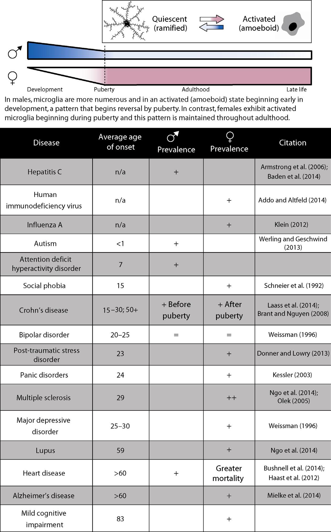
Figure 1.2 Sex-dependent differences in immune cell activation throughout development may account for differences in immune-related disease prevalence. While males express a higher degree of activated microglia during development, this pattern switches such that females express more activated microglia in later life (top). Although the exact role of microglial activation within immune-related diseases is unclear, the pattern of activation corresponds to sex differences in disease prevalence (bottom).
complex and context specific (Levite, 2008). Receptor subtype, immune cell type, activation state of the cell, and dose of neurotransmitter all contribute to differential response and functional outcome.
In rodent models, prenatal exposure to immune pathogens has been shown to alter the adult expression of various monoamines, their metabolites, as well as excitatory and inhibitory amino acids in a sex-specific fashion. In a study by Bitanihirwe et al. (2010), an immune challenge late in gestation decreased dopamine in the medial prefrontal cortex (mPFC) in both males and females while increasing serotonin (5-hydroxytryptamine) in both the amygdala and nucleus accumbens in females only. Similarly, excitatory glutamate was decreased in the mPFC of males only, and inhibitory GABA was decreased in the dorsal hippocampus of females only. Whether such effects translate to humans remains an unanswered question; however, it is plausible that prenatal immune exposure and subsequent neurochemical abnormalities may underlie sex-dependent immune responses and susceptibilities to immune-related diseases.
4.5 X-linked regulation of immunity
While the majority of sexually dimorphic genes are autosomal, sex chromosome-linked genes also regulate the differential expression of immune-related genes and immune disorders within the population. Of the 1100 genes in the human X chromosome and 100 genes in the human Y chromosome, only 54 are homologous between males and females (Dai and Ahmed, 2014). In females, random X-inactivation of one X chromosome prevents duplication of X chromosome genes. Variations in this process and escape from X inactivation occur in an estimated 15% of X chromosome-linked genes and may account for traits, including those related to immune function, to be different by sex (Buckberry et al., 2014; Dai and Ahmed, 2014). The X chromosome is home to several immune-related genes including TLR7, TLR8, FOXP3 gene, CD40, IL-1, IkB kinase g, and cytokine receptors (IL9R), to name a few (Dai and Ahmed, 2014; Van Lunzen and Altfeld, 2014). Mutations and varied expression of these genes are linked to disease susceptibility and presentation, as reviewed by Dai and Ahmed (2014).
Interestingly, the X chromosome is enriched in microRNAs (miRNAs) and suppression of several relevant target genes located on the X chromosome may occur through failure of X chromosome silencing (Sharma and Eghbali, 2014). Duplicate dosing of miRNA information within the X chromosomes may account for heightened immune responses in females.
4.6 MicroRNAs
MicroRNAs are small nonprotein-coding RNAs that regulate gene expression at the posttranscriptional level. Dysregulation of miRNAs are a factor in several disease states, including immune-related disorders (Dai and Ahmed, 2014; Dai et al., 2013; Sharma
and Eghbali, 2014), and sex differences in miRNA expression relating to inflammatory signaling are observed in females at birth and males thereafter (Murphy et al., 2014).
Several studies have documented the sex-dependent expression of miRNAs in gonadal tissue as well as in the brain, liver, and lungs (Dai and Ahmed, 2014). Within the brain, differential expression of miRNAs was detected, particularly in the cortex, with the majority of upregulated sequences found in women during adolescence. Furthermore, differentially expressed miRNAs were related to Wnt signaling and transforming growth factor-b pathways – two pathways implicated in neurological disease (Ziats and Rennert, 2014).
In a rodent model of lupus (murine NZB/WF1 model), distinct sex differences in the expression of miRNAs are noted. Male NZB/WF1 mice administered estrogen experience increases in lupus symptoms as well as lupus-related miRNAs (Sharma and Eghbali, 2014). One such miRNA, miR-155, plays an important role in the regulation of adaptive and innate immune processes and has been shown to alter levels of inflammation within disease states such as experimental autoimmune encephalomyelitis and rheumatoid arthritis (Dai et al., 2013). Immune-related disorders appear to upregulate expression of miRNAs, and administration of sex hormones, such as estrogen, exacerbates immune responses and further miRNA dysregulation, suggesting an interaction of sex hormones and miRNA regulation in the susceptibility to, or progression of, immune-related disorders.
4.7 Microbiome
The intestinal biota play a role in the balance of the immune system and the response to immune challenges. Trillions of bacterial species found within the human gut interact with the host immune system to shape and maintain homeostasis. Further, new findings suggest that the immune system has evolved to accommodate such colonization by bacterial communities and that imbalances within this relationship may influence the immune response and the development of immune-related disorders (Cerf-Bensussan and Gaboriau-Routhiau, 2010). Following exposure to a pathogen (Mycobacterium avium subsp. paratuberculosis), male and female mice mount a sex-dependent gut immune response such that levels of IL-1a, IL-1b, IL-17, IL-10, and IL-6 differ by sex (Karunasena et al., 2014). Further, females exposed to a pathogen, probiotics, or a combination of pathogen and probiotics exhibited increased levels of IL-6 and IL-1a, but decreased levels of IL-10 compared with exposed males and control mice illustrating an overall decrease of anti-inflammatory mediators (Karunasena et al., 2014). Within this study, increases in the ratios of specific disease-related bacterial species (Stenotrophomonas sp. and Eubacterium contortum related to ulcerative colitis, inflammatory bowel disease, and Crohn’s disease) were detected in females compared with males, and maintenance on probiotics reversed this effect (Karunasena et al., 2014). Additional evidence for sexdependent control over the gut microbiota is illustrated in nonobese diabetic (NOD)
mice in which males and females have equal incidence of diabetes under germ-free conditions. In contrast, under specific pathogen-free conditions, females develop diabetes at twice the rate of male NOD mice, demonstrating a differential control of commensal microbes, which is often related to autoimmunity (Chervonsky, 2010). These findings are important given the interaction of the gut microbiota with the immune response and suggest that sex-dependent factors within the gut environment influence overall immune health and susceptibility.
5 CONSEQUENCES OF SEX DIFFERENCES WITHIN CENTRAL NERVOUS SYSTEM TRAUMA AND DISEASE
5.1 Introduction
Exposure to immunogens during gestation, or critical periods during development, may incur susceptibilities to certain systemic or neurological pathologies. Distinct sex differences are noted within the susceptibilities and incidences of these pathologies and the contribution of sex steroids to these disease states are of interest. Early immune activation serves as a risk factor for adult schizophrenia and autism, namely in males (Schwarz and Bilbo, 2012; Rana et al., 2012). Moreover, early immune activation of microglia has been shown to play a role in brain aging and increasing susceptibility to Alzheimer’s disease (Bitanihirwe et al., 2010). Indeed, several rodent models have demonstrated the impact of early life immune challenges. Such consequences to the offspring include effects on behavior, cognition, learning, memory (Rana et al., 2012; Bilbo et al., 2006), cytoarchitecture, and neurochemical release (see Section 4.4; Rana et al., 2012; Madhusudan et al., 2013; Bitanihirwe et al., 2010). Important to keep in mind is that the factors that underlie these late-life susceptibilities are multifarious and highly dependent on both the timing and nature of the immune challenge as well as on the animal species and maternal environment. Furthermore, many effects of prenatal immune exposure are tissue or brain-region specific, adding another layer of complexity. Though model animals systems have highlighted that early immune activation leads to lifelong alterations to several bodily systems, much work is necessary to tease apart such effects.
5.2 Sex differences in behavior
Male and female behavioral responses to immune activation are also sex-dependent. Male offspring of pregnant rats exposed to an LPS-immune challenge show reduced social play behavior during PND 26 to 40 compared with control rats, while female rats were less susceptible to behavioral change (Taylor et al., 2012). Further, these sexdependent responses to early life immune challenges can persist into adulthood. For example, in a mouse model of late prenatal immune exposure, the sex-dependent effects on adult conditioned avoidance learning were assessed. Males exposed to polyriboinosinic-polyribocytidylic acid (Poly-I:C) on gestational day 17 showed enhanced
latent inhibition in adulthood compared with control mice of both sexes and Poly-I:C exposed females, demonstrating a delayed learning ability specific to male exposed offspring (Bitanihirwe et al., 2010). In a separate assessment, rats treated with LPS on PND 3 and 5 show altered sex-dependent behavior when exposed to a subsequent challenge in adulthood. Upon the second LPS challenge, males demonstrated reduced locomotor activity in a non-novel open field compared with males who did not undergo a neonatal immune challenge. In contrast, females did not exhibit differences in locomotor activity regardless of previous immune exposures (Tenk et al., 2008).
Early life immune challenges have been shown to further alter neurochemical responses in a sex-dependent manner. Although increased locomotor activity is commonly observed in rats after exposure to a dopaminergic agonist, females previously exposed to LPS exhibit a greater increase in locomotor activity following dopaminergic agonist treatment compared with males and saline treated controls (Tenk et al., 2007). Furthermore, males exposed to LPS on PND 3 and 5 experienced a greater degree of anxietylike behaviors than saline treated controls while females did not exhibit this difference (Walker et al., 2009). Although this apparent sex bias for behavioral alterations could be attributed partially to lack of study in females, some studies, as discussed at the beginning of this section, do compare both sexes, and these findings underscore the potential increased susceptibility of males to immune activation at a young age.
The pattern of increased susceptibility to immune activation in young males is not specific to mice and rats. Juvenile immune activation impairs learning in a sexspecific manner in birds as well. For example, juvenile male zebra finches exposed to an LPS challenge had impaired performance in a novel foraging task; females did not experience the same deficiencies (Grindstaff et al., 2013 ). Similarly, when male white-crowned sparrows were exposed to LPS on long and short days, mimicking seasonal differences, males exposed to LPS on long days lost more weight than males exposed to LPS on short days, while females did not exhibit such differences ( OwenAshley et al., 2006 ).
In contrast to the discussion above, exposure later in life seems to impact females more severely. Female rats that experience a repeated immune challenge in adulthood exhibit increased depressive-like behaviors compared with untreated females and immunechallenged males (Tonelli et al., 2008). Similarly, females (at or after PND 74) who experienced repeated LPS administration in the nasal cavity had reduced escape behavior and increased immobility compared with both males and females not treated with LPS (Tonelli et al., 2008). These data provide additional support for a sex-dependent variation of temporal susceptibility to immune activity. Consistently, and in a separate model, experimentally induced gastritis in both rats and mice caused increased depressive- and anxiety-like symptoms in females compared with males when tested in adulthood (Luo et al., 2013; Painsipp et al., 2007). This hypothesis of temporal sensitivity is further discussed in the review by Schwarz and Bilbo (2012).
Environmental factors also contribute to the apparent sex difference in immune response. Male rats exposed to LPS in adulthood who were single-housed reduced the number of times they visited the food hopper while LPS-group-housed and control males did not. Furthermore, LPS-exposed females who were group-housed decreased their food hopper visits while single-housed and control females did not (Yee and Prendergast, 2010).
Finally, in short-term measures of immune sickness behavior, females may fare better. Adult females exposed to mild LPS treatment fared better in the forced swim test than their male counterparts while other measures such as sucrose consumption and food intake were not different between males and females (Pitychoutis et al., 2009). These findings, though in contrast, may highlight an important distinction between males and females and their responses to immune activity in the long and short term.
5.3 Sex differences in other comorbidities
A growing body of evidence exists implicating the immune system in many diseases that initially were not believed to hold immune relevance. With better understanding of the impact of immune system activation in a multitude of different disorders, sex differences in immune function are becoming more widely recognized as a possible mechanism of the sex bias in prevalence of these diseases.
The discrepancy in cellular makeup (discussed in Sections 3 and 4.2) at different stages of life likely underlies differences in susceptibility to immune-activity-related comorbidities. Males may be more susceptible to immune events and diseases that develop during early life while females appear to be more susceptible to diseases that manifest in adulthood. As referenced earlier, immune challenges in young males lead to long-term memory and learning impairments with no effect to females (Schwarz and Bilbo, 2012). Conversely, immune challenges after adolescence may be more impactful in females due to increased cellular activity in the female brain during these later stages of life. For example, disorders such as autism and schizophrenia, which are more frequently observed in males, are thought to be promoted by early life immune stress, while diseases such as depression and anxiety, which manifest later in life, have a female sex bias promoted by susceptibilities to later life immune stress (Schwarz and Bilbo, 2012). Early life immune exposure and later risk for disease is well documented in studies of influenza during pregnancy. Women who experience influenza while pregnant run a fourfold increased risk of their offspring developing bipolar disorder, and similar risks are associated with schizophrenia (Parboosing et al., 2013). Patients with schizophrenia have higher circulating levels of IL-6 and soluble IL-6 receptor protein compared with healthy controls (Lin et al., 1998; Maes et al., 1994), and both depression and obsessivecompulsive disorder are similarly associated with immune activation and the presence of high levels of immune molecules such as IL-12, IL-1b, IL-8, and TNF-a (Ganguly and Brenhouse, 2014). Differing levels of immune molecules implicated in psychiatric
disorders may stem from baseline sex differences or differential responses to immune stress previously discussed. In animal models, neonatal male rats challenged with LPS experienced increased levels of TNF-a in adulthood while females did not, highlighting the sex-dependent and long-term molecular changes that occur in response to immune insults. As these molecular effectors manifest in a sex-dependent manner, so does their impact on the differential incidences of psychiatric disorders (Walker et al., 2010).
An alternate theory has been proposed to account for increased psychiatric disease in women, highlighting the similarity between psychiatric diseases and immune-sickness behavior. It is suggested that in both of these conditions, the presence of inflammatory cytokines in the brain is responsible for the characteristic symptoms of disorders such as depression and anxiety. Symptoms such as lack of appetite, lack of interest in social activities, and fatigue are associated with immune system sickness, but are also characteristics of many psychiatric disorders. The similarity of symptoms suggests that increased sustained inflammatory cytokines signaling could be a mechanism of development of mental health disorders. It is possible, therefore, that sustained female neuroimmune activation throughout adulthood contributes to the increased frequency with which particular mental health disorders manifest in females (Schwarz and Bilbo, 2012; Dantzer and Kelley, 2007).
Autism spectrum disorders (ASD) have been associated with both increased cytokine activity as well as an increased density of microglial cells (Schaafsma and Pfaff, 2014).
ASD affects approximately four males for every one female, and several theories have been put forth to explain the sex bias of ASD. While most of these theories depend on sex differences alone, immune activity may be involved. The influence of sex hormones over both immune and glial activity in patients with ASD has not gone unnoticed and it is likely that a balance between sex hormones and immune activity plays a role, at least in part, in the risk and severity of ASD (Werling and Geschwind, 2013).
Alzheimer’s disease (AD) also shows a strong sex bias in incidence. Several genetic risk factors have been identified in the susceptibility to developing AD, and many of these carry profound sex differences. Expression of apolipoprotein ε (APOE) is one of the strongest known risk factors for AD and shown to confer a fourfold increased risk of AD in women, but only a slight increased risk in males (Mielke et al., 2014). Expression of APOE is also associated with an earlier age of onset for AD. Similarly, decreased expression of brain-derived neurotrophic factor (BDNF) is an important risk factor for the development of AD, and BDNF expression is tightly regulated by estrogen (Mielke et al., 2014). Decreases in estrogen in aged women may therefore increase the risk for AD via reductions in BDNF. Though more work needs to be done to establish the mechanisms of sex differences in AD, gonadal hormone regulation of various immune and regulatory factors likely underlies these discrepancies, at least in part. Other variables, including the increased lifespan of women versus men certainly contributes to increased prevalence of AD; however, overall incidence as well as differences in age of onset support a biological basis for sex differences within AD.