Previous editions Copyrighted 2016 by Elsevier, A division of Reed Elsevier India Private Limited. All rights reserved.
ISBN: 978-81-312-5694-7
e-book ISBN: 978-81-312-5705-0
No part of this publication may be reproduced or transmitted in any form or by any means, electronic or mechanical, including photocopying, recording, or any information storage and retrieval system, without permission in writing from the publisher. Details on how to seek permission, further information about the Publisher’s permissions policies and our arrangements with organizations such as the Copyright Clearance Center and the Copyright Licensing Agency, can be found at our website: www.elsevier.com/permissions.
This book and the individual contributions contained in it are protected under copyright by the Publisher (other than as may be noted herein)
Notice
Practitioners and researchers must always rely on their own experience and knowledge
in evaluating and using any information, methods, compounds or experiments described herein Because of rapid advances in the medical sciences, in particular, independent verification of diagnoses and drug dosages should be made. To the fullest extent of the law, no responsibility is assumed by Elsevier, authors, editors or contributors in relation to the adaptation or for any injury and/or damage to persons or property as a matter of products liability, negligence or otherwise, or from any use or operation of any methods, products, instructions, or ideas contained in the material herein
Dedicated to My Mother, Late Smt. Ganga Devi Singh
My Father, Late Shri Hari Ram Singh
My Students, Past, and Present
Foreword to the first edition
Professor (Dr) VK Arora, MD, DCD, CTC&E (JAPAN) FNCCP, FIMSA, FGSI, Vice Chancellor, Santosh Medical College, Santosh University, Ghaziabad, NCR, Delhi, ExAdditional Director General of Health Services Government of India
It gives me a great pleasure to write the Foreword for Professor Vishram Singh’s book Selective Anatomy Prep Manual for Undergraduates. There was a long-felt need for a suitable book on anatomy in question-answer format to help students not only to revise vast course of anatomy before examination in limited time but also present their knowledge in an easy format.
It is a herculean task to select the frequently asked questions in examinations of various universities and answer them in a manner as expected by an examiner.
Professor Vishram Singh is an eminent and highly regarded Anatomist. He has authored about a dozen books and published a number of research papers in national and international journals
This book is in two volumes in question-answer format. Volume I covers the complete syllabus of Paper I and Volume II, the syllabus of Paper II. The book is profusely illustrated by four-color line diagrams which can be easily reproduced by the students during examination.
This book is an appropriate comprehensive manual for university examination, thus I strongly recommend it to the undergraduate medical students.
Wishing Professor Vishram Singh for his future endeavor.
Preface to the second edition
Vishram Singh
It is with great pleasure that I present the second edition of Selective Anatomy: Prep Manual for Undergraduates, which is widely used by the undergraduate medical students as well as dental, paramedical and nursing students.
This book is in question–answer format and set in 2 volumes – Volume I covers the syllabus of Paper I and Volume II the syllabus of Paper II. The popularity of this book reflects the appeal of its concept-building approach written with my vast experience of teaching about 45 years. Efforts have been made very carefully to present the text in concise manner that will be acceptable to most of the examiners. In fact, the huge syllabus of anatomy is beyond the comprehension of students in 1 year. The main purpose of this book is to relieve the students of pre-examination stress while revising the syllabus paper-wise in short available time However, the students should be aware that this book is meant only for the purpose of revision and does not replace the standard textbook.
This book is liked and well appreciated by the students all over India. Based on enormous suggestions from the students and fellow academicians, many new questions and answers along with figures and tables have been added in this edition. The previous text has been thoroughly revised and most of the diagrams have been completely revised for easy understanding and reproducibility in the examination by the students.
I strongly feel that the book in its present form will be more useful than the previous one to students and teachers alike.
I will highly appreciate the comments and suggestions from both students and teachers for further improvement of this book
“Nothing is permanent in life except change.”
Preface to the first edition
Vishram Singh
The Medical Council of India has reduced the duration of teaching of 1st year MBBS course from 1½ years to 1 year. It has also introduced the specific pattern of questions such as long and short answer questions, short notes, drawing and labeling of diagrams, providing anatomical, embryological, and genetic basis of clinical problems and MCQs.
Each student tries his/her best to clear the examination However, many students do not know how to present the answers considering the marks allotted.
This book is in question-answer format in 2 volumes. Volume I covers the syllabus of Paper I, while Volume II will deal with the syllabus of Paper II.
Having 40 years of teaching experience and being an examiner in various medical colleges and institutions, I have put my best effort in selecting frequently asked questions (FAQs) and tried to answer them in a concise manner acceptable to most of the examiners. Most of the diagrams are drawn by myself to ensure the accuracy and to see that they can be easily reproduced by the students in examination.
Although, initially I was a bit hesitant to write a book in question and answer format but later my conscience allowed me to do so because the sole aim of a teacher is to solve the problems faced by the students and inspire them to become good doctors
I hope that this book will definitely solve the problems of students and relieve them from pre-examination stress. However, the student should be aware that this book is meant only for revision purpose and not to replace the standard textbook.
I am confident this book will serve the purpose for which it meant.
Lastly I will highly appreciate comments both good and bad about the book from both students and faculty because that will help me to improve the book in future
“Necessity is the mother of invention. ”
Acknowledgements
I sincerely thank my colleagues in the Department, especially Prof. Mangla M. Pai (HOD) and Prof. Latha V. Prabhu and Associate Prof. Murli Manju for their cooperation and appreciation of my work.
I highly appreciate the help provided by Associate Prof Preeti Srivastava, NDMC Medical College and Hindu Rao Hospital, Delhi, for going through the proofs of this book. I am also thankful to Assistant Prof. Krishna G., Department of Anatomy, Rajarajeswari Medical College, Bengaluru, Karnataka for providing feedback from students.
I gratefully acknowledge the feedback and support of all my fellow colleagues in Anatomy throughout India, particularly:
• Prof. N.C. Goel (Vice principal and former Head of the Department), Hind Institute of Medical Sciences, Barabanki, Lucknow, Uttar Pradesh.
• Prof Punita Manik (Head of the Department), King George Medical College, Lucknow, Uttar Pradesh.
• Prof. P.K. Sharma (Head of the Department), Era Medical College, Lucknow, Uttar Pradesh.
• Prof. Poonam Kharb (Head of the Department), ITS Dental College, Ghaziabad, Uttar Pradesh
• Prof. T.C. Singel, Zydus Medical College, Dahod, Gujarat.
• Prof. T.S. Roy (Head of the Department), AIIMS, New Delhi.
• Profs Vandana Mehta (Head of the Department) and Hitendra Lohiya, Vardhman Mahavir Medical College and Safdarjang Hospital, New Delhi.
• Prof. Vanita Gupta (Head of the Department), Rama Medical College, Hapur, Uttar Pradesh
• Profs Deepa Singh (Head of the Department) and Akshya Dubey, Himalayan Institute of Medical Sciences, Jolly Grant, Dehradun, Uttarakhand.
• Prof. W.M.S. Johnson (Dean), Sree Balaji Medical College, Chennai.
• Prof. Suniti Pandey (Head of the Department), GSVM Medical College, Kanpur.
• Prof (Dr) S L Jethani (Medical Superintendent and Former Head of the Department of Anatomy), Himalayan Institute of Medical Sciences, Dehradun, Uttarakhand.
• Prof. G.M. Mahesh (Head of the Department), Basaveshwara Medical College, Chitradurga, Karnataka.
• Profs Avinash Chandra Agrawal (Head of the Department) and A.K. Srivastava, Prasad Institute of Medical Sciences, Banthra, Lucknow
• Prof. Sneh Agrawal (Head of the Department), Lady Harding Medical College,
New Delhi.
• Prof. Sneha Guruprasad Kalthur (Head of the Department) and Dr Prakash Babu, KMC, Manipal, Karnataka.
• Prof. Emeritus S.D. Joshi, Sri Aurobindo Institute of Medical Sciences, Indore, Madhya Pradesh.
Lastly, I thank my daughter Dr Rashi Singh, son Dr Gaurav Singh and daughter-inlaw Anupama Singh for helping me in the preparation of this manuscript. I gratefully acknowledge the help and cooperation received from the staff of RELX India Pvt Ltd, especially Arvind Koul (Content Strategist), Shabina Nasim (Head of Content Project Management) and Goldy Bhatnagar (Content Project Manager), in completing the project on time.
SECTION I
Thorax
OUTLINE
1. Thoracic cavity
2. Mediastinum and pleura
3. Lungs
4. Pericardium and heart
5. Superior vena cava, aorta, pulmonary trunk, and thymus
6. Trachea, esophagus, thoracic duct, and azygos vein
1
Thoracic cavity
❖ Define thoracic cavity and give its boundaries and contents. AN21.3 It is a cavity of thorax. It is enclosed in an elastic osseocartilaginous framework which helps in both increasing and decreasing the volume of thoracic cavity.
Boundaries
Anterior: Sternum.
Lateral:
12 ribs with their costal cartilages and intercostal spaces containing intercostal muscles, membranes, nerve and vessels.
Posterior:
Bodies of 12 thoracic vertebrae and the intervening intervertebral discs
Contents
The major contents are:
• Lungs, chief organs of respiration
• Heart, chief organ of circulation
• Trachea and oesophagus
• Major blood vessels associated with heart
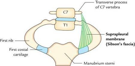
❖ Write a short note on the thoracic inlet. AN21.3 It is a superior aperture of the thoracic cavity. It is reniform in shape and measuring 10 cm in transverse plane and 5 cm in anteroposterior plane The inlet slopes downwards and anteriorly at an angle of 45°. It is partially closed on each side by a suprapleural membrane (also called Sibson’s fascia).
Boundaries
Posterior:
Body of first thoracic vertebra.
Oneachside:
First rib and its costal cartilage.
Anterior:
Upper border of manubrium sterni.
❖ Enumerate the structures passing through the thoracic inlet. AN21.3
The major structures are:
Twotubes:
Trachea and esophagus
Twosetsofarteries
• Branches of arch of aorta viz. brachiocephalic trunk, left common carotid, and left subclavian
• Right and left internal thoracic arteries
Foursetsofneuralstructures:
• Right and left vagus nerves
• Right and left phrenic nerves
• Right and left sympathetic trunks
• Right and left first thoracic nerves (ventral rami)
N.B.
Apices of the lungs covered by cervical pleura also project upward through inlet into the root of the neck.
❖ Write a short note on the suprapleural membrane (Sibson’s fascia). AN21.3
It is a tough triangular membrane, which on either side partly separates the thoracic cavity from the neck.
Features(fig.1.1)
• Its apex is attached to the tip of the transverse process of the seventh cervical (C7) vertebra.
• Its base is attached to the inner border of the first rib and its cartilage.
• Its inferior surface is fused with cervical pleura.
• Its superior surface is related to subclavian vessels
N.B.
Morphologically, Sibson’s fascia represents the degenerated tendon of the scalenus minimus (pleuralis) muscle.
Appliedanatomy
• It protects the apex of lung from injury
• It prevents the puffing of root of neck during respiration.
❖ Describe the thoracic outlet in brief. AN21.3 It is the inferior aperture of the thoracic cavity and closed completely by thoracoabdominal diaphragm (or diaphragm).
Boundaries
Anterior:
Xiphoid process.
Anterolaterally:
Costal margin formed by the union of 7th to 9th costal cartilages
Posterolaterally:
Tips of 11th and 12th ribs.
Posterior:
Body of T12 vertebra.
N.B.
The structures forming the boundaries of thoracic outlet form a osteocartilaginous rim for the origin of the thoracoabdominal diaphragm. The structures freely pass to and
FIG 11 Suprapleuralmembrane
fro between thoracic and abdominal cavities through openings in the thoracoabdominal diaphragm.
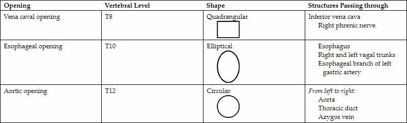
❖ Enumerate the major (large) openings in the diaphragm and structures passing through them in tabular form. AN47.13
There are 3 major (large) openings in the diaphragm. The details of these openings and structures passing through them are given in Table 1.1.
It is an extra rib which develops from costal element of the transverse process of the C7 vertebra. It is present in about 0.2–0.5% of the cases. Its posterior end is attached to transverse process of the C7 vertebra, and its distal extremity is free or attached to the first rib
Appliedanatomy
The cervical rib reduces the size of scalene triangle and may cause thoracic outlet syndrome Clinically it presents as:
• Tingling and numbness along the medial side of the hand and little finger, due to compression of the lower trunk of the brachial plexus
• Pallor and coldness of the upper limb due to compression of subclavian artery
• Reduction in radial pulse pressure
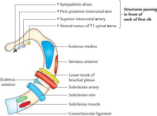
❖ Enumerate the structures passing in front of the neck of the first rib. AN21.1 From medial to lateral side, these are (Fig. 1.2):
FIG 12 Superiorviewoffirstribshowing special feature Notethestructurespassinginfront oftheneckofthefirstrib
• Sympathetic chain
• First posterior intercostal vein
• Superior intercostal artery
• First thoracic nerve (ventral ramus)
MNEMONIC: Chain pulls VAN. Here C of chain stands for sympathetic chain.
❖ Write a short note on the sternal angle (angle of Louis). AN21.1 It is a horizontal bony angulation formed at the junction of manubrium and body of sternum. It can be palpated in a living person as a bony transverse ridge about 5 cm below the suprasternal notch.
Anatomicaleventstakingplaceatsternalangleare:
• Articulates on either side with the costal cartilage of the 2nd rib which helps in counting the ribs in clinical practice
• Ascending aorta ends at this level
• Arch of aorta begins and ends at this level
• Descending aorta begins at this level
• Pulmonary trunk divides into two pulmonary arteries at this level
• Azygos vein enters the superior vena cava at this level
• Trachea divides into two principal bronchi at this level
• Marks the junction between superior and inferior mediastinum
• Marks the junction between two discontinuous dermatomes C4 (above) and T2
(below)
• Superior vena cava pierces the fibrous pericardium at this level
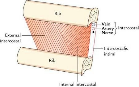
❖ Define intercostal space and enumerate its contents. AN21.4 It is a space between two consecutive ribs and their costal cartilages It extends anteriorly upto the lateral border of sternum and posteriorly upto the body of corresponding thoracic vertebra.
Contents
A typical intercostal space contains (Fig 1 3):
FIG.1.3 Contentsoftheintercostalspace
• 3 intercostal muscles.
• A neurovascular bundle.
The intercostal muscles are arranged in 3 layers. From superficial to deep, these are:
• External intercostal
• Internal intercostal
• Transversus thoracis
N.B.
• The transversus thoracis is divided into 3 parts: sternocostalis, intercostalis intimi, and subcostalis
• The neurovascular bundle consists of intercostal nerve, intercostal vein, and intercostal artery. The neurovascular bundle runs between the middle and inner layers of intercostal muscles and lies in the subcostal groove on the inner surfaces of the rib near its lower border.
❖ Give the direction of fibres of intercostal muscles. AN21.4
These are as follows:
Muscle
Direction of Fibres
External intercostal Downward, forward, and medially
Internal intercostal Downward, backward, and laterally
Transversus thoracis Downward, backward, and laterally
N.B.
The intercostal muscles are supplied by the intercostal nerves.
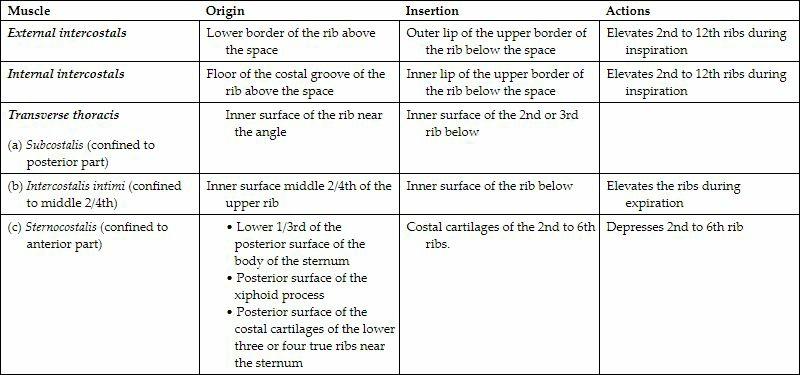
❖ Give the origin, insertion, and actions of intercostal muscles in tabular form. AN21.4
These are given in Table 1.2.
TABLE1.2
Origin,Insertion,andActionsofIntercostalMuscles
❖ Write a short note on the intercostal nerves. AN21.5
The intercostal nerves are anterior primary rami of thoracic spinal nerves and are located in the intercostal spaces. Thus, there are 11 intercostal nerves in the thoracic wall
Uniquefeature
The intercostal nerves retain their segmental character unlike the anterior primary rami
of other regions where they form nerve plexuses, viz. cervical, brachial, lumbar, and sacral.
Classificationofintercostalnerves
They are divided into two types
Typicalintercostalnerves:
They remain confined within the respective intercostal spaces of thoracic wall, viz. 3rd, 4th, 5th, and 6th intercostal nerves.
Atypicalintercostalnerves:
They extend beyond the thoracic wall, e.g., 1st, 2nd, 7th, 8th, 9th, 10th, and 11th intercostal nerves.
N.B.
The 7th to 11th intercostal nerves are called thoracoabdominal nerves as they leave the thoracic wall to supply the anterior abdominal wall.
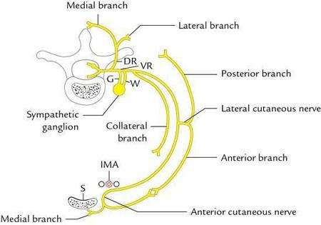
❖ Write a short note on a typical intercostal nerve. AN21.5
The anterior primary rami of the 3rd to 6th thoracic spinal nerves are termed as typical intercostal nerves and supply the muscles of the corresponding intercostal space and skin of the thoracic wall
Course(fig.1.4)
Each intercostal nerve emerges through the respective intervertebral foramen and enters into respective intercostal space. The courses in the intercostal space are as follows:
• In the posterior part of intercostal space, it runs between the pleura and posterior intercostal membrane as far as the angle of the rib.
• Thereafter, it continues its course in the costal groove between internal intercostal and intercostalis intimi muscles.
• Finally, it runs between the internal intercostal and sternocostalis muscles.
• At the anterior end of intercostal space, the nerve passes in front of internal mammary artery and runs forward, piercing the internal intercostal muscle, anterior intercostal membrane to become the anterior cutaneous nerve.
• White ramus communicans to the sympathetic trunk.
• Collateral branch: It arises in the posterior part of the intercostal space and runs along the inferior margin of the space along the upper border of rib below.
• Lateral cutaneous branch: It appears in midaxillary line and divides into anterior and posterior branches.
• Anterior cutaneous nerve (terminal branch) appears on the side of sternum and divides into medial and lateral branches.
• Muscular branches
Appliedanatomy
• Irritation of intercostal nerves causes severe pain, which is referred to the front and side of the chest
• Pus from tubercular abscess/cold abscess of the vertebral column tracks around the thoracic wall along the intercostal neurovascular bundle and points on the surface of the thoracic wall at 3 sites of exit of posterior, lateral, and anterior cutaneous branches of the spinal nerves (Fig. 1.5).