The most current, evidence-based answers available for every medical and surgical specialty.
2 Trusted Answers
Content supplied by Elsevier, the world’s leading provider of health and science information.
3 Unrivaled Speed to Answer
Faster, more relevant clinical answers, so you can spend less time searching and more time caring for patients.
Start searching with ClinicalKey today! Visit ClinicalKey.com for more information and subscription options.
Sectional Anatomy by MRI and CT
This page intentionally left blank
Sectional Anatomy by MRI and CT Fourth Edition
Mark W. Anderson, MD
Harrison Distinguished Teaching Professor of Radiology Chief, Musculoskeletal Imaging Professor of Orthopaedic Surgery University of Virginia Charlottesville, Virginia
Michael G. Fox, MD
Associate Professor of Radiology and Medical Imaging Associate Professor of Orthopaedic Surgery University of Virginia Charlottesville, Virginia
No part of this publication may be reproduced or transmitted in any form or by any means, electronic or mechanical, including photocopying, recording, or any information storage and retrieval system, without permission in writing from the publisher. Details on how to seek permission, further information about the Publisher’s permissions policies and our arrangements with organizations such as the Copyright Clearance Center and the Copyright Licensing Agency, can be found at our website: www. elsevier.com/permissions.
This book and the individual contributions contained in it are protected under copyright by the Publisher (other than as may be noted herein).
Notices
Knowledge and best practice in this field are constantly changing. As new research and experience broaden our understanding, changes in research methods, professional practices, or medical treatment may become necessary.
Practitioners and researchers must always rely on their own experience and knowledge in evaluating and using any information, methods, compounds, or experiments described herein. In using such information or methods they should be mindful of their own safety and the safety of others, including parties for whom they have a professional responsibility.
With respect to any drug or pharmaceutical products identified, readers are advised to check the most current information provided (i) on procedures featured or (ii) by the manufacturer of each product to be administered, to verify the recommended dose or formula, the method and duration of administration, and contraindications. It is the responsibility of practitioners, relying on their own experience and knowledge of their patients, to make diagnoses, to determine dosages and the best treatment for each individual patient, and to take all appropriate safety precautions.
To the fullest extent of the law, neither the Publisher nor the authors, contributors, or editors, assume any liability for any injury and/or damage to persons or property as a matter of products liability, negligence or otherwise, or from any use or operation of any methods, products, instructions, or ideas contained in the material herein.
Previous editions copyrighted 2007, 1995, and 1990.
Library of Congress Cataloging-in-Publication Data
Names: Anderson, Mark W., 1957- , author. | Fox, Michael G., author. | El-Khoury, Georges Y. Sectional anatomy by MRI and CT. Preceded by (work):
Title: Sectional anatomy by MRI and CT / Mark W. Anderson, Michael G. Fox. Description: Fourth edition. | Philadelphia, PA : Elsevier, [2017] | Includes index. | Preceded by Sectional anatomy by MRI and CT / Georges Y. El-Khoury, William J. Montgomery, Ronald A. Bergman. 3rd ed. 2007. Identifiers: LCCN 2015049199 | ISBN 9780323394192 (hardcover : alk. paper) Subjects: | MESH: Anatomy, Regional | Magnetic Resonance Imaging | Tomography, X-Ray Computed | Atlases
LC record available at http://lccn.loc.gov/2015049199
Content Strategist: Robin Carter
Content Development Specialist: Kathryn DeFrancesco
Publishing Services Manager: Catherine Jackson
Senior Project Manager: Daniel Fitzgerald
Designer: Paula Catalano
Printed in China
Preface
With the explosion of cross-sectional imaging, the accessibility of a high quality anatomic atlas has become essential and it is with great pleasure that we introduce the fourth edition of this classic atlas.
Since it was first published in 1990, it has become a standard anatomic reference source. The first three editions were masterfully edited by Drs. Georges El-Khoury, Ronald Bergman, and William Montgomery, and we are honored to be able to continue the tradition of excellence that they established.
New features in this fourth edition include color-coded labeling and a corresponding online version that allows for easy access anytime/anywhere and provides features such as scroll, zoom, and search functions that should further enhance the user’s experience.
We hope that you will find this new edition to be an integral and valuable addition to your practice.
Mark W. Anderson, MD Michael G. Fox, MD
Acknowledgments
We are indebted to Drs. El-Khoury and Bergman for their prior efforts in producing and improving this text and for allowing us to continue along the path of excellence they established. We also thank Robin Carter, Katie DeFrancesco, and Dan Fitzgerald
from Elsevier for helping to bring this project to fruition. Without their invaluable assistance, it wouldn’t have happened!
Mark W. Anderson, MD
Michael G. Fox, MD
Contents
SECTION I UPPER EXTREMITY
Chapter 1 MRI of the Pectoral Girdle and Chest Wall, 3
Axial, 4
Sagittal, 14 Coronal, 24
Chapter 2 MRI of the Shoulder, 34
Axial, 36
Oblique Sagittal, 46
Oblique Coronal, 56
Chapter 3 MR Arthrography of the Shoulder, 66
Axial, 67
Oblique Sagittal, 72
Oblique Coronal, 78
ABER (Abduction and External Rotation), 84
Chapter 4 MRI of the Arm, 90
Axial, 92
Sagittal, 98
Coronal, 105
Chapter 5 MRI of the Elbow, 114
Axial, 115
Oblique Sagittal, 125
Oblique Coronal, 134
Chapter 6 MRI of the Forearm, 143
Axial, 146
Sagittal, 156
Coronal, 162
Chapter 7 MRI of the Wrist, 170
Axial, 171
Sagittal, 180
Coronal, 189
Chapter 8 MRI of the Hand, 196
Axial, 198
Sagittal, 205
Coronal, 214
SECTION II LOWER EXTREMITY
Chapter 9 MRI of the Hip, 221
Axial, 224 Sagittal, 233 Coronal, 243
Chapter 10 MR Arthrography of the Hip, 250
Axial, 251 Sagittal, 257 Coronal, 262
Chapter 11 MRI of the Thigh, 267
Axial, 269 Sagittal, 277 Coronal, 286
Chapter 12 MRI of the Knee, 294
Axial, 295 Sagittal, 302 Coronal, 311
Chapter 13 MRI of the Leg, 321
Axial, 324 Sagittal, 334 Coronal, 342
Chapter 14 MRI of the Ankle, 348
Axial, 349
Oblique Axial, 359 Sagittal, 365 Coronal, 372
Chapter 15 MRI of the Foot, 381
Axial, 385 Sagittal, 391 Coronal, 399
SECTION III SPINE AND BACK
Chapter 16 MRI of the Thoracic Spine, 411
Axial, 415
Sagittal, 417
Coronal, 420
Chapter 17 MRI of the Lumbar Spine, 424
Axial, 425
Sagittal, 429
Coronal, 432
SECTION IV THORAX
Chapter 18 CT of the Thorax, 441
Axial, 442
Sagittal, 449
Coronal, 453
Chapter 19 MRI of the Heart, 459
Axial, 460
Sagittal, 464
Coronal, 467
SECTION V ABDOMEN
Chapter 20 CT of the Abdomen, 473
Axial, 475
Sagittal, 481
Coronal, 487
Chapter 21 MRI of the Abdomen, 493
Axial, 494
Sagittal, 502
Coronal, 510
SECTION VI PELVIS
Chapter 22 CT of the Male Pelvis, 521
Axial, 522
Sagittal, 528
Coronal, 531
Chapter 23 CT of the Female Pelvis, 537
Axial, 538
Sagittal, 543
Coronal, 546
Chapter 24 MRI of the Male Pelvis, 552
Axial, 553
Sagittal, 559
Coronal, 565
Chapter 25 MRI of the Female Pelvis, 571
Axial, 572
Sagittal, 576
Coronal, 580
Index, 585
Upper
Extremity I Section
This page intentionally left blank
MRI of the Pectoral Girdle and Chest Wall 1 Chapter
AXIAL
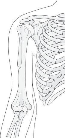
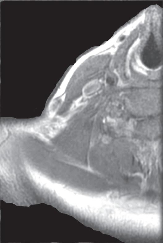
Figure 1.1.1
Sternocleidomastoid m
Anterior scalene m
Middle scalene m
Posterior scalene m
Levator scapulae m
Trapezius m
Platysma m Internal jugular v
Infrahyoid mThyroid cartilage
Inferior constrictor m
Carotid a
Vertebral a C5-6 disc level
Multifidus m
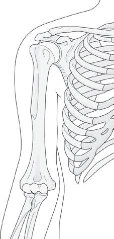
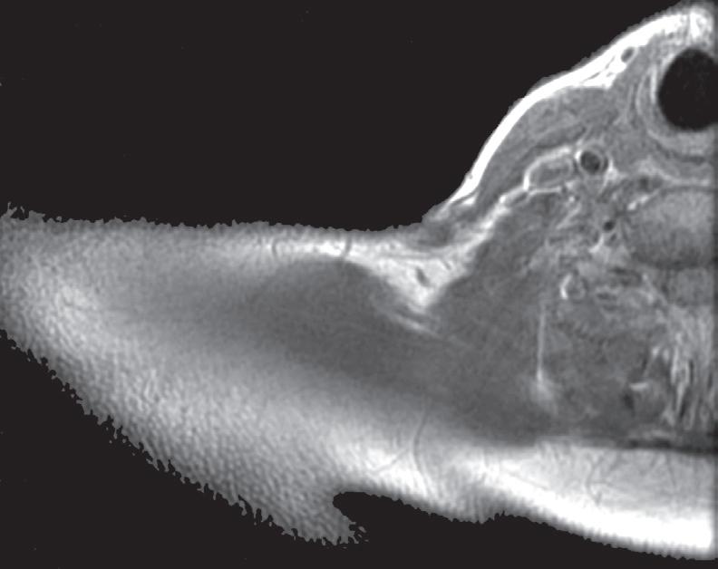
1.1.2
Posterior scalene m
Trapezius m
Splenius cervicis and splenius capitis mm
Semispinalis cervicis and semispinalis capitis mm
Figure
Infrahyoid m
Platysma m
jugular v Sternocleidomastoid m
scalene
Levator scapulae m
Splenius cervicis and splenius capitis mm
Semispinalis cervicis and semispinalis capitis mm
Multifidus m
C6-7 disc level
Longus colli m
Internal carotid a
Trachea
Figure 1.1.3
Distal clavicle
Middle scalene m Supraspinatus m
Anterior scalene m
Sternocleidomastoid m
Anterior jugular v
Transverse cervical vessels
Rhomboid minor m Splenius m
Trachea
Thyroid gland
Esophagus
Longus colli m
C7 vertebral body
Figure 1.1.4
Acromion
Clavicle
Acromioclavicular joint
Externa jugular v
Middle scalene m
Serratus anterior m
Internal jugular v
Anterior scalene m
Posterior scalene m
Sternocleidomastoid m
Serratus posterior superior m
Thyroid
Trachea
Common carotid a
Longus colli m
Posterior scalene m
Semispinalis capitis m
Splenius capitis and splenius cervicis mm
Trapezius m
Levator scapulae m
Trapezius mLevator scapulae m
Scapular spine Supraspinatus m
Multifidus m
Figure 1.1.5
Middle deltoid m
Supraspinatus m
Anterior deltoid m
Figure 1.1.6
Posterior deltoid m
Clavicle
External jugular v
Sternocleidomastoid m, clavicular head
Sternocleidomastoid m, sternal head
Anterior scalene m
Supraspinatus t
Deltoid m, anterior head
Scapular spine
Thoracoacromial a, acromial branch
Greater tuberosity of humerus Coracoid
Humeral head
Deltoid m
m
Trapezius m Levator scapulae m
Subclavius m
Subscapularis m
Clavicle
Anterior jugular v
Common carotid a
Internal jugular v
Longus colli m
Middle scalene m
Serratus anterior m
Semispinalis capitis m
Splenius capitis and splenius cervicis mm
Rhomboid minor m
Anterior scalene m
Serratus anterior m
Sternocleidomastoid m, clavicular head
Sternocleidomastoid m, sternal head
Glenoid
Infraspinatus m
Scapular spine
Supraspinatus m
m
Rhomboid minor m
Splenius capitis and splenius cervicis mm
Sternohyoid m
Common carotid a
Anterior jugular v Internal jugular v
Costovertebral joint
Subclavian a
Costotransverse joint
Rib
Semispinalis capitis m
Trapezius
Trapezius
Figure 1.1.7
Conjoined t of coracobrachialis m and biceps brachii m, short head Coracoid process
Pectoralis minor t Cephalic v Cephalic v
Subscapularis m
Subclavius m Clavicle
Deltoid m, anterior head
Biceps brachii t, long head
Greater tuberosity of humerus
Humeral head
Glenoid
Posterior deltoid m
Figure 1.1.8
Deltoid m
Glenohumeral joint
Glenoid
Anterior scalene m
Sternocleidomastoid m, sternal head
Suprascapular neurovascular bundle in suprascapular notch
Infraspinatus m
Coracobrachialis t Humeral head
Biceps brachii t, long head
Scapular body
Serratus anterior m
Sternohyoid m
Anterior jugular v
Sternothyroid m
Common carotid a
Subclavian a
Thoracoacromial a
Semispinalis thoracis m
Semispinalis capitis m
Splenius capitis and splenius cervicis mm
Rhomboid major m
Trapezius m
Pectoralis major m, clavicular head Pectoralis minor m Cephalic v
Axillary v
Biceps brachii t, short head Clavicle
Subclavius m
Infraspinatus m
Suprascapular a and n in spinoglenoid notch
Infraspinatus mSubscapularis m Serratus anterior m Trapezius m
Sternocleidomastoid m, sternal head
Anterior jugular v
Sternohyoid m
Brachiocephalic a Brachiocephalic v
Trachea
Subclavian v
Axillary a
Right lung
Semispinalis thoracis m
Trapezius m Rhomboid major m
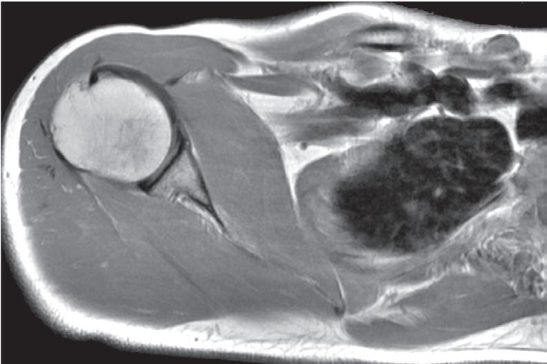
Figure 1.1.9
Biceps brachii t, long head Lesser tuberosity of humerus
Pectoralis minor m Subscapularis m
Biceps brachii t, short head Coracobrachialis m
Pectoralis major m, clavicular head Axillary v Axillary a
Sternocleidomastoid m, sternal head
Deltoid
Greater tuberosity of humerus
Infraspinatus mGlenoidSerratus anterior m
Scapular body, medial border Serratus anterior m Trapezius m
Rhomboid major m
Clavicle
Sternothyroid m
Sternohyoid m
Brachiocephalic a
Brachiocephalic v
Right lung
Rib
Erector spinae m
Semispinalis thoracis m
Trapezius m
Splenius capitis and splenius cervicis mm
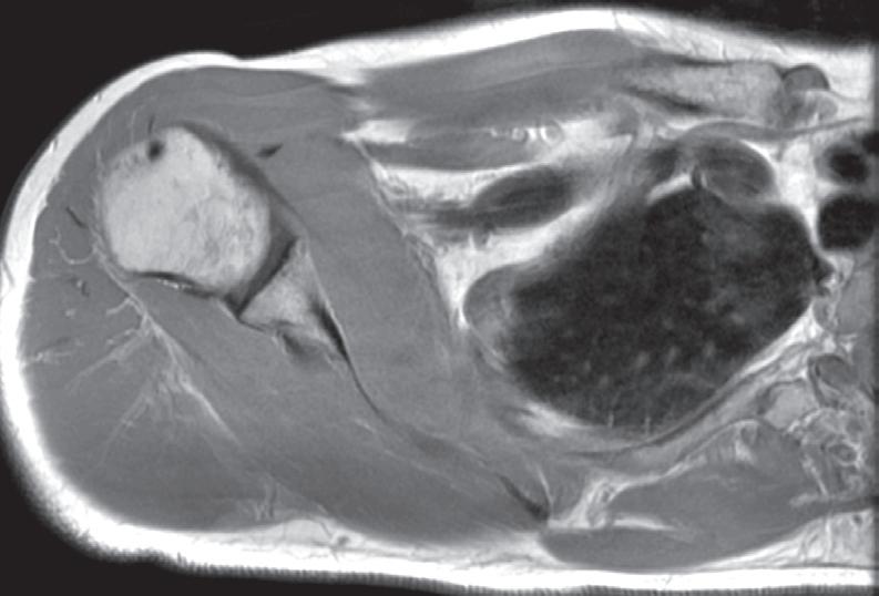
Figure 1.1.10
Biceps brachii t, long head
Surgical neck of humerus
Deltoid m, anterior head Biceps brachii t, short head
Coracobrachialis m and biceps brachii m, short head
Pectoralis major m, clavicular head
Deltoid m
Quadrangular space
Posterior circumflex humeral a and branches and axillary n and branches
Pectoralis minor m
Subscapularis m Teres minor m Triceps brachii m, long head
Axillary v Axillary a
Sternoclavicular joint
Left brachiocephalic v
Brachiocephalic a
Right brachiocephalic v
Trachea
Costovertebral joint
Rib
Semispinalis thoracis m
Splenius capitis and splenius cervicis mm
Rhomboid major m
Infraspinatus m
Trapezius m Serratus anterior m
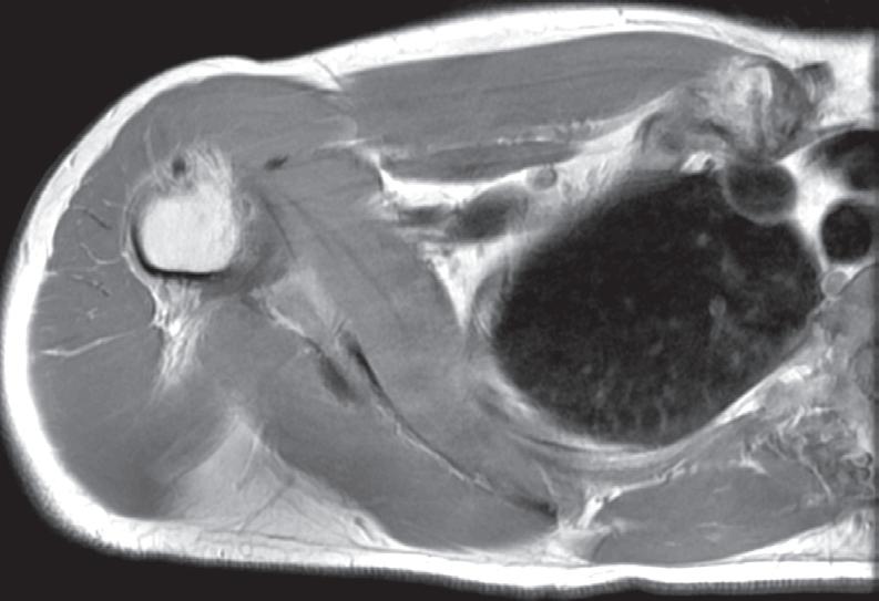
Figure 1.1.11
Biceps brachii t, long head
Humerus
Deltoid m
Biceps brachii t, short head
Coracobrachialis m Axillary a
Pectoralis major m, clavicular head
Pectoralis minor m Axillary v
Figure 1.1.12
Quadrangular space
Axillary n and posterior circumflex humeral a
Costochondral cartilage, first rib
Triceps brachii m, long head Teres minor m Teres major m
Biceps brachii t, long head
Deltoid m
Superior portion of t of latissimus dorsi m
Triceps m, lateral head
Latissimus dorsi m
Triceps brachii m, long head
Teres major m
Biceps brachii m, short head
Infraspinatus m
Coracobrachialis m
Scapular body
Axillary a
Sternum
Left brachiocephalic v
Brachiocephalic a
Right brachiocephalic v
Trachea
Thoracic vertebral body
Subscapularis m
Rib
Rhomboid major m
Trapezius m
Serratus anterior m
Pectoralis major m, sternoclavicular head
Circumflex scapular a
Teres minor m
Subscapular a
Infraspinatus m
Sternum
Ascending aorta
Superior vena cava
Pectoralis minor m
Trachea
Right lung
Subscapularis m
Rhomboid major m
Trapezius m Serratus anterior m
Figure 1.1.13
Biceps brachii m, short head
Pectoralis major t
Biceps brachii m, long head
Deltoid m
Latissimus dorsi and teres major tt
Triceps brachii m, lateral head
Axillary n, posterior branch
Triceps brachii m, long head
Coracobrachialis m
Axillary neurovascular bundle
Pectoralis major m, sternocostal head
Pectoralis minor m
Sternum
Ascending aorta
Superior vena cava
Right mainstem bronchus
Subscapularis m
Vertebral body Rib
Serratus anterior m
Trapezius m Rhomboid major m
Infraspinatus m
Figure 1.1.14
Cephalic v
Pectoralis major t
Biceps brachii m, long head
Deltoid m
Humeral diaphysis
Latissimus dorsi t and teres major m
Triceps brachii m, lateral head
Axillary n, posterior branch
Triceps brachii m, long head
Biceps brachii m, short head
Pectoralis minor m Coracobrachialis m
Pectoralis major m, costosternal head
Axillary neurovascular bundle
Internal thoracic a and v
Ascending aorta Superior vena cava Right lung
Subscapularis m Teres major m
Semispinalis thoracis m
Erector spinae m Trapezius m
m
Inferior scapula Teres minor m
Latissimus dorsi m Teres major m
Figure 1.1.15
Biceps brachii m, long head
Cephalic v
Pectoralis major t
Deltoid m
Triceps brachii m, lateral head
Brachial neurovascular bundle
Latissimus dorsi m
Triceps brachii m, long head
Biceps brachii m, short head Coracobrachialis m Pectoralis minor m
Subscapularis m
Figure 1.1.16
Biceps brachii m, long head
Cephalic v
Biceps brachii m, short head
Deltoid m
Humeral diaphysis
Triceps brachii m, lateral head
Triceps brachii m, long head
Radial n and deep brachial a
Pectoralis minor m Coracobrachialis m
Pectoralis major m, costosternal head
main pulmonary a Thoracic vertebral body Rib
Semispinalis thoracis m
spinae m
Pectoralis major m, costosternal head Internal thoracic a and v Sternum Sternum Ascending aorta
Internal thoracic a and v
m Erector spinae m
Semispinalis thoracis m
Trapezius m Rhomboid major m Serratus anterior m
Inferior scapular body
Teres major m
Rhomboid major m Serratus anterior m
Inferior scapula
Teres major m
Latissimus dorsi m
Figure 1.1.17
Biceps brachii m, short head
Pectoralis minor m Coracobrachialis m
Pectoralis major m, costosternal head
Internal thoracic a and v
Figure 1.1.18
Biceps brachii m, long head
Cephalic v
Humeral diaphysis
Triceps brachii m, medial head
Radial n Deltoid m
Triceps brachii m, lateral head
Triceps brachii m, long head
Cephalic v
Biceps brachii m, long head
Deltoid m
Triceps brachii m, medial head
Radial n
Triceps brachii m, lateral head
Triceps brachii m, long head
Biceps brachii m, short head
Sternum
Semispinalis thoracis m
Trapezius m
Erector spinae m
Rhomboid major m
Inferior medial scapula Serratus anterior m Teres major m
Latissimus dorsi m Neurovascular bundle
Brachial neurovascular bundle
Latissimus dorsi m
Long thoracic n and a Serratus anterior m
Sternum
Pectoralis major m
Internal thoracic a and v
Right lung
Pectoralis minor m Coracobrachialis m
Trapezius m
Erector spinae m
Intercostal m
Inferior medial scapula
Internal
a and v
Biceps brachii m, long head
Cephalic v
Coracobrachialis m
Deltoid m
Radial n and deep brachial a
Triceps brachii m, lateral head
Triceps brachii m, long head
Trapezius m
Vertebral body Costovertebral joint
Erector spinae m
Rib Latissimus dorsi m
Latissimus dorsi m Serratus anterior m
Triceps brachii m, medial head Brachial neurovascular bundle
Sternum
thoracic
Pectoralis major m, costosternal head
Pectoralis minor m
Biceps brachii m, short head
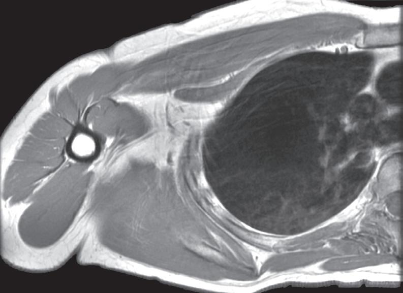
SAGITTAL
Figure 1.2.1
Figure 1.2.2
Deltoid m
Infraspinatus t
Triceps brachii m, long head
Deltoid m
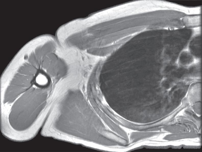
Deltoid
Triceps brachii m,
Deltoid m
Triceps brachii m, long head
Deltoid m
Cephalic v
Triceps brachii m, lateral head
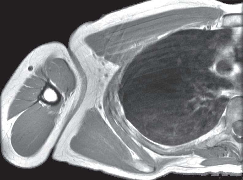
Figure 1.2.3
Lateral edge of acromion
Supraspinatus t
Humeral head
Biceps brachii t, long head
Deltoid m
Cephalic v
Deltoid tuberosity
Biceps brachii m, long head
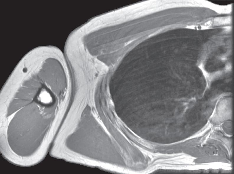
Figure 1.2.4
Supraspinatus t
Humeral head
Subscapularis t
Lesser tuberosity of humerus
Deltoid m
Anterior circumflex humeral a
Cephalic v
Pectoralis major t
Biceps brachii m, short head
Biceps brachii m, long head
Infraspinatus t
Teres minor m and t
Deltoid m
Posterior circumflex humeral a and axillary n
Teres major m
Triceps brachii m, lateral head
Humeral diaphysis
Triceps brachii m, medial head
Triceps brachii m, long head
Infraspinatus t
Deltoid m
Teres minor m
Posterior circumflex a and axillary n
Teres major m
Radial n and deep brachial a
Triceps brachii m, medial head
Triceps brachii m
Acromion
Figure 1.2.5
Acromion
Coracoacromial lig
Humeral head
Deltoid m
Pectoralis major m
Posterior circumflex a and axillary n
Biceps brachii m, short head, and coracobrachialis m