The right of Jay H. Lefkowitch to be identified as author of this work has been asserted by him in accordance with the Copyright, Designs and Patents Act 1988.
No part of this publication may be reproduced or transmitted in any form or by any means, electronic or mechanical, including photocopying, recording, or any information storage and retrieval system, without permission in writing from the publisher. Details on how to seek permission, further information about the Publisher’s permissions policies and our arrangements with organizations such as the Copyright Clearance Center and the Copyright Licensing Agency, can be found at our website: www.elsevier.com/permissions
This book and the individual contributions contained in it are protected under copyright by the Publisher (other than as may be noted herein).
Notices
Practitioners and researchers must always rely on their own experience and knowledge in evaluating and using any information, methods, compounds or experiments described herein. Because of rapid advances in the medical sciences, in particular, independent verification of diagnoses and drug dosages should be made. To the fullest extent of the law, no responsibility is assumed by Elsevier, authors, editors or contributors for any injury and/or damage to persons or property as a matter of products liability, negligence or otherwise, or from any use or operation of any methods, products, instructions, or ideas contained in the material herein.
ISBN: 9780702075841
Content Strategist: Michael Houston
Content Development Specialist: Joanne Scott
Project Manager: Beula Christopher
Design: Brian Salisbury
Illustration Manager: Narayanan Ramakrishnan
Illustrator: Deborah Maizels
Marketing Manager: Claire McKenzie Printed in China
Preface
In the seemingly few short years since the previous edition of this book, there have been dramatic changes in hepatology and in the practice of liver pathology. Probably the most striking of these changes is the disappearing culture of the liver biopsy for chronic hepatitis C. Formerly a staple of daily biopsy reporting sessions, biopsies from patients with chronic hepatitis C for grading and staging have all but disappeared with the advent of directacting antiviral agents and the potential for sustained viral response and cure. With a global prevalence of some 71 million HCV-viraemic individuals, the World Health Organization initiative of 90% reduction of new or current HCV infections by 2030 is possibly reachable, but the reality is that global diagnosis of hepatitis C virus infection and access to antiviral drugs are not universal and highly dependent on variations in economies and in public health policies throughout the world. Another change since the ninth edition of Scheuer’s Liver Biopsy Interpretation is the changed nomenclature of the disorder PBC: formerly primary biliary cirrhosis, PBC is now ‘primary biliary cholangitis’. The original name ‘primary biliary cirrhosis’ hailed from 1950, and as morphologists, liver pathologists in particular know that the early and progressive stages of this disease have little to do with cirrhosis, and that it may take decades before cirrhosis actually develops. And, as the late Professor Sheila Sherlock advocated, relieving PBC patients of the burden and stigma of having a condition with the term ‘cirrhosis’ embedded in it is a good thing.
Genomic medicine looms large in pathologists’ practices these days, and in liver pathology this constitutes the third of the dramatic changes in our practices since the ninth edition of this book. We are now all too frequently asked to pursue genomic evaluations of liver specimens in order to identify mutations with new or known treatment options. Consequently, those who examine liver biopsies should have a fundamental knowledge of known genomic changes among common liver tumours. To this end, there is now augmented coverage of genomic correlates throughout this 10th edition and specifically in Chapter 11 (Neoplasms and Nodules).
Despite the new or refocused directives cited above, the goal of the 10th edition is, as before, to be a practical and concise ‘bench book’ for use at the microscope. As Professor Scheuer wrote in the Preface to the 3rd Edition, ‘the main purpose of this book, to help those who need to interpret liver biopsies’, remains unaltered. By introducing new text coverage and new photomicrographs I hope this edition will provide the proper foundation by which to address the specific diagnostic questions we face in liver pathology today.
Jay H. Lefkowitch
In memory of Peter J. Scheuer, M.D. ‘a man of an angel’s wit and singular learning. I know not his fellow. For where is the man of that gentleness, lowliness and affability? And, as time requireth, a man of marvellous mirth and pastimes, and sometime of as sad gravity. A man for all seasons’.
Robert Whittington (1520)
Peter J Scheuer attended the Royal Free Hospital School of Medicine in London, UK, where he later became Professor of Pathology and Chairman of the Department of Histopathology. The first edition of Professor Scheuer’s Liver Biopsy Interpretation was published in 1968, only a decade after Menghini first introduced the technique of needle liver biopsy. Professor Scheuer’s many publications on hepatobiliary disease included seminal papers on primary biliary cirrhosis, histological grading of hepatic iron and the classification of chronic hepatitis. He collaborated extensively with his esteemed colleague Professor Dame Sheila Sherlock and the clinical Liver Unit, further establishing the Royal Free Hospital as a major international destination for patients with liver disease and for trainees in clinical hepatology and liver pathology.
N.B. For a brief history of Hepatology at The Royal Free Hospital, London, see Campollo O. 50 years of Hepatology: The Royal Free Hospital School of Hepatology. Ann Hepatology 2020; 19: 113–116.
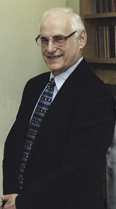
Peter J Scheuer, MD, 1928–2006. (Photograph by Charles Manley, Columbia University.)
Acknowledgements
Over 50 years ago, Peter Scheuer wrote the first book on liver biopsy interpretation based upon what was then a decade of microscopic experience with needle liver biopsies. This 10th edition notably contains many blocks of the original text as he wrote it back in 1968, still as accurate and concise as if newly minted. My first acknowledgement, therefore, must be of the many enduring wisdoms he embedded in the first edition of Liver Biopsy Interpretation that continue as important guideposts of today. I was most fortunate to have Peter Scheuer as my mentor during my fellowship training, and thereby also to have the unique opportunity of working in proximity to Professor Dame Shelia Sherlock, a formidable presence in clinical hepatology. Their luminous contemporary, if slightly more senior, colleague, Professor Hans Popper, completed the trinity of superb teachers upon whom I relied for my education in liver disease and to whom I am forever indebted.
Closer to home, my colleagues in the Pathology Department who are involved with the liver pathology service provide a stimulating and intellectually challenging environment in which to work, always bringing intriguing diagnostic questions to the table. Our superb histology technical staff, led by Sunilda Valladares-Silva, and our immunohistochemistry staff under Yuis Jimenez-Cortez consistently impress us with the outstanding quality of their work. Academic and diagnostic life for me at Columbia is greatly enriched by having colleagues like Drs Lorna Dove, Elizabeth Verna, Paul Gaglio and Alyson Fox, whose expertise in hepatology and devotion to their patients and to teaching are in a special class altogether. I must also thank those pathologists and hepatologists at other institutions who have requested my opinion on liver biopsies. These cases are never less than intriguing and provide a forum for continuing education for me, as well as for those who have asked for my input.
Michael Houston has been the major guiding force at Elsevier for Scheuer’s Liver Biopsy Interpretation, and I am greatly appreciative of his enthusiasm for bringing out this 10th edition. I am grateful to once again be working with Joanne Scott, Deputy Content Development Manager, whose expertise in textbook publication and rigorous attention to detail and deadlines provide an ideal working environment. Thanks also to Beula Christopher King, our Project Manager in Chennai for the 10th edition, for her creative work.
Lastly, I must mention the death in October 2018 of Dr Donald West King, M.D., the former Chairman of the Department of Pathology at Columbia’s College of Physicians and Surgeons. Dr King, who was my mentor from medical school through residency and beyond, was singlehandedly responsible for my setting out on a career as a liver pathologist and made my fellowship under Peter Scheuer in London possible. In so many ways, each new edition of Scheuer’s Liver Biopsy Interpretation has served as a testament to the importance of understanding and elucidating the ‘new’ in biology: ‘new’ pathogenetic pathways, ‘new’ morphologic correlates of disease. Understanding the ‘new’ was the way of life for Dr King.
General Principles of Biopsy Assessment
Introduction
Liver biopsy is one of many diagnostic tools used in the evaluation and management of patients with liver disease. It continues to play an important role because the concepts and classifications of liver disease are rooted in morphology. Moreover, looking at a liver biopsy specimen through the microscope is a very direct way of visualising the morphological changes that affect the liver in disease. The pathologist’s interpretation (rather than mere enumeration) of these changes is used to answer important clinical questions such as disease causation and activity, and is important in therapeutic decision making.1 A thorough and informed interpretation of liver biopsy findings therefore stands to have substantial impact on patient care. It bears emphasising that the evidence base2 for much of liver biopsy interpretation rests on the large body of important observations reported in the pathology literature during the past 60 years since Menghini in 1958 first introduced the technique of percutaneous needle biopsy.3 Questions of a more basic pathobiological nature can also be addressed by applying contemporary techniques of molecular and genomic medicine to liver biopsy material. There are many reasons for performing liver biopsy (Box 1.1), as will be apparent from the contents of this book. In many instances in both adult and paediatric patients, the diagnosis is uncertain from clinical and radiological data, and liver biopsy provides the direct answer. Establishing a tissue diagnosis of neoplastic disease, evaluation of jaundice of uncertain cause and assessment of pyrexia of unknown aetiology are other important diagnostic problems. In the present era of emerging personalised and precision medicine, liver biopsy for tumour diagnosis (especially for hepatocellular carcinoma) optimises the possibility of genetic and molecular analysis for targeting therapy.4,5 Pathologists are well familiar with the need for formal grading and staging of chronic hepatitis (covered in Ch. 9), although the availability of direct-acting antiviral therapy for hepatitis C virus has greatly reduced the volume of such biopsies. The ubiquitous ‘elevated liver function tests’ inscribed on biopsy requisitions are now very often explained by steatosis, steatohepatitis or related conditions (Ch. 7) stemming from the wide prevalence of obesity, diabetes, hyperlipidaemia and metabolic
Box 1.1 Reasons for liver biopsy
Evaluation of abnormal liver function tests
Investigation of pyrexia of unknown aetiology
Diagnosis of neoplasms
Evaluation of ascites and portal hypertension
Grading and staging of chronic hepatitis
Documentation of steatosis and its possible complications
Evaluation of liver dysfunction after liver, kidney and bone marrow transplantation
Determination of stage of fibrosis/cirrhosis for candidate who may require combined organ (e.g., heart-liver) transplantation.
Investigation of jaundice of unclear aetiology
Determination of the effects of therapy
syndrome. Indeed, in evaluating abnormal liver function tests in patients with negative serological studies, liver biopsy is rarely normal.6 Even when normal or ‘near-normal’, later follow-up biopsy and/or clinical data may well provide a diagnosis of autoimmune hepatitis, primary biliary cholangitis, non-alcoholic fatty liver disease or another liver disease in a significant number of these individuals.7–9 The workup of liver dysfunction following liver, kidney or haematopoietic cell transplantation is also reliant on information from liver biopsies, which must be reported promptly and with due consideration that the pathological changes in these patients may reflect more than one aetiological factor.
In contemporary hepatology, there are various non-invasive methods for assessing hepatic fibrosis and necroinflammatory activity which potentially might obviate the need for liver biopsy, but these methods also have recognised pitfalls.10
Thin needle with ultrasound/computed tomography guidance
Laparoscopic
Operative wedge
Fine-needle aspiration
Type and adequacy of liver biopsy specimen
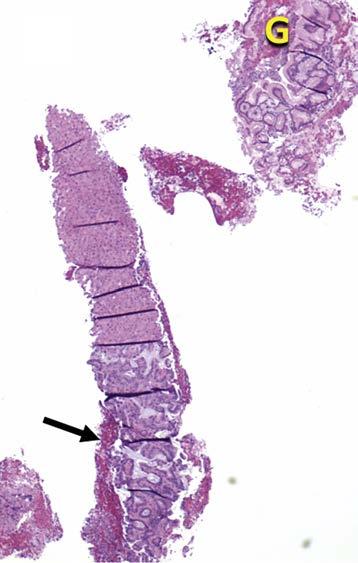
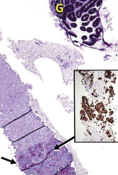
Several liver biopsy techniques and routes are now available for use (Box 1.2), each with inherent diagnostic advantages and disadvantages.1 Liver biopsy is an invasive technique which requires a skilled operator and all possible safeguards to minimise the risk of complications. Precise guidelines vary from one centre to another.11 Following the biopsy procedure the needle track may be plugged with gelatin sponge (Fig. 1.1) or other materials12 (Fig. 1.2) to prevent bleeding.13 The standard percutaneous suction needle biopsy popularised by Menghini3 continues to be in active use, while biopsy samples obtained with thin needles under computed tomography guidance and by the transjugular route14 are now seen more often. Transjugular needle core biopsies occasionally are suboptimal because the specimen contains more vein wall than liver parenchyma (Fig. 1.3). Transgastric liver biopsy obtained during endoscopic ultrasonography is an alternative method (Fig. 1.4) that is useful in the diagnosis of hepatic masses and for certain lesions that are inaccessible by the percutaneous route.15 Whatever method is chosen, the operator should carefully consider whether the specimen obtained is likely to be adequate for the intended purpose. For example, a small needle specimen obtained with a small-bore needle guided by ultrasound imaging may be adequate for the diagnosis of hepatocellular carcinoma, but not necessarily suitable for the diagnosis and histological evaluation of chronic hepatitis.16 With needles of the Menghini type the biopsy core is aspirated and may fragment if the liver is cirrhotic. This is discussed further in Chapter 10. Cutting needles have been reported to produce better specimens,17 but, in patients with focal lesions, aspiration needles often sample both the lesion itself and the adjacent liver; this is helpful in planning treatment.
Biopsy pathology differs from autopsy pathology in that there are pitfalls peculiar to small samples. A needle biopsy specimen of liver represents perhaps one fifty-thousandth of the whole organ and there is thus an obvious possibility of sampling error. Some diseases of the liver are diffuse and involve every acinus, so that sampling error is unlikely; these can be diagnosed with confidence even in small specimens. A diagnosis of acute viral hepatitis can be established in a needle specimen only a few millimetres long, whereas a specimen of similar size may not be adequate for the accurate diagnosis and evaluation of chronic liver disease, for assessment of bile duct numbers, for assessing the full extent of steatosis18 or for the detection of focal lesions such as tumour deposits or granulomas. Focal or unevenly distributed lesions cannot be entirely excluded on the basis of their
Box 1.2 Liver biopsy techniques
absence from an unguided needle biopsy specimen. When focal lesions are suspected, multiple biopsies may help to reduce sampling error.
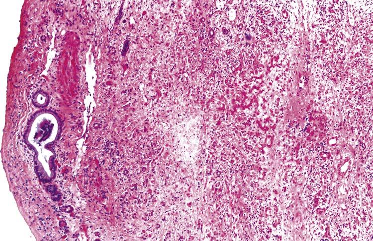
Chronic hepatitis and cirrhosis present particular sampling problems. In some patients with hepatitis there is a zone of extensive necrosis immediately adjacent to the capsule, whereas the deeper parenchyma is less severely affected. A small specimen consisting of tissue from the subcapsular zone of the liver would then give a misleadingly pessimistic
Fig. 1.1 Foreign material. This is absorbable gelatin which was used to plug a needle track. A small amount of liver tissue is seen at the point of the arrow. (Needle biopsy, H&E.)
Fig. 1.2 Foreign material. Material used to plug a needle track has here escaped and produced a peritoneal foreignbody giant-cell reaction.12 (H&E.)
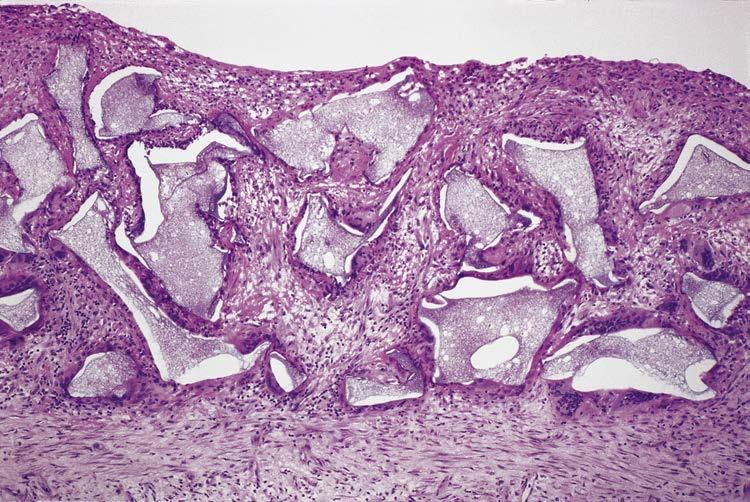
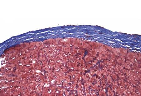
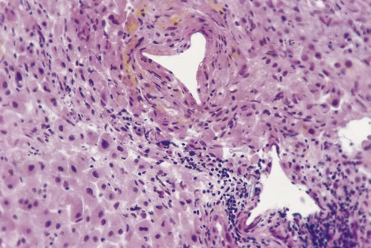
Fig. 1.3 Transjugular vs. percutaneous needle biopsy. A and B: Transjugular biopsy. A: This needle core was obtained by the transjugular route and only half the width of the core is liver tissue, with the remainder along the right side occupied by the wall of a large vein. B: The stroma of the vein wall contains muscle, in contrast to the non-muscular liver capsule. C and D: Percutaneous needle biopsy. C: Percutaneous biopsies usually have capsule at one end of the core. D: The capsule is composed of collagen fibres and fibroblasts, without muscle. (A&C: haematoxylin and eosin stain; B&D: Masson trichrome stain.)
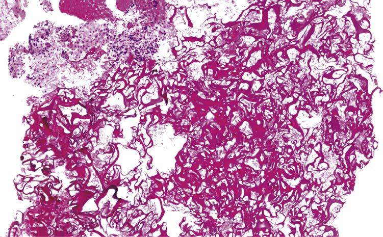
impression (Fig. 1.5). In cirrhosis the structure of a nodule is sometimes very similar to that of normal liver, so that a sample consisting almost entirely of the parenchyma from within a nodule may present serious diagnostic difficulties (Fig. 1.6). These are accentuated by the resistance of dense fibrous tissue; in a patient with cirrhosis an aspiration biopsy needle may glance off fibrous septa and selectively sample the softer nodular parenchyma. For this reason, some clinicians prefer to use cutting needles in patients with suspected cirrhosis.19
Abnormalities in a liver biopsy may represent changes remote from a pathological lesion rather than the lesion itself. In large bile-duct obstruction, for example, the results of the obstruction are clearly seen in the biopsy sample, whereas the cause of the obstruction is usually not visible. The biopsy may be taken from the vicinity of a focal liver lesion such as metastatic carcinoma, and present one or more of a range of pathological features, often puzzling to the interpreter (Fig. 1.7). Similarly, disease elsewhere in the body may give rise to reactive changes in the liver; biopsy appearances are not normal but at the same time do not indicate primary liver disease.
Biopsies reveal lesions or diseases rarely seen at autopsy because of their relatively benign course, such as sarcoidosis. In other conditions the evolution of a disease to an end stage means that the earlier and more characteristic pathological features are rarely seen at autopsy or even at liver transplantation. In such cases liver biopsies provide valuable insights into the pathology of the disease.
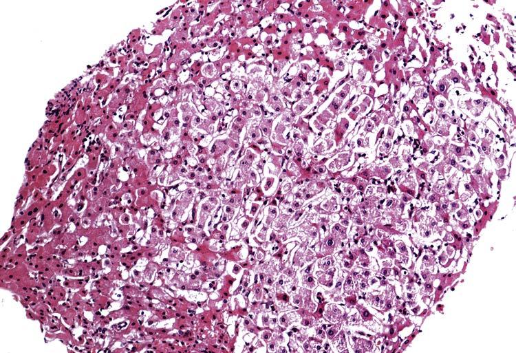
Fig. 1.4 Transgastric liver biopsy. A: The specimen includes fragments of gastric mucosa (G), blood clot and, at the bottom, a core of liver tissue infiltrated by adenocarcinoma (arrow). B: PAS (Periodic acid-Schiff) stain darkly stains the mucin within the gastric epithelium (G) and also within the infiltrating adenocarcinoma (arrow). Inset: The infiltrating adenocarcinoma, clinically suspicious for cholangiocarcinoma, is strongly positive on the immunostain for cytokeratin 19. (A: Needle biopsy, H&E; B: diastase-PAS; inset: specific immunoperoxidase.)
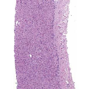
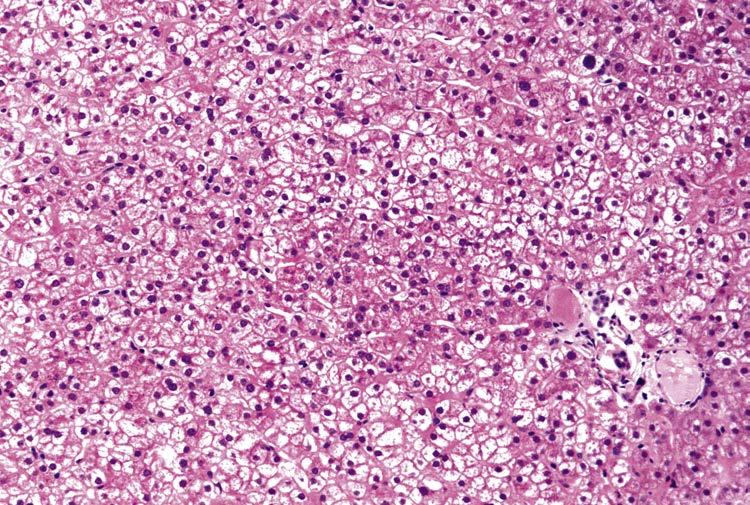
Fig. 1.5 Subcapsular necrosis. There is a zone of multiacinar necrosis immediately deep to the liver capsule (right) in this patient with chronic hepatitis. The changes are less severe in the deeper tissue to the left. A small superficial sample would have presented problems of interpretation. (Needle biopsy, H&E.)
Fig. 1.7 Changes near metastatic tumour. Portal changes like those of biliary obstruction are seen (left and top right), and there is sinusoidal dilatation in the perivenular area (bottom right). (Needle biopsy, H&E.)
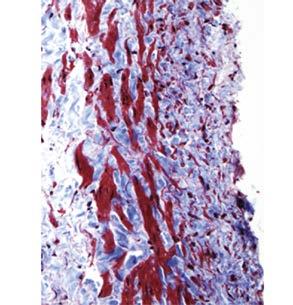
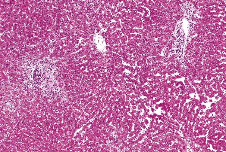
Fig. 1.6 Cirrhosis. Appearances are nearly normal because the sample is from the centre of a nodule and does not include septa. A portal tract (at right, below centre) is small and poorly formed. (Needle biopsy, H&E.)
The specimen at the bedside and in the laboratory
Liver biopsy does not always provide a final or complete diagnosis. Sometimes it even fails to give helpful information. In most cases, however, an adequate and properly processed biopsy is an important item among the diagnostic tests to which the patient is subjected. The relatively limited range of morphological reactions of the liver to injury determines a need for full clinical, biochemical, immunological and imaging data to complement the biopsy findings. Pathologists need this information in order to avoid writing clinically unhelpful, or even misleading, reports, though they may prefer to read the slides before the clinical data to avoid bias.20 Conversely, it is important that pathologists should produce clear and full reports on the biopsy findings for their clinical colleagues. Every report should attempt to answer one or more clinical questions, whether or not these are explicitly stated on the request form. The use of a standardised checklist has been advocated as a means of ensuring that no potentially useful information is omitted.21 However, most pathologists currently write unstructured reports. These can be supplemented by a summary giving the essential message which the pathologist wants to convey.
The specimen at the bedside and in the laboratory
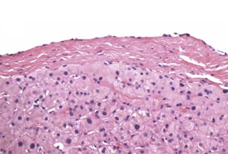
Before a liver biopsy is undertaken, the clinician may wish to discuss with the pathologist the need for any special treatment of the specimen, such as freezing part of the specimen or taking tissue for electron microscopy.20 Accurate assessment of the often subtle changes in a liver biopsy requires sections of high quality. The pathologist is usually aware of possible artefacts in liver biopsy material, as in any histological specimen. Artefacts should obviously be avoided whenever possible, and recognised as such when they do occur. A biopsy of adequate size may be made undiagnosable by rough handling (Fig. 1.8), poor fixation, overheating, poor microtome technique and bad staining, all of which can obscure the criteria on which histological diagnoses are based. Poor fixation coupled with prolonged saline immersion sometimes leads to potentially confusing liver-cell swelling and widespread separation of hepatocytes and distortion of the liver-cell plate structure (Fig. 1.9).
Fig. 1.8 Traumatic artefact. The triangular spaces, which slightly resemble blood vessels, are artefacts caused by rough packing of the specimen between pieces of foam sponge. (H&E.)
Fig. 1.9 Fixation artefact. Hepatocytes in the central part of the specimen are swollen and palestaining because of poor fixation. Prolonged saline immersion has separated and created widened spaces between hepatocytes. (Needle biopsy, H&E.)
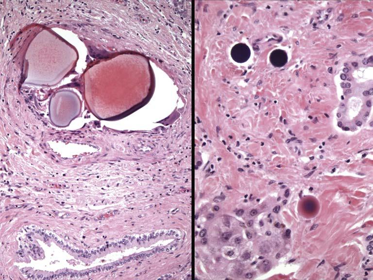
False-positive staining for iron is unrelated to particular cells or structures, or is in a different focal plane from the tissue. Foreign materials injected radiologically may appear puzzling to the pathologist due to unfamiliarity or because of localisation in unexpected organs which have been unintentionally embolised. Primary and metastatic tumours of the liver are often treated with drug-eluting chemoembolic gels via transarterial chemoembolisation (TACE) or by Yttrium 90 microspheres in selective internal radiation therapy (SIRT). TACE gels are large (300 μm) and typically lodge within medium-size hepatic artery branches within portal tracts,22 while Yttrium 90 microspheres, due to their considerably smaller size (30–40 μm), may migrate from portal tract arteries to small portal microvessels, periportal inlet vessels and sinusoids23 (Fig. 1.10).
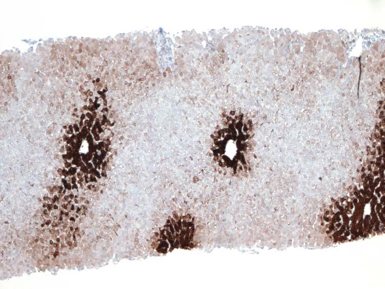
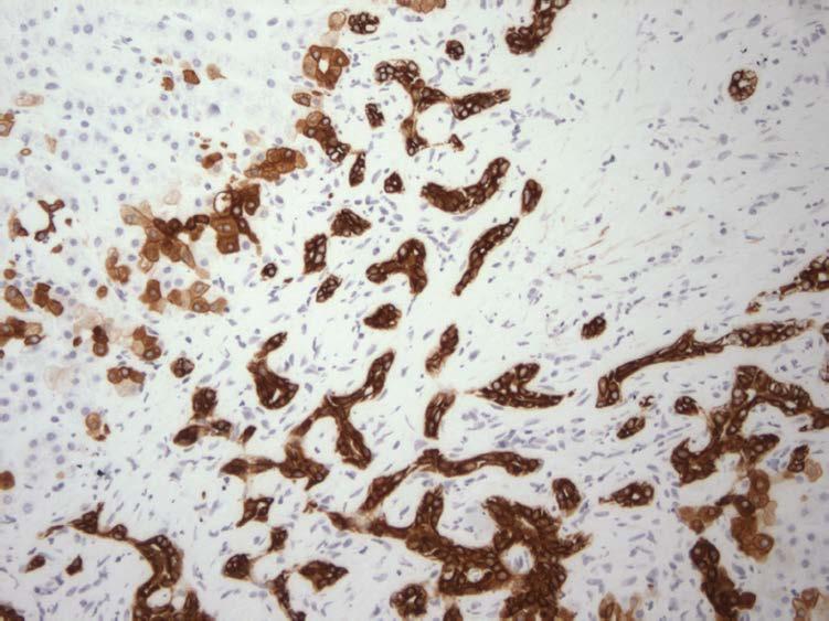
This book is mainly about changes seen in conventionally stained paraffin sections and cytological preparations. There are many other ways of looking at or investigating a tissue sample, some of them helpful in routine diagnosis. Immunohistochemistry is frequently an essential aspect of liver biopsy evaluation. Its value in individual diseases is covered in the subsequent chapters. One example is use of immunostains to address the functional heterogeneity and ‘zonation’ of the normal liver lobule or acinus (which is based upon oxygenation24 and Wnt/β-catenin signalling25,26). The liver’s zonation can be demonstrated using immunohistochemical stains for enzymes localised to particular acinar zones. A striking example is glutamine synthetase, which is involved in ammonia metabolism and is only present in the several layers of hepatocytes surrounding efferent venules (Fig. 1.11). Demonstration of the ductular reaction using antibody to cytokeratin 7 (or 19) (Fig. 1.12) is important in several chronic biliary tract diseases, in fibrosing cholestatic hepatitis after liver transplantation and in the progression of fibrosis in steatohepatitis and other conditions.27 Immunostains are useful in demonstrating viral hepatitis antigens (Ch. 9) and are the most accurate way of diagnosing α1-antitrypsin deficiency morphologically. Immunohistochemistry is used extensively in the evaluation of primary and secondary tumours of the liver (Ch. 11). Electron microscopy has a well-defined place in liver pathology and is dealt with in the final chapter.
The specimen at the bedside and in the laboratory
Fig. 1.11 Immunohistochemistry and the functional heterogeneity of the liver lobule. Glutamine synthetase immunostain shows positivity for this urea cycle enzyme localised to a rim of perivenular hepatocytes, while the mid-zone and periportal regions are negative. P, portal tract.
Fig. 1.10 Chemoembolic gels and Yttrium 90 microspheres. A: Chemoembolic gels are present within a mediumsized hepatic artery branch. Foreignbody giant cells have gathered around the gels (arrows). B: Yttrium 90 microspheres are present within microvessels in and adjacent to the portal tract. (Explant livers, H&E.)
Fig. 1.12 Cytokeratin 7 immunostain in biliary tract disease. A vigorous ductular reaction has developed in this case of primary sclerosing cholangitis, as demonstrated with cytokeratin 7 immunostain. The native bile duct (bd) is also identified with this method.
In situ hybridisation has been applied to liver tissue for the identification or assessment of replication of hepatitis viruses and cytomegalovirus. The polymerase chain reaction (PCR) can be applied to liver tissue, and provides more direct evidence of virus infection in the liver than serum PCR. DNA extracted from biopsy tissue can be used in analysis of viral infections and several inherited metabolic diseases.
Part of the biopsy specimen can be analysed for copper, iron or abnormally stored substances, and enzyme activities can be assayed by micromethods. In the case of copper and iron, these measurements can, if necessary, be made after paraffin embedding, as discussed in Chapter 14. Elution of Sirius red from sections provides an accurate method for the measurement of tissue collagen,28 and this stain is also used for image analysis of collagen.29,30 In situ demonstration of enzymes can be achieved by immunocytochemical methods or by means of enzyme histochemistry, as has been described in the functional zonation of human liver.31,32
Well-established techniques of morphometry and image analysis have been applied to tissue sections to obtain data on relative volumes of tissue components in normal human liver33,34 and in disease. Three-dimensional reconstruction using a computer has helped in the understanding of disease processes and of the relationship between anatomical structures.35–37
2. Crawford JM. Evidence-based interpretation of liver biopsies. Lab Invest. 2006;86:326–334.
3. Menghini G. One-second needle biopsy of the liver. Gastroenterology. 1958;35:190–199.
4. Torbenson M, Schirmacher P. Liver cancer biopsy—back to the future?! Hepatology 2015;61:431–433.
5. Sherman M, Bruix J. Biopsy for liver cancer: how to balance research needs with evidence-based clinical practice. Hepatology 2015;61:433–437.
6. Skelly M M , James P D , Ryder S D . Findings on liver biopsy to investigate abnormal liver function tests in the absence of diagnostic serology. J Hepatol . 2001;35: 195–199.
7. Czeczok TW, Van Arnam JS, Wood LD, et al. The almost-normal liver biopsy: presentation, clinical associations, and outcome. Am J Surg Pathol. 2017;41:1247–1253.
8. Strasser M, Stadlmayr A, Haufe H, et al. Natural course of subjects with elevated liver tests and normal liver histology. Liver Int 2016;36:119–125.
9. Prati D, Colli A. When a liver biopsy is ‘normal’. Liver Int. 2016;36:21–23.
10. Tapper EB, Lok ASF. Use of liver imaging and biopsy in clinical practice. N Engl J Med. 2017;377:756–768.
11. Sue M, Caldwell SH, Dickson RC, et al. Variation between centers in technique and guidelines for liver biopsy. Liver 1996;16:267–270.
12. Thompson NP, Scheuer PJ, Dick R, et al. Intraperitoneal Ivalon mimicking peritoneal malignancy after plugged percutaneous liver biopsy. Gut. 1993;34:16–35.
13. Sawyer AM, McCormick PA, Tennyson GS, et al. A comparison of transjugular and plugged-percutaneous liver biopsy in patients with impaired coagulation. J Hepatol 1993;17:81–85.
14. Stift J, Semmler G, Walzel C, et al. Transjugular aspiration liver biopsy performed by hepatologists trained in HVPG measurements is safe and provides important diagnostic information. Dig Liver Dis. 2019;51: 1144–1151.
15. Nakanishi Y, Mneimneh WS, Sey M, et al. One hundred thirteen consecutive transgastric liver biopsies for hepatic parenchymal diseases. A single-institution study. Am J Surg Pathol 2015;39:968–976.
16. Petz D, Klauck S, Röhl FW, et al. Feasibility of histological grading and staging of chronic viral hepatitis using specimens obtained by thin-needle biopsy. Virchows Arch 2003;442:238–244.
17. Sada PN, Ramakrishna B, Thomas CP, et al. Transjugular liver biopsy: a comparison of aspiration and Trucut techniques. Liver 1997;17:257–259.
18. Ratziu V, Charlotte F, Heurtier A, et al. Sampling variability of liver biopsy in nonalcoholic fatty liver disease. Gastroenterology. 2005;128:1898–1906.
19. Gerber MA, Thung SN, Bodenheimer Jr HC, et al. Characteristic histologic triad in liver adjacent to metastatic neoplasm. Liver. 1986;6:85–88.
20. Desmet VJ. What more can we ask from the pathologist? J Hepatol. 1996;25(suppl 1):25–29.
21. Foschini M, Sarti F, Dina RE, et al. Standardized reporting of histological diagnoses for nonneoplastic liver conditions in needle biopsies. Virchows Arch. 1995;426:593–596.
22. Panaro F, Ramos J, Gallix B, et al. Hepatic artery complications following liver transplantation. Does preoperative chemoembolization impact the postoperative course? Clin Transplant. 2014;28:598–605.
23. Luo D-L, Chan JKC. Basophilic round bodies in gastric biopsies little known by pathologists: iatrogenic Yttrium 90 microspheres deriving from selective internal radiation therapy. Int J Surg Pathol. 2013;21:535–537.
24. Jungermann K, Kietzmann T. Oxygen: modulator of metabolic zonation and disease of the liver. Hepatology. 2000;31:255–260.
25. Burke ZD, Reed KR, Phesse TJ, et al. Liver zonation occurs through a β-catenindependent, c-Myc-independent mechanism. Gastroenterology. 2009;136:2316–2324.
26. Yang J, Mowry LE, Nejak-Bowen KN, et al. Betacatenin signaling in murine liver zonation and regeneration: a Wnt-Wnt situation! Hepatology 2014;60:964–976.
27. Williams MJ, Clouston AD, Forbes SJ. Links between hepatic fibrosis, ductular reaction, and progenitor cell expansion. Gastroenterology 2014;146:349–356.
28. Jimenez W, Pares A, Caballeria J, et al. Measurement of fibrosis in needle liver biopsies: evaluation of a colorimetric method. Hepatology. 1985;5:815–818.
29. Pape L, Olsson K, Petersen C, et al. Prognostic value of computerized quantification of liver fibrosis in children with biliary atresia. Liver Transplant. 2009;15:876–882.
30. Sandrini J, Boursier J, Chaigneau J, et al. Quantification of portal-bridging fibrosis area more accurately reflects fibrosis stage and liver stiffness than whole fibrosis or perisinusoidal fibrosis areas in chronic hepatitis C. Mod Pathol. 2014;27:1035–1045.
31. Lamers WH, Hilberts A, Furt E, et al. Hepatic enzymic zonation: a reevaluation of the concept of the liver acinus. Hepatology 1989;10:72–76.
32. Sokal EM, Trivedi P, Cheeseman P, et al. The application of quantitative cytochemistry to study the acinar distribution of enzymatic activities in human liver biopsy sections. J Hepatol. 1989;9:42–48.
33. Ranek L, Keiding N, Jensen ST. A morphometric study of normal human liver cell nuclei. Acta Pathol Microbiol Scand A 1978;83:467–476.
34. Rohr HP, Luthy J, Gudat F, et al. Stereology: a new supplement to the study of human liver biopsy specimens. In: Popper H, Schaffner F, eds. Progress in Liver Diseases. Vol. V. 1st ed. New York, NY: Grune & Stratton; 1976:24.
35. Yamada S, Howe S, Scheuer PJ. Threedimensional reconstruction of biliary pathways in primary biliary cirrhosis: a computer-assisted study. J Pathol. 1987;152:317–323.
36. Nagore N, Howe S, Boxer L, et al. Liver cell rosettes: structural differences in cholestasis and hepatitis. Liver. 1989;9:43–51.
37. Ludwig J, Ritman EL, LaRusso NF, et al. Anatomy of the human biliary system studied by quantitative computer-aided threedimensional imaging techniques. Hepatology 1998;27:893–899.
Laboratory Techniques
Processing of the specimen
As soon as a needle biopsy specimen is obtained from the patient, it should be expelled gently into fixative or onto a piece of glass, card or wood. Filter paper is less suitable because fibres tend to adhere to the tissue and may interfere with sectioning. The specimen must be treated with great care, and excessive manipulation should be rigorously avoided; distortion of the specimen by rough handling at this stage may seriously interfere with accurate diagnosis, because diagnosis often depends on subtle criteria. At this stage, minute pieces can be put into an appropriate fixative for electron microscopy (Ch. 17), preferably by an operator experienced in this technique, and samples taken for chemical analysis or freezing. Frozen sections may be needed for demonstration of lipids. If porphyria is suspected, a very small amount of the unfixed tissue can be examined under ultraviolet light or with a suitable quartz halogen source, either whole or smeared onto a glass slide.
Tissue for paraffin embedding should be transferred to a fixative as soon as possible. When transit to the laboratory is likely to involve much movement, it is helpful to fill the container to the brim with fixative. Buffered formalin and formol saline are both suitable for routine fixation, which is accomplished after 3 h at room temperature or less at higher temperatures (Table 2.1). Operative wedge biopsies and larger specimens need longer fixation. Fixatives other than formalin are successfully used in some centres; handbooks of laboratory techniques should be consulted for optimum times and conditions for each fixative.
Minute fragments can be hand-processed more quickly than larger pieces, and this also avoids undue shrinkage and hardening. Automated vacuum embedding allows the time of processing of needle specimens to be drastically reduced, as shown in Table 2.1; the ultrarapid method by which a good section can be produced in about 2 h has become important because of the need for rapid decisions on treatment in patients who have undergone liver transplantation. Frozen sections, occasionally needed for a decision at surgery, can be cut by a standard method using a cryostat. They are sometimes adequate for diagnosis of obvious lesions such as neoplasms, but are unsuitable for recognition of subtle changes, and can even be dangerously misleading.
The exact number of sections routinely cut from a block varies widely from one laboratory to another. In Scheuer’s former laboratory at the Royal Free Hospital in London, 10 or more consecutive sections 3–5 μm thick are cut from each block and alternate sections used for the staining procedures outlined in the next paragraphs. The remaining sections are stored. Step sections are used when discrete lesions such as granulomas or tumour deposits are suspected or for identification of bile ducts when duct paucity is suspected. Serial or near-serial sections are helpful when utilising multiple immunohistochemical stains.
Agent
Manual overnight automatic (vacuum)*
Routine overnight automatic
Routine automatic (vacuum)* Ultrarapid
*All at 50°C except for wax step.
Choice of stains
The stains routinely applied to liver biopsies vary according to local custom. The minimum advised is haematoxylin and eosin (H&E) and a reliable method for connective tissue. The author prefers a silver preparation for reticulin as the principal method for showing connective tissue, for reasons discussed below, but trichrome stains also have important applications and can reveal changes not easily seen in a reticulin, such as the pericellular fibrosis of steatohepatitis. Routine staining for iron enables the biopsy to be used to screen for iron storage disease and the periodic acid–Schiff (PAS) stain after diastase digestion (DPAS or PASD) provides a relatively crude, but practicable, screening procedure for α1-antitrypsin deficiency as well as showing activated macrophages and bile-duct basement membranes. Stains for copper-associated protein, elastic fibres and hepatitis B surface antigen are useful (orcein and Victoria blue methods can stain all three of these) and arguably essential additions to the routine list. Rhodanine stain is an excellent method for demonstrating copper itself. Some pathologists like to see two H&E-stained sections, one from the beginning and the other from the end of a series of consecutive sections. Other methods are used as required for particular purposes. The extent to which ‘special’ stains form part of the routine set must be decided by each pathologist. The volume of liver biopsy specimens and the institutional resources have an impact on which set of stains is adopted.1
A reticulin preparation is important for accurate assessment of structural changes. Without it, thin layers of connective tissue and hence cirrhosis may be missed, as may foci of well-differentiated hepatocellular carcinoma in which the reticulin structure is often highly abnormal (see Fig. 11.13). Counterstaining is sometimes used, but is apt to distract rather than help, bearing in mind that the chief function of the reticulin preparation is to provide a sensitive low-power indicator of structural changes.
Stains for collagen such as chromotrope–aniline blue (CAB) are important for the detection of new collagen formation, especially in alcoholic steatohepatitis and its imitators (Ch. 7). Collagen staining is therefore advised for any biopsy showing substantial steatosis. It also helps to show blocked veins within scars; these are easily missed on H&E staining. It is therefore wise to use a trichrome stain when vascular disease is suspected.
A stain for elastic fibres such as the orcein stain, Victoria blue or elastic–Van Gieson is also useful to identify blocked vessels. The stains often enable the pathologist to distinguish
Table 2.1 Sample tissue schedules for liver biopsies.
Choice of stains
between recent collapse and old fibrosis, since only the latter is positive (Ch. 6). Again, this distinction may be very difficult to make on H&E and even with the help of stains for collagen and reticulin. The orcein and Victoria blue also show copper-associated protein and hepatitis B surface material.
Staining for iron by Perls’ method or another similar technique enables not only iron but also bile, lipofuscin and other pigments to be evaluated, as discussed in Chapter 3. Counterstaining should be light to avoid obscuring small amounts of pigment.
Staining of glycogen by means of the PAS method or Best’s carmine demonstrates the extent of any liver cell loss, and shows focal areas devoid of hepatocytes such as granulomas. Glycoproteins may be demonstrated by the PAS method after digestion with diastase to remove glycogen. This stain serves to accentuate hypertrophied macrophages, such as Kupffer cells filled with ceroid pigment after an acute hepatitis or episode of cholestasis. Alpha1-antitrypsin bodies stain strongly, but the stain is not sufficiently sensitive to enable all examples of α1-antitrypsin deficiency to be detected.
Staining for copper is mainly used in suspected Wilson’s disease, although, as explained in Chapter 14, it is not always helpful and may even be negative. The rhodanine method is preferred because it is easy to distinguish the orange-red colour of copper from bile, a distinction which is occasionally difficult with rubeanic acid. In Wilson’s disease, there is variable correlation between the presence of stainable copper and staining for copper-associated protein. In chronic cholestasis, however, the two usually correspond. Table 2.2 shows the special stains used at our institution as a regular panel for liver specimens, along with their specific utility.
Other non-immunological methods useful on occasion include the Ziehl–Neelsen stain for mycobacteria and for the ova of Schistosoma mansoni. Specific staining for bilirubin is rarely necessary, but conjugated bilirubin is stained a bright green colour by the Van Gieson method (see Fig. 4.10) and a forest green colour by Hall’s stain. Amyloid is stained by the usual techniques.
For immunohistochemical staining, standard techniques are applied. Among antibodies that are helpful in everyday practice are those against components of the hepatitis B virus, the delta agent, cytomegalovirus and α1-antitrypsin. Neoplasms of doubtful histogenesis or differentiation are investigated by appropriate panels of antibodies, as in any other organ. In hepatocellular carcinoma, bile canaliculi between tumour cells may stain with a polyclonal anti-CEA (carcinoembryonic antigen) antibody, cross-reacting with a canalicular antigen. Assessment of bile-duct loss may require staining of cytokeratins 7 and 19, characteristic of bile-duct rather than liver-cell cytoplasm and of the ductular reaction (Ch. 4). The application of immunohistochemistry and of other modern techniques is discussed in more detail in Chapter 17.
Most of the staining methods mentioned above are used routinely in many laboratories, and can be found in the books listed under ‘General reading’ at the end of this chapter. A selection of methods is given below (Box 2.1).
Stain Utility in identifying specific structure(s) and/or process
Trichrome • Fibrosis:
- Portal/periportal: in chronic hepatitis; in chronic biliary tract disease, chronic liver allograft rejection, other conditions.
- Perivenular: cardiac sclerosis; after central perivenulitis of allograft cellular rejection; after variant form of autoimmune hepatitis; after steatohepatitis (alcoholic or non-alcoholic type).
- Perisinusoidal: steatohepatitis-related (alcoholic or non-alcoholic fatty liver disease) (zone 3); ‘diabetic hepatosclerosis’ [non-zonal]; hypervitaminosis A (diffuse); congenital syphilis (diffuse)
Table 2.2 Special stains in evaluating liver biopsies.
Table 2.2 Continued
Stain Utility in identifying specific structure(s) and/or process
Silver impregnation for reticulin fibres (Gordon & Sweets)
1. Bring section to distilled water.
2. Treat with acidified potassium permanganate for 10 min; wash in distilled water.
3. Leave section in 1% oxalic acid until pale (about 1 min). Wash well in several changes of distilled water.
4. Mordant in 2.5% iron alum for 10 min. Wash in several changes of distilled water.
5. Treat with silver solution until section is transparent (about 10–15 s). Wash in several changes of distilled water.
6. Reduce in 10% formalin (4% aqueous solution of formaldehyde) for 30 s. Wash in tap water followed by distilled water.
7. Tone if desired in 0.2% gold chloride for 1 min. Rinse in distilled water.
8. Fix in 2.5% sodium thiosulphate for 5 min. Wash several times in tap water.
9. Transfer section to ethanol, clear and mount.
Reticulin appears black. The colour of the collagen varies according to whether step 7 is used; in untoned preparations it is yellow-brown.
Silver solution
To 5 ml of 10% aqueous silver nitrate, add strong ammonia (specific gravity 0.88) drop by drop until the precipitate which forms is just dissolved. Add 5 ml of 3% sodium hydroxide. Add strong ammonia drop by drop until the resulting precipitate dissolves. The solution does not clear completely. Make up to 50 ml with distilled water. Scrupulously clean glassware should be used throughout.
Acidified potassium permanganate
To 95 ml of 0.5% potassium permanganate, add 5 ml of 3% sulphuric acid.
Chromotrope–aniline blue (CAB) method for collagen and Mallory bodies
(As used at Mount Sinai Hospital, New York; modified from Roque2 and Churg & Prado3)
1. Bring section to water.
2. Stain nuclei by the celestine blue–Lillie Mayer sequence or other method. Rinse in distilled water.
3. Immerse in 1% phosphomolybdic acid for 1–3 min. Rinse well in distilled water.
4. Stain with CAB solution for 8 min. Rinse well in distilled water. Blot.
5. Dehydrate quickly, clear and mount.
Collagen is stained blue. Mallory bodies stain blue or sometimes red. Giant mitochondria stain red.
CAB solution
Aniline blue (1.5 g) is dissolved in 2.5 ml HCl and 200 ml distilled water with gentle heat; 6 g chromotrope 2R is added. The pH should be 1.0.
Orcein stain for copper-associated protein, elastic fibres and hepatitis B surface material4
1. Bring section to water.
2. Treat with acidified potassium permanganate for 15 min.
3. Rinse in water and decolorise in 2% oxalic acid.
4. Rinse in distilled water, then wash in tap water for 3 min.
5. Stain in commercial orcein solution for 30–60 min, at room temperature.
6. Rinse in water, then differentiate if necessary in 1% HCl in 70% ethanol.
7. Dehydrate, clear and mount.
Elastic fibres, copper-associated protein and hepatitis B surface material (HBsAg) stain brown. The method is less sensitive for HBsAg than immunohistochemical techniques. However, of the components listed, copper-associated protein is often the most difficult to stain reliably. Natural orceins seem to be more satisfactory than synthetic ones, but are difficult or impossible to obtain. In case of difficulty, doubling the concentration of orcein and the amount of HCl may help (Hans Popper, personal communication).
Acidified potassium permanganate
To 95 ml of 0.5% potassium permanganate, add 5 ml of 3% sulphuric acid. Box 2.1 Continued
Rhodanine stain for copper5,6,7
1. Bring section to distilled water.
2. Incubate in rhodanine working solution for 18 h at 37°C or 3 h at 56°C.
3. Rinse in several changes of distilled water and stain with Carazzi’s haematoxylin for 1 min.
4. Rinse with distilled water and then quickly in borax solution. Rinse well in distilled water.
5. Dehydrate, clear and mount.
Copper deposits stain bright red. Bile stains green. Weakly positive stains tend to fade, but fading can be reduced by staining at the higher temperature and by using certain mounting media (e.g. Ralmount (Raymond A. Lamb), DPX or Diatex). Note the two alternative times and temperatures for the rhodanine working solutions. The staining time can be shortened further.5
Rhodanine stock solution
p-Dimethylaminobenzylidene rhodanine 0.2 g
Ethanol
The working solution is prepared by diluting 3 ml of the well-shaken stock solution with 47 ml distilled water.
Borax solution
Disodium tetraborate
0.5 g
Distilled water 100 ml
Victoria blue method for copper-associated protein, elastic fibres and hepatitis B surface material8
1. Bring section to distilled water.
2. Treat with acidified potassium permanganate (see Gordon & Sweets’ reticulin, above) for 5 min.
3. Treat with 4% aqueous sodium metabisulphite for 1 min.
4. Wash in running tap water.
5. Wash well with 70% ethanol.
6. Stain in Victoria blue solution in a Coplin jar for a minimum of 4 h, and preferably overnight.
7. Wash well with 70% ethanol. This is the differentiation step; ensure that the background of the section is clear.
8. Wash in running tap water for 1 min.
9. Stain with nuclear fast red solution for 5 min.
10. Wash in running water for 2 min.
11. Dehydrate, clear and mount.
Copper-associated protein, elastic fibres and HBsAg are stained blue on a pink background.
Victoria blue solution
Distilled water
Dextrine
200 ml
g
Box 2.1 Continued
Slowly warm the mixture of the above until it boils. Gradually add 25 ml of boiling 29% ferric chloride solution and boil for a further 3 min. Cool and filter through fine paper. Dry the filtrate on the filter paper to complete dryness in a 56°C oven. Dissolve the filtrate in 400 ml 70% ethanol. Finally add 4 ml concentrated HCl and 6 g phenol. The solution is best left for 2 weeks before use.
Nuclear fast red
Dissolve 0.1 g nuclear fast red in 100 ml warmed 5% aluminium sulphate. Filter when cool.
References
1. Clark I, Torbenson MS. Immunohistochemistry and special stains in medical liver pathology. Adv Anat Pathol. 2017;24:99–109.
2. Roque AL. Chromotrope aniline blue method of staining Mallory bodies of Laennec’s cirrhosis. Lab Invest. 1953;2:15–21.
3. Churg J, Prado A. A rapid Mallory trichrome stain (Chromotrope–aniline blue). Arch Pathol. 1956;62:505–506.
4. Shikata T, Uzawa T, Yoshiwara N, et al. Staining methods of Australia antigen in paraffin section – detection of cytoplasmic inclusion bodies. Jpn J Exp Med. 1974;44:25–36.
5. Lindquist RR. Studies on the pathogenesis of hepatolenticular degeneration. II. Cytochemical methods for the localization of copper. Arch Pathol. 1969;87:370–379.
General reading
Clark I, Torbenson MS. Immunohistochemistry and special stains in medical liver pathology. Adv Anat Pathol. 2017;24:99–109.
Savarna KS, Layton C, Bancroft JD Bancroft’s Theory and Practice of Histological Techniques. 8th ed. Edinburgh: Elsevier Churchill Livingstone; 2018.
Kiernan JA Histological and Histochemical Methods: Theory and Practice. 3rd ed. Oxford: Butterworth-Heinemann; 1999.
6. Emanuele P, Goodman ZD. A simple and rapid stain for copper in liver tissue. Ann Diagn Pathol 1998;2:125–126.
7. Mounajjed T, Oxentenko AS, Qureshi H, Smyrk TC. Revisiting the topic of histochemically detectable copper in various liver diseases with special focus on venous outflow impairment. Am J Clin Pathol. 2013;139:79–86.
8. Tanaka K, Mori W, Suwa K. Victoria bluenuclear fast red stain for HBs antigen detection in paraffin section. Acta Pathol Jpn 1981;31:93–98.
Lefkowitch JH. Special stains in diagnostic liver pathology. Semin Diagn Pathol 2006;23:190–198.
Polak JM, van Noorden S Introduction to Immunocytochemistry. 2nd ed. Microscopy Handbooks 37, Oxford: Bios Scientific Publishers; 1997.
Prophet EB, Mills B, Arrington JB Laboratory Methods in Histotechnology. Washington, DC: American Registry of Pathology; 1992.