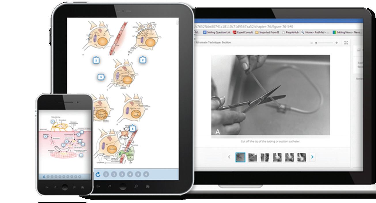
Activate the eBook version of this title at no additional charge.
Elsevier eBooks for Practicing Clinicians gives you the power to browse and search content, view enhanced images, highlight and take notes—both online and offline.
Unlock your eBook today.
1. Visit expertconsult.inkling.com/redeem
2. Scratch box below to reveal your code
3. Type code into “Enter Code” box
4. Click “Redeem”
5. Log in or Sign up
6. Go to “My Library” It’s that easy!
Place Peel Off Sticker Here
For technical assistance: email expertconsult.help@elsevier.com call 1-800-401-9962 (inside the US) call +1-314-447-8300 (outside the US)
Rich’s Vascular Trauma
This page intentionally left blank
Rich’s Vascular Trauma
FOURTH EDITION
Todd E. Rasmussen, MD, FACS
Colonel (Ret.) USAF MC
Professor of Surgery and Senior Associate Consultant Division of Vascular and Endovascular Surgery
Mayo Clinic Rochester, Minnesota
Nigel R.M. Tai, MB, BS, MS, FRCS
Colonel, Late RAMC
Consultant Trauma & Vascular Surgeon
Royal Centre for Defence Medicine (Research & Clinical Innovation) HQ Defence Medical Services Birmingham, United Kingdom; Vascular Clinical Lead
Previous editions copyrighted 2016, 2004, and 1978
ISBN: 978-0-323-69766-8
No part of this publication may be reproduced or transmitted in any form or by any means, electronic or mechanical, including photocopying, recording, or any information storage and retrieval system, without permission in writing from the publisher. Details on how to seek permission, further information about the Publisher’s permissions policies and our arrangements with organizations such as the Copyright Clearance Center and the Copyright Licensing Agency, can be found at our website: www.elsevier.com/permissions
This book and the individual contributions contained in it are protected under copyright by the Publisher (other than as may be noted herein).
Notices
Practitioners and researchers must always rely on their own experience and knowledge in evaluating and using any information, methods, compounds or experiments described herein. Because of rapid advances in the medical sciences, in particular, independent verification of diagnoses and drug dosages should be made. To the fullest extent of the law, no responsibility is assumed by Elsevier, authors, editors or contributors for any injury and/or damage to persons or property as a matter of products liability, negligence or otherwise, or from any use or operation of any methods, products, instructions, or ideas contained in the material herein.
Library of Congress Cataloging Number: 2021942088
Content Strategist: Jessica L. McCool
Content Development Specialist: Kevin Travers
Content Development Manager: Meghan B. Andress
Publishing Services Manager: Shereen Jameel
Senior Project Manager: Umarani Natarajan
Design Direction: Margaret Reid
Christopher Aylwin, MBBS, MA, FRCS
Consultant Vascular and Trauma Surgeon
Major Trauma Centre
Imperial College Healthcare NHS Trust London, United Kingdom
Ed B.G. Barnard, BM, BS, BMedSci(Hons), PhD, FRCEM, FIMC, RCSEd
Senior Lecturer
Academic Department of Military Emergency Medicine
Royal Centre for Defence Medicine (Research & Clinical Innovation)
Birmingham, United Kingdom; Honorary Consultant in Emergency Medicine
Cambridge University Hospitals NHS Foundation Trust Cambridge, United Kingdom
Andriy I. Batchinsky
Director, Autonomous Reanimation and Evacuation Program
Brooks City Base San Antonio, Texas; Senior Principal Investigator The Geneva Foundation Tacoma, Washington; Manager, Extracorporeal Life Support Capability Area
U.S. Army Institute of Surgical Research Battlefield Health and Trauma Research Institute Fort Sam Houston, Texas
Kenneth Boffard, MB, BCh, FRCS, FRCS(Edin), FRCPS(Glas), FCS(SA), FACS Professor Emeritus Department of Surgery University of the Witwatersrand Johannesburg, South Africa; Professor and Academic Head Trauma and Critical Care Milpark Academic Trauma Centre Johannesburg, South Africa
Jeremy W. Cannon, MD
Associate Professor of Surgery Department of Surgery
Perelman School of Medicine at the University of Pennsylvania Philadelphia, Pennsylvania; Adjunct Associate Professor of Surgery Department of Surgery
Uniformed Services University of the Health Sciences Bethesda, Maryland
List of Contributors
Ravi Chauhan, FRCA, FCAI, MBChB, Dip IMC, RCSEd Intensive Care and Anaesthesia Consultant
Intensive Care
Royal Centre of Defence Medicine Birmingham, United Kingdom
Kenneth J. Cherry, MD
Edwin P. Lehman Professor of Surgery
Emeritus University of Virginia Medical Center Charlottesville, Virginia; Consultant, Sentara Vascular Specialists Sentara
Norfolk General Hospital Eastern Virginia Medical School
Norfolk, Virginia
Kevin K. Chung, MD Professor and Chair Department of Medicine
Uniformed Services University of the Health Sciences Bethesda, Maryland
Ian D. Civil, MBChB, FRACS, FACS Director of Trauma Services
Auckland City Hospital Auckland, New Zealand
Jon Clasper, CBE, DSc, DPhil, DM, LLM, FRCSEd(Orth)
Professor Emeritus & Consultant Orthopaedic Surgeon Visiting Professor in Bioengineering Imperial College London; Clinical Lead
The Royal British Legion Centre for Blast Injury Studies London, United Kingdom
William Darrin Clouse, MD Professor of Surgery
Chief, Division of Vascular & Endovascular Surgery University of Virginia Charlottesville, Virginia
Lazar B. Davidovic, MD, PhD, FETCS Head of the Clinic
Clinic for Vascular and Endovascular Surgery
Clinical Center of Serbia Belgrade, Serbia; Full Professor of Vascular Surgery
Faculty of Medicine
University of Belgrade Belgrade, Serbia
David L. Dawson, MD
Clinical Professor
Texas A&M University Temple, Texas; Vascular Surgeon
Baylor Scott & White Health Temple, Texas
Demetrios Demetriades, MD, PhD, FACS Professor of Surgery University of Southern California Los Angeles, California
Joseph J. Dubose, MD, Col, MC, USAF Professor of Surgery
University of Maryland School of Medicine Baltimore, Maryland; Director, C-STARS
R Adams Cowley Shock Trauma Center University of Maryland Medical Center Baltimore, Maryland
Philip M. Edmundson, MD Division of Trauma and Emergency Surgery
UT Health San Antonio San Antonio, Texas
Timothy Fabian, MD Professor Emeritus
University of Tennessee Health Science Center Memphis, Tennessee
David V. Feliciano, MD
Clinical Professor
Department of Surgery University of Maryland School of Medicine Baltimore, Maryland; Attending Surgeon Shock Trauma Center
University of Maryland Medical Center Baltimore, Maryland
Charles James Fox, MD
Associate Professor of Surgery
Baltimore Shock Trauma Center Division of Vascular Surgery
University of Maryland School of Medicine Baltimore, Maryland
Michaela Gaffley, MD
General Surgery Resident Wake Forest University School of Medicine
Winston-Salem, NC, United States
Shaun M. Gifford, MD, MS, RPVI
Chief, Vascular Surgery
David Grant Medical Center
Travis Air Force Base California
Elon Glassberg, MD, MHA, MBA
Medical Corps Israeli Defense Forces Bar-Ilan University Faculty of Medicine
Safed
Israel
The Uniformed Services University of the Health Sciences Bethesda, Maryland
Peter Gogalniceanu, MEd, FRCS
Senior Surgical Registrar
Departments of Trauma and Vascular Surgery
The Royal London Hospital London, United Kingdom
Matthew A. Goldshore Department of Surgery Perelman School of Medicine at the University of Pennsylvania Philadelphia, Pennsylvania
Eitan Heldenberg, MD
Head Department of Vascular Surgery
Hillel Yaffe Medical Center Hadera, Israel
Joseph A. Herrold, MD, MPH Assistant Professor R Adams Cowley Shock Trauma Center Baltimore, Maryland
Shehan Hettiaratchy, MA, DM, FRCS(Plast)
Lead Surgeon Imperial College Healthcare NHS Trust St Mary’s Hospital London, United Kingdom
Tal M. Hörer, MD, PhD
Associate Professor Surgery Department of Cardiothoracic and Vascular Surgery Örebro University Hospital and Univeristy Faculty of Life Sceince Örebro, Sweden
Kenji Inaba, MD, FACS Professor of Surgery University of Southern California Los Angeles, California
Robert H. James, BSc, FRCEM, FIMC, RCSEd, RAF Consultant in Emergency Medicine & Pre-hospital Emergency Medicine
JHG(SW), University Hospitals Plymouth & Devon Air Ambulance;
Honorary Lecturer in Military Emergency Medicine and Pre-hospital
Retrieval and Transfer Medicine
Royal Centre for Defence Medicine & University of Plymouth Devon, United Kingdom
Jan O. Jansen, MBBS, PhD
Center for Injury Sciences, Department of Surgery
University of Alabama at Birmingham Birmingham, Alabama
Donald H. Jenkins, MD
Professor of Surgery, Division of Trauma and Emergency Surgery
UT Health San Antonio San Antonio, Texas;
Betty and Bob Kelso Distinguished Chair in Burn and Trauma Surgery
Division of Trauma and Emergency Surgery
Associate Deputy Director, Military Health Institute Division of Trauma and Emergency Surgery
UT Health San Antonio San Antonio, Texas
Michael Jenkins, BSc, MS, FRCS, FRCS, FEBVS
Consultant Vascular Surgeon
Regional Vascular Unit
Imperial College Healthcare NHS Trust London, United Kingdom
David S. Kauvar, MD, MPH
Vascular Surgery Service
Brooke Army Medical Center Fort Sam Houston, Texas; Associate Professor Department of Surgery
Uniformed Services University of the Health Sciences Bethesda, Maryland
Alexander Kersey, MD
General Surgery Resident
Walter Reed National Military Medical Center Bethesda, Maryland
Alexis Lauria, MD
General Surgery Resident
Walter Reed National Military Medical Center Bethesda, Maryland
Gregory A. Magee, MD, MSc
Assistant Professor of Surgery Department of Surgery University of Southern California Los Angeles, California
James E. Manning, MD
Professor of Emergency Medicine (Ret.)
Emergency Medicine University of North Carolina Chapel Hill, North Carolina
Miroslav Markovic, MD, PhD, FETCS, FIUA Professor of Surgery Faculty of Medicine University of Belgrade Belgrade, Serbia; Vascular Surgeon
Clinic for Vascular and Endovascular Surgery
Clinical Center of Serbia Belgrade, Serbia
Ernest E. Moore, MD
Distinguished Professor and Vice Chair for Research
University of Colorado Denver Denver, Colorado; Director of Research Surgery
Ernest E Moore Trauma Shock Center Denver, Colorado
Laura J. Moore, MD
Professor of Surgery & Chief of Surgical Critical Care
The University of Texas McGovern Medical School
Houston, Texas; Medical Director
Shock Trauma Intensive Care Unit
The Red Duke Trauma Institute Memorial Hermann Hospital—Texas Medical Center
Houston, Texas
Jonathan J. Morrison, PhD, FRCS, FEBVS, FACS Assistant Professor and Chief of Endovascular Surgery
R Adams Cowley Shock Trauma University of Maryland Baltimore, Maryland
Sanjeewa Heman Munasinghe, RWP, RSP,VSV, USP, MBBS, MD, FSLCR
Secretary to the Ministry of Health and Indigenous Medical Services Colombo, Sri Lanka; Consultant Radiologist Army Hospital Colombo, Sri Lanka
Rossi Murilo, superior, mestrado Professor of Surgery
University of Valença School of Medicine Valença–UNIFAA
Rio de Janeiro, Brazil; Director Executive of FES State Health Foundation
Rio de Janeiro, Brazil; Master’s Vascular Surgery UFRJ (Federal University of Rio de Janeiro)
Rio de Janeiro, Brazil
David M. Nott
Consultant Vascular and Trauma Surgeon
Regional Vascular Unit and Major Trauma Centre
Imperial Healthcare NHS Trust London, United Kingdom
Carlos A. Ordoñez, MD, FACS
Chief, Division of Trauma and Acute Care Surgery
Fundación Valle del Lili
Cali, Colombia; Professor of Surgery, Trauma and Critical Care
Trauma and Acute Care Surgery Fellowship Universidad del Valle Cali, Colombia
Allan Pang, MBChB, FRCA
Academic Department Military Anaesthesia and Critical Care
Royal Centre for Defence Medicine Birmingham, United Kingdom; Specialist Anaesthesia Trainee Anaesthestic Department
James Cook University Hospital Middlesbrough, United Kingdom
Michael W. Parra, MD
Trauma Research Director
Trauma-Critical Care Broward Health Level I Trauma Center Fort Lauderdale, Florida
Douglas M. Pokorny, MD
Division of Trauma and Emergency Surgery UT Health San Antonio San Antonio, Texas
Rina Porta, MD, PhD
Vascular Interventionist Radiology
Vascular Surgery
Department of Vascular and Endovascular Surgery
Clínicas Hospital—School of Medicine University of São Paulo—FMUSP
São Paulo, Brazil
Brandon W. Propper, MD
Vascular Surgery Program Director
Walter Reed National Military Medical Center Associate Professor of Surgery Uniformed Services University Bethesda, Maryland
Amila Sanjiva Ratnayake, MBBS, MS Consultant General Surgeon Military Hospital Colombo, Sri Lanka; Adjunct Associate Professor
Uniformed Services University of the Health Sciences Bethesda, Maryland
Viktor A. Reva, MD, PhD
Assistant Professor Department of War Surgery
Kirov Military Medical Academy Saint-Petersburg, Russian Federation;
Assistant Professor Department of Polytrauma
Dzhanelidze Research Institute of Emergency Medicine
Saint-Petersburg, Russian Federation
Norman Minner Rich, MD, DMCC
Professor Emeritus in Surgery
USU Walter Reed Surgery
Uniformed Services University of the Health Sciences Bethesda, Maryland
Igor M. Samokhvalov, MD, PhD, Prof., Colonel MC (Ret)
Dzhanelidze Research Institute of Emergency Medicine
Saint-Petersburg, Russian Federation
James B. Sampson, MD
Colonel USAF MC
Air Force Medical Readiness Agency San Antonio, Texas
Stephanie Savage, MD, MS Professor of Surgery Department of Surgery University of Wisconsin Madison, Wisconsin
Thomas M. Scalea, MD, FACS
Francis X. Kelly Professor of Trauma Surgery, Director of Program in Trauma, Physician-in-Chief
The University of Maryland School of Medicine
R Adams Cowley Shock Trauma Center Baltimore, Maryland
David Schechtman, MD
General Surgery Resident Department of General Surgery
Brooke Army Medical Center San Antonio, Texas; Teaching Fellow
Department of Surgery
Uniformed Services University Bethesda, Maryland
Daniel J. Scott, MD, RPVI
Deputy Chief, Vascular Surgery
San Antonio Military Medical Center
Texas
Niten Singh, MD Professor of Surgery Division of Vascular Surgery University of Washington Seattle, Washington
Michael J. Sise, MD , FACS
Senior Trauma and Vascular Surgeon Scripps Mercy Hospital San Diego, California
Jason E. Smith, MBBS, MSc, MD, FRCP, FRCEM
Consultant in Emergency Medicine Defence Medical Services United Kingdom; Defence Professor of Emergency Medicine Academic Department of Military Emergency Medicine
Royal Centre for Defence Medicine Birmingham, United Kingdom; Honorary Consultant in Emergency Medicine Emergency Department University Hospitals Plymouth NHS Trust Plymouth, United Kingdom
Ian J. Stewart, MD
Deputy Vice Chair of Research Department of Medicine
Uniformed Services University of the Health Sciences Bethesda, Maryland
Peep Talving, MD, PhD, FACS Professor of Surgery Institute of Clinical Medicine University of Tartu Tartu, Estonia; Director Division of Acute Care Surgery North Estonia Medical Center Tallinn, Estonia
Sujeewa P.B. Thalgaspitiya, MBBS, MS Head, Senior Lecturer Department of Surgery Faculty of Medicine and Allied Sciences Rajarata University of Sri Lanka Anuradhapura, Sri Lanka; Consultant Surgeon Teaching Hospital Anuradhapura Anuradhapura, Sri Lanka
Rebecca Joy Ur, MD Vascular Surgery
Vascular Institute of the Rockies Denver, Colorado
Pirkka Vikatmaa, MD, PhD
Section Chief Vascular Emergencies Department of Vascular Surgery
Helsinki University Hospital and University of Helsinki Helsinki, Finland
Matthew Vuoncino, MD
Integrated Vascular Surgery Resident University of California—Davis and Travis Air Force Base
Sacramento, California
Carl Magnus Wahlgren, MD, PhD
Chief, Senior consultant Department of Vascular Surgery
Karolinska University Hospital
Adjunct Professor Karolinska Institute Stockholm, Sweden
Fred A. Weaver, MD, MMM
Professor and Chief
Division of Vascular Surgery and Endovascular Therapy
Keck School of Medicine, University of Southern California
Los Angeles, California
Joseph M. White, MD, FACS, FSVS
Associate Professor of Surgery
The Department of Surgery
Uniformed Services University of the Health Sciences and Walter Reed National Military Medical Center
Bethesda, Maryland
Paul W. White, MD
Program Director, Vascular Surgery Fellowship
Walter Reed National Military Medical Center
Bethesda, Maryland; Associate Professor
Uniformed Services University of the Health Sciences
Bethesda, Maryland
Consultant to the Surgeon General for Vascular Surgery
United States Army
Timothy K. Williams, MD
Associate Professor
Vascular and Endovascular Surgery
Wake Forest Baptist Health
Winston-Salem, NC, United States
Tom Woolley, MD, FRCA, MBBS
Defence Professor
Anaesthetics and Critical Care
Academic Department Military Anaesthesia and Critical Care
Royal Centre for Defence Medicine
Birmingham, United Kingdom
Jeniann A. YI, MD, MSCS Senior Fellow Department of Surgery
University of Colorado Anschutz Medical Campus
Aurora, Colorado
*
One day, this comprehensive, up to date, and carefully refreshed (see Preface) account of the management of vascular trauma in the second decade of the 21st century will move from the shelves of volumes that constitute the medical school curricula around the world to the quieter library stacks of medical history. The emerging hot topics explored on its pages will have been resolved and incorporated into the clinical mainstream. The innovations and new assessments described in each of its chapters will have become common practice, and the evolving systems will have been consolidated and implemented as standard. The calls for new management strategies to fill the gaps in current capabilities will have been answered. Vascular surgery, the youngest of the 10 surgical specialties, will have grown into all its potential.
Rich’s Vascular Trauma , in this current and three previous editions, provides the textual infrastructure that has enabled this remarkable disciplinary growth. When, eventually, it is replaced by successor volumes, its value will be transformed. Its contents will assume a different responsibility: that of providing a definitive historical record of the creation of vascular surgery in the modern era. Each revised edition contributes to the challenging task of focused and sustained tracking of an intricate, highly technical surgical specialty that has developed at extraordinary speed. Additionally, this fourth edition contributes a truly international dimension, drawing on testimony and evidence from vascular specialists with regional and national specificities in their provision that has contributed to the global development of the discipline and its community of practice.
EMILY MAYHEW*
Imperial College London, 2021
HARRY PARKER** London, 2021
The features that make this work essential for vascular specialists also secure its particular interest for medical historians. It pays respectful attention to the practices of the past that would eventually coalesce into the discipline of vascular surgery and the formal management of vascular injury. The historical review picks up the first signs of the integration of military and civilian medical practice in vascular repair to show that it is a fascinating constant of vascular surgery that alliances forged by military medics in times of war were consolidated in peace. Despite the unprecedented scale and pace of military casualty, lessons from field surgery were learned, transmitted, and applied consistently in civilian practice. Within the medical sector, it is rare to see progress maintained and stabilized across periods of transition. A key consequence of this extraordinary success is that both clinician and patient expectations of survivability were revised significantly, and remain undiminished. This work provides evidence and exemplar of disciplinary progress and good historical practice, as well as a crucial reminder that there are responsibilities to be respected when the stakes of survival are renegotiated. One element will never change no matter the century or the mechanism of vascular injury. Survivors, whether unexpected or anticipated, will seek to understand the process by which their lives were secured. This is a useful dimension of the work that we suggest might receive additional consideration. Rich’s Vascular Trauma is a resource that enables professional development, historical reflection, and, above all, answers to that most important and complicated question asked by the patient from their life beyond survival: “what happened to me?”
** Harry Parker is a writer and artist and lives in London. He joined the British Army when he was 23 and served in Iraq in 2007 and Afghanistan in 2009 as a Captain in 4th Battalion The Rifles. His debut novel, Anatomy of a Soldier is published by Faber and Faber.
Emily Mayhew is Historian in Residence in the Centre for Blast Injury Studies, Department of Bioengineering at Imperial College London. She is the author of Wounded: From Battlefield to Blighty, 1914-1918 published by Vintage and The Four Horsemen: War, Pestilence, Famine and Death and the Hope of a New Age, published by Riverrun.
Preface to the Fourth Edition of Rich’s Vascular Trauma
NORMAN M. RICH and KENNETH J. CHERRY
The first two decades of the 21st century saw the military surgical communities of the United States of America, the United Kingdom, and other allied counties respond with determination and innovation to the challenges faced by those caring for patients with life-and-limb threatening vascular trauma. Air superiority during the Afghanistan and Iraq Wars, combined with sophisticated field and enroute treatment protocols, allowed rapid evacuation of the injured to definitive surgical centers within the theater of war. Stabilized patients were repatriated rapidly to military hospitals back home, half-way around the world from their original point of injury. Deployed teams cared for patients who, in previous conflicts, may never have reached surgical care alive. Killed-in-action and casefatality rates decreased as clinical experience and new systems of care and innovative approaches and products were applied.
Implementing a process that the National Academy of Medicine referred to as focused empiricism, military surgeons managed a once-in-a-generation burden of vascular injury within a new and evolving global trauma system.1 Newly designed tourniquets, balanced transfusion of blood products, damage control surgery, including the use of temporary vascular shunts, and selective venous and tibial artery repair were among the approaches that became standard during the wars. For the first time a closed, negative pressure wound dressing technology was used to control soft tissue injuries associated with vascular trauma and endovascular devices were applied to select injury patterns in frontline surgical hospitals.
Unable to perform traditional randomized, controlled research on these approaches, surgeons relied on registrybased study and international collaboration to develop real-world evidence that was applied within a system of data-driven performance improvement. Throughout this period, military techniques and protocols for vascular trauma were scrutinized, adjusted based on the best available evidence, and shared with civilian surgeons, as part of the constructive exchange of breakthroughs that accompanies the unearthing of fresh knowledge in either setting.1
The challenge now is to preserve and sustain the progress in vascular trauma care made since the beginning of this century; progress that the Third Edition of Rich’s Vascular Trauma did much to capture when it was published in 2015, toward the closure of the Iraq and Afghanistan Wars. Six years on, this carefully refreshed Fourth Edition is a commendable addition to the toolbox required to address that ever-urgent task of avoiding the so-called Walker Dip2; where peacetime or inter-war periods see atrophy of military
surgical readiness, with the cost of that inertia shored up and eventually born by those injured in wars of the future. More than ever, we are convinced that the answer to this conundrum lies in purposeful collaboration and shared endeavors across all stakeholders charged with the responsibility of surgical care: civilian and military surgical communities, trauma and vascular surgeons, prehospital and in-hospital specialists, global health, humanitarian and military providers, and across international borders.
We are delighted to see that, in the Fourth Edition, the Editors have again assembled contributions from an array of talented practitioners and leaders who have wedded state-of-the-art technical insight to hard-won wisdom, divined from a range of practice settings: an approach which sees the Fourth Edition endorsed and adopted by the Society for Vascular Surgery. Todd Rasmussen of the United States Air Force and Uniformed Service University has been an effective leader, role model, and respected mentor in all of this experience, forging an effective partnership with his counterpart Nigel Tai of the British Army and UK Defence Medical Services—a partnership borne out of the recent wars that has now served two Editions of this textbook.
These two Editors continue the important work of forerunners Frank Spencer, Ken Mattox, and Asher Hirschberg, whose foundational Editorship proved to be the shoulders upon which subsequent editions rest. The work of the contributors within these pages consolidates and continues the themes and perspectives that Michael E. DeBakey, Carl W. Hughes, and others took from their respective service in World War II, the Korean Conflict, and Vietnam, and that Colonels Todd Rasmussen3 and Nigel Tai took from theirs.
Finally, with the publication of this Fourth Edition we would like to acknowledge our friend and military surgical colleague Surgeon Vice-Admiral Alasdair Walker CB OBE QHS FRCS, who died in 2019. Admiral Walker completed his surgical research fellowship at the Uniformed Services University and was a key mentor and contributor to the Third Edition of this textbook. As Surgeon General to the UK Armed Forces, Admiral Walker worked tirelessly to mitigate the insidious effects of the phenomenon that he defined (The Walker Dip). Admiral Walker was a lion of military surgery who had immense character and unrivaled experience in a career spanning the 1982 War in the South Atlantic to the 2009 fighting season in Helmand Province, Afghanistan. Despite daunting bona fides and ascension to the highest levels of military leadership, Admiral Walker was unpretentious in conversation, reassuring in mentorship, and ever the advocate for the next
generation of physician and surgeon. His untimely death is a loss to current and future generations of surgeons and those whom they serve.
The success that we have no doubt will accompany this latest edition of Rich’s Vascular Trauma was, to a large degree, set by Admiral Walker’s tireless groundwork in strengthening and renewing the bonds of surgical kinship between his country’s military, ours, and that of countless allies along the way. He leaves us a rich legacy of union and friendship, upon which this and future Editions of this textbook will surely capitalize in the pursuit of ever-better outcomes for our deserving patients.
References
1. National Academies of Sciences, Engineering, and Medicine. A National Trauma Care System: Integrating Military and Civilian Trauma Systems to Achieve Zero Preventable Deaths After Injury. Washington, DC: National Academies Press; 2016.
2. Expounded on at the 2013 meeting of the Military Health Services Research Symposium meeting in Fort Lauderdale, FL using the example of the Crimean War to illustrate his point. The phenomenon can be found in almost all historical antecedents. Military Med. 2014;179:477–482.
3. Rich NM, Carl W, Hughes CW, De Bakey ME. Recognition of Air Force surgeons at Wilford Hall Medical Center-supported 332nd EMDG/Air Force Theater Hospital, Balad Air Base, Iraq. J Vasc Surg. 2007;46(6):1312–1313.
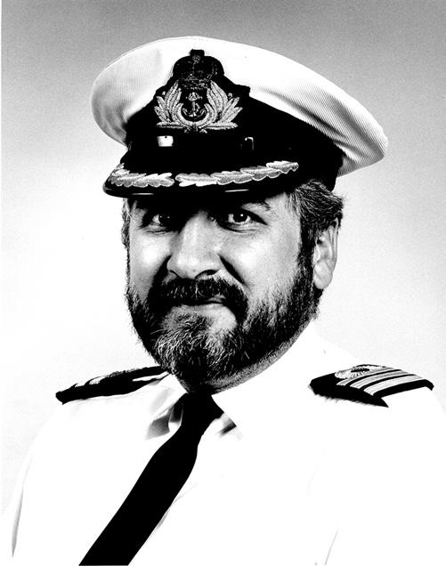
Vice-Admiral Alasdair Walker, CB, OBE, QHS, FRCS, RN 22 June 1956–1 June 2019
Alasdair Walker qualified from the University of Glasgow in 1979. He deployed to the South Atlantic during the Falklands War in 1982 and led Commando Forward Surgical Goup 2 during the Iraq War in 2003. He was Senior Surgeon in the Role 3 Hospital at Camp Bastion in 2009. Subsequent appointments included Medical Director (2009), Director of Medical Policy and Operational Capability for the Surgeon General (2011), Assistant Chief of the Defence Staff for Health (2014), Medical Director General (Navy), and Surgeon General in 2015. He retired from the Royal Navy in May 2019 as Surgeon Vice-Admiral.
The portrait above was taken during his time as International Scholar in the Department of Surgery at Uniformed Services University of Health Sciences, Bethesda, Maryland, United States in 1992.
Table of Contents
Surgical Trainee’s Perspective, 1
ALEXANDER KERSEY and ALEXIS LAURIA
SECTION 1
Setting the Stage, 11
1 The Vascular Injury Legacy, 12
NORMAN M. RICH and KENNETH J. CHERRY
2 Epidemiology of Vascular Trauma, 23
PETER GOGALNICEANU, TODD E. RASMUSSEN, and NIGEL R.M. TAI
3 Systems of Care in the Management of Vascular Injury, 34
DONALD H. JENKINS, DOUGLAS M. POKORNY, and PHILIP M. EDMUNDSON
4 Training Paradigms for Vascular Trauma, 42
PAUL W. WHITE and JAMES B. SAMPSON
SECTION 2
Immediate Management and Diagnostic Approaches, 55
5 Prehospital Management of Vascular Injury, 56
ROBERT H. JAMES and JASON E. SMITH
6 Damage Control and Immediate Resuscitation for Vascular Trauma, 70
TOM WOOLLEY, RAVI CHAUHAN, and ALLAN PANG
7 Diagnosis of Vascular Injury, 82
MICHAEL J. SISE
8 Imaging for the Evaluation and Treatment of Vascular Trauma, 91
DAVID L. DAWSON
SECTION 3
Emerging Technologies and New Approaches to Vascular Trauma and Shock, 107
9 Endovascular Suites and the Emergency Vascular Service, 108
JOSEPH A. HERROLD, THOMAS M. SCALEA, and JONATHAN J. MORRISON
10 Stent-Grafts, Coils, and Plugs, 114
DAVID SCHECHTMAN and BRANDON W. PROPPER
11 Resuscitative Endovascular Balloon Occlusion of the Aorta, 126
JENIANN A. YL, CHARLES JAMES FOX, and ERNEST E. MOORE
12 Endovascular Variable Aortic Control, 137
MICHAELA GAFFLEY and TIMOTHY K. WILLIAMS
13 Selective Aortic Arch Perfusion, 144
JAMES E. MANNING and ED B.G. BARNARD
14 Endovascular to Extracorporeal Organ Support for Vascular Trauma and Shock, 158
KEVIN K. CHUNG, ANDRIY I. BATCHINSKY, and IAN J. STEWART
15 Gathering the Evidence: Clinical Study of New Technologies, 166
LAURA J. MOORE and JAN O. JANSEN
SECTION 4
The Management of Vascular Trauma, 170
16 Cardiac, Great Vessel, and Pulmonary Injuries, 171
DAVID V. FELICIANO and JOSEPH J. DUBOSE
17 Blunt Thoracic Aortic Injury, 199
DEMETRIOS DEMETRIADES, PEEP TALVING, and KENJI INABA
18 Abdominal Aortic Trauma, Iliac and Visceral Vessel Injuries, 212
CHRISTOPHER AYLWIN and MICHAEL JENKINS
19 Inferior Vena Cava, Portal, and Mesenteric Venous Systems, 226
TIMOTHY FABIAN and STEPHANIE SAVAGE
20 Neck and Thoracic Outlet, 241
GREGORY A. MAGEE and FRED A. WEAVER
21 Upper Extremity and Junctional Zone Injuries, 252
MATTHEW VUONCINO, JOSEPH M. WHITE, and WILLIAM DARRIN
CLOUSE
22 Lower Extremity Vascular Trauma, 273
DAVID S. KAUVAR and BRANDON W. PROPPER
23 Surgical Damage Control and Temporary Vascular Shunts, 288
DANIEL J. SCOTT and SHAUN M. GIFFORD
24 Considerations for Conduit Repair of Vascular Injury, 300
NITEN SINGH and REBECCA JOY UR
25 Management of Pediatric Vascular Injury, 312
MATTHEW A. GOLDSHORE and JEREMY W. CANNON
26
Soft-Tissue and Skeletal Wound Management in the Setting of Vascular Injury, 321
SHEHAN HETTIARATCHY and JON CLASPER
27 Vascular Surgery in the Austere Environment, 332
DAVID M. NOTT
SECTION 5
Global Perspectives on Vascular Trauma, 352
28 Australia and New Zealand, 353 IAN D. CIVIL
29 Sri Lanka, 357 AMILA SANJIVA RATNAYAKE, SANJEEWA H. MUNASINGHE, and SUJEEWA P.B. THALGASPITIYA
30 Vascular Trauma in Finland, 365 PIRKKA VIKATMAA
31 Sweden, 370 TAL M. HÖRER and CARL MAGNUS WAHLGREN
32 Russia, 374 IGOR M. SAMOKHVALOV and VIKTOR A. REVA
33 Serbia, 377 LAZAR B. DAVIDOVIC and MIROSLAV MARKOVIC
34 Israel, 388 EITAN HELDENBERG and ELON GLASSBERG
35 South Africa, 391 KENNETH BOFFARD
36 Colombia: Don’t Dread the Popliteal and Axillary Fossa, 396 CARLOS A. ORDOÑEZ and MICHAEL W. PARRA
37 Brazil, 401 ROSSI MURILO and RINA PORTA Index, 407
This page intentionally left blank
Surgical Trainee’s Perspective
ALEXANDER KERSEY and ALEXIS LAURIA
Introduction
The aim of this section is to provide a concise, residentfocused, overview of a select number of chapters in the textbook. For each of the selected chapters, the topics are broken down by a general introduction (contextualizing the subject), surgical approach (detailing patient evaluation and prioritization) and tactics (providing helpful tips – the “Do’s and Don’ts” of safe surgical care) needed to get the best outcomes.
The section will be most useful for the busy Resident needing to quickly review the fundamentals of a vascular trauma topic, priming them for a later and more comprehensive review of the relevant chapters when their schedule permits.
Subject: Critical First Steps and Damage Control Resuscitation (Chapters 5 and 6)
GENERAL
n Damage control resuscitation (DCR) begins in the prehospital arena and is continued via reception into the Emergency Department (ED), transfer to surgery or interventional radiology (IR) suite, and within the Critical Care Unit.
n DCR prioritizes use of tourniquets, hemostatic dressings, temporizing procedures (resuscitative endovascular balloon occlusion of the aorta [REBOA]) and balanced transfusion of blood products to mitigate the consequences of hemorrhage and shock.
n DCR generates surgical options by restoring physiological normality.
SURGICAL APPROACH
n Prior preparation in the ED is essential: muster team members, assign roles, pre rehearse likely scenarios, prepare equipment and drugs, order blood products, don PPE, forewarn onward destinations (operating room (OR), IR suite, intensive care unit (ICU), etc.) and anticipated consultants.
n Use C-ABC framework to prioritize immediate steps in management:
n (C) Control of catastrophic bleeding – tourniquet, hemostatic dressings, REBOA
n Consider tranexamic acid (TXA) as part of the resuscitation protocol based on local practice.
n Consider broad spectrum antibiotics and tetanus where indicated.
n Match anesthesia-induction strategy to patient physiology (use cardio-stable induction agents to avoid catastrophic loss of cardiac output).
n Keep patient warm.
SURGICAL TACTICS IN THE ED
n Do:
n Familiarize yourself with the type of tourniquet and hemostatic dressings used by your prehospital care providers – how they are applied and released.
n Understand how long the warm ischemia time has been and ensure it is recorded.
n Release tourniquets only for specific purpose (e.g., immediately prior to diagnostic angiography; to assess likelihood of arterial injury, etc.). If no gross hemorrhage, leave down but be prepared to tighten again if further hemorrhage occurs.
n Remove dressings if ineffective or if examining wound will change operative decision-making.
n Re-assess tourniquets/hemostatic dressings after patient movement to ensure proper function.
n Monitor coagulopathy through early use of thromboelastography (TEG)/rotational thromboelastometry (ROTEM); monitor physiology through multiple feeds (trends in hemodynamic variables, lactate, urine output, etc.).
n Refine anticipated management plan as results from examination and investigations accrue and communicate accordingly.
n Establish and maintain hierarchy of open, closedloop communication and task allocation (i.e., identify team lead but enable all to have a voice).
n Use checklists and regular, formalized briefing opportunities (SNAP brief, STACK brief, ‘time outs’) to review progress and before major interventions (Chapter 6, p. 68)
n Do Not:
n Lose situational awareness and become task fixated.
n Become distracted by a prominent injury and fail to appropriately assess whole patient in stepwise fashion.
n Fail to obtain and document a brief but thorough extremity motor and sensory examination prior to intubation if safe to do so.
n Fail to consider ethics issues and/or fail to set ceilings of care for patients where continued medical intervention is likely to be futile.
n Fail to consider requirements of associated members of the attending trauma team (e.g., Orthopedics) and not integrate these points in to the overall DCR strategy.
Subject: Injury Identification and Diagnostic Workup (Chapters 7 and 8)
GENERAL
n Early identification of vascular injury is crucial to preventing long-term morbidity, loss of limb, or loss of life
n Chapter 7, Box 7.1 (Checklist for Prompt Recognition of Vascular Injury)
n Modalities for diagnosis include detailed injury history, bedside examinations (pulse examination, Doppler examination, point-of-care ultrasound, Ankle Brachial Index (ABI)), radiographic imaging (CT angiography [CTA]) and formal angiography.
n Approach is based on: (1) patient stability, (2) concomitant injuries, and (3) availability/feasibility of diagnostic modalities (i.e., contrast allergies).
DIAGNOSTIC APPROACH
n Any exsanguinating hemorrhage should be temporized per C-ABC with adjuncts (as described previously).
n Hard signs of vascular injury, CTA or angiography positive for vascular injury→to the OR
n Chapter 7, Fig. 7.5 (Algorithm of the indications for immediate operation and the role of imaging modalities)
n Hard signs of vascular injury include:
n Pulsatile hemorrhage
n Expanding hematoma
n Bruit or thrill over area of injury
n Absent pulse
n ABI < 0.9
n Soft signs of vascular injury include:
n History of hemorrhage at scene
n Wounds of neck/extremities with unexplained hemorrhagic shock
n Neurologic deficit in peripheral nerve in proximity to vessels
n High risk fracture, dislocation, or penetrating proximity wound
n Presence of a pulse does not rule out a vascular injury. Conversely, a normal ABI makes the likelihood of a vascular injury much lower.
n Consider use of checklist adjuncts to “clear” the patient of vascular injuries in various cavities or prompt further imaging.
n Chapter 7, Box 7.3 (Clearing Trauma Patients for Presence of Vascular Injury)
TACTICS
n Do:
n Consider injury mechanism when evaluating for occult injury.
n Be cognizant of additive use of contrast.
n Discuss injury concerns with radiologist so that vascular workup can be integrated with planned imaging to avoid multiple trips to the radiology suite.
n Frequently reassess for changes in pulse examination throughout the resuscitation as hypotension, vasopressors may confound examination.
n Do Not:
n Take an unstable patient to imaging.
n Delay operative intervention for imaging if the imaging will not add to or change decision-making.
n Hesitate to confirm questionable pulse examinations –either with second provider or Doppler examination.
n Fail to consider the risks associated with each imaging modality (contrast reactions, renal dysfunction, access site complications, time requirements, risks of radiation).
Subject: Resuscitative Endovascular Balloon Occlusion of the Aorta (REBOA) (Chapter 11)
GENERAL
n Minimally invasive alternative to thoracotomy and aortic clamping for temporization of exsanguinating subdiaphragmatic hemorrhage.
n Carries systemic consequences due to lower body and visceral ischemia but these are probably less than standard emergency department thoracotomy (EDT) and cross-clamping. These consequences may be off-set through the development of partial occlusion or intermittent occlusion techniques.
SURGICAL APPROACH
n The first step is percutaneous femoral artery cannulation, which should be done under ultrasound (US) guidance to maximize chances of success and minimize complications.
n The artery is accessed via hollow-needle and wire 2 to 3 cm below the mid-inguinal ligament. Seldinger technique is used to place a sheath, which is used to position a guidewire over which a compliant occlusion balloon can be placed into zone I or III as required, using predetermined standard insertion lengths (Chapter 11, Fig. 11.1). The wireless ER REBOA system (Prytime Medical; Chapter 11, Fig. 11.2) does not require wire guidance and employs a 7-Fr sheath.
n The aorta is divided into three zones; zone I ((left subclavian to celiac), zone II (celiac to lowest renal artery) and zone III (infrarenal aorta). Chapter 11, Fig. 11.4).
n The length of catheter insertion needed to reach each zone can be approximated using anatomic landmarks:
n Zone I: femoral access site to sternal notch
n Zone III: femoral access site to umbilicus
n The occlusion balloon is positioned in zone III for pelvic hemorrhage and distal zone I for intraabdominal bleeding.
n Position of the balloon may be confirmed with plain x-ray.
n The balloon is inflated with a contrast/saline mix:
n Inflate balloon until recognizable hemodynamic response (increased central/upper extremity blood
pressure, absent or decreased distal pulses, step-up in waveform proximal to balloon)
n Tactile feedback during inflation is important in recognizing aortic wall tension and avoiding injury –if resistance is met, inflation should stop
n The REBOA is secured to prevent balloon migration and the patient can be taken to the right place to stop the hemorrhage (IR suite, OR).
n Balloon time must be assiduously tracked. Zone I aortic occlusion time must be kept to less than 30 minutes to reduce the chances of spinal cord or visceral ischemia/ tissue infarction. Longer occlusion times may be tolerated for zone III inflation.
SURGICAL TACTICS
n Do:
n Match the need for REBOA to a good understanding of patient physiology:
n Physiologically stable patients with potential for sudden deterioration may have a sheath inserted as a prelude to REBOA deployment if there is deterioration.
n Unstable patients may have sheath insertion and balloon inflation to allow safer transfer to IR/OR.
n Deflate the balloon slowly (consider 1–2 mL every 2–3 minutes) to prevent rapid hemodynamic changes and catastrophic ischemia-reperfusion injury to the heart, giving plenty of warning to anesthetic colleagues.
n Consider adding medications or common rescue needs (bicarb, blood and/or crystalloid, pressors, management of hyperkalemia) in anticipation of reperfusion injury after balloon deflation.
n Employ REBOA as part of a comprehensive DCR paradigm.
n Do Not:
n Use REBOA as a “bridge” if the destination is not determined or the hemorrhage control strategy not clear – always have a plan as to next steps (including definitive hemorrhage control).
n Persist at multiple attempts at groin cannulation. Resort to surgical cutdown early if groin cannulation is not possible.
n Longitudinal incision extending inferiorly from the midpoint between the pubic symphysis and anterior superior iliac spine (ASIS).
n Use REBOA for intrathoracic hemorrhage.
n Fail to evaluate the patient for complications, particularly lower limb ischemia due to peri-sheath thrombosis, and have a plan for dealing with them.
Subject: Temporary Vascular Shunts (Chapter 23)
GENERAL
n Use of shunts is a desirable option to bridge damaged vessels in the extremities, junctional areas, and trunk to enable early reperfusion of tissue.
n Consider shunting when definitive repair must be deferred due to:
n physiological instability (i.e., damage control surgery)
n need to complete other life-saving interventions
n requirement to perform skeletal fixation
n absence of sufficient expertise or materials
n Shunting is applicable to both arteries and veins.
n Shunt dwell times of 2 to 5 hours are typical; however, some scenarios require longer times. The goal should be to remove shunts and perform definitive revascularization as early as the patient’s status, resources, and technical expertise allow.
SURGICAL APPROACH
n Shunts should be used as part of a comprehensive vascular management plan and require a technically experienced team with adequate resources.
SURGICAL TACTICS
n Do:
n Get the preliminaries right: adequate proximal and distal vascular exposure; injury definition/débridement; assessment of inflow and backflow; Fogarty sweep; heparinized saline flush.
n Ensure that vascular injury downstream of the shunt has been ruled out to prevent hemorrhage once flow restored – consider angiography.
n Choose a shunt and position that is right for the vessel caliber and injury:
n In-line – short segmental defect or small working area (Chapter 23, Figs. 23.3 and 23.4)
n Looped – long segment defect, large working area (Chapter 23, Fig. 23.5).
n Remember standard sequencing: [Shunt]→[Fracture Reduction and Fixation]→[Definitive Vascular Repair]→[Fasciotomy].
n Have a plan for definitive management, know when/ where necessary resources are available.
n Be aware of and have a plan for shunt-related complications (dislodgement, luminal injury, thrombosis, kinking, etc.) and communicate this to other relevant members of the patient care team.
n Do Not:
n Inadequately secure the shunt.
n Fail to consider collaterals and branch points within the injury/shunt zone and ligate these as needed.
n Fail to give anesthesia colleagues warning of reperfusion prior to shunt clamp removal.
n Routinely employ systemic anticoagulation.
n Routinely use shunts to bridge defects in small vessels below the elbow or the knee (increased likelihood of thrombosis).
Subject: Neck Injury (Chapter 20)
GENERAL
n A unique, compacted and congested anatomical zone with multiple vital structures.
n Neck trauma is divided into three zones based on anatomic landmarks (Chapter 20, Fig. 20.1):
n Zone I – sternal notch to cricoid cartilage
n Zone II – cricoid cartilage to angle of mandible
n Zone III – angle of mandible to base of skull
n Wide spectrum of presentation from exsanguinating hemorrhage to subtle clinical or imaging findings that can lead to delayed stroke.
n Carotid injuries may present with contralateral extremity weakness, aphasia, or Horner’s syndrome.
n Vertebral injuries are rarely symptomatic on presentation.
n Increased prevalence of CTA has resulted in more blunt injuries (blunt carotid and vertebral injury – BCVI) being identified.
n Risk factors for BCVI that should prompt screening include:
n Head and neck trauma associated with severe neck hyperextension and rotation or hyperflexion
n Lefort II or III fracture
n Basilar skull fracture involving the carotid canal
n Closed head injury consistent with diffuse axonal injury presenting with Glasgow coma scale (GCS) score <6
n• Cervical vertebral body or transverse foramen fracture, subluxation, or ligamentous injury at any level or any fracture of C1–C3
n A seat-belt or other clothesline-type injury with significant cervical pain, swelling, or altered mental status
SURGICAL APPROACH
n Physical examination is extremely important with identification of hard vascular signs an indication for airway control and operative exploration. For stable patients, CTA is the next step. It is crucial to pick-up pathologic neurological signs prior to intervention.
n Repair of penetrating carotid trauma in a patient with neurological deficit has attracted controversy, but there are no absolute contraindications. However, a delay of more than 3 hours from coma onset and large areas of cerebral infarct seen on initial CT scan are reasons to consider what reperfusion is likely to achieve.
n Catheter-based angiography and endovascular stenting is suited to distal carotid (zone III) hemorrhage where surgical access is difficult. Similarly, very proximal lesions (zone I) that would otherwise mandate median sternotomy for proximal control may be managed via covered stents.
n Operative positioning and draping should take into account the potential need to open the chest and possible vein harvest.
n Carotid injuries are exposed via a standard sternal notch to mastoid process incision, via a plane that lies medial to the sternocleidomastoid muscle. The internal jugular vein should be mobilized laterally away from the carotid; dividing the facial vein facilitates this. Exposing the upper parts of the internal carotid (zone III) requires preservation of the XII cranial nerve, division of the occipital artery, and mobilization of the posterior belly of the digastric, protecting the IX and XI cranial nerves.
n Options for carotid repair include:
n Primary repair with monofilament suture (rarely advisable)
n Patch angioplasty with bovine pericardium or vein graft
n Interposition graft with saphenous vein preferably or PTFE (graft generally required for >2-cm length defects)
n Use of a Fogarty occlusion balloon, carefully inflated, is a facile means of gaining distal control in zone III injuries. If possible, repairs of the internal carotid artery (ICA) should be undertaken using a shunt to maintain prograde flow. If backflow cannot be obtained after gentle Fogarty thrombectomy, there is little advantage attempting repair and ligation is advisable.
n Vertebral artery injuries are challenging to repair due to difficult access; management concentrates on injury definition (CTA), and control of hemorrhage (ligation, embolization) where this is significant, accepting the risk of posterior circulation stroke.
n Injuries to the vertebral artery as it passes through the transverse processes of the cervical vertebra are approached by the same route as exposure of the carotid artery. Hematoma will displace the carotid sheath anteriorly; the carotid artery and internal jugular vein must be displaced to allow access to the injury tract, longus colli muscle, and injured vertebral artery. Gaining proximal and distal control requires removal of the anterior tubercle of the transverse process which is difficult to accomplish in the midst of hemorrhage.
n Alternatively, tamponading the surgically exposed injury tract with hemostatic material or the balloon of a Fogarty catheter while addressing balanced transfusion and judicious use of time may be sufficient to allow bleeding to stop as a prelude to follow-up catheter-based embolization.
SURGICAL TACTICS
n Do:
n Screen aggressively for BCVI; management is almost always nonoperative with antithrombotic therapy and follow-up CTA the mainstay of treatment. Enlarging pseudoaneurysms that develop during follow-up can be selectively managed with stenting or coiling.
Grade Description
I Intimal injury with <25% luminal narrowing
II Dissection or hematoma with >25% luminal narrowing
Management
Antithrombotic therapy
Antithrombotic therapy
III Pseudoaneurysm Antithrombotic therapy; consider endovascular management
IV Occlusion
V Transection
Antithrombotic therapy
Operative intervention (endovascular if inaccessible)
n Antithrombotic therapy (either anticoagulation or antiplatelet) is chosen empirically based on injury pattern, provider experience, and institutional guidelines.
n Do Not:
n Fail to assess penetrating injuries of the neck for presence of tracheal and esophago-pharyngeal injury, through rigorous surgical exploration at the time of vascular repair and combination of rigid esophagoscopy + esophagography + tracheo-laryngoscopy if any doubt exists.
n Fail to repeat CTA 7 to 10 days after injury for nonoperatively managed patients to assess for resolution or progression
Subject: Upper Extremity and Junctional Zone Injuries (Chapter 21)
GENERAL
n Upper limb vascular trauma can lead to life-threatening hemorrhage and tissue ischemia, neuropathy and ischemia reperfusion injury.
n Junctional trauma may require control from within the chest; is technically challenging to manage and may be associated with gross shock.
n Where possible, injuries to the subclavian artery (SCA) can be managed with covered stents.
SURGICAL APPROACH
n Junctional penetrating trauma may present with a combination of upper limb signs (loss or function, reduced pulse through to obvious ischemia), local signs (pulsatile periclavicular hemorrhage or expanding hematoma), chest signs (massive hemothorax).
n A variety of surgical approaches exist for management of junctional injury:
Supraclavicular extension, resection of clavicular head
Extend with median sternotomy and supraclavicular incision (may be referred to as trapdoor incision)
Clavicular resection, infraclavicular incision
n Ligation of the SCA or axillary is unlikely to lead to limb loss or crippling ischemia due to collaterals but may result in functional impairment; repair or shunting is advised if possible. Brachial artery injuries should be repaired.
n Injuries to single forearm arteries may be ligated if there is good flow in the intact remaining vessel verified intraoperatively via Doppler.
SURGICAL TACTICS
n Do:
n Prep widely considering all possible approaches for proximal and distal control. Include the hand and forearm to allow for intraoperative Doppler interrogation and possible fasciotomy.
n Be cognizant of critical structures (brachial plexus, vagus, phrenic nerves).
n Use CTA to confirm site of probable junctional vascular injury if the patient is stable.
n Make liberal use of shunts for complex injuries, especially where conjoined orthopedic fixation is anticipated.
n Remember to perform forearm fasciotomy if compartment syndrome is anticipated (Chapter 21, Box. 21.1 and Fig. 21.15).
n Do Not:
n Use limb viability trauma scoring systems (Chapter 21, Tables 21.2 and 21.3) as an absolute driver of decision-making, but instead as a cue/prompting measure to consider all elements contributing to injury burden.
n Hesitate to gain the second opinion of a colleague when considering amputation.
n Cover a dominant vertebral artery when stenting the SCA, or fail to follow-up on patients with covered stents (where long-term outcomes are unknown).
n Blunt thoracic aortic injury (BTAI) typically occurs in the aorta distal to the origin of the left subclavian artery, and ranges from intimal tear only (minimal aortic injury) to pseudoaneurysm and complete transection (with lethal and unconstrained hemorrhage). CTA is the standard diagnostic tool.
Mid-distal brachial Incision over medial bicipital groove
Ulnar, radial Longitudinal forearm incision
SCA, Subclavian artery.
Lateral extension onto arm
S-shaped extension over antecubital crease to expose distal brachial artery
n Most cases that survive to reach surgical care can be temporized through vigorous blood pressure control (beta blockade) and careful monitoring while arrangements are optimized for definitive management or other more life-threatening injuries addressed.
n In general, goal SBP <120 mm Hg
n Esmolol drip is most commonly used due to rapid onset, ease of titration
n Aortic endovascular stenting (thoracic endovascular aortic repair (TEVAR); Chapter 17, Figs. 17.9, 17.10) has become a prevailing mode of treatment, with open
or hybrid repairs reserved for injuries involving the aortic arch or where endovascular resources are limited.
SURGICAL APPROACH
n Open repair is accomplished by the clamp and sew approach via left posterolateral thoracotomy and distal perfusion of the aorta to reduce the chance of spinal cord ischemia (Chapter 17, pp. 5–7, Fig. 17.8).
n Proper sizing of stents is key in preventing TEVAR-related complications (Chapter 17, Table 17.3 and Figs. 17.11–17.13). Bird’s neck deformity can be avoided by using new generation devices that allow for the curvature of the aorta in young patients.
n Most patients tolerate covering of the origin of the left subclavian well. Carotid-subclavian bypass can be undertaken for patients who develop subclavian steal.
SURGICAL TACTICS
n Do:
n Base the timing of definitive management on the nature and extent of the BTAI lesion, other associated injuries, and facility expertise.
n Consider conservative management (with early CTA follow-up) for patients with minor lesions (intimal tear, small pseudoaneurysm).
n Consider screening for blunt cardiac injury with electrocardiogram (EKG) monitoring.
n Do Not:
n Fail to ensure that all TEVAR patients are submitted to life-long surveillance programs to ensure stent complications are identified.
Subject: Cardiac, Great Vessel and Pulmonary Injuries (Chapter 16)
GENERAL
n Surgery is infrequently needed for thoracic injury; patients with penetrating injuries to the heart and great vessels usually do not reach the surgeon alive.
n The most common indications for thoracotomy are hemorrhage from the lung, major arterial injury in the arch or root-of-neck vessels, or a penetrating cardiac wound.
SURGICAL APPROACH
n Any penetrating injury between the nipples from the sternal notch to xiphoid process (known as the “cardiac box”) or encompassing the left chest, should be evaluated for potential cardiac injury.
n Site large-bore access venous access sites on the contralateral side to any injury, and consider using the common femoral veins.
n Critically shocked patients with evidence of massive hemothorax (chest x-ray appearance, immediate drainage of 1200–1500 mL of blood via tube thoracostomy); or cardiac tamponade (diagnosed on US); or visible hemorrhage from the root of the neck should undergo
emergent endotracheal intubation in the ER and transfer expeditiously to the OR for surgery.
n Where physiology becomes agonal, perform EDT (left anterolateral thoracotomy or clamshell thoracotomy if suspected right-sided injury) (Chapter 16, pp. 4–6, Figs. 16.1–16.4) with the aim of:
n Confirming the diagnosis.
n Performing pericardiotomy if tamponade is present.
n Via longitudinal incision in the pericardium above the left phrenic nerve.
n Controlling hemorrhage from a wound to the heart, great vessels, or lung.
n Catastrophic lung hemorrhage may be temporized with use of hilar clamping or lung twist (Chapter 16, p. 25). These require division of the inferior pulmonary ligament.
n Clamping the descending thoracic aorta to preserve circulating volume and perfuse the coronary and carotid arteries.
n The inferior pulmonary ligament can be taken down to aid in visualization.
n Undertaking internal cardiac massage.
n Less critically disturbed patients whose physiology stabilizes with resuscitation can be more thoroughly worked up (CT chest, CTA arch vessels) and then moved to critical care for close observation. Conservative management should be abandoned if tube thoracostomy output continues (>200 mL/h over 2–4 hours) or if volume requirements become elevated.
SURGICAL TACTICS
n Do:
n Make use of the subxiphoid pericardial window technique to rule out tamponade during trauma laparotomy (Chapter 16, p. 8).
n Match the repair technique to the location of the heart injury: clamp and suture (permanent monofilament) for atria; unpledgeted repair to right ventricle; pledgeted repair to left ventricle.
n Consider temporizing cardiac injuries in extremis with skin staples, Foley balloon, or other adjuncts (Chapter 16, Table 16.2).
n Use partial isolation (Satinsky clamp) to deal with simple penetrating arch injury.
n Use debranching techniques to manage complex injuries to the branch vessels (i.e., control the injury→sew proximal end of prosthetic graft onto arch→sew distal end of graft to cut distal end of the disrupted arch vessel).
n Use lung-sparing techniques (suture, stapled wedge, tractotomy) when dealing with pulmonary hemorrhage (Chapter 16, pp. 25–26).
n Consider endovascular techniques to manage great vessel injuries.
n Do Not:
n Undertake futile resuscitative thoracotomy (blunt mechanism of injury [MOI] with no signs of life in ER, penetrating MOI with no signs of life in the field).
n Fail to consider packing the chest as a damage control measure.
n Injure the intercostal arteries arising from the posterior aspect of the thoracic aorta when applying a cross-clamp.
n Lose sight of location of coronary arteries and inadvertently ligate or disrupt coronary arteries while attempting to repair a cardiac injury.
n Forget to get postop echocardiography to evaluate for valvular injury post cardiac repair.
Subject: Aortic, Iliac and Visceral Arterial Injuries (Chapter 18)
GENERAL
n Wide variety of presentation with potential for rapid deterioration and exsanguination.
n Vascular injuries in the abdomen are categorized according to anatomical location, defined within three retroperitoneal zones: zone I – midline, zone II – lateral, zone III – pelvic (Chapter 18, Fig. 18.1).
n CTA is the gold standard investigation for stable casualties; exploratory laparotomy for unstable patients (i.e., no or short-lived response to initial resuscitation).
SURGICAL APPROACH
n Skin preparation and draping should take account of the potential need for left anterolateral thoracotomy and vascular control at the groins.
n Evisceration, four-quadrant packing, and sequential removal of packs removes blood and allows a methodical start to challenging surgery.
n Any zone I retroperitoneal hematoma will require exploration as there is a high chance of it involving the aorta or its branches, or the inferior vena cava (IVC). Left or right medial visceral rotation respectively for aorta (hematoma biased to left of midline) and IVC (hematoma biased to right of midline) are the key maneuvers to expose the injury, though consideration should be given to obtaining supraceliac aortic control in very unstable patients beforehand (Chapter 18, Figs. 18.2–18.4).
n Large zone I hematomas that are present in the supracolic compartment may be better controlled through clamping of the thoracic aorta via left anterolateral thoracotomy.
n Zone II and zone III hematomas may be managed more judiciously, with exploration reserved for ongoing bleeding (expanding hematoma or the presence of physiological instability). Some also include penetrating trauma as an indication, particularly for pelvic hematoma where the iliac vessels may have been injured.
SURGICAL TACTICS
n Do:
n Do consider preplacement of a REBOA catheter prelaparotomy to enable rapid aortic control should this be required.
n Branches of the celiac artery can be ligated proximally with low risk of end-organ ischemia; injury to the peripancreatic superior mesenteric artery (SMA) should be repaired or shunted in order to avoid catastrophic mid-gut infarction. Approach the celiac via
the lesser sac or left medial visceral rotation; approach the SMA via the lesser sac (with stapled division of the pancreas in-extremis), via left medial visceral rotation or via the root of the small bowel mesentery. The inferior mesenteric artery can be ligated. Injuries to the renal arteries usually result in ligation and probable nephrectomy.
n Mobilize the cecum or sigmoid colon to visualize the common and external iliac vessels, avoiding the ureter, and be prepared to achieve distal control at the groin if unfavorable pelvic anatomy is present.
n Do Not:
n Fail to consider endovascular treatment of pelvic vessel injury (covered stent) if the situation permits (i.e., consider operating in a hybrid room if possible).
n Fail to consider preperitoneal packing as a means to temporize pelvic bleeding associated with pelvic fractures prior to embolization.
n Fail to consider the likelihood of abdominal compartment syndrome and the advantage of temporary laparostomy to prevent this and allow assessment of visceral viability at a subsequent planned relook procedure.
n As with injury to the aorta and its branches, injuries to the IVC, portal vein, and mesenteric vessels are highly lethal.
n CTA is the investigation of choice, with hematomas around the ascending colon and duodenum fairly specific for IVC injuries as well as caval filling defect.
n Resuscitative thoracotomy or zone I REBOA are valid means of controlling aortic inflow in order to manage the critically deteriorating patient.
n Selected patients, without hemodynamic disturbance, and where the hematoma is small-to-moderate on CT scan, may be monitored and observed; assuming there is no other reason to pursue laparotomy (e.g., blunt injury; or, if penetrating, no violation of peritoneum or signs of peritonism).
SURGICAL APPROACH
n Cava:
n Utilize right medial visceral rotation (Chapter 19, Fig. 19.2), with extensive kocherization of the duodenum, for infra- and suprarenal IVC and control. Use digital pressure or careful application of swabs/ sponges on sticks to occlude the IVC either side of an injury rather than attempting encirclement with risk to the lumbar vessels.
n Retrohepatic injuries to the cava, heralded by dark blood continuously welling up from behind the liver, should be managed via manual compression of the liver against the cava and thence appropriate packing and/ or clamping of the portal triad (Pringle maneuver).
n If this fails to control bleeding, the laparotomy incision should be extended to the right chest via right anterolateral thoracotomy, with control of the suprahepatic IVC from within the pericardium, prior to full mobilization of the liver, and exposure of the injured retrohepatic cava. Alternatively, seek to place occlusion balloons in the cava via percutaneous means to isolate the liver prior to mobilization.
n Portal Vein
n Approach by clamping the portal triad proximal to the injury, taking down the hepatic flexure of the colon, and performing a wide Kocher maneuver, then releasing the clamp to facilitate digital control of the injury and gentle dissection of the vein away from the hepatic artery and common bile duct to allow injury definition and placement of vessel loops.
n Be prepared to divide the neck of the pancreas (to the left of the SMA/superior mesenteric vein [SMV]) using a linear cutter stapler in order to expose the most proximal portion of the portal vein if proximal control is not achievable otherwise.
n Superior Mesenteric Vein
n Injuries are associated with central hematoma at the base of the small bowel mesentery (at the fusion of the peritoneum overlying junction of the transverse colon mesentery).
n Medial visceral rotation from the right with kocherization allows the operator to place a hand behind the mesentery containing the injured vein. This allows for application of digital control and dissection of the vessel/ clamping/repair, or ligation prior to hematoma entry.
SURGICAL TACTICS
n Do:
n Consider getting aortic control before opening a large central hematoma.
n Handle the portal vein and SMV carefully; they are thin-walled and tear easily.
n Anticipate the splanchnic sequestration effects of portal vein or SMV ligation and ensure aggressive volume resuscitation in the postoperative period. Plan for early relook laparotomy to assess for potential bowel infarction.
n Do Not:
n Expect to see contrast extravasation on the initial CT abdomen in venous injury.
n Ligate the suprarenal IVC – this is not tolerated and will lead to acute renal failure.
n Spend too much time attempting complex repair of the portal vein – damage control adjuncts such as ligation or shunting should be considered early to avoid extensive bleeding.
n The lower limb is the most frequent site of arterial injury in both civilian and military trauma.
n Documenting a complete lower extremity neurovascular examination prior to intervention assists in determination of injury pattern as well as evaluation of possible postoperative complications.
n High energy mechanisms of injury, especially explosions, result in complex multisegmental injury with disruption of soft tissue, bone, and skin.
n Limb salvage and vascular reconstruction must only be considered in the context of the totality of injury and associated physiological disturbance. Interventions that save life and restore homeostasis should be prioritized accordingly.
n The presence of a vascular injury can be discerned from hemorrhage, ischemia, or signs found on CTA. The latter, as a preprocedural investigation, is especially useful in situations where several levels of vascular damage may be present (shotgun injury, multiple long bone fracture).
n Warm ischemic time is very important and must be carefully monitored, driving urgency of revascularization. Aim to restore perfusion within 3 hours of injury –although classic teaching is 6 hours, recent data indicates this is too long.
n Primary amputation is a difficult decision to undertake –a second opinion, obtained from an experienced colleague brought to the OR for this purpose can help in decision-making.
SURGICAL APPROACH
n Proximal tourniquet for control of ongoing hemorrhage until proximal control is achieved via exposure of vessels above the injury zone, with subsequent distal control and thence entry into the hematoma and evaluation of the injury. Prep tourniquet into field to allow for intraoperative manipulation.
n Injuries at the groin may not be amenable to tourniquet control; sponge-stick or digital control of external hemorrhage should be maintained while a retroperitoneal approach is used to effect access to the external iliac artery (EIA). A curvilinear skin incision, from the mid-inguinal point superior to the anterior superior iliac spine, can be employed, dividing aponeurosis of the external oblique, splitting the underlying fibers of the internal oblique and transversus abdominis, reaching the preperitoneal plane, and developing this medially to reach the EIA.
n In general, standard vascular axial exposures (midinguinal, anteromedial thigh, medial calf; Chapter 22, pp. 9–13) can be used to deal with injuries to the common, profunda and superficial femoral arteries, and the popliteal and posterior tibial vessels. There should be no hesitation in extending these incisions proximally or distally as the situation dictates.
n The orthodox preliminaries of injury definition, evaluation of inflow and backflow, and Fogarty catheter thrombectomy of upstream and downstream vessels are required before considering whether the patient requires a shunt or immediate definitive repair.
n Ensure adequate healthy tissue coverage over any shunt/ repair after the wound is appropriately débrided.