Instant digital products (PDF, ePub, MOBI) ready for you
Download now and discover formats that fit your needs...
Radiographic Imaging and Exposure E Book 5th Edition, (Ebook PDF)
https://ebookmass.com/product/radiographic-imaging-and-exposure-ebook-5th-edition-ebook-pdf/
ebookmass.com
Essentials of Radiographic Physics and Imaging E Book 2nd Edition, (Ebook PDF)
https://ebookmass.com/product/essentials-of-radiographic-physics-andimaging-e-book-2nd-edition-ebook-pdf/
ebookmass.com
Principles of Radiographic Imaging: An Art and A Science 6th Edition Richard R. Carlton
https://ebookmass.com/product/principles-of-radiographic-imaging-anart-and-a-science-6th-edition-richard-r-carlton/
ebookmass.com
Analysis and Design of Prestressed Concrete Di Hu
https://ebookmass.com/product/analysis-and-design-of-prestressedconcrete-di-hu/
ebookmass.com

Belonging to the Rancher Sam Crescent
https://ebookmass.com/product/belonging-to-the-rancher-sam-crescent/
ebookmass.com
Dark Harmony Laura Thalassa
https://ebookmass.com/product/dark-harmony-laura-thalassa-5/
ebookmass.com
The Palgrave Handbook of Corporate Sustainability in the Digital Era Seung Ho Park
https://ebookmass.com/product/the-palgrave-handbook-of-corporatesustainability-in-the-digital-era-seung-ho-park/
ebookmass.com
Organizational Behavior (18th Edition) (Whatu2019s New in Management) (eBook PDF)
https://ebookmass.com/product/organizational-behavior-18th-editionwhats-new-in-management-ebook-pdf/
ebookmass.com
Tribals and Dalits in Orissa: Towards a Social History of Exclusion, c. 1800-1950 Biswamoy Pati
https://ebookmass.com/product/tribals-and-dalits-in-orissa-towards-asocial-history-of-exclusion-c-1800-1950-biswamoy-pati/
ebookmass.com


(eBook PDF) Writing and Reporting for the Media 12th
https://ebookmass.com/product/ebook-pdf-writing-and-reporting-for-themedia-12th-edition/
ebookmass.com


and Electromagnets / 67
/ 8

Pair Production / 199
Photodisintegration / 199
Effect on Technical Factor Selection / 200
CHAPTER 13
Minimizing Patient Dose 204
Estimating Approximate Entrance Skin Exposure / 205
Typical Entrance Skin Exposure / 208
Reducing Patient Dose with Communication / 209
Reducing Patient Dose with Positioning / 209
Reducing Patient Dose with Technical Factors / 209
Discussing Radiation Risk Versus Benefit With Patients / 213 UNIT III Creating
CHAPTER 14
Vision and Perception 220
Image Perception / 221
Controlling the Image in Space / 226
Radiography as an Art Form / 227
CHAPTER 15
Beam Restriction 233
Controlling Scatter / 234
Beam Restrictors / 236
Ancillary Devices / 238
CHAPTER 16
The Patient as a Beam Emitter 240
Attenuation / 241
The Human Body as an Attenuator / 241
The Patient’s Relationship to Image Quality / 243
CHAPTER 17 The Pathology Problem 246
Pathology and Radiation Absorption / 247
Increased Attenuation (Additive) Conditions / 250
Decreased Attenuation (Destructive) Conditions / 253
CHAPTER 18
The Grid 257
Purpose of the Grid / 258
Grid Construction / 259
Grid Patterns / 262
Grid Types / 262
Grid Uses / 263
Grid Selection/Conversions / 264
Grid Performance Evaluation / 265
Grid Errors / 265
An Alternate Scatter Reduction Method—The Air-Gap Technique / 269
CHAPTER 19
Radiographic Film 273
Construction / 274
Latent Image Formation / 278
Types of Film / 279
Film Storage and Handling / 280
Film Identification / 280
CHAPTER 20
Film Processing 283
Developing / 284
Fixing / 287
Archiving / 288
Automatic Processing / 288
Darkroom / 290
Silver Recovery Systems / 291
CHAPTER 21
Film Sensitometry 294
Sensitometric Equipment / 295
The D Log E Curve / 298
Film Characteristics / 299
CHAPTER 22
Intensifying Screens and Film/Screen Combinations 308
Construction / 309
Phosphors / 311
Characteristics / 312
Cassettes and Holders / 315
Care / 316
Emission Spectra / 316
Characteristics / 317 CHAPTER 23
Digital Radiography 323
Historical Development / 324
Digital Image Formation / 324
Image Processing Operations / 327
Digital Image Quality / 332
Exposure Indicators / 333
Computed Radiography (PSP) Systems / 339
Flat Panel Digital Radiography / 353
Digital Radiography Artifacts / 356
24 Picture Archiving and Communications Systems (PACSs)
Informatics / 363
Image Acquisition / 365
Image Distribution / 365
Image Display / 366 Storage / 369
Process / 376
/ 380
CHAPTER 33
Automatic Exposure Controls 506
Ionization Chambers / 507
Positioning Skills / 509
CHAPTER 34
Exposure Conversion Problems 514
Standard Conversion Relationships and Tables / 515
Solving Complex Exposure Problems / 517
UNIT VI
SPeCiaL imaging SyStemS anD mODaLitieS
CHAPTER 35
Mobile Radiography 523
Special Patient Considerations / 524
Special Radiation Protection Considerations / 527
Types of Equipment / 527
Special Technical Factor Selection Considerations / 528
CHAPTER 36
Fluoroscopy 533
Historical Development / 534
Fluoroscopic Uses / 534
Types of Equipment / 534
Fluoroscopic X-Ray Tubes / 535
Image Intensification Tubes / 536
Image Quality / 540
Video Viewing Systems / 541
Digital Fluoroscopy / 542
Recording the Fluoroscopic Image / 542
Mobile Fluoroscopic Equipment / 543
Radiation Protection During Fluoroscopy / 543 CHAPTER 37
Tomography and Digital
Tomosynthesis 548
The Tomographic Principle / 549
Tomographic Quality / 551
Types of Motion / 554
Digital Tomosynthesis / 555
Tomographic Procedures / 556 CHAPTER 38 Mammography 561
Historical Development / 562
Generator Characteristics / 567
X-Ray Tube / 571
Accessories / 581
Resolution / 584
Digital Mammography / 584
Quality Control / 585 CHAPTER 39
History and Overview / 590
Bone Science / 591
Osteoporosis / 592
Diagnostic Medical
Sonography 724
Introduction / 725
Sound Waves / 726
Attenuation / 735
Range / 736
Field / 736
Doppler / 737
Harmonics / 737
Transducer and Components / 738
Instrumentation / 741
What?
Arlene Adler and Rick Carlton publishing another revision! I know it is hard to believe—again the two of them. How fortunate for medical imaging sciences professionals to have such hard-working, dedicated scholars, who, by their very nature, continually strive to provide the most comprehensive and practical text of relevant and essential principles for the medical imaging sciences professional community.
As one of the most popular imaging sciences texts on the market, this fifth edition of Principles of Radiographic Imaging includes the quality attributes and fundamentals of prior editions, preserving essential concepts and condensing and deleting older conventional content while addressing newer, more modern imaging principles.
Noteworthy to the fifth edition is a considerable rework and expansion of chapters on digital imaging principles containing solidified terminologies. Educators throughout our profession have hungered for reliable, comprehensive, and practical learning resources for digital imaging technologies, and Carlton and Adler’s fifth edition has definitely delivered. Significant expansion of digital radiography content produced a need to revise nearly all, and in some cases, consolidate, principles chapters. Additional content areas address other newer imaging equipment, such as digital tomosynthesis and PACS.
As educators are keenly aware, visuals included in prior editions have always positively
reinforced student learning. The new edition is no exception, as illustrative art throughout the text has been expanded and updated. Of course, the accompanying laboratory manual and instructor resources have undergone appropriate revisions to complement updates incorporated in the text.
The nationally recognized curriculum requires educators to support student learning in other related imaging modalities and disciplines. To this end, this fifth edition of Principles of Radiographic Imaging includes chapters that address essentials of imaging sciences instrumentation used in bone densitometry, nuclear medicine and PET scanning, radiation therapy, and diagnostic medical sonography.
As technologists writing for technologists, Mr. Carlton and Ms. Adler danced a delicate dance to determine what to condense, what to delete, and how to expand. They have, yet again, served up a respected authoritative teaching and learning resource for medical imaging sciences professionals—one that is concise yet thorough, and one that certainly broadens the scope of understanding of imaging principles for the entire profession.
Denise Moore, M.S., R.T.(R) Professor Emeritus Radiologic Technology
Sinclair Community College Dayton, Ohio
enhanced by the addition of digital factors that now impact on all imaging decisions.
OrganizatiOn
The overall design of the book separates the 45 chapters into six units: Creating the Beam, Protecting Patients and Personnel, Creating the Image, Analyzing the Image, Comparing Exposure Systems, and Special Imaging Systems and Modalities. We are pleased to offer four new chapters that address the instrumentation for bone densitometry, nuclear medicine and PET scanning, radiation therapy, and diagnostic medical sonography. Along with the chapters on mammography, vascular imaging equipment, computed tomography, and magnetic resonance imaging, we now offer framework information on all radiologic and imaging sciences modalities and treatments. This design helps organize the content for students by following a logical progression from introductory physics through the production and control of the beam to advanced modality systems. We remain extremely pleased that our book remains one of the resources listed in the Radiography Curriculum of the American Society of Radiologic Technologists, and we have long been pleased that this book continues to be recommended by the Canadian Association of Medical Radiation Technologists for preparation for its certification in Radiological Technology.
neW tO thiS eDitiOn
The rapid changes in technology present a challenge to textbook authors who are committed to providing current information for learners. The authors and contributors to the fifth edition carefully reviewed all content to identify areas requiring updating or new topics. As a result, numerous changes were made. The more significant changes are as follows:
◾ Significant rewrite of digital radiography imaging principles with appropriate condensation of conventional radiography.
◾ New chapters on radiologic and imaging sciences instrumentation used for bone densitometry, nuclear medicine and PET scanning, radiation therapy, and diagnostic medical sonography.
◾ Updates on new imaging equipment such as digital tomosynthesis and PACS, as well as making sure information on modalities such as CT and MRI remain current.
FeatureS
In addition to the updated and new content, this new edition continues to feature the following learning aids and critical content:
◾ Physical concepts are clearly explained and illustrated with many high-quality figures.
◾ Effects of changing parameters on image quality are carefully described and illustrated with numerous images.
◾ Criteria for image analysis are presented to help learners develop analytical skills.
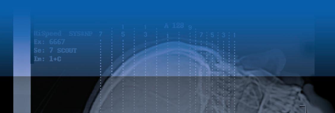
◾ High-quality radiographs are included throughout the text.
◾ Radiation protection concepts and procedures are emphasized for both patients and radiographic personnel.
◾ Chapter-end summaries provide a quick reference to critical concepts and developments in the science of radiography.
◾ Numerous troubleshooting tips are included to ensure quality radiographs.
◾ Extensive references and recommended readings provide a historical perspective and provide learners a means to expand their understanding of concepts and systems.
◾ Epigraphs and historical photos help trace the evolution of radiography to the present.
◾ Unique emphasis on the art versus the science of radiography illustrates the broad applications of the technology.
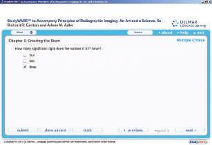
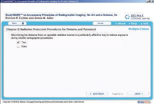
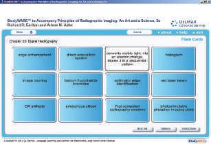
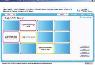
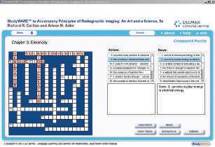
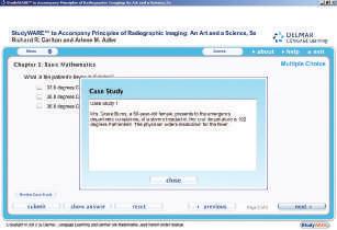
StudyWARE
The StudyWARE software includes interactive quizzes and activities that provide additional practice, and fun, while learning. Activities include games such as concentration and hangman as well as flashcards, crossword puzzles, and image labeling exercises. See “How to Use the StudyWARE” on page xxx for details.
Workbook
(ISBN: 978-1-4390-5870-1)
The workbook was created as a series of activities, both laboratories and worksheets, to provide higher-level synthesis and analysis for each chapter in the textbook. There are a total of 99 exercises: 62 are laboratories and 37 are worksheets.
Premium Website (ISBN: 978-1-1337-0276-4)
A Premium Website is available to accompany the text that includes the StudyWARE™, slide presentations in PowerPoint ®, and an image library.
Redeeming an Access Code:
1. GO TO: http://www.CengageBrain.com.
2. ENTER THE Access code in the Prepaid Code or Access Key field, REDEEM.
3. REGISTER as a new user or LOG IN as an existing user if you already have an account with Cengage Learning or CengageBrain.com.
4. SELECT Go to MY Account.
5. OPEN the product from the My Account page.
Also Available:
◾ Premium Website IAC to accompany Principles of Radiographic Imaging: An Art and a Science, Fifth Edition (ISBN: 978-1-1337-0275-7)
Instructor Resources CD-ROM (ISBN: 978-1-4390-5869-5)
The Instructor Resources CD-ROM is a robust computerized tool designed to meet your instructional needs. A must-have for all instructors, this comprehensive and convenient CD-ROM contains the following components.
Instructor’s Manual
The Instructor’s Manual includes answers to the review questions contained within the textbook. The review questions are designed to assess the students’ accomplishment of the chapter objectives and may be useful as a homework assignment or simply as a self-assessment tool for the student. The manual also provides the instructor with additional student assignment ideas, which may be used to assess students’ understanding of the content and/or help expand their knowledge of the content beyond the scope of the text.
Answers are also provided for the worksheets and laboratory exercises in the workbook, with the exception of laboratories in which the student answer determines the results.
PowerPoint® Presentations
More than 1,700 PowerPoint slides are designed to aid you in planning your class presentations. If a student misses a class, a printout of the slides for a lecture provides a helpful review page. Instructors, please feel free to add your own slides for additional topics you introduce to the class.
Computerized Test Bank in ExamView®
The test bank includes approximately 1,770 test questions. These include multiple-choice, shortanswer, matching, and completion questions, as well as problems. Users can add their own questions. This software allows the user to create tests in less than 5 minutes, with the ability to print them out in a variety of layouts. It also has electronic “take-home testing” (put test on disk) and Internetbased testing capabilities.
Image Library
The image library includes more than 600 files containing images from the textbook.
Instructor Companion Site
An Instructor Companion Site is available that includes the Instructor Resources.
To access the Instructor Companion site, go to http://login.cengage.com.
◾ If you have a Cengage SSO account: Sign in with your e-mail address and password.
◾ If you do not have a Cengage SSO account: Click Create My Account and follow the prompts.
WebTUTOR™
(WebTUTOR™ on Blackboard
ISBN: 978-1-4390-5867-1)
Designed to complement the textbook, WebTUTORTM is a content-rich, Web-based teaching and learning aid that reinforces and clarifies complex concepts. The BlackboardTM platform also provides rich communication tools to instructors and students, including a course calendar, chat, email, and threaded discussions.
CourseMate
(CourseMate PAC (printed access card)
ISBN: 978-1-1337-0273-3)
(CourseMate IAC (instant access code)
ISBN: 978-1-1337-0274-0)
Principles of Radiographic Imaging: An Art and a Science, Fifth Edition includes CourseMate, a complement to your textbook. CourseMate includes:
◾ An interactive eBook
◾ Interactive teaching and learning tools, including:
◾ Quizzes
◾ Flashcards
◾ PowerPoint slides
◾ Glossary
◾ and more
◾ Engagement Tracker, a first-of-its-kind tool that monitors student engagement in the course
To access these materials online, visit http:// login.cengage.com.
Statement OF COntent aCCuraCy
Although we assume full responsibility for any errors, including those that may be construed as arising from quoting other works out of context, we have made every effort to ensure the accuracy of the information. However, appropriate information sources should be consulted, especially for new or unfamiliar procedures. It is the responsibility of every practitioner to evaluate the appropriateness of a particular procedure in the context of actual clinical situations. Therefore, neither the authors nor the publisher take responsibility or accept any liability for the actions of persons applying the information contained herein in an unprofessional manner. This information is designed to supplement and enhance the instructional methodologies of educators in JRCERT (Joint Review Committee on Education in Radiologic Technologies [USA]) and CAMRT (Canada) approved radiography programs and should not be applied, especially to human subjects, without this background. In committing this book to print we fully realize that it is never finished, merely suspended for the time being.
Finally, as a reader your perceptions are important to us. We encourage you to communicate with us regarding facets of the book you appreciate or would like to see changed. We especially appreciate constructive comments and notice of errors. Our intention is to present the principles of radiography in an interesting format that provides a base from which true professionalism can develop. Any commentary readers care to make toward this end will be valued and welcomed.
Richard R. Carlton
Grand Valley State University
Radiologic and Imaging Sciences
Center for Health Sciences
Suite 455
301 Michigan St. NE
Grand Rapids, MI 49503
616-331-5600
Arlene M. Adler
Indiana University Northwest 3400 Broadway Gary, IN 46408 1219-980-6540 aadler@iun.edu