No part of this publication may be reproduced or transmitted in any form or by any means, electronic or mechanical, including photocopying, recording, or any information storage and retrieval system, without permission in writing from the publisher. Details on how to seek permission, further information about the Publisher’s permissions policies and our arrangements with organizations such as the Copyright Clearance Center and the Copyright Licensing Agency, can be found at our website: www.elsevier.com/permissions.
This book and the individual contributions contained in it are protected under copyright by the Publisher (other than as may be noted herein).
Notice
Practitioners and researchers must always rely on their own experience and knowledge in evaluating and using any information, methods, compounds or experiments described herein. Because of rapid advances in the medical sciences, in particular, independent verifi cation of diagnoses and drug dosages should be made. To the fullest extent of the law, no responsibility is assumed by Elsevier, authors, editors or contributors for any injury and/or damage to persons or property as a matter of products liability, negligence or otherwise, or from any use or operation of any methods, products, instructions, or ideas contained in the material herein.
Manager, Content Strategy: Nimisha Goswami
Content Project Manager: Anand K Jha
Sr Graphic Designer: Milind Majgaonkar
Sr Production Executive: Ravinder Sharma
Laser typeset by GW India
Printed in India by
Dedication
Dedicated to my angels … Sanjana and Sidharth my beloved husband … Gopi Krishna my parents … Ambuja and Shiva Subramanian and to the Almighty …
Contributors
S Lakshmi, MDS, MICOI (USA), Professor, Department of Prosthodontics and Implantology, Meenakshi Ammal Dental College, Chennai - 600095
CS Raj Mohan, MDS, Course Leader—Fixed Prosthodontics, Adult Restorative Dentistry, Oman Dental College, Muscat, Oman
Preface to the third edition
S Lakshmi
The third edition of the Preclinical Manual of Prosthodontics maintains our commitment to guide dental students of preclinical and clinical years. We have ensured that the descriptions are precise, and well supported with illustrations. The preclinical students can use the book as a hand guide manual for their preclinical sessions.
In this edition, we have introduced a section for the preclinical students to get an orientation of a clinical procedure in the rehabilitation of completely edentulous patients with a removable prosthesis. We have also added newer dental materials and instruments in Chapter 2 Instruments and Materials, for the preclinical students to be aware of the latest trends. Preclinical exercises of teeth preparation have been included in Chapter 3 Preclincal Exercise which is a practical exam exercise for the clinical students. Newer preclinical exercises have been included which will help students to do the laboratory related work during the clinical sessions of the course. We are confident that the students will be benefitted in understanding the basic concepts of prosthodontics.
Preface to the first edition
S Lakshmi
It is a privilege and an honour to have had an opportunity to bring out a manual of Preclinical Prosthodontics for the preclinical students of dental schools. The subject of Prosthodontics is introduced to the students of dentistry in the first year of the curriculum. The students appear for a preclinical Prosthodontics examination in the second year of BDS course before entering into the clinical prosthodontics in the third year.
During the first year and second year of the BDS course, students are required to do certain preclinical exercises so that they are trained to do these procedures which will be a part of the clinical sessions in the third and final year of the course. Although there are a lot of books prescribed for the final year clinical students, it has been my observation that the preclinical students find it difficult to read from a Prosthodontic book meant for a final year student. This was the motivation to bring out a manual on Preclinical Prosthodontics for the preclinical dental students.
The Preclinical Manual of Prosthodontics consists of 5 chapters. Chapter 1: Synopsis of the Preclinical Prosthodontics deals with a brief description on Prosthodontic subject with coloured photographs and illustrations for a basic understanding of the subject for the preclinical student (Complete denture prosthodontics, Removable partial prosthodontics, Fixed Prosthodontics). Chapter 2: Instruments and Materials contains the description of the dental instruments and materials used during the Preclinical Prosthodontic session. The coloured pictures of the instruments and materials are followed by a brief description which will be useful for the students. In the preclinical
examination, one of the exercises is to identify instruments and materials and this section would be of help in this regard. Chapter 3: Preclinical Prosthodontic Exercise deals with preclinical exercises which the students are required to do during the preclinical Prosthodontic sessions. A step-by-step procedure with photographs and the description is given for all the preclinical exercises. Chapter 4: Common Viva Question deals with frequently asked questions in the viva voce while Chapter 5: Glossary of Terms gives a comprehensive glossary of prosthodontic terms which a student should be well versed with.
Acknowledgements
This book reflects a ‘team effort’. I have been blessed to have an extraordinary support team of teachers, students, colleagues and family who have helped me in making every step of my dream into a reality.
First of all, I would like to express my gratitude and Pranams to my teacher Dr E Munirathnam Naidu, who was instrumental in making me choose Prosthodontics as my field of specialization and inspired me to be a teacher.
I would like to thank my entire team at Meenakshi Ammal Dental College. I thank Dr Annapoorni, my teacher and Head of the Department of Prosthodontics and Implantology for giving me all the freedom to work and execute this project in our department. She has always been an energetic supporter for me.
I thank Dr Sendhilnathan, a talented teacher for his dedication, support and his outstanding contribution in shaping the book. I also thank Dr Raj Mohan, an able co-ordinator for his contributions. He has been highly remarkable in executing the work during the progress of the book. I acknowledge and appreciate my postgraduate students, Dr Anoop, Dr Vignesh, Dr Gayathri, Dr Divya, Dr Durga Prasad, Dr Ravikiran, Dr Vinod, Dr Gaurav Gupta, Dr Karthikeyan who have been actively helping me throughout the writing of this manual. I thank Dr Sivapathasundharam, the Principal and my colleagues Dr Abby, Dr Valli, Mr Vivek for all their help and support.
I express my thanks to Vitallium lab for helping in the laboratory processing and Netway Babu for their professional inputs.
I am thankful to the Elsevier India team of Ms Nimisha Goswami and Mr Anand K Jha for their faith in me and in their untiring efforts of giving the life and body to this manual.
I express my respect, love and gratitude for my parents-in-law Mrs Sulochana and Dr M Velayutham who have been supporting me by taking care of my children and without whom I could not have pursued my professional career.
One of my greatest blessings in life are my parents, my mother Ambuja and my father Shiva and my sister Latha who has been a pillar of strength to me.
Special hugs to my children Sanjana and Sidharth who bring enormous joy to me.
Words are not adequate to express my admiration and gratitude to my husband Dr Gopi Krishna for his continuous encouragement, support and bringing out my hidden potentials and for his bountiful love and affection on me and our children that has been everything to me.
Synopsis of preclinical prosthodontics
S Lakshmi, CS Raj Mohan
CHAPTER OUTLINE
1.1. Complete Denture, 1
1.1.1. Introduction to Prosthodontics, 1
1.1.2. Anatomical Landmarks of Maxillary Arch and Mandibular Arch, 6
1.1.3. Impression Trays, 14
1.1.4. Impressions in Complete Denture, 18
1.1.5. Dental Casts, 21
1.1.6. Record Bases, 24
1.1.7. Occlusion Rim, 28
1.1.8. Articulators, 30
1.1.9. Artificial Teeth, 37
1.1.10. Occlusion in Complete Denture, 39
1.1.11. Parts and Surfaces of a Complete Denture, 41
1.1.12 Clinical Steps in the Fabrication of Removable Complete Denture Prosthesis, 45
1.2. Removable Partial Denture, 52
1.2.1. Classification of Partially Edentulous Arch and Applegate’s Rules, 52
1.2.2. Components of Cast Partial Denture, 55
1.2.3. Rests, 64
1.2.4. Direct Retainers, 66
1.2.5. Indirect Retainers, 73
1.2.6. Surveyors, 74
1.3. Fixed Partial Denture, 78
1.3.1. Parts of a Bridge or Fixed Partial Dental Prosthesis, 78
1.1. Complete denture
1.1.1. Introduction to prosthodontics
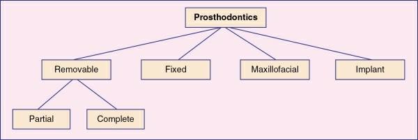
Prosthodontics
Prosthodontics is a branch of dentistry pertaining to the diagnosis, treatment planning, rehabilitation and maintenance of the oral function, comfort, appearance and health of the patients with clinical conditions associated with missing or deficient teeth and/or maxillofacial tissues using biocompatible substitutes.
Branches of prosthodontics
Removable prosthodontics
This branch of prosthodontics is concerned with the replacement of missing teeth and adjacent structures for edentulous and partially edentulous patients by artificial prosthesis that can be removed by the patient.
Fixed prosthodontics
This branch of prosthodontics pertains to the replacement of missing teeth by artificial substitute that cannot be removed by the patient.
Maxillofacial prosthodontics
This branch of prosthodontics deals with replacement of the stomatognathic and craniofacial structures.
Implant prosthodontics
This branch of prosthodontics deals with replacement of missing teeth and associated structures by restorations that are retained by the dental implants.
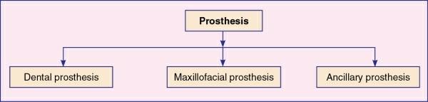
Prosthesis
Prosthesis may be defined as an artificial replacement of a missing part of the human body.
Types of Prosthesis
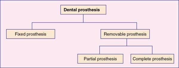
i. Dental prosthesis
ii. Maxillofacial prosthesis
iii. Ancillary prosthesis
Dental prosthesis
An artificial replacement of one or more teeth and associated dental/alveolar structures.
Fixed
dental prosthesis
Any dental prosthesis that is cemented, screwed or attached to the retained natural teeth or roots.
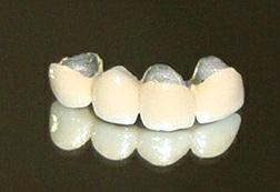
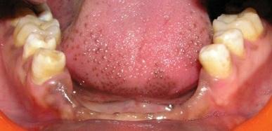
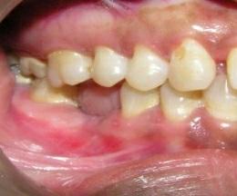
Any dental prosthesis that replaces some of the missing teeth in a partially edentulous arch (Figs 1.1.1.4 and 1.1.1.5).
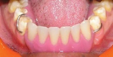
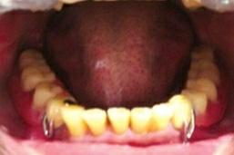
Removable partial dental prosthesis (RPDP)
The prosthesis that replaces some of the teeth in a partially edentulous arch and that can be removed from the mouth by the patient. It can be a simple
FIG 1.1.1.4 Missing lower anterior teeth.
FIG 1.1.1.5 A patient wearing removable temporary prosthesis.
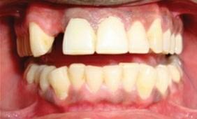
removable partial denture fabricated in acrylic resin called temporary partial denture. A removable partial denture fabricated in cast metal alloy and acrylic resin is called cast partial denture (Figs 1.1.1.6–1.1.1.8).
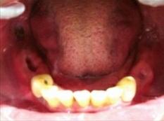
1.1.1.6 Partially edentulous arch denture.
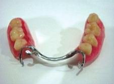
1.1.1.7 Cast partial denture.
FIG 1.1.1.8 Patient wearing cast partial denture.
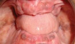
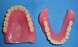
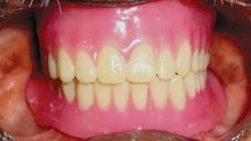
Removable complete dental prosthesis
The prosthesis that replaces the entire dentition and associated structures of maxilla and mandible (Figs 1.1.1.9–1.1.1.11).
FIG
FIG
1.1.1.9 Completely edentulous arch.
1.1.1.10 Complete denture.
1.1.1.11 Patient wearing a complete denture.
Maxillofacial prosthesis
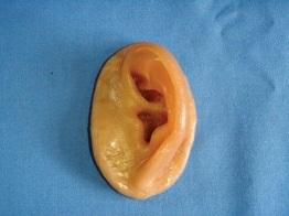
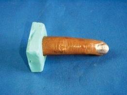
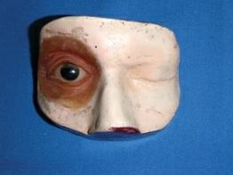
The prosthesis that is used to replace a part or all of any stomatognathic or craniofacial structures. Examples of the maxillofacial prosthesis are auricular prosthesis, orbital prosthesis, nasal prosthesis and facial prosthesis (Figs 1.1.1.12–1.1.1.14).
FIG
FIG
FIG
FIG 1.1.1.12 Auricular prosthesis.
FIG 1.1.1.13 Finger prosthesis.
FIG 1.1.1.14 Orbital prosthesis.
Implant supported prosthesis
A prosthesis that is used to replace missing teeth, which is retained by dental implants (Figs 1.1.1.15–1.1.1.18).
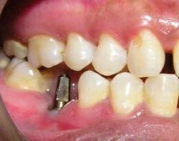
FIG 1.1.1.15 Missing tooth number 46.
FIG 1.1.1.16 Dental implant placed in 46 region.
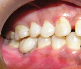
FIG 1.1.1.17 46 crown retained by implant.
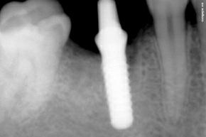
FIG 1.1.1.18 IOPA radiograph of implant placed in 46 region.
Ancillary prosthesis
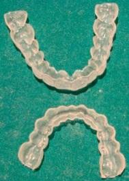
A type of dental prosthesis that is used in the field of prosthodontics, which is used for a short-term or special usage. Examples of such prosthesis are stents, splints and guide (Fig. 1.1.1.19).
FIG 1.1.1.19 Upper and lower splints.
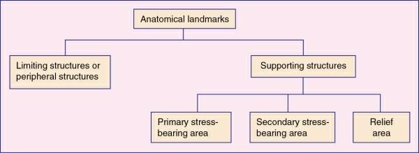
1.1.2 Anatomical landmarks of maxillary arch and mandibular arch
Anatomical landmarks of maxillary and mandibular arch
1. Anatomy
2. Clinical significance
Anatomical landmarks of maxillary arch
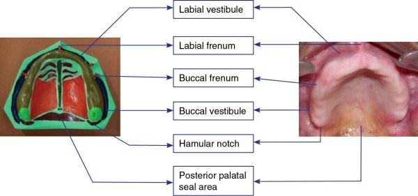
Limiting structures or peripheral structures (figs 1.1.2.1 and 1.1.2.2)
• Labial frenum
• Labial vestibule
• Buccal frenum
• Buccal vestibule
• Hamular notch
• Posterior palatal seal
• Fovea palatinae
FIG 1.1.2.1 Parts of the edentulous oral cavity.
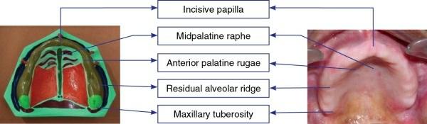
Supporting structures
Primary stress-bearing area
• Hard palate
• Posterior lateral slopes of the residual alveolar ridge
Secondary stress-bearing area
• Rugae
Relief area
• Incisive papilla
• Midpalatine raphe
Labial frenum
Anatomy
• The fibrous band covered by mucous membrane that extends from labial aspect of the residual alveolar ridge to the lip.
• It may be single or double and narrow or broad.
• It contains no muscle fibres.
Clinical significance
• Relief for the labial frenum must be given during final impression procedure.
• When relief is not given in the denture, it results in pain and it may cause dislodgement of the denture.
FIG 1.1.2.2 Parts of the edentulous oral cavity – relief areas.