No part of this publication may be reproduced or transmitted in any form or by any means, electronic or mechanical, including photocopying, recording, or any information storage and retrieval system, without permission in writing from the publisher. Details on how to seek permission, further information about the Publisher’s permissions policies and our arrangements with organizations such as the Copyright Clearance Center and the Copyright Licensing Agency, can be found at our website: www.elsevier.com/permissions
This book and the individual contributions contained in it are protected under copyright by the Publisher (other than as may be noted herein).
Notices
Knowledge and best practice in this field are constantly changing. As new research and experience broaden our understanding, changes in research methods, professional practices, or medical treatment may become necessary.
Practitioners and researchers must always rely on their own experience and knowledge in evaluating and using any information, methods, compounds, or experiments described herein. In using such information or methods they should be mindful of their own safety and the safety of others, including parties for whom they have a professional responsibility.
With respect to any drug or pharmaceutical products identified, readers are advised to check the most current information provided (i) on procedures featured or (ii) by the manufacturer of each product to be administered, to verify the recommended dose or formula, the method and duration of administration, and contraindications. It is the responsibility of practitioners, relying on their own experience and knowledge of their patients, to make diagnoses, to determine dosages and the best treatment for each individual patient, and to take all appropriate safety precautions.
To the fullest extent of the law, neither the Publisher nor the authors, contributors, or editors, assume any liability for any injury and/or damage to persons or property as a matter of products liability, negligence or otherwise, or from any use or operation of any methods, products, instructions, or ideas contained in the material herein.
Previous editions copyrighted 2011 and 2005.
Library of Congress Cataloging-in-Publication Data
Names: Leslie, Kevin O., editor. | Wick, Mark R., 1952- editor. | Preceded by (work): Leslie, Kevin O. Practical pulmonary pathology.
Title: Practical pulmonary pathology : a diagnostic approach / [edited by] Kevin O. Leslie, Mark R. Wick.
Other titles: Practical pulmonary pathology (Leslie) | Pattern recognition series.
Description: Third edition. | Philadelphia, PA : Elsevier, [2018] | Series: Pattern recognition series | Preceded by Practical pulmonary pathology / Kevin O. Leslie, Mark R. Wick. 2nd ed. c2011. | Includes bibliographical references and index.
Classification: LCC RC733 | NLM WF 600 | DDC 616.2/4075–dc23 LC record available at https://lccn.loc.gov/2017024025
Content Strategist: Michael Houston
Senior Content Development Specialist: Laura Schmidt
Publishing Services Manager: Patricia Tannian
Senior Project Manager: Amanda Mincher
Design Direction: Ashley Miner
Printed in China
Last digit is the print number: 9 8 7 6 5 4 3 2 1
This work is dedicated to my wife, Peggy, and our children, Katie and Amy, whose support and tolerance over the years have made this work possible. I am also thankful for my good fortune in knowing Dr. Tom Colby, longtime friend, colleague, and mentor, and for the hundreds of pathologists and pulmonologists whose patients have provided me with insight and inspiration over the years.
—KOL
Many thanks are due to my wife, Jane, and my children, Morgan, Robert, and Kellyn, for generously giving of their time with me so that edition three could be completed. In addition, I would like to dedicate the current text to the memory of Philip E. Bernatz, MD (1921–2010), who was a wonderful mentor, colleague, and friend.
—MRW
Contents
Pattern-Based Approach to Diagnosis xv
lung Anatomy 1
Kevin O. leslie and Mark R. Wick
Pulmonary Function Testing for Pathologists 15
Imre Noth
Optimal Processing of Diagnostic lung Specimens 21
Staci Beamer, Dawn E. Jaroszewski, Robert W,Viggiano, and Maxweill. Smith
computed Tomography of Diffuse lung Diseases and Solitary Pulmonary Nodules 35
Giorgia Dalpiaz
Developmental and Pediatric lung Disease 99
Megan K. Dishop
II
Acute lung Injury 125
Oi-Yee Cheung, Paolo Graziano, and Maxweill. Smith
lung Infections 147
Ann E. McCullough and Kevin O.leslie
Chronic Diffuse lung Diseases 227
Mikiko Hashisako, Junya Fukuoka, and Maxweill. Smith
II
Nonneoplastic Pathology of the large and Small Airways 299
Mattia Barbareschi and Alberto Cavazza
Pneumoconioses 335
Kelly 1. Butnor and Victor l. Roggli
Pulmonary Vasculitis and Pulmonary Hemorrhage 365
William David Travis, Kevin O. leslie, and Mary Beth Beasley
Pulmonary Hypertension 401
Andrew Churg and Joanne l. Wright
Pathology of lung Transplantation 421
Andras Khoor
Neuroendocrine Neoplasms of the lung 439
Alain C. Borczuk
Sarcomas and Sarcomatoid Neoplasms of the lungs and Pleural Surfaces 467
Mark R. Wick, Kevin O. leslie, and Mark H. Stoler
Hematolymphoid Disorders 527
Madeleine D. Kraus and Mark R. Wick
Nonneuroendocrine Carcinomas (Excluding Sarcomatoid Carcinoma) and Salivary Gland Analogue Tumors of the lung 573
Philip T. Cagle, Ross A. Miller, and Timothy Craig Allen
Metastatic Tumors in the lung: A Practical Approach to Diagnosis 597
Kim R. Geisinger and Stephen Spencer Raab
Pseudoneoplastic lesions of the lungs and Pleural Surfaces 643
Mark R. Wick, Timothy Craig Allen, Jon H. Ritter, and Osamu Matsubara
Benign and Borderline Tumors of the lungs and Pleura 665
Mark R. Wick and Stacey E. Mills
II
Malignant and Borderline Mesothelial Tumors of the Pleura 723
Mark R. Wick, Kevin O. leslie, Jon H. Ritter, and Stacey E. Mills
Appendix 763
Kevin O.leslie
Index 781
Contributors
Timothy Craig Allen, MD, JD
Professor
Department of Pathology
Director of Anatomic and Surgical Pathology
The University of Texas Medical Branch Galveston, Texas
Mattia Barbareschi, MD
Director, Unit of Surgical Pathology
Santa Chiara Hospital Trento, Italy
Staci Beamer, MD
Department of Surgery
Mayo Clinic Arizona Phoenix, Arizona
Mary Beth Beasley, MD
Professor of Pathology
Mount Sinai Medical Center
New York, New York
Alain C. Borczuk, MD
Department of Pathology and Laboratory Medicine
Weill Cornell Medicine
New York, New York
Kelly J. Butnor, MD
Department of Pathology and Laboratory Medicine
University of Vermont Medical Center
Burlington, Vermont
Philip T. Cagle, MD
Director, Pulmonary Pathology
Pathology and Genomic Medicine
Houston Methodist Hospital
Houston, Texas;
Professor of Pathology
Pathology and Laboratory Medicine
Weill Cornell Medical College
New York, New York
Alberto Cavazza, MD
Director, Unit of Surgical Pathology
Santa Maria Nuova Hospital/IRCCS
Reggio Emilia, Italy
Oi-Yee Cheung, MD Consultant
Department of Pathology
Queen Elizabeth Hospital Hong Kong, China
Andrew Churg, MD Pathologist
Vancouver General Hospital; Professor of Pathology
University of British Columbia Vancouver, British Columbia, Canada
Giorgia Dalpiaz, MD
Physician
Radiology
Bellaria Hospital, Bologna Bologna, Italy
Megan K. Dishop, MD
Medical Director of Anatomic Pathology
Children’s Hospitals and Clinics of Minnesota Minneapolis, Minnesota
Junya Fukuoka, MD, PhD
Professor and Department Chair Department of Pathology
Nagasaki University Graduate School of Biomedical Sciences; Chair
Nagasaki University Hospital
Nagasaki Educational and Diagnostic Center of Pathology; Assistant Dean
Nagasaki University School of Medicine; Professor
Nagasaki University School of Tropical Medicine and Global Health Sakamoto, Nagasaki, Japan
Contributors
Kim R. Geisinger, MD Professor Department of Pathology University of Mississippi Medical Center Jackson, Mississippi
Paolo Graziano, MD Unit of Pathology Foundation, Scientific Institute for Research and Health Care (IRCCS) San Giovanni Rotondo (FG), Italy
Mikiko Hashisako, MD, PhD Assistant Professor Department of Pathology Nagasaki University Hospital Nagasaki, Japan
Dawn E. Jaroszewski, MD Department of Surgery Mayo Clinic Arizona Phoenix, Arizona
Andras Khoor, MD Consultant and Chair Laboratory Medicine and Pathology Mayo Clinic Jacksonville, Florida
Madeleine D. Kraus, MD Director of Hematopathology Nemours Children’s Hospital Orlando, Florida
Kevin O. Leslie, MD Professor of Pathology Department of Laboratory Medicine and Pathology Mayo Clinic Arizona Scottsdale, Arizona
Osamu Matsubara, MD, PhD Professor of Pathology Department of Basic Pathology National Defense Medical College Namiki, Tokorozawa-shi Saitama, Japan
Ann E. McCullough, MD Chair, Division of Anatomic Pathology Mayo Clinic Arizona Scottsdale, Arizona
Ross A. Miller, MD Assistant Professor Pathology and Genomic Medicine Houston Methodist Hospital Houston, Texas
Stacey E. Mills, MD University of Virginia Medical Center Charlottesville, Virginia
Imre Noth, MD Professor of Medicine Pulmonary and Critical Care University of Chicago Chicago, Illinois
Stephen Spencer Raab, MD Professor Department of Pathology University of Mississippi Medical Center Jackson, Mississippi
Jon H. Ritter, MD Professor and Director of Surgical Pathology Washington University Medical Center St. Louis, Missouri
Victor L. Roggli, MD Department of Pathology Duke University Medical Center Durham, North Carolina
Maxwell L. Smith, MD Department of Laboratory Medicine and Pathology Mayo Clinic Arizona Scottsdale, Arizona
Mark H. Stoler, MD Professor of Pathology and Clinical Gynecology Department of Pathology University of Virginia Health System Charlottesville, Virginia
William David Travis, MD Attending Thoracic Pathologist Department of Pathology Memorial Sloan Kettering Cancer Center New York, New York
Robert W. Viggiano, MD Pulmonary Medicine Mayo Clinic Arizona Phoenix, Arizona
Mark R. Wick, MD Professor of Pathology University of Virginia Health System Charlottesville, Virginia
Joanne L. Wright, MD Pathologist St Paul’s Hospital, Vancouver Professor of Pathology University of British Columbia Vancouver, British Columbia, Canada
Series Preface
It is often stated that anatomic pathologists come in two forms: “Gestalt”based individuals who recognize visual scenes as a whole and match them unconsciously with memorialized archives; and criterion-oriented people who work through images systematically in segments and tabulate the results—internally, mentally, and quickly—as they go along in examining a visual target. These approaches can be equally effective, and they are probably not as dissimilar as their descriptions would suggest. In reality, even “Gestaltists” subliminally examine details of an image, and, if asked specifically about particular features of it, they are able to say whether one characteristic or another is important diagnostically.
In accordance with these concepts, in 2004 we published a textbook titled Practical Pulmonary Pathology: A Diagnostic Approach (PPPDA). That monograph was designed around a pattern-based method, wherein diseases of the lung were divided into six categories on the basis of their general image profiles. Using that technique, one can successfully segregate pathologic conditions into diagnostically and clinically useful groupings.
The merits of such a procedure have been validated empirically by the enthusiastic feedback we have received from users of our book. In addition, following the old adage, “imitation is the sincerest form of flattery,” since our book came out, other publications and presentations have appeared in our specialty and have used the same approach.
After publication of the PPPDA text, representatives at Elsevier, most notably William Schmitt, were enthusiastic about building a series of texts around pattern-based diagnosis in pathology. To this end we have recruited a distinguished group of authors and editors to accomplish
that task. Because a panoply of patterns is difficult to approach mentally from a practical perspective, we have asked our contributors to be complete and yet to discuss only principal interpretative images. Our goal is to eventually provide a series of monographs that, in combination with one another, will allow trainees and practitioners in pathology to use salient morphologic patterns to reach with confidence final diagnoses in all organ systems.
As stated in the introduction to the PPPDA text, the evaluation of dominant patterns is aided secondarily by the analysis of cellular composition and other distinctive findings. Therefore, within the context of each pattern, editors have been asked to use such data to refer the reader to appropriate specific chapters in their respective texts.
We have also stated previously that some overlap is expected between pathologic patterns in any given anatomic site; in addition, specific disease states may potentially manifest themselves with more than one pattern. At first, those facts may seem to militate against the value of pattern-based interpretation. However, pragmatically, they do not. One can often narrow diagnostic possibilities to a very few entities using the pattern method, and sometimes a single interpretation will be obvious. Both of those outcomes are useful to clinical physicians caring for a given patient.
It is hoped that the expertise of our authors and editors, together with the high quality of morphologic images they present in this Elsevier series, will be beneficial to our reader-colleagues.
Kevin O. Leslie, MD
Mark R. Wick, MD
It has been 12 years since Practical Pulmonary Pathology: A Diagnostic Approach (PPPDA) was first published. We are happy to report that the original version of this book was warmly received, with a distribution of approximately 8000 copies. Readers seemed to find our pattern-based approach to be a useful one in the daily practice of anatomic pathology, judging by the direct feedback we received. We also were honored when PPPDA won the 2005 Textbook of the Year Award from the Royal Society of Medicine and Royal Society of Authors.
In light of these successes, and in view of the fact that hospital pathology continues to grow rapidly in scope and complexity, we decided to prepare a second and now third edition of our book. Several features are new to this edition. A new chapter (Chapter 2) on pulmonary function for pathologists has been added, authored by renowned pulmonary and critical care specialist Dr. Emre Noth. This chapter succeeds and compliments the second edition chapter on chest imaging patterns authored initially by international expert radiologists Drs. Maffessanti and Dalpiaz, now updated under the sole authorship of Dr. Dalpiaz. Both of these chapters help round out the pathologist’s understanding of lung diseases and are critical to the book. Inevitably there have been additions to, and revisions of, the prior text because of advances in our understanding of the pertinent disease processes. Corresponding references have been added, and they are current through 2016. Moreover, many illustrative photomicrographs have been changed in an effort to improve the visual presentation of the topics discussed. Finally, self-assessment questions tied to all the chapters in the current book have been compiled and are available online. It is hoped that these questions will be useful to pathologists in their maintenance of certification and as a reflection of their mastery of the information in the book.
As before, we begin with the general patterns of disease and then add key morphologic findings that assist the reader in focusing on appropriate sections of the book where similar findings are discussed. This approach is facilitated by a structural overlay that limits the patterns. We have found that six general patterns occur, and these are best appreciated at scanning magnification with the microscope. We could begin at an even lower “magnification” using the high-resolution computed tomogram (CT), and this is what our radiology colleagues commonly do as they assemble a differential diagnosis based on observed findings in this medium (see Chapter 4). However, in practice, the CT
images may not be readily available to the pathologist at the time the biopsy is interpreted, so for our six pathology patterns, we begin with a tissue section mounted on a glass slide. To help the pathologist in practice correctly identify diseases within patterns, we have included a simple worksheet that emphasizes the importance of knowing the clinical, imaging, and pathologic features in order to arrive at the most appropriate diagnostic category (page xvi).
An overview of the six patterns is presented, and each pattern is then illustrated in the pages that follow. Most of the patterns were devised to navigate the diffuse lung diseases commonly referred to as interstitial lung diseases or ILD. Given the tumefactive nature of neoplasms, these are heavily represented in Pattern 5 (Nodules), but some nonneoplastic diseases, such as sarcoidosis, nodular infections, granulomatosis with polyangiitis, and certain pneumoconioses, may also manifest as a nodular pattern. Rarely, neoplasms can present as diffuse interstitial lung disease clinically and radiologically.
A basic knowledge of the two-dimensional structure of the lung is essential for accurately assessing patterns of disease. We assume that the reader is familiar with basic lung anatomy by the time a diagnostic problem is being evaluated in the patient care setting, but a brief review is always helpful (see Chapter 1).
Once the overriding or dominant pattern is recognized, the diagnostician assesses the cellular composition and any other distinctive findings that accompany the pattern. In the case of a tumor forming a nodular mass, the presence of prominent spindled cells, or large granular cells, or clear cells provides a direction for creating a differential diagnosis. Within each pattern, we have attempted to use such qualifying elements to direct the reader to the appropriate chapter for further study, reasonably confident that the answer will lie within. For the unusual finding not identified in the list for a given pattern, the reader is directed to the appendix where we have assembled a “visual encyclopedia” of distinctive findings and artifacts.
Naturally, overlap occurs between patterns, and this too can be a useful guide to the correct diagnosis. For example, some infections are both nodular and have airspace filling (e.g., botryomycosis, aspiration pneumonia), whereas others are characterized by acute lung injury and diffuse airspace filling (e.g., pneumococcal pneumonia, pneumocystis pneumonia.) In fact, some diffuse inflammatory conditions in the lung
Preface
may manifest five of the six patterns in different areas of the same biopsy (e.g., rheumatoid lung). Nevertheless, as more and more information is accrued from the biopsy, the differential diagnosis becomes more limited. In some cases it may be necessary to include several possibilities in the final diagnosis, especially for the nonneoplastic diseases where the effect of ancillary data not available at the time of diagnosis may be very large. Once again, we are grateful to all of the authors who generously and diligently updated their chapters in the third edition of PPPDA. In
addition, many thanks are due to our colleagues at the Mayo Clinic and the University of Virginia for their strong support of this project. Finally, this work could not have reached fruition without the valuable help of our editor, William Schmitt of Elsevier, and the editorial and production expertise of Laura Schmidt and Amanda Mincher.
Kevin O. Leslie, MD
Mark R. Wick, MD
Pattern-Based Approach to Diagnosis
A fundamental truth about medical textbooks is that they are often not read from beginning to end once a student of medicine has progressed beyond the basic medical school curriculum. In the practice of medicine, textbooks are more commonly used as references for learning about a disease or entity that a clinician suspects a patient may have based on history, physical findings, and imaging/laboratory data gleaned from an initial screening evaluation. The disease-based textbook is analogous to a dictionary or encyclopedia, both of which are much easier to use if a person already has a good idea of what he or she is investigating.
Today, the vast majority of diagnosis-oriented medical textbooks continue to exist as compendia of individual diseases, more or less grouped by the anatomic compartment or structure affected (e.g., brainstem diseases, bile duct diseases, glomerular diseases) or a common mechanism if one is discernible (e.g., inflammatory diseases, neoplastic diseases). Typically, the discussion of each disease begins with a historical introduction, continues with the characteristics of the disease, and ends with the treatment and prognosis. This book is no different, but the authors have added this introductory material as a tool to help navigate the contents. The approach is based on the premise that six primary histopathologic patterns exist for all lung diseases. Identifiable using the low-magnification microscope objective lens, these patterns serve as the introductory image of the disease process. (In truth, chest imaging with high-resolution computed tomography is an even better place to begin—see Chapter 4). Once the primary pattern is recognized, the histopathologist must collect additional findings from the biopsy specimen. With the primary pattern and secondary attributes in hand, a cogent differential diagnosis can be proffered. This process is significantly enhanced by knowledge of the clinical presentation and imaging characteristics, but if these are not available when the slides are being examined, they can still be useful for narrowing the differential diagnosis after the histopathology has been evaluated. A detailed analysis of the use of clinical, radiologic, and histopathologic data in the evaluation of the diffuse medical lung diseases (often referred to as interstitial lung diseases, or ILDs) is available for the interested reader (open access file for download).*
A basic knowledge of the two-dimensional structure of the lung is essential for accurately assessing patterns of disease. We assume that
the reader is familiar with basic lung anatomy by the time a diagnostic problem is being evaluated in the patient care setting, but a brief review is always helpful (see Chapter 1). An overview of the six major patterns is provided (see Table 1), followed by illustrations of each pattern. The pattern-based approach presented here was devised mainly to assist in the interpretation of the diffuse lung diseases, commonly referred to as ILDs. Given the tumefactive nature of neoplasms, these are heavily represented in Pattern 5 (Nodules), but some nonneoplastic diseases, such as sarcoidosis, nodular infections, granulomatosis with polyangiitis, and certain pneumoconioses, may also manifest a nodular pattern. Rarely, neoplasms can present as diffuse ILD clinically and radiologically (e.g., lymphangitic carcinoma, intravascular lymphoma). Within each of the major patterns, the authors have provided the reader with the appropriate chapters and relevant pages in the book for further study, reasonably confident that the answer (or approach) to a particular diagnostic problem will be present. There are diagnostic considerations for which no specific chapter or page number is provided. Some of these may require reference to another source. For the distinctive or unusual finding not identified in the list for a given major pattern, the reader is directed to the Appendix, where the authors have assembled a “visual encyclopedia” of distinctive findings and artifacts encountered in the course of microscopic evaluation.
As every diagnostic pathologist knows, overlap occurs between diseases, and sometimes this overlap can be useful in establishing the correct diagnosis. For example, some infections both are nodular (Pattern 5) and have airspace filling (e.g., botryomycosis, aspiration pneumonia), whereas others are characterized by acute lung injury and diffuse airspace filling (e.g., pneumococcal pneumonia, pneumocystis pneumonia). In fact, some diffuse inflammatory conditions of the lung may manifest all of the six patterns in different areas of the same biopsy (e.g., rheumatoid lung). In some cases, it may be necessary to include several possibilities in the final diagnosis, especially for the nonneoplastic diseases, where the effect of ancillary data not available at the time of diagnosis may be very large. The exposition begins with Pattern 1 (Acute Lung Injury) because this is the pattern that dominates all others and is most often the reason a biopsy was performed at all.
*See Leslie KO: My approach to interstitial lung disease using clinical, radiological and histopathologic patterns. J Clin Pathol. 2009;62(5):387–401. The Worksheet for the Pattern-Based Approach to Lung Disease, located on page xvi, is a printable form for organizing these data.
Patient Information
Worksheet for the Pattern-Based Approach to Lung Disease
Age: __________ Gender: Male Female
Disease Onset
Acute (hours to days) Subacute (weeks to a few months) Chronic (months to years)
Chronic small airways disease (as constrictive bronchiolitis)
Vasculopathic diseases
Lymphangioleiomyomatosis (LAM)
Other rare cystic diseases
Pattern 1 Acute Lung Injury
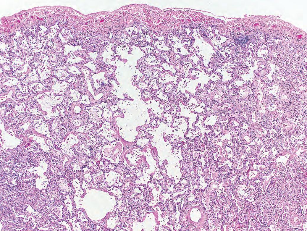
Elements of the pattern: The lung biopsy shows patchy or diffuse edema, fibrin, and reactive type 2 cell hyperplasia. The dominance of noncellular, protein-rich material imparts an overall red or pink appearance to the biopsy at scanning magnification (in routine hematoxylin-eosin stained sections).
Special stains for organisms are required for all lung specimens that show acute injury.
Additional Findings Diagnostic Consideration
Hyaline membranes
Necrosis in parenchyma
Necrosis in bronchioles
Fibrin in alveoli
Eosinophils in alveoli
Siderophages in alveoli
Fibrinous pleuritis
Neutrophils
Atypical cells
Fibrin + vacuolated macrophages
Diffuse alveolar damage
Infection
Some tumors
Infarct
Infections
Acute aspiration
Diffuse alveolar damage
Drug toxicity
Connective tissue disease
Infection
Eosinophilic lung diseases
Diffuse alveolar hemorrhage
Drug toxicity
Infarct
Connective tissue diseases
Eosinophilic pneumonia
Pneumothorax
Infections
Capillaritis in diffuse alveolar hemorrhage
Acute lung injury
Viral infections
Leukemias
Intravascular lymphoma
Infection
Drug toxicity
Connective tissue diseases
Pattern 1 Acute Lung Injury
Ch. 5:110; Ch. 6:125
Ch. 6:130
Ch. 17:586
Ch. 11:390
Ch. 6:133; Ch. 9:312
Ch. 9:306
Ch. 6:128
Ch. 6:136
Ch. 6:134
Ch. 6:133; Ch. 7:203
Ch. 6:139; Ch. 8:255
Ch. 6:140; Ch. 11:393
Ch. 11:394
Ch. 7:152; Ch. 11:390
Ch. 6:134
Ch. 6:139
Ch. 8:276
Ch. 6:143
Ch. 11:395
Ch. 6:142
Ch. 6:143
Ch. 16:528
Ch. 16:548
Ch. 7:174
Ch. 6:136
Ch. 6:136
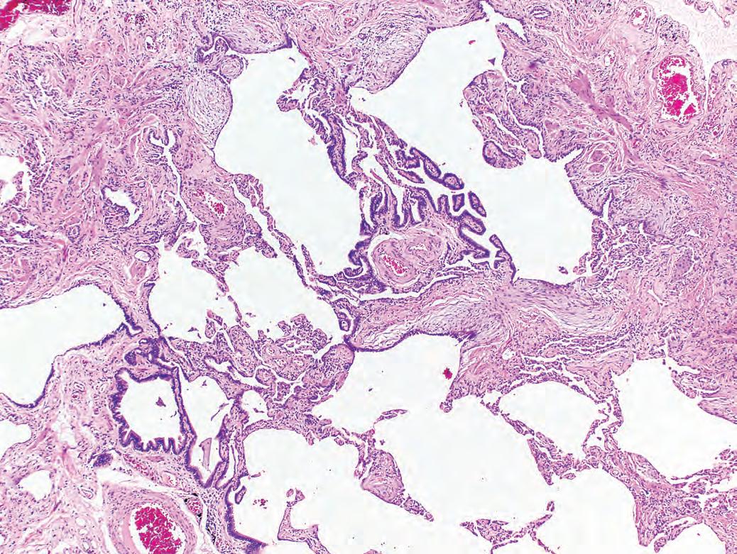
Pattern 2 Fibrosis
Elements of the pattern: The lung biopsy is involved by variable amounts of fibrosis. As in Pattern 1, the biopsy tends to be more pink than blue at scanning magnification, as a result of collagen deposition (in routine hematoxylin-eosin stained sections). Some fibrosis patterns are accompanied by chronic inflammation that may impart a blue tinge to the process, or even dark blue lymphoid aggregates.
Significant lung fibrosis is always associated with some degree of structural remodeling. Avoid diagnosing “fibrosis” on transbronchial biopsies.
Additional Findings
Hyaline membranes
Microscopic honeycombing
Prominent bronchiolization
Uniform alveolar septal fibrosis
Peripheral lobular fibrosis
Siderophages in alveoli
Fibrinous pleuritis
Prominent nonnecrotizing granulomas
Many vacuolated cells
Prominent chronic inflammation
Airway-centered scarring
Pattern 2 Fibrosis
“Acute on chronic” disease
Infection on fibrosis
Drug toxicity on fibrosis
Connective tissue disease in “exacerbation”
Acute exacerbation of idiopathic pulmonary fibrosis (IPF)
Usual interstitial pneumonia (UIP)
Hypersensitivity pneumonitis
Connective tissue disease
Pulmonary Langerhans cell histiocytosis
Respiratory bronchiolitis ILD
Connective tissue diseases
Chronic hypersensitivity pneumonitis
Small airways disease
Chronic aspiration
Connective tissue diseases
Postirradiation
UIP/IPF
Erdheim Chester disease
Rosai-Dorfman disease
Chronic eosinophilic pneumonia
Chronic cardiac congestion
Chronic venous outflow obstruction
Chronic hemorrhage in connective tissue disease
Chronic hemorrhage in bronchiectasis
Pneumoconiosis
Pulmonary Langerhans cell histiocytosis
Smoking-related interstitial lung disease
Chronic renal dialysis
Idiopathic pulmonary hemosiderosis
Connective tissue disease
Eosinophilic pleuritis in pneumothorax
Sarcoidosis
Chronic airway obstruction
Drug toxicity
Hermansky-Pudlak syndrome
Genetic storage diseases
Nonspecific interstitial pneumonia (NSIP)
Rheumatoid arthritis and other connective tissue diseases
Pulmonary Langerhans cell histiocytosis
Pneumoconiosis
Chronic hypersensitivity pneumonitis
Connective tissue diseases
Idiopathic airway-centered fibrosis
Idiopathic pleuroparenchymal fibroelastosis
Chronic aspiration
Ch. 5:110
Ch. 6:128
Ch. 6:136
Ch. 6:140
Ch. 8:234
Ch. 8:229
Ch. 8:269
Ch. 8:247
Ch. 8:272
Ch. 8:240
Ch. 8:247
Ch. 8:269
Ch. 9:317
Ch. 8:267;
Ch. 8:247
Ch. 9:312
Not specifically addressed
Ch. 8:229
Ch. 8:276
Ch. 19:650
Ch. 8:255
Ch. 5:114
Not specifically addressed
Ch. 8:250
Ch. 11:390
Ch. 10:339
Ch. 8:272
Ch. 8:243
Not specifically addressed
Ch. 11:395
Ch. 8:247
Ch. 8:276; Appendix:770
Ch. 8:266
Ch. 8:272
Ch. 8:289
Ch. 8:279
Ch. 5:120
Ch. 8:235
Ch. 8:247
Ch. 8:272
Ch. 9:320
Ch. 8:269
Ch. 8:247
Ch. 8:288
Ch. 8:246
Ch. 8:267; Ch. 9:312
Pattern 3 Chronic Cellular Infiltrates
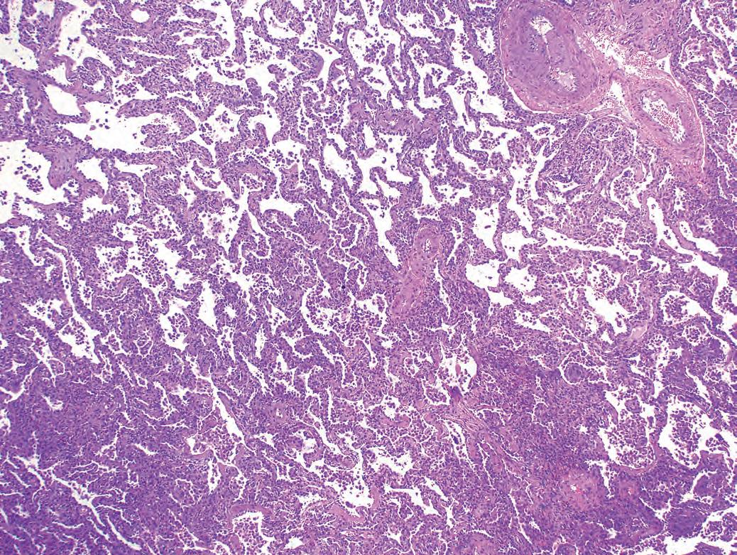
Elements of the pattern: The lung biopsy is dominated by interstitial chronic inflammation and variable reactive type 2 cell hyperplasia. The dominance of mononuclear infiltrates may impart an overall blue appearance to the biopsy at scanning magnification (in routine hematoxylin-eosin stained sections).
Additional Findings
Hyaline membranes
Necrosis in parenchyma
Necrosis in bronchioles
Poorly formed granulomas (small and nonnecrotizing)
Well-formed necrotizing granulomas
Eosinophils in alveoli
Siderophages in alveoli
Fibrinous/chronic pleuritis
Patchy organizing pneumonia
Atypical cells
Multinucleated giant cells
Dense mononuclear infiltration
Pattern 3 Chronic Cellular Infiltrates
Lymphoid aggregates/germinal centers
Diagnostic Consideration
“Acute on chronic” connective tissue disease
Drug toxicity
Diffuse alveolar hemorrhage
Viral and fungal infections
Aspiration
Infarction in antiphospholipid syndrome
Viral infections
Aspiration
Hypersensitivity pneumonitis (subacute)
Atypical mycobacterial infection
“Hot tub” lung
Lymphoid interstitial pneumonia
Drug toxicity
Infections
Rare drug reactions
Necrotizing sarcoidosis
Middle lobe syndrome
Eosinophilic lung diseases
Smoking-related lung diseases
Diffuse alveolar hemorrhage
Chronic cardiac congestion
Drug toxicity
Connective tissue diseases
Thoracic trauma/infection
Pancreatitis-associated pleuritis
Drug toxicity
Connective tissue diseases
Infections
Cryptogenic organizing pneumonia
Diffuse alveolar hemorrhage
Aspiration
Viral infections
Lymphangitic carcinoma
Hard metal disease
Mica pneumoconiosis
Hypersensitivity pneumonitis
Intravenous drug abuse
Drug toxicity
Aspiration pneumonia
Eosinophilic pneumonia
Lymphomas
Lymphoid interstitial pneumonia
Connective tissue diseases
Hypersensitivity pneumonitis
Certain infections (the atypical pneumonias)
Connective tissue diseases
Diffuse lymphoid hyperplasia
Lymphoid interstitial pneumonia
Follicular bronchiolitis
Ch. 6:134
Ch. 6:136
Ch. 11:393
Ch. 7:178, 199
Ch. 7:161; Ch. 9:306
Ch. 8:251
Ch. 7:199
Ch. 7:161; Ch. 9:306
Ch. 8:269
Ch. 8:270
Ch. 7:175
Ch. 8:244
Ch. 8:259
Ch. 7:177
Not specifically addressed
Ch. 11:383
Ch. 9:303
Ch. 6:139; Ch. 8:255
Ch. 8:243
Ch. 11:393
Ch. 5:114
Ch. 8:259
Ch. 8:247
Ch. 8:248
Not specifically addressed
Ch. 8:259
Ch. 8:247
Ch. 8:239
Ch. 8:237
Ch. 11:393
Ch. 7:161; Ch. 9:306
Ch. 7:199
Ch. 8:246
Ch. 10:354
Ch. 10:347
Ch. 8:269
Ch. 8:263
Ch. 8:259
Ch. 7:161; Ch. 9:306
Ch. 6:139; Ch. 8:255
Ch. 16:542
Ch. 8:244
Ch. 8:247
Ch. 8:269
Ch. 7:162
Ch. 8:247
Ch. 8:245; Ch. 16:537
Ch. 8:244
Ch. 9:308
Pattern 4 Alveolar Filling
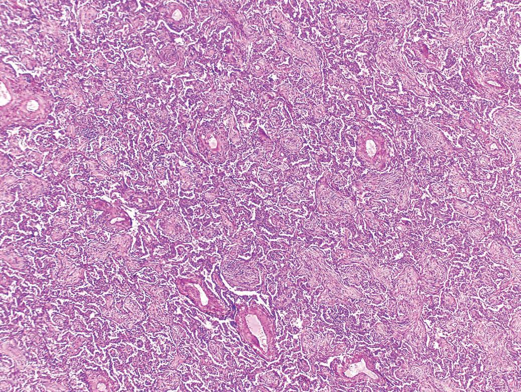
Elements of the pattern: The dominant finding is alveolar spaces filled with cells or noncellular elements.
Elements of the pattern: One, or many, nodules of variable size and shape. An interface between the nodular lesion and more normal lung should be discernible. In the case of very large nodules encompassing the entire specimen, radiologic imaging can be used as part of the definition.
Additional Findings
Large neoplastic lymphoid cells
Small lymphoid cells without germ centers
Small lymphoid cells with germ centers
Giant multinucleated neoplastic cells
Primitive small round neoplastic cells
Spindled or fusiform neoplastic cells
Large pink epithelioid neoplastic cells
Large clear epithelioid neoplastic cells
Large basophilic epithelial cells with peripheral palisade