Practical Pulmonary Pathology A Diagnostic Approach
Third Edition
Kevin O. Leslie, MD Professor of Pathology Department of Laboratory Medicine and Pathology Mayo Clinic Arizona Scottsdale, Arizona
Mark R. Wick, MD Professor of Pathology University of Virginia Health System Charlottesville, Virginia
Contents
Pattern-Based Approach to Diagnosis xv
lung Anatomy 1
Kevin O. leslie and Mark R. Wick
Pulmonary Function Testing for Pathologists 15
Imre Noth
Optimal Processing of Diagnostic lung Specimens 21
Staci Beamer, Dawn E. Jaroszewski, Robert W,Viggiano, and Maxweill. Smith
computed Tomography of Diffuse lung Diseases and Solitary Pulmonary Nodules 35
Giorgia Dalpiaz
Developmental and Pediatric lung Disease 99
Megan K. Dishop
II
Acute lung Injury 125
Oi-Yee Cheung, Paolo Graziano, and Maxweill. Smith
lung Infections 147
Ann E. McCullough and Kevin O.leslie
Chronic Diffuse lung Diseases 227
Mikiko Hashisako, Junya Fukuoka, and Maxweill. Smith
II
Nonneoplastic Pathology of the large and Small Airways 299
Mattia Barbareschi and Alberto Cavazza
Pneumoconioses 335
Kelly 1. Butnor and Victor l. Roggli
Pulmonary Vasculitis and Pulmonary Hemorrhage 365
William David Travis, Kevin O. leslie, and Mary Beth Beasley
Pulmonary Hypertension 401
Andrew Churg and Joanne l. Wright
Pathology of lung Transplantation 421
Andras Khoor
Neuroendocrine Neoplasms of the lung 439
Alain C. Borczuk
Sarcomas and Sarcomatoid Neoplasms of the lungs and Pleural Surfaces 467
Mark R. Wick, Kevin O. leslie, and Mark H. Stoler
Hematolymphoid Disorders 527
Madeleine D. Kraus and Mark R. Wick
Nonneuroendocrine Carcinomas (Excluding Sarcomatoid Carcinoma) and Salivary Gland Analogue Tumors of the lung 573
Philip T. Cagle, Ross A. Miller, and Timothy Craig Allen
Metastatic Tumors in the lung: A Practical Approach to Diagnosis 597
Kim R. Geisinger and Stephen Spencer Raab
Pseudoneoplastic lesions of the lungs and Pleural Surfaces 643
Mark R. Wick, Timothy Craig Allen, Jon H. Ritter, and Osamu Matsubara
Benign and Borderline Tumors of the lungs and Pleura 665
Mark R. Wick and Stacey E. Mills
II
Malignant and Borderline Mesothelial Tumors of the Pleura 723
Mark R. Wick, Kevin O. leslie, Jon H. Ritter, and Stacey E. Mills
Appendix 763
Kevin O.leslie
Index 781
Series Preface
It is often stated that anatomic pathologists come in two forms: “Gestalt”based individuals who recognize visual scenes as a whole and match them unconsciously with memorialized archives; and criterion-oriented people who work through images systematically in segments and tabulate the results—internally, mentally, and quickly—as they go along in examining a visual target. These approaches can be equally effective, and they are probably not as dissimilar as their descriptions would suggest. In reality, even “Gestaltists” subliminally examine details of an image, and, if asked specifically about particular features of it, they are able to say whether one characteristic or another is important diagnostically.
In accordance with these concepts, in 2004 we published a textbook titled Practical Pulmonary Pathology: A Diagnostic Approach (PPPDA). That monograph was designed around a pattern-based method, wherein diseases of the lung were divided into six categories on the basis of their general image profiles. Using that technique, one can successfully segregate pathologic conditions into diagnostically and clinically useful groupings.
The merits of such a procedure have been validated empirically by the enthusiastic feedback we have received from users of our book. In addition, following the old adage, “imitation is the sincerest form of flattery,” since our book came out, other publications and presentations have appeared in our specialty and have used the same approach.
After publication of the PPPDA text, representatives at Elsevier, most notably William Schmitt, were enthusiastic about building a series of texts around pattern-based diagnosis in pathology. To this end we have recruited a distinguished group of authors and editors to accomplish
that task. Because a panoply of patterns is difficult to approach mentally from a practical perspective, we have asked our contributors to be complete and yet to discuss only principal interpretative images. Our goal is to eventually provide a series of monographs that, in combination with one another, will allow trainees and practitioners in pathology to use salient morphologic patterns to reach with confidence final diagnoses in all organ systems.
As stated in the introduction to the PPPDA text, the evaluation of dominant patterns is aided secondarily by the analysis of cellular composition and other distinctive findings. Therefore, within the context of each pattern, editors have been asked to use such data to refer the reader to appropriate specific chapters in their respective texts.
We have also stated previously that some overlap is expected between pathologic patterns in any given anatomic site; in addition, specific disease states may potentially manifest themselves with more than one pattern. At first, those facts may seem to militate against the value of pattern-based interpretation. However, pragmatically, they do not. One can often narrow diagnostic possibilities to a very few entities using the pattern method, and sometimes a single interpretation will be obvious. Both of those outcomes are useful to clinical physicians caring for a given patient.
It is hoped that the expertise of our authors and editors, together with the high quality of morphologic images they present in this Elsevier series, will be beneficial to our reader-colleagues.
Kevin O. Leslie, MD
Mark R. Wick, MD
It has been 12 years since Practical Pulmonary Pathology: A Diagnostic Approach (PPPDA) was first published. We are happy to report that the original version of this book was warmly received, with a distribution of approximately 8000 copies. Readers seemed to find our pattern-based approach to be a useful one in the daily practice of anatomic pathology, judging by the direct feedback we received. We also were honored when PPPDA won the 2005 Textbook of the Year Award from the Royal Society of Medicine and Royal Society of Authors.
In light of these successes, and in view of the fact that hospital pathology continues to grow rapidly in scope and complexity, we decided to prepare a second and now third edition of our book. Several features are new to this edition. A new chapter (Chapter 2) on pulmonary function for pathologists has been added, authored by renowned pulmonary and critical care specialist Dr. Emre Noth. This chapter succeeds and compliments the second edition chapter on chest imaging patterns authored initially by international expert radiologists Drs. Maffessanti and Dalpiaz, now updated under the sole authorship of Dr. Dalpiaz. Both of these chapters help round out the pathologist’s understanding of lung diseases and are critical to the book. Inevitably there have been additions to, and revisions of, the prior text because of advances in our understanding of the pertinent disease processes. Corresponding references have been added, and they are current through 2016. Moreover, many illustrative photomicrographs have been changed in an effort to improve the visual presentation of the topics discussed. Finally, self-assessment questions tied to all the chapters in the current book have been compiled and are available online. It is hoped that these questions will be useful to pathologists in their maintenance of certification and as a reflection of their mastery of the information in the book.
As before, we begin with the general patterns of disease and then add key morphologic findings that assist the reader in focusing on appropriate sections of the book where similar findings are discussed. This approach is facilitated by a structural overlay that limits the patterns. We have found that six general patterns occur, and these are best appreciated at scanning magnification with the microscope. We could begin at an even lower “magnification” using the high-resolution computed tomogram (CT), and this is what our radiology colleagues commonly do as they assemble a differential diagnosis based on observed findings in this medium (see Chapter 4). However, in practice, the CT
images may not be readily available to the pathologist at the time the biopsy is interpreted, so for our six pathology patterns, we begin with a tissue section mounted on a glass slide. To help the pathologist in practice correctly identify diseases within patterns, we have included a simple worksheet that emphasizes the importance of knowing the clinical, imaging, and pathologic features in order to arrive at the most appropriate diagnostic category (page xvi).
An overview of the six patterns is presented, and each pattern is then illustrated in the pages that follow. Most of the patterns were devised to navigate the diffuse lung diseases commonly referred to as interstitial lung diseases or ILD. Given the tumefactive nature of neoplasms, these are heavily represented in Pattern 5 (Nodules), but some nonneoplastic diseases, such as sarcoidosis, nodular infections, granulomatosis with polyangiitis, and certain pneumoconioses, may also manifest as a nodular pattern. Rarely, neoplasms can present as diffuse interstitial lung disease clinically and radiologically.
A basic knowledge of the two-dimensional structure of the lung is essential for accurately assessing patterns of disease. We assume that the reader is familiar with basic lung anatomy by the time a diagnostic problem is being evaluated in the patient care setting, but a brief review is always helpful (see Chapter 1).
Once the overriding or dominant pattern is recognized, the diagnostician assesses the cellular composition and any other distinctive findings that accompany the pattern. In the case of a tumor forming a nodular mass, the presence of prominent spindled cells, or large granular cells, or clear cells provides a direction for creating a differential diagnosis. Within each pattern, we have attempted to use such qualifying elements to direct the reader to the appropriate chapter for further study, reasonably confident that the answer will lie within. For the unusual finding not identified in the list for a given pattern, the reader is directed to the appendix where we have assembled a “visual encyclopedia” of distinctive findings and artifacts.
Naturally, overlap occurs between patterns, and this too can be a useful guide to the correct diagnosis. For example, some infections are both nodular and have airspace filling (e.g., botryomycosis, aspiration pneumonia), whereas others are characterized by acute lung injury and diffuse airspace filling (e.g., pneumococcal pneumonia, pneumocystis pneumonia.) In fact, some diffuse inflammatory conditions in the lung
Patient Information
Worksheet for the Pattern-Based Approach to Lung Disease
Age: __________ Gender: Male Female
Disease Onset
Acute (hours to days) Subacute (weeks to a few months) Chronic (months to years)
Chronic small airways disease (as constrictive bronchiolitis)
Vasculopathic diseases
Lymphangioleiomyomatosis (LAM)
Other rare cystic diseases
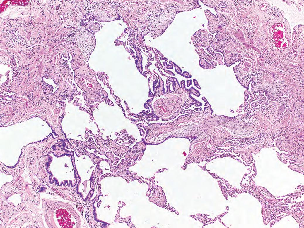
Pattern 2 Fibrosis
Elements of the pattern: The lung biopsy is involved by variable amounts of fibrosis. As in Pattern 1, the biopsy tends to be more pink than blue at scanning magnification, as a result of collagen deposition (in routine hematoxylin-eosin stained sections). Some fibrosis patterns are accompanied by chronic inflammation that may impart a blue tinge to the process, or even dark blue lymphoid aggregates.
Significant lung fibrosis is always associated with some degree of structural remodeling. Avoid diagnosing “fibrosis” on transbronchial biopsies.
Additional Findings
Hyaline membranes
Microscopic honeycombing
Prominent bronchiolization
Uniform alveolar septal fibrosis
Peripheral lobular fibrosis
Siderophages in alveoli
Fibrinous pleuritis
Prominent nonnecrotizing granulomas
Many vacuolated cells
Prominent chronic inflammation
Airway-centered scarring
Pattern 2 Fibrosis
“Acute on chronic” disease
Infection on fibrosis
Drug toxicity on fibrosis
Connective tissue disease in “exacerbation”
Acute exacerbation of idiopathic pulmonary fibrosis (IPF)
Usual interstitial pneumonia (UIP)
Hypersensitivity pneumonitis
Connective tissue disease
Pulmonary Langerhans cell histiocytosis
Respiratory bronchiolitis ILD
Connective tissue diseases
Chronic hypersensitivity pneumonitis
Small airways disease
Chronic aspiration
Connective tissue diseases
Postirradiation
UIP/IPF
Erdheim Chester disease
Rosai-Dorfman disease
Chronic eosinophilic pneumonia
Chronic cardiac congestion
Chronic venous outflow obstruction
Chronic hemorrhage in connective tissue disease
Chronic hemorrhage in bronchiectasis
Pneumoconiosis
Pulmonary Langerhans cell histiocytosis
Smoking-related interstitial lung disease
Chronic renal dialysis
Idiopathic pulmonary hemosiderosis
Connective tissue disease
Eosinophilic pleuritis in pneumothorax
Sarcoidosis
Chronic airway obstruction
Drug toxicity
Hermansky-Pudlak syndrome
Genetic storage diseases
Nonspecific interstitial pneumonia (NSIP)
Rheumatoid arthritis and other connective tissue diseases
Pulmonary Langerhans cell histiocytosis
Pneumoconiosis
Chronic hypersensitivity pneumonitis
Connective tissue diseases
Idiopathic airway-centered fibrosis
Idiopathic pleuroparenchymal fibroelastosis
Chronic aspiration
Ch. 5:110
Ch. 6:128
Ch. 6:136
Ch. 6:140
Ch. 8:234
Ch. 8:229
Ch. 8:269
Ch. 8:247
Ch. 8:272
Ch. 8:240
Ch. 8:247
Ch. 8:269
Ch. 9:317
Ch. 8:267;
Ch. 8:247
Ch. 9:312
Not specifically addressed
Ch. 8:229
Ch. 8:276
Ch. 19:650
Ch. 8:255
Ch. 5:114
Not specifically addressed
Ch. 8:250
Ch. 11:390
Ch. 10:339
Ch. 8:272
Ch. 8:243
Not specifically addressed
Ch. 11:395
Ch. 8:247
Ch. 8:276; Appendix:770
Ch. 8:266
Ch. 8:272
Ch. 8:289
Ch. 8:279
Ch. 5:120
Ch. 8:235
Ch. 8:247
Ch. 8:272
Ch. 9:320
Ch. 8:269
Ch. 8:247
Ch. 8:288
Ch. 8:246
Ch. 8:267; Ch. 9:312
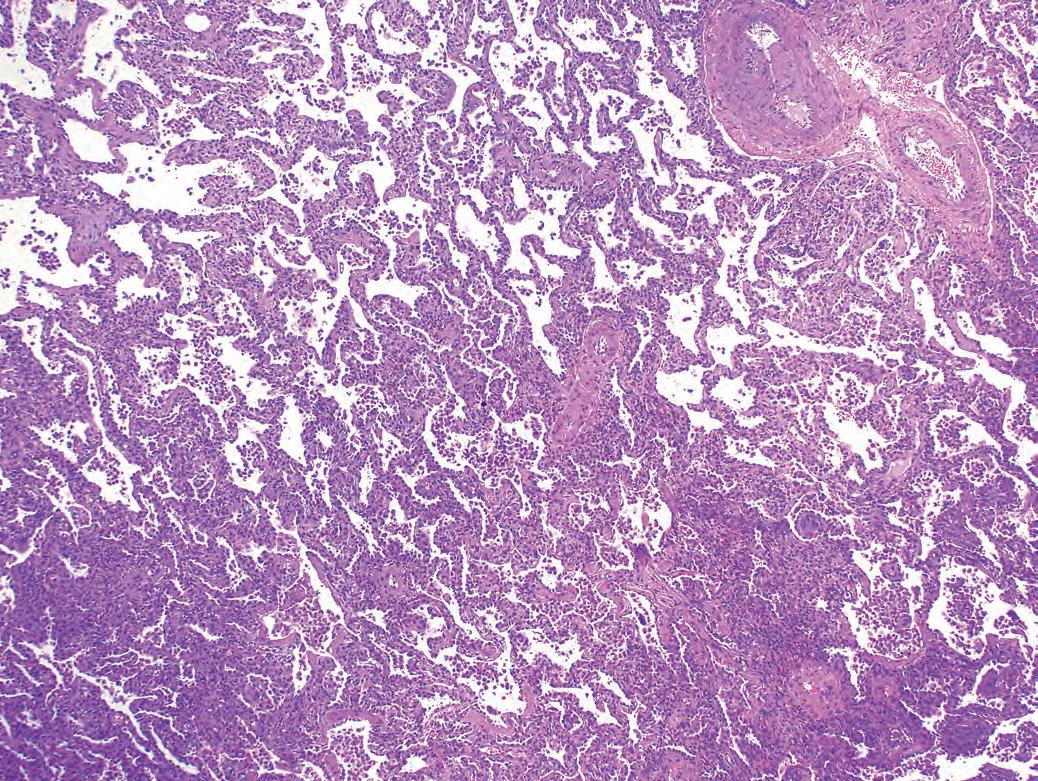
Pattern 3 Chronic Cellular Infiltrates
Elements of the pattern: The lung biopsy is dominated by interstitial chronic inflammation and variable reactive type 2 cell hyperplasia. The dominance of mononuclear infiltrates may impart an overall blue appearance to the biopsy at scanning magnification (in routine hematoxylin-eosin stained sections).
Additional Findings
Hyaline membranes
Necrosis in parenchyma
Necrosis in bronchioles
Poorly formed granulomas (small and nonnecrotizing)
Well-formed necrotizing granulomas
Eosinophils in alveoli
Siderophages in alveoli
Fibrinous/chronic pleuritis
Patchy organizing pneumonia
Atypical cells
Multinucleated giant cells
Dense mononuclear infiltration
Pattern 3 Chronic Cellular Infiltrates
Lymphoid aggregates/germinal centers
Diagnostic Consideration
“Acute on chronic” connective tissue disease
Drug toxicity
Diffuse alveolar hemorrhage
Viral and fungal infections
Aspiration
Infarction in antiphospholipid syndrome
Viral infections
Aspiration
Hypersensitivity pneumonitis (subacute)
Atypical mycobacterial infection
“Hot tub” lung
Lymphoid interstitial pneumonia
Drug toxicity
Infections
Rare drug reactions
Necrotizing sarcoidosis
Middle lobe syndrome
Eosinophilic lung diseases
Smoking-related lung diseases
Diffuse alveolar hemorrhage
Chronic cardiac congestion
Drug toxicity
Connective tissue diseases
Thoracic trauma/infection
Pancreatitis-associated pleuritis
Drug toxicity
Connective tissue diseases
Infections
Cryptogenic organizing pneumonia
Diffuse alveolar hemorrhage
Aspiration
Viral infections
Lymphangitic carcinoma
Hard metal disease
Mica pneumoconiosis
Hypersensitivity pneumonitis
Intravenous drug abuse
Drug toxicity
Aspiration pneumonia
Eosinophilic pneumonia
Lymphomas
Lymphoid interstitial pneumonia
Connective tissue diseases
Hypersensitivity pneumonitis
Certain infections (the atypical pneumonias)
Connective tissue diseases
Diffuse lymphoid hyperplasia
Lymphoid interstitial pneumonia
Follicular bronchiolitis
Ch. 6:134
Ch. 6:136
Ch. 11:393
Ch. 7:178, 199
Ch. 7:161; Ch. 9:306
Ch. 8:251
Ch. 7:199
Ch. 7:161; Ch. 9:306
Ch. 8:269
Ch. 8:270
Ch. 7:175
Ch. 8:244
Ch. 8:259
Ch. 7:177
Not specifically addressed
Ch. 11:383
Ch. 9:303
Ch. 6:139; Ch. 8:255
Ch. 8:243
Ch. 11:393
Ch. 5:114
Ch. 8:259
Ch. 8:247
Ch. 8:248
Not specifically addressed
Ch. 8:259
Ch. 8:247
Ch. 8:239
Ch. 8:237
Ch. 11:393
Ch. 7:161; Ch. 9:306
Ch. 7:199
Ch. 8:246
Ch. 10:354
Ch. 10:347
Ch. 8:269
Ch. 8:263
Ch. 8:259
Ch. 7:161; Ch. 9:306
Ch. 6:139; Ch. 8:255
Ch. 16:542
Ch. 8:244
Ch. 8:247
Ch. 8:269
Ch. 7:162
Ch. 8:247
Ch. 8:245; Ch. 16:537
Ch. 8:244
Ch. 9:308
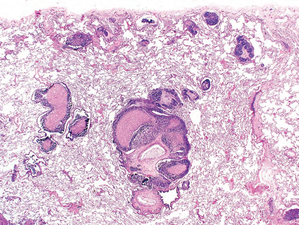
Pattern 5 Nodules
Elements of the pattern: One, or many, nodules of variable size and shape. An interface between the nodular lesion and more normal lung should be discernible. In the case of very large nodules encompassing the entire specimen, radiologic imaging can be used as part of the definition.
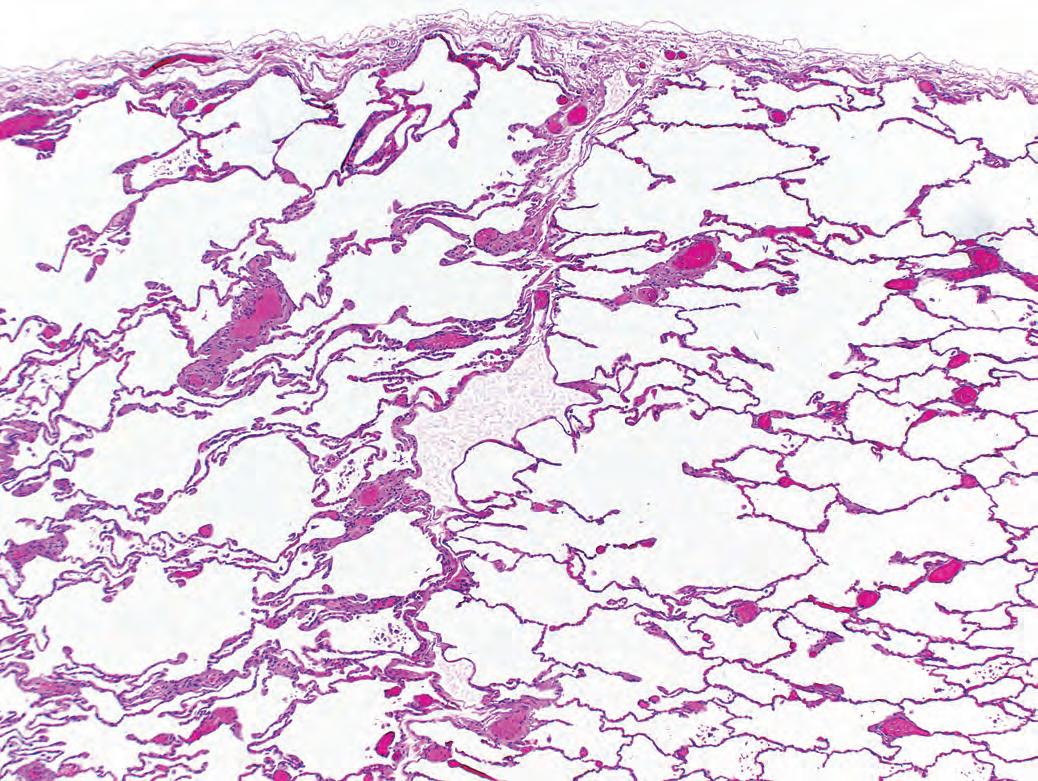
Pattern 6 Nearly Normal Lung
Elements of the pattern: The lung biopsy has little or no disease evident at scanning magnification.