Cambridge University Press is part of Cambridge University Press & Assessment, a department of the University of Cambridge.
We share the University’s mission to contribute to society through the pursuit of education, learning and research at the highest international levels of excellence.
www.cambridge.org
Information on this title: www.cambridge.org/9781108970617
This publication is in copyright. Subject to statutory exception and to the provisions of relevant collective licensing agreements, no reproduction of any part may take place without the written permission of Cambridge University Press & Assessment.
First Edition 2014
Second Edition 2024
Printed in the United Kingdom by TJ Books Limited, Padstow Cornwall
A catalogue record for this publication is available from the British Library.
A Cataloging-in-Publication data record for this book is available from the Library of Congress.
ISBN 978-1-108-97061-7 Paperback
Cambridge University Press & Assessment has no responsibility for the persistence or accuracy of URLs for external or third-party internet websites referred to in this publication and does not guarantee that any content on such websites is, or will remain, accurate or appropriate.
Every effort has been made in preparing this book to provide accurate and up-to-date information that is in accord with accepted standards and practice at the time of publication. Although case histories are drawn from actual cases, every effort has been made to disguise the identities of the individuals involved. Nevertheless, the authors, editors, and publishers can make no warranties that the information contained herein is totally free from error, not least because clinical standards are constantly changing through research and regulation. The authors, editors, and publishers therefore disclaim all liability for direct or consequential damages resulting from the use of material contained in this book. Readers are strongly advised to pay careful attention to information provided by the manufacturer of any drugs or equipment that they plan to use.
Eighteen months ago, Richard Montgomery kindly agreed to foreword the Second Edition of our Postgraduate Paediatric Orthopaedic book.
All the Postgraduate Orthopaedic forewords for each book published are very carefully chosen. The surgeons we ask are inspirational, widely respected and have influenced practice nationally and internationally.
Richard was very kind and helpful to us as junior consultants, always offering expert advice and guiding us. He was never critical, always encouraging and a mentor to us. Unfortunately, at the time of completing 1st proof corrections, he passed away suddenly. He will be a greatly missed colleague, teacher, and friend.
In loving memory of Richard Montgomery
List of Contributors ix
Foreword to the First Edition xi
Foreword to the Second Edition xii
Preface xiii
Acknowledgements xiv
Interactive Website xv
List of Abbreviations xvi
Section 1 General Introduction
1. Introduction and General Preparation 1
Paul A. Banaszkiewicz
2. Clinical Assessment 7
Stan Jones and Sattar Alshryda
3. Normal Lower Limb Variants in Children 24
Manoj Ramachandran and Gregory B. Firth
Section 2 Regional Paediatric Orthopaedics
4a. Slipped Upper Femoral Epiphysis 37
Sattar Alshryda and Paul A. Banaszkiewicz
4b. Legg–Calvé–Perthes Disease 52
Sattar Alshryda and Paul A. Banaszkiewicz
4c. Developmental Dysplasia of the Hip 66
Sattar Alshryda and Paul A. Banaszkiewicz
4d. Miscellaneous Hip Disorders 86
Sattar Alshryda and Paul A. Banaszkiewicz
5. Traumatic Hip Disorders 92
Sean Duffy and Fergal Monsell
6. Orthopaedic Knee Disorders 108
Sattar Alshryda and Fazal Ali
7. Traumatic Knee Disorders 129
Nick Nicolaou and Joanna Thomas
8. Orthopaedic Foot and Ankle Disorders 144
Anthony Cooper, Akinwande Adedapo, and Stan Jones
9. Traumatic Foot and Ankle Disorders 170
Vittoria Bucknall and Mohammed Al-Maiyah
10. Orthopaedic Spine Disorders 180
Ashley A. Cole and Lee M. Breakwell
11. Traumatic Spine Disorders 200
Ashley A. Cole and Lee M. Breakwell
12. Orthopaedic Shoulder Disorders 210
Om Lahoti and Tony Antonios
13. Traumatic Shoulder Disorders 222
Talal Al-Jabri, Sandeep Gokhale, and Clare Carpenter
14. Orthopaedic Elbow Disorders 233
Om Lahoti and Matt Nixon
15. Traumatic Elbow Disorders 241
Thomas Dehler and John Davies
16. Orthopaedic Hand and Wrist Disorders 263
Sara Dorman and Dean E. Boyce
17. Traumatic Hand and Wrist Disorders 286
Ehab Aldlyami and Khalid Alawadi
Section 3 Core Paediatric Orthopaedics
18a. Neuromuscular Conditions/Lower Limbs 297
Simon L. Barker and Sattar Alshryda
18b. Neuromuscular Conditions/Upper Limbs 322
Bavan Luckshman and Rachel Buckingham
18c. Gait Analysis and Orthoses 327
Jennifer Walsh, Syed Kazmi, and Tahani Al Ali
19. Musculoskeletal Infection 338
Mark Gaston, Richard Gardner, and Simon Kelley
20. Musculoskeletal Tumours 347
Richard Gardner, Gino R. Somers, and Sevan Hopyan
21. Skeletal Dysplasia 369
Anish P. Sanghrajka and James A. Fernandes
22. Metabolic Bone Disease 385
Richard Hutchinson, Mubashshar Ahmad, and Gavin DeKiewiet
23. Deformity Correction 396
Stan Jones, Farhan Ali, Anthony Cooper, and Alwyn Abraham
24. Orthopaedic-Related Syndromes 413
Deborah M. Eastwood
25. Miscellaneous Paediatric Orthopaedic Conditions 424 Ben Marson and Kathryn Price
Additional videos can be found at www.cambridge .org/alshryda Index 443
Contributors
Alwyn Abraham BSc MBChB FRCS (Tr & Orth)
University Hospitals of Leicester NHS Trust, Leicester, UK
Akinwande Adedapo MBBS FRCS (Eng) FRCS (Glas)
James Cook University Hospital, Middlesbrough, UK
Mubshshar Ahmad MBBS FRCS (Tr & Orth)
University Hospital of North Durham,UK
Tahani Al Ali BSc MSc
Al Jalila Children Speciality Hospital, Dubai Academic Health Corporation, Dubai, UAE
Norfolk and Norwich University Hospitals, Norwich, UK
Gino R. Somers MBBS PhD FRCPA Hospital for Sick Children, Toronto, Canada
Joanna Thomas MBBS MSc FRCS (Tr & Orth)
University Hospital Southampton NHS Foundation Trust, Southampton, UK
Jennifer Walsh BEng PGDip Stat PhD Astley Ainslie Hospital, Edinburgh, UK
Foreword to the First Edition
Since 1998, I have convened an annual core curriculum lecture course in paediatric orthopaedics at Alder Hey Children’s Hospital. Over the years, we have frequently been asked to recommend books that succinctly cover all of the necessary information and I now believe that we have found such a book in Postgraduate Paediatric Orthopaedics: The Candidate’s Guide to the FRCS (Tr & Orth) Examination. As the title suggests, the text is targeted toward trainees sitting the FRCS (Tr & Orth) examination but the book would also be useful for those who seek to enhance or maintain their paediatric orthopaedic knowledge base, including practicing orthopaedic surgeons, GPs, paediatricians and specialist physiotherapists. The text is
much more than lecture notes and covers all of the major subjects with sufficient information to keep the reader interested, while still delivering the required facts to an examination candidate as quickly as possible. I congratulate the editors and the authors for producing such a useful text.
Colin E. Bruce Consultant
Children’s Orthopaedic Surgeon Alder Hey Children’s Hospital Liverpool, UK
President of the British Society of Children’s Orthopaedic Surgery (BSCOS)
Foreword to the Second Edition
As a past paediatric orthopaedic examiner and Chair of the Intercollegiate Specialty Board for the FRCS Trauma & Orthopaedics exam, I was very interested to see drafts of the text and was honoured to be asked to write this foreword.
I have known Sattar, Paul and Stan and also several of the other distinguished contributors for many years. They are some of the most able, enthusiastic and industrious medical educators that I have met. Their long experience in assisting candidates to prepare for the exam is obvious in this book.
When candidates are preparing for the exam there is a tendency to cram in knowledge in as many fields as possible. However, if that knowledge is not structured, it may be difficult to recall it and to present it to the examiners in a logical way. Recalling the facts, and being able to present them to the examiner in a logical way is vital to exam success.
What the authors provide in this book is a distillation of the key facts and classifications as well as a structure for organizing knowledge about paediatric orthopaedic conditions. I wish it had been available when I was training!
Prof. Richard Montgomery MB, BS, FRCS(Ed), FRCS(Eng)
Visiting Professor, School of Health & Social Care, University of Teesside
Past Chair, Intercollegiate Specialty Board in T&O Surgery, for the FRCS T&O examination
Past President of the British Limb Reconstruction Society
Preface
Why another exam-related FRCS (Tr & Orth) book? Don’t we cover paediatrics in the chapters of the other Postgraduate Orthopaedics books?
We always felt the need for a more definitive guide to the paediatric component of the FRCS exam.
We were never entirely happy that the FRCS (Tr & Orth) paediatric syllabus was particularly well written or developed in a number of orthopaedic books. Most lacked the specific subject focus that candidates needed to pass the FRCS (Tr & Orth) exam.
General orthopaedic books tended to scratch the surface of a difficult area of orthopaedics that needs to be learnt well for the exam. Specialized books in paediatrics meant you could lose all focus of the subject’s relevance and end up not extracting out the relevant/specific detail required to pass the exam. Moreover, you could end up spending a lot of unnecessary time and effort drowning in these specialized textbooks and not have enough time left to read the basic science, trauma, or hands sections.
Our aim with this book was to make it all-encompassing, so that it covers everything you need to know to pass the FRCS (Tr & Orth) section of the exam, without having to cross-reference from other larger textbooks – a tall order, but one which we hope we manage to succeed in doing.
We were careful not to make the book too detailed, so it ends up being like a subspecialty book in paediatrics. At
the same time, we didn’t want it to become too flimsy, such that you felt you were missing something and you needed to repeatedly go to the bigger specialized textbooks of paediatric orthopaedics.
Special mention about the unusual time of Covid-19 that occupied a large part of the book writing process. Whilst some authors may say the extra time in lockdown gave them the opportunity to complete tasks, they were unlikely to have done otherwise this wasn’t particularly applicable to us. Despite the extra time of lockdown with elective surgery cancelled extra time doesn’t always equate with productive efficient time usage.
As with all the Postgraduate Orthopaedics book series, we make no claim for the originality of the material contained in the text. This material is available in the larger orthopaedic community. We have simply distilled and focused this knowledge down into something that will hopefully get you through the exam.
We hope you find this book useful in preparing for the exam and we wish you every success. We hope that in some small (or large) way the book will make the difference between you passing and failing the exam
Sattar Alshryda
Stan Jones
Paul A. Banaskiewicz
Acknowledgements
Special thanks to all the authors involved with the Postgraduate Orthopaedics book series over the years. Without your input, no book would be possible.
Special thanks to Jessica Papworth and Beth Sexton at Cambridge University Press for their help, guidance, and patience with this second edition of Postgraduate Paediatric Orthopaedics
Special thanks to our medical artist Biswa Prakash Sahoo from India who did a great job of drawing the book illustrations.
Great appreciation for Anthony Michael Rex (Consultant Spine Surgeon), Metwally Sayed Ahmad(Consultant Orthopaedic Surgeon,Kuwait) Karim Khalil (Paediatric Orthopaedic Specialist) and Yasir Adil Al-Humairi (T&O Resident) for their help in the proofreading. Thanks to Faizan Jabbar for
coordinating our website structure and to our web designer Farrakh, who has helped us develop and progress our website through the years.
Special thanks to Kath McCourt CBE, Nick Caplan and Dianna Ford at Northumbria University for their help and support through the years.
As ever, thanks to Jo McStea who keeps the whole PGO setup rolling along.
Thanks to James Coey,Assistant Dean Basic Sciences St. George’s University for photography,inspiration and just being there to help and guide.
Special thanks to all of our orthopaedic trainers who helped guide and nurture our development through the many years of our own orthopaedic training.
Interactive Website
www.postgraduateorthopaedics.com
This website accompanies the textbook series Postgraduate Orthopaedics. It includes:
• Postgraduate Orthopaedics: The Candidate’s Guide to the FRCS (Tr & Orth) Examination, 3rd edition
• Postgraduate Orthopaedics: Viva Guide for the FRCS (Tr & Orth) Examination
• Postgraduate Paediatric Orthopaedics
• Postgraduate Orthopaedics. SBAs for the FRCS (Tr&Orth) Examination
The aim is to provide additional information and resources in order to maximize the learning potential of each book. The book comes with 20 educational videos. More videos will be added regularly on the accompanying YouTube channel https://www.youtube.com/@ChildrenOrthopaedics
Additional areas of the website provide supplementary orthopaedic material, updates, and web links. Meet the Editorial Team provides a profile of authors who were involved in writing the books. Details of up-and-coming courses are provided. Details of the next diet of exams are provided.
There are many single best answer questions that are exam focused along with case-based discussions, podcasts and webinars.
It is very important our readership gives us feedback. Please e-mail us if you have found any errors in the text that we can correct.In addition, if we have not included an area of orthopaedics that you feel we should cover, please let us know. Likewise any constructive suggestions for improvement would be most welcome.
There is also a list of Postgraduate Orthopaedics courses available for candidates to fine-tune their examination skills.
Abbreviations
AA anatomical axis
AAOS American Academy of Orthopaedic Surgeons
ABC aneurysmal bone cyst
AC acromioclavicular
ACA acquired coxa vara; axis of correction of angulation
ACL anterior cruciate ligament
ADI anterior atlanto-dens index
ADM abductor digiti minimi
ADTA anterior distal tibial angle
AER apical ectodermal ridge
AFO ankle foot orthosis
AHA Assisting Hand Assessment
AI acetabular index
AIIS anterior inferior iliac spine
AIS adolescent idiopathic scoliosis
aLDFA anatomical lateral distal femoral angle
ALP alkaline phosphatase
ALPSA anterior labral periosteum sleeve avulsion
AMC arthrogryposis multiplex congenita
aMPFA anatomical medial proximal femoral angle
AOFAS American Orthopedic Foot and Ankle Score
AP anteroposterior
ASIA American Spinal Injury Association
ASIS anterior superior iliac spine
ATD articulotrochanteric distance
ATFL anterior talofibular ligament
ATiFL anterior tibiofibular interosseous ligament
ATLS Adult Trauma Life Support
ATR angle of trunk rotation
AVN avascular necrosis
BD Blount’s disease
BMD bone mineral density
BMP 2 bone morphogenetic protein 2
Ca calcium
cAMP cyclic adenosine monophosphate
CAPTA Child Abuse Prevention and Treatment Act
CCBS congenital constriction band syndrome
CCV congenital coxa vara
CDH congenital dislocation of the hip
CDK congenital dislocation of the knee
CEA centre edge angle
cEDS classic Ehlers–Danlos syndrome
CFD congenital femoral deficiency
CFL calcaneofibular ligament
CI confidence interval
CKD chronic kidney disease
CMCJ carpometacarpal joint
CMT Charcot–Marie–Tooth
CNP C-type natriuretic peptide
COMP cartilage oligomeric matrix protein
CORA centre of rotation of angulation
CP cerebral palsy
CPIP Cerebral Palsy Integrated Pathway
CRP C-reactive protein
CSVL Central sacral vertical line
CtE Commission through Evaluation (trial)
CTEV congenital talipes equinovarus
CV coxa vara
CVS chorionic villus sampling
CVT congenital vertical talus
DCV developmental coxa vara
DDH developmental dysplasia of the hip
DEXA dual-energy X-ray absorptiometry
dGEMRIC delayed gadolinium-enhanced MRI of cartilage
DIPJ distal interphalangeal joint
DMAA distal metatarsal articular angle
DMD Duchenne muscular dystrophy
DRUJ distal radioulnar joint
ECU extensor carpi ulnaris
EDF elongation, derotation, and flexion
EDS Ehlers–Danlos syndrome
EI extensor indicis
EMA epiphyseal–metaphyseal angle; European Medicines Agency
EMG electromyogram
EMI extended matching item
EPB extensor pollicis brevis
EPL extensor pollicis longus
ERT enzyme replacement therapy
ESIN elastic stable intramedullary nail
ESR erythrocyte sedimentation rate
EXT exostosin
FAI femoroacetabular impingement
FCU flexor carpi ulnaris
FD fibrous dysplasia
FDA Food and Drug Administration
FDP flexor digitorum profundus
FDS flexor digitorum superficialis
FES functional electrical stimulation
FFCD focal fibrocartilaginous dysplasia
FGF-23 fibroblast growth factor 23
FGFR-23 fibroblast growth factor receptor 23
FMDA Femoral–metaphyseal–diaphyseal angle
FPA foot progression angle
FTR femoral: tibial ratio
GA general anaesthesia
GABA gamma-aminobutyric acid
GLAD glenolabral articular disruption
GMC General Medical Council
GMFCS Gross Motor Function Classification System
GMFM Gross Motor Function Measure
GRAFO ground reaction ankle foot orthoses
HA hydroxyapatite
HAGL humeral avulsions of the glenohumeral ligament
HEA Hilgenreiner epiphyseal angle
HKAFO hip, knee, ankle, and foot orthosis
HLH haemophagocytic lymphohistiocytosis
HSCT haematopoietic stem cell transplant
HSMN hereditary sensorimotor neuropathy
HVA hallux valgus angle
IIS infantile idiopathic scoliosis
IL-6 interleukin-6
IMA intermetatarsal angle
IP interphalangeal
IPA interphalangeal angle
IPJ interphalangeal joint
IT iliotibial
ITFJ inferior tibiofibular joint
JCIE Joint Committee on Intercollegiate Examinations
The same sentiments still apply from our first edition’s chapter that general FRCS (Tr & Orth) exam guidance material can become a little dull and tedious to most candidates. We again have tried to avoid any unnecessary repetition of material, concentrating on the important details vital for exam success.
Since we wrote the introduction chapter for the first edition book several years ago, candidate preparation for the FRCS (Tr & Orth) exam has significantly altered in two major ways. The first is the established use of WhatsApp groups for exam preparation, and the second is an even bigger more widespread reliance on being part of a study group for exam preparation.
One of the major concerns for most candidates is to know the most likely paediatric viva questions that regularly appear in the exam. Equally important is to be aware of any unusual clinical cases that have unexpectedly appeared in the Section 2 exam. This is especially important for the intermediate cases as a difficult, unusual condition can sink your day.
Facioscapulohumeral muscular dystrophy is a fairly rare case that, for most candidates, is off their radar for the clinical exam. In several consecutive diets of exams, this disorder ended up as an intermediate case. Candidates who had no clue about the condition almost invariably failed badly, whilst those in the know usually performed very well. There was usually a large discrepancy in performance between the two types of candidates. A candidate’s performance on this particular intermediate case could be the defining feature of whether a candidate passed or failed their entire exam.
In the last five years, there has been widespread normalization in the use of WhatsApp groups for exam preparation. At its most basic level, this involves candidates listing their own exam experiences almost immediately after their exam is completed. As such, questions are widely circulated and freely available for both trainees and non-trainees to digest.
The problems that existed with the ‘candidate accounts’ that floated around on the Internet still exist with the WhatsApp accounts. Most ‘candidate experiences’ are all written in a very similar vein and after reading the first two or three, very little extra new material is then uncovered. Also these accounts become just an endless list of topics without a structured answer to the question being provided.
Study groups are now more than ever vital for exam success, with many groups scheduling 1–2 hours of exam discussion and practice on the Internet most nights.
The aims of the exam are to see if you have enough knowledge to practise safely as a day-one orthopaedic consultant in a district general hospital in the generality of orthopaedics. The
exam is not set out to test you in microscopic detail about trivial irrelevancies. The exam is not even designed to test for subspecialty interest. The other quoted analogy that is often used by Royal Colleges is that the exam should be viewed as a mature conversation between two consultant colleagues. I have never bought into this comparison but you will equally find many who accept this metaphor.
The first day you are on call as a consultant, your registrar may phone you up about a child with a painful hip in casualty. A child with knock knees may have been wrongly referred to your adult knee clinic. Your trauma practice may cover children and you may worry about risks of growth arrest with particular fracture patterns.
The History of the FRCS (Tr & Orth) Exam
In the late 1970s, the old-style FRCS ceased to mark the end of training and had become the entry into higher surgical training. The only exam in Britain devoted exclusively to orthopaedics was the MChOrth from the University of Liverpool. To take this exam, you generally had to work in or around the Mersey Deanery.
The situation was clearly unsatisfactory and, under the guidance of the Royal College of Surgeons in Edinburgh, a Specialty Fellowship exam in orthopaedics was introduced in 1979. This exam was optional but soon became established as a benchmark of completion of training and a quality assurance measure. It was an entirely clinical exam with a viva voce format. The standard was high, and the pass mark variable. It was not an easy exam to pass, but it became accepted that recognition of the standard of higher surgical training by assessment in the form of an exam is essential in orthopaedics. This is, in fact, applicable to all surgical specialties, not just orthopaedics.
In time, the exam was accepted by all four Royal Colleges, and in 1990, a new intercollegiate exam was introduced. This originally took place twice a year in each of the colleges in turn. This exam became a requirement for accreditation, together with the satisfactory completion of training in an approved programme that had been inspected and approved by the Specialist Advisory Committee.
For many years, it was difficult to get hold of any valuable exam guidance. The exam appeared to be surrounded in secrecy. Despite a curriculum and syllabus, many candidates entered the exam not really knowing what to expect. The usual line was that if you had undertaken good clinical work, read the appropriate literature and had a sound grasp of basic sciences, you would be expected to pass.
It was generally difficult to get useful information and tips from previous candidates, such as the expected standard or the
questions likely to be asked. Another fact – now easily forgotten –was that the Internet was in its infancy and there simply was not the candidate support network that there is today.
There were not a large number of courses available to guide a candidate on the expected standard, and some courses set the level far too high. The idea was that you were panicked into hitting the books, as you perceived that your knowledge was not up to the required standard. This was fine if you had a year or so to go before the exam and you could plan a more intensive schedule of revision, but not so good if your exam was sooner.
The situation began to change around the turn of the millennium. A number of candidates began writing down their own experiences as a revision tool for the next wave of candidates sitting the exam. A small select number of candidates in larger training programmes began to form study groups. These study groups acquired, and circulated, these candidate accounts among themselves to help with exam preparation. The deal was that once you had passed, you wrote your own account for those candidates coming after you to use for their preparation. In time, these candidate experience reports began to circulate more freely in a wider domain.
Today there are numerous websites containing candidates’ exam experiences. These include the British Orthopaedic Trainee Association, various regional training programme sites, and lastly individual accounts from successful candidates. The major problem with many candidate experiences is that they deal with specific viva or clinical questions in a rather superficial way, mainly with bullet point headings. Also, we have yet to see an unsuccessful candidate’s experience posted on the Internet. Candidates generally learn more from what went wrong than if only successful accounts are presented. WhatsApp groups and Telegram are now replacing the Internet as means for trainees to quickly communicate to each other exam tips and tricks or to discuss difficult learning points.
The standard of FRCS (Tr & Orth) exam courses has, by and large, significantly improved and, in general, candidates are much more informed and have a better idea of what types of question tend to get asked. So one of the most major changes with the FRCS (Tr & Orth) exam in the last 10 years is that the mystery surrounding it has completely evaporated away.
The old-style viva with a variable number of questions is definitely a thing of the past. The viva is now standardized for candidates, with similar questions being asked for each topic covered. This leads to a much fairer exam, with much less potential for any discrimination.
Exam Format
The current FRCS (Tr & Orth) exam encompasses two sections: Section 1 is a written exam, and Section 2 the clinical and oral exams. For further information, and to make sure that your information is up to date, we suggest that you carefully review the Intercollegiate Specialty Board website (www.jcie.org.uk).
Section 1
Section 1 is the written component of the Intercollegiate Examination in Trauma and Orthopaedic Surgery. In 2018, the Joint Committee on Intercollegiate Examinations (JCIE) agreed
to phase out extended matching item (EMI) questions. When compared to single best answer (SBA) questions, EMI questions were less able to differentiate between candidates and were difficult to construct. EMI questions have not featured in the FRCS (Tr & Orth) examinations from January 2021 onwards.
Section 1 exam will consist of two papers as follows:
Paper 1 (2 hours and 15 minutes)
SBA – 120 questions
Paper 2 (2 hours and 15 minutes)
SBA – 120 questions
Total 4 hours and 30 minutes – 240 questions.
Candidates will have a two-year period from their first attempt to pass the Section 1 exam, with a maximum of four attempts with no re-entry. Details are available on the JCIE website (www.jcie.org.uk). Candidates with proven dyslexia may be eligible for the Section 1 examination times to be extended and this should be highlighted in advance of the exam.
There is no negative marking; therefore, all questions should be attempted. Sample questions can be viewed on the JCIE website. Experienced examiners perform a formal process of standard setting to decide the final pass mark for each paper. SBA questions are subject to quality assurance procedures, including feedback from both examiners and candidates. Difficulty level, content, discrimination index, and internal consistency are analysed. Ambiguous questions, or those deemed insufficient to differentiate between candidates, are removed through this process.
SBA questions consist of an introductory theme, a question stem, and five possible responses (listed as A to E), of which one is the most appropriate answer. SBA questions are exactly what the name suggests: candidates choose the best from five possible answers. It is important to note that this is not a ‘single correct answer’, but a ‘single best answer’. Moreover, all five possible answers could be considered correct, but candidates are asked which is best, or most appropriate, given the information provided. As questions are designed to test higher-order thinking, this could mean that limited or irrelevant information is provided. Questions require a judgement based on interpretation of the available evidence. Questions that candidates later complain about, for example ‘there was more than one correct answer’ or that a question was ‘too ambiguous’, can often prove the best performing questions.
For more detailed information on the dynamics of the Section 1 paper, candidates should read Chapters 1 and 2 of SBAs for the FRCS (Tr&Orth) Examination: A Companion to Postgraduate Orthopaedics Candidate’s Guide.
Section 2
This underwent major changes in 2020. For some time, the General Medical Council (GMC) had been keen to remove altogether the clinical examination component from the Section 2 exam. There were considerable ongoing discussions and disagreement among the GMC, the four Royal Colleges, and various stakeholders. A compromise was reached in that although the short cases would be replaced by short case clinical examination technique vivas, the intermediate cases involving patients would remain. There would be four intermediate cases, with one of them being simulated.
The introduction of this new Section 2 clinical exam was significantly disrupted with the Covid-19 pandemic, with several exam diets being cancelled. There was an urgent need to begin examining a large backlog of trainees to prevent the orthopaedic training system from grinding to a halt. It was therefore decided to temporarily hold the clinical component without direct patient involvement using iPads. Six short case examination vivas were introduced, along with the temporary use of simulated intermediate cases, both showing candidates a series of clinical photographs on iPads on which questions were based. When the pandemic settles, it is expected that patient involvement with the intermediate cases will resume.
The clinical component was often viewed as the most difficult part of the whole exam to pass and these changes had a major impact on how candidates prepare for the exam. Although the Royal Colleges remain positive and upbeat, a significant number of consultants are disappointed with the changes. If you are an educationalist, you are likely to follow the party line and state that the exam assessment will remain as robust as ever. In practice, there is a huge difference in skills, professionalism, and knowledge required to examine a patient with a rotator cuff tear, and elicit and interpret positive clinical signs, as opposed to just describing how you would go about examining for this in a viva situation. If you have prelearnt the talk and gone through some practice runs in your study group, you should be more than halfway towards passing the viva.
The short case clinical examination technique viva involves candidates being shown a clinical case and describing how they would go about examining that patient – for paediatrics, it could be a picture of a young child with a unilateral pes planus deformity, perhaps an obvious tarsal coalition – and describing their approach to that particular patient.
Superficially, this viva is similar to the oral topics viva, but the Joint Surgical Colleges Fellowship Examination (JSCFE) Committee are at great pains to point out that the clinical short case vivas are very different. There are no questions on management of the condition and the specific aim of the viva is to work through clinical examination. Be prepared to be grilled in detail about how you perform a particular clinical test, and the theory behind it. More important than how you perform a particular test is how the test will change your management of the condition. For example, if the Coleman block test demonstrates that the hindfoot is rigid, how will this change your management of the pes cavus deformity?
A decision has been reached to minimize the use of radiographs or scans in this viva, as this will avoid steering the conversation on to management.
Candidates’ feedback for the paediatric short case clinical vivas suggests that these are particularly difficult to answer. Previously, unless the exam was being held near a paediatric tertiary centre, the amount of complex paediatric cases brought into the exam hall was usually limited. Now a photograph of a very rare paediatric condition can be shown and candidates may struggle to piece together a structured examination format to satisfy the examiners. Added to this, the general unfamiliarity of this new exam format means that stress levels are much greater in paediatric clinicals than was ever the case.
Clinicals
The clinicals are divided into six five-minute clinical examination technique vivas, including upper and lower limb, as well as spine, vivas.
At present, there are four intermediate cases, 15 minutes each (five-minute history, five-minute clinical examination, five-minute discussion). These involve patients.
Orals
The oral component is divided into four 30-minute viva sections:
• Basic science
• Trauma, including spine
• Adult elective orthopaedics, including spine
• Paediatric orthopaedics and hand surgery, including shoulder and elbow.
Paediatric Section
The paediatric oral section is combined with the hands and upper limbs section. The examiners have to introduce themselves to the candidate and remind the candidate which oral he or she is about to be examined on, to allow the candidate time to settle. Feedback is given where appropriate such as: ‘OK, let’s move on’ or ‘We have covered this area, let’s go on’. Examiners are encouraged to avoid remarks such as ‘Excellent’, ‘Well done’, ‘That’s great’, or ‘Fantastic’.
Props, such as radiographs, pictures, and charts, are usually used to lead into a question.
Three paediatric topics are discussed – these usually cover a trauma-type question, one big (A-list) topic, and a less obvious clinical topic.
Hammering on when a candidate could not answer a question used to be a common candidate complaint, but examiners are now actively dissuaded from this practice.
All candidates are treated in exactly the same manner and marks are based on performance only. Examiners are instructed to allow for candidates’ nervousness and are told not to respond to inappropriate behaviour by a candidate. Inappropriate behaviour would include rudeness or sarcastic remarks to the examiners, impoliteness, and bad-mannered or derogatory comments about facilities or organization issues.
A significant change is that viva questions are now more clinically orientated and relevant to the types of situation that may present to a consultant orthopaedic surgeon in clinical practice. For this reason, potential exam questions are now significantly more scrutinized than previously, before being approved by the exam committee for inclusion in the exam.
When to Sit the Exam
It is generally accepted that you will need about one full year of preparation before you will feel confident to sit the exam.
In theory, it should be relatively easy for you to decide if you have enough experience and have prepared in sufficient detail to sit the exam. In practice, a multitude of competing issues usually complicate this decision.
If you are a trainee, you will have been sitting the UK In-Training Exam for the last three or four years and should know your annual scores. Many training programmes also have
yearly mock clinical and viva exams, and will not let you sit the real exam unless you have achieved a good enough pass in these mock tests.
In the past, you may have had a charitable training programme director who was willing to take a chance with you, but this is now less likely, as it may have a direct bearing on the number of trainees allocated to a region.
As a general rule, do not use up an attempt if you are underprepared and unlikely to pass, as you have only four attempts at the exam and you are essentially throwing away one opportunity. The old advice that it would be good practice and allow you to sail through the second attempt is a wrong tactic, and is out-of-date advice from a different time.
Study Groups
A key factor for your success will be the formation of a study group that meets regularly, discusses various topics, and arranges practice viva sessions.
Study groups now more than ever are very important and the key to successfully and as painlessly as possible passing the exam. They offer support and help to an otherwise long, lonely and difficult exam journey. Just as important is the opportunity that study groups allow for individuals to talk through and conceptualize difficult topics. A large part (but not all) of the exam is about how well you can communicate to the examiners. The more practice you get, the more skilled you will be with this.
There are a number of factors that will contribute to a study group’s success and also some aspects that you should avoid. The group should comprise 3–4 candidates who all need to get along well with each other and should not be too far apart from one another in terms of knowledge. If there are significant rivalries and petty jealousies within the group, with trainees trying to score points off each other, the group is not going to work out. Be careful of candidates who think that they are too good for the group; they are likely to let you down near the end and do their own thing.
Also, be careful with candidates who are unlikely to pass the exam first time round and who are just too far behind with their studies to contribute significantly to group discussions. Give these candidates the benefit of the doubt, as surprises do happen, but be concerned if you draw repeated blanks with large gaps in core knowledge. Politely sideline such candidates if the extra input is significantly affecting the group’s performance. In general, do not include candidates who are a few months, or perhaps a year, off sitting the exam. They are unlikely to have sufficient motivation and drive at this stage in their revision.
Study groups have refined their study techniques in that there is more savvy use of the Internet late at night and at weekends to have in-depth discussions of topics and viva practice. Particularly with some like-minded trainees working in various parts of the country, they may organize regular Zoom meetings to go through topics.
Some trainees arrange to meet up with each other for 2–3 days before the actual exam to practise their own viva exam approach.
Covid-19 and Webinars
With the Covid-19 pandemic, there has been a huge explosion in the amount of online webinars available to candidates. A substantial number are directed towards the FRCS (Tr & Orth) exam. As a resource for learning, webinars are controversial and have had a mixed response. Some trainees definitely prefer this learning medium and believe that it is the way forward for the future. Others view the whole thing as passive learning, with trainees at home, with their feet up on a couch, not really actively involved with the process.
Webinars can be presented live or be pre-recorded. Prerecorded webinars allow the lecture to be broadcast at a later date and means that the lecture can be redone a few times until it is fit for purpose. Live is more spontaneous, but mistakes can occur in delivery and there are always some delays with screen sharing. It is also possible to have breakout groups and smaller group teaching sessions.
Some accounts are free but usually have limited facilities available, whilst to stream professionally can be very expensive. Quality can vary, depending on Internet connectivity and whether a professional webcam is used.
The four Royal Colleges have recorded many webinars, which are now available on their website, as an educational resource. With so many different webinars available, candidates need to be focused and efficient in their use of study time.
There are also large sources of YouTube videos and podcasts available to trainees. Some sources are excellent, and others less so, but it is important that trainees are ruthless in weighing up whether to use that particular source and the benefit it will provide.
Last-Minute Preparation
In the last 2–3 weeks leading up to the exam, try not to panic and attempt to go over all your revision again. This will not work and will just lead to you getting even more stressed and irrational. Use this time for quick, focused revision.
Attending a last-minute revision course as a sort of dry run a couple of weeks before the real exam is becoming more popular. This only really works if you are not too far away from the required standard and the dry run is used to iron out, finetune, and rehearse your performance. Hoping to get lucky with a sort of quick revision before the real exam, but with significant knowledge gaps, is unlikely to be successful.
Exam Tactics
Dress sensibly: no loud ties, short shirts, or Vivienne Westwood high heels. Everything straight down the line. Do not stand out. Do not smell of cigarettes, as this is very off-putting for most people. A bit of cologne is OK, but unless you have a body odour problem, be careful not to use too much, as this may also be offputting to examiners.
If you tend to sweat a lot under your armpits, get yourself a strong proven deodorant to avoid this. We see a small number of candidates in the exam where their armpits are covered in huge wet patches of sweat. This must be uncomfortable for candidates
and off-putting to patients. In simple terms, although this should not affect your mark, it just does not look good.
If you are one of a small number of candidates who are significantly affected by exam stress, it may be reasonable to get some professional help. The scenario during the exam would be extreme nervousness, difficulty focusing on what question is being asked, wet armpits, and sweat pouring off your face. This situation is very uncomfortable and will affect your performance. A beta-blocker will probably have no significant physiological affect but psychologically may help to calm you down and improve your overall exam performance. We would suggest speaking to your GP for advice.
Book a hotel fairly near the exam venue, preferably within walking distance, although this may not always be possible.
Allow plenty of time to arrive promptly at the exam. We know the arguments of turning up too early and getting freaked out by other candidates talking too much and winding you up. This is irritating, but a fair amount less stressful than leaving your arrival to the last minute and risking that you get caught up in traffic and turn up late.
Keep Your Distance
A piece of advice that we keep repeating is to get away from other exam candidates as quickly as possible after completing the various exam sections. It is extremely questionable whether anything useful can be gained by hanging around to chat to other candidates after completing the clinicals or vivas.
At best, this will unnerve you and can make you feel uncomfortable; at worst, it will put you off for the remaining parts of the exam. Even worse, you may end up in a bar afterward, drowning the sorrows of a perceived poor performance and ruin any chances of that last-minute brush-up of key topics you had planned for later that evening.
Stay focused during the exam period. Don’t let your guard down; don’t relax, and don’t be fooled into a false sense of security.
At the same time, and in equal measure, don’t get paranoid, edgy, and uptight, as this is just as counterproductive. You need to come across as relaxed and professional, as someone who is in control and who can be relied on. This mindset is much easier to achieve if you stay clear of other candidates. Perhaps the only exception should be candidates in your study group; you could chat to them for a few minutes after each exam section.
Suggested Reading
Which orthopaedic paediatric books to use for preparation for the paediatric section of the FRCS (Tr & Orth) exam is very much a matter of personal preference and choice. However, some books are more suited and better to use than others.
[1] Practice of Pediatric Orthopaedics
The numerous illustrations are first class, and the book has excellent recommendations and reviews. It is easy to read and fairly comprehensive. In view of the changes in the clinicals with the introduction of short case examination vivas, the large volume of pictures are especially useful to help with pattern recognition and spot diagnoses.
[2] Joseph’s Paediatric Orthopaedics
We were a little guarded in our previous review of the first edition, but the book has gone onto a second edition, so perhaps we were being too harsh with our criticisms. Overall, it is an excellent book to borrow from a library, and perhaps buy if you can get a discounted copy, rather than having to buy the more expensive hardback edition.
The book is targeted at higher surgical trainees and younger consultants. It is written by paediatric orthopaedic surgeons from four different continents. Although this gives the book a truly international flavour, in the highly focused world of FRCS (Tr & Orth) exams, this can be a drawback.
It is firmly emphasized on treatment, allowing trainee orthopaedic surgeons to make an informed contribution during their time in the paediatrics department and to speak confidently about the approach to individual patients during their specialty exams.
[3] Pediatric Orthopaedic Secrets, 3rd ed
By and large, the Secrets series has improved greatly in recent years. Some of the material in the paediatric book does not particularly match the FRCS (Tr & Orth) syllabus and the format is only loosely applicable to the exam. That said, we have come across a number of candidates who swear by the Secrets series. We advise that you borrow one from the library before buying. It has good reviews. The book has not been updated since 2007 but does not feel out of date just yet. It has a question-and-answer format that some candidates may find useful.
[4] Oxford Textbook of Trauma and Orthopaedics, 2nd ed
This is more of a reference book with a fairly detailed paediatric section. Reducing the three-volume first edition into a single volume in the second edition was a masterstroke and makes the book much easier to read.
[5] Miller’s Review of Orthopaedics
This has a reasonably good paediatric section. As the text is listed, the section is probably best suited for revision for Section 1 of the FRCS (Tr & Orth) exam.
[6] AAOS Comprehensive Orthopaedic Review
This book is similar in style to Miller’s, but more comprehensive. It has excellent reviews and is recommended for FRCS (Tr & Orth) exam preparation. The biggest drawback is the price.
[7] Paediatric Orthopaedics in Clinical Practice
This book is great, but whilst reading, a candidate may get an uneasy feeling that there are more focused paediatric FRCS (Tr & Orth) exam-related books that should be taking priority. It is a broad-based book that can be used by a number of different doctors dealing with paediatric orthopaedics.
[8] Pediatric Orthopedics in Practice
This is a comprehensive book that is very well written and easily digestible. It is expensive and has a European slant,
so it is unlikely to be many candidates’ first choice of book to buy. Worth a look-through if you need a slightly different approach to that hard-to-understand topic with which you may be struggling.
[9] Paediatrics for the FRCS (Tr + Orth)
Examination
This is a great book written for the FRCS (Tr & Orth) exam. Lots of hidden gems within and exam-orientated viva questions to keep you occupied. Highly recommended.
[10] Core Knowledge in Orthopaedics: Pediatric Orthopaedics
We bought this book several years ago, and it was a reasonable book at the time to use as a guide for a number of different paediatrics-related projects with which we were involved. The book is beginning to date and one to perhaps pass over, as there are
References
1. Diab M, Staheli LT. (2015) Practice of Pediatric Orthopaedics, 3rd ed. Philadelphia, PA: Lippincott Williams & Wilkins.
2. Joseph B, Nayagam S, Loder R, Torode I. (2009) Paediatric Orthopaedics: A System of Decision-Making. London: Hodder Arnold.
3. Staheli LT, Song KM. (2007) Pediatric Orthopaedic Secrets, 3rd ed. Philadelphia, PA: Elsevier Saunders.
4. Bulstrode C, Wilson-MacDonald J, Eastwood D, et al. (2017) Oxford Textbook of Trauma and Orthopaedics,
now many more books better suited for studying for the FRCS (Tr & Orth) exam.
[11] Pediatrics (Orthopaedic Surgery Essentials Series)
This is a great series that include many fantastic subject titles such as basic science and lower limb arthroplasty. The book is written by American authors so is not well suited to the UK FRCS (Tr & Orth) exam. The book does need to be updated. One to buy second-hand if going for a bargain on Amazon.
[12] Orthopaedic Knowledge Update: Pediatrics
Some candidates like these books. We have used this series in the past but have become a bit indifferent to it. It does have its moments and candidates may find some chapters useful for consolidation purposes. An American book that is expensive to buy whilst attempting to create a monopoly on education.
2nd ed. Oxford: Oxford University Press.
5. Miller MD. (2012) Review of Orthopaedics, 6th ed. Philadelphia, PA: Saunders.
6. Lieberman JR. (2009) AAOS Comprehensive Orthopaedic Review. Rosemont, IL: American Academy of Orthopaedic Surgeons.
7. Aresti NA, Ramachandran M, Paterson M, Barry M. (2016) Paediatric Orthopaedics in Clinical Practice, 1st ed. London: Springer.
8. Gelfer Y, Eastwood D, Daly K. (2018) Pediatric Orthopaedics in Practice, 1st ed. Oxford: Oxford University Press.
9. Gelfer Y, Eastwood D, Daly K. (2018) Paediatrics for the FRCS (Tr + Orth) Examination, 1st ed. Oxford: Oxford University Press.
10. Dormans JP. (2005) Core Knowledge in Orthopaedics: Pediatric Orthopaedics, 1st ed. St Louis, MO: Mosby.
11. Cramer KE, Scherl SA, Einhorn TA, Tornetta P. (2003) Pediatrics (Orthopaedic Surgery Essentials Series), 1st ed. Boston, MA: Lippincott Williams and Wilkins.
12. Martus JE. (2003) Orthopaedic Knowledge Update: Pediatrics 5 Rosemont, IL: American Academy of Orthopaedic Surgeons.
2 Clinical Assessment
Stan Jones and Sattar Alshryda
Introduction
Assessing a child with orthopaedic problems is more challenging than assessing adults. Children are poor historians and parents are usually emotional. Establishing a rapport during the first visit (and in the exam) is essential – although not always easy or possible. It gets easier with experience though.
The initial contact with the child and family involves introducing oneself to all the family members, including the child. This should be carried out in a professional, yet friendly, manner. The cultural background of the family should be considered and it is important to conform to gender order for introductions. Whenever possible, the discussion should be held with the child, whilst allowing the parents to add any relevant details or clarify some ambiguity.
The next stage of the assessment should aim to allay the anxiety/fear of the child. This can be done in a variety of ways and depends on the age of the child. In a younger child, the introduction to toys may be all that is required, whilst in the older child, it may involve talking about friends, sports, movies, school, or a piece of clothing.
History
Presenting Complaint
The three main complaints that bring children to a clinic (and exam) are:
1. Pain
2. Deformities (including physiological and pathological ones)
Ten relevant questions should be asked about the main complaint:
1. Duration of symptoms
2. Severity
3. Mode of onset
4. Any history of any injury or provoking factors
5. Frequency and timing of symptoms
6. Aggravating and relieving factors, and progression
7. Any functional impairment
8. Impact on carers, school, sports, and social life
9. Previous investigations and results
10. Treatment received and their effects.
It is also important to consider the presenting complaints in relation to the age of the child; for example, Perthes disease has to be considered as a differential in a young child (4–8 years of age) with a history of hip and knee pain, whereas in an adolescent with similar complaints, one has to think of slipped upper femoral epiphysis (SUFE).
Normal variants, including in-toeing, out-toeing, bowed legs, knocked knees, and flat feet, are common ‘deformities’ that trigger consultation. These must be confidently differentiated from similar pathological deformities such as Blount’s disease, congenital vertical talus, and tarsal coalition.
Deformity that is unilateral, or asymmetric or progressive, is often pathological. Severe deformities (more than two standard deviations (SDs) to what is accepted for the same age and gender), associated short stature, overweight, or syndromes could be pathological.
Children’s functions improve naturally as they get older (see Table 2.1 for key motor and social skills). Regression should trigger search for progressive neurodegenerative diseases and Rett’s syndrome. Delayed milestones should trigger a thorough history and neurological examination before reassuring parents as within normal. Hand preference during the first two years of life is a sign of hemiplegic cerebral palsy (CP).
Table 2.1 Normal developmental milestones
3 months Lifts head up when prone Smiles when spoken to
6 months Sits with support, head steady when sitting Laughs and smiles spontaneously
9 months Sits without support Waves ‘bye-bye’, vocalizes ‘ma-ma’ or ‘da-da’
1 year Walks with one-hand support Starts cooperating with dressing
2 years Runs forward Uses three-word sentences, matches colours
3 years Jumps in place Dresses oneself, puts on own shoes
5 years Hops Names four colours; counts 10 objects correctly
6 years Skips Does small buttons on shirt; ties bows on shoes
Birth History and Developmental Milestones
A history of antenatal problems, such as bleeding during pregnancy, maternal diabetes, and premature and/or difficult labour, is common in children with CP. Reduced fetal movements during late pregnancy has been associated with arthrogryposis. First pregnancy, breech presentation, and oligohydramnios have been linked to developmental dysplasia of the hip (DDH). Nowadays, club foot and major congenital limb deformities are diagnosed during antenatal fetal ultrasound.
Enquire about developmental milestones, for example when the child first sat and walked. In a third of late walkers, the cause is pathological, for example CP [1].
Family History
A number of genetic or non-genetic orthopaedic conditions run in some families. DDH and club foot are common in families who have first-degree members with the same condition. Multiple bony exostoses and osteogenesis imperfecta are genetic diseases and therefore are common among relatives. Finally, a history of past illnesses and hospitalizations completes the history.
Clinical Examination
Examination of a child commences as soon as the child and the family enter the consulting room. The child must continue to be observed, whilst the history is taken, as valuable clues can be gained.
The child should be undressed appropriately, but with their cooperation, and must be kept warm at all times. The modesty of the older child should always be respected, that is, by providing a gown.
The infant can be examined on the parent’s lap.
Examination should be focused and relevant to the presenting complaint. A systematic approach is important, but this can be modified according to the child’s preference or the situation. The examination generally involves:
• Specific assessment of joints or parts (the ‘look, feel, move, and special tests’ approach).
General Assessment/Screening
1. Inspection of the child’s (or parents’) face may reveal the following:
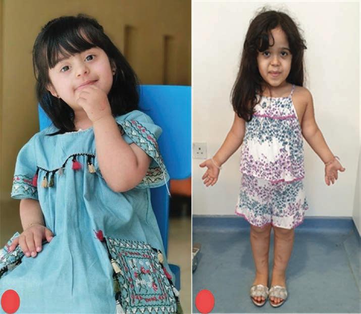
a. Typical features such as in trisomy 21 (flat face with upward and slanted palpebral fissures or epicanthic folds, high-arched palate). Recognized dysmorphic features suggestive of a syndrome or skeletal dysplasia such as achondroplasia (average-sized trunk, short upper arms and thighs, large head with prominent forehead) (Figure 2.1)
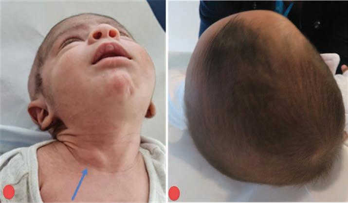
b. Plagiocephaly and congenital torticollis are signs of packaging disorders; check for hip dysplasia (Figure 2.2)
Figure 2.1 A typical facial appearance can give a clue for the underlying diagnosis and problems. (A) shows a child with trisomy 21, and (B) shows a child with achondroplasia.
c. Blue eyes – a parent with blue eyes may clinch the diagnosis of osteogenesis imperfecta (see Chapter 25, Figure 25.11)
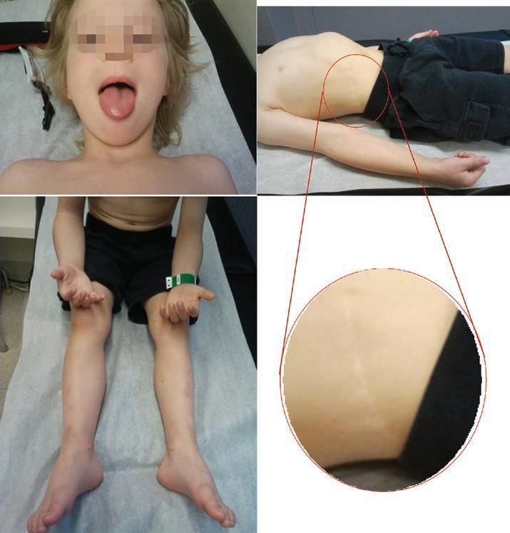
d. Facial asymmetry, large (asymmetric) tongue – may suggest Beckwith–Wiedemann syndrome (Figure 2.3)
e. Observe for wheelchairs, walking aids, orthoses, and shoes
f. Look for skin changes, café-au-lait spots, axillary freckling, and neurofibromas (see Chapter 24, Figure 24.4) – are suggestive of neurofibromatosis; vascular
Figure 2.2 (A) Congenital torticollis with typical sternomastoid tumour (blue arrow – not always present). (B) Positional plagiocephaly. Both signs have been associated with DDH. It is important to differentiate the latter from craniosynostosis, which is a premature closure of one of the skull sutures. In positional plagiocephaly, both fontanelles are open, and posterior and anterior head flattening is parallel and usually gets better quickly over a few weeks. If in doubt, refer to neurosurgery.
Figure 2.3 A child with Beckwith–Wiedemann syndrome. There is hemihypertrophy of the right side of the body, including the face and tongue. There is a right lower abdominal scar from a previous nephrectomy.