Effect of plant extracts against Alzheimer’s disease
Introduction
Pathology
Alzheimer’s disease (AD) is an age-linked disease that more commonly affects the elderly. Usually, AD diagnosis before age 65 is significantly low and found in a small percentage of people (2%–5% of all cases) possessing genetic mutations in corresponding genes (Bekris et al., 2010). Although AD is found in families, with genetic approach on one hand but surpassing the role of genetics on other hand, mounting evidence has shown that the genetic risk factors account for only one-third of brain modifications that occur with age (Small et al., 2000; Cole et al., 2019). The other two-thirds of nongenetic factors probably are based on lifestyle and the environment. The APOE gene on chromosome 19, such as the APOE4 gene, encodes a protein that shows a crucial role in cholesterol metabolism. Nevertheless, in contrast to amyloid precursor protein (APP) and presenilins, this gene merely signifies a risk factor for late-onset AD in 60% of cases (Bird, 2008; Liu et al., 2013; Zhou et al., 2019).
Amyloid beta (Aβ) and tau start aggregating in the brain many years before the diagnosis of clinical symptoms. Age-associated plaques are noticed in brain areas including the hippocampus, amygdala, and neocortex (Kodali et al., 2015; Subramaniam, 2019). The Aβ peptide is a small peptide obtained from the proteolytic breakdown of APP by β-secretase and γ-secretase through the secretory amyloidogenic pathway occurring in several neuronal sections, such as axons, nerve terminals, and dendrites (Yamin et al., 2008; Poddar et al., 2019). These pathological events occur several years before the onset of the characteristic plaques, and the accumulation of Aβ takes place many years before the progression of clinical dementia and can be ante-mortem as shown by PET-amyloid imaging of the brain of AD subjects (Rodrigue et al., 2009; Rowe and Villemagne, 2011; Serrano-Pozo et al., 2011).
Copyright © 2022 Elsevier Inc. All rights reserved.
In addition, studies on postmortem brain tissue have demonstrated that a decrease in the activity of a few enzymes of the tricarboxylic acid cycle is noticed in AD patients (Sorbi et al., 1983; Butterworth and Besnard, 1990; Mastrogiacoma et al., 1996). Bubber et al. (2005) studied a total evaluation of the activity of the complete enzymes of the Krebs cycle in AD brain tissue and corroborated the reduction in the activity of pyruvate dehydrogenase and α-ketoglutarate dehydrogenase complexes, together with a profound decrease in the isocitrate dehydrogenase activity. Conversely, the succinate dehydrogenase and malate dehydrogenase enzymes exhibited enhanced activity. The role of oxidative phosphorylation (OXPHOS) complexes is ambiguous, although the functional irregularities of the Krebs cycle and the respiratory chain certainly stimulate a modification of the energy metabolism and result in an enhanced generation of reactive oxygen species (ROS), thus making the mitochondrial membrane permeable and provoking programmed cell death (Bubber et al., 2005; Atlante et al., 2017). Since the etiopathogenesis of AD is complex, the utilization of incredible natural compounds to treat neurodegeneration in AD is very essential (Mancuso et al., 2012; Rahman et al., 2017; Nasrullah et al., 2017; Uddin et al., 2018a,b, 2019a,b,c).
Plant extracts
Diet
Diet enriched with vegetables and fruits offer several health benefits, according to voluminous epidemiological studies (Bergamini, 2010; Atlante et al., 2020).
Diet also has an ability to induce disease. Healthy eating averts AD, and interestingly, a diet that keeps Alzheimer’s at bay is nearly similar to what makes the heart healthy, lowers cholesterol, prevents cancer, and balances glucose levels (Brown, 2015; Crimmins, 2015). Indeed, green leafy vegetables and fruits present significant benefits to health, but several other foods do substantially improve the human brain. Therefore, it can be concluded that exact dietary suggestions for AD patients are currently gaining ground (Barnard et al., 2014; Cremonini et al., 2019; Amini et al., 2020).
In general, the existence of high oxidizable content like lipid milieu of the myelin membrane of neurons render the brain vulnerable to oxidative injury. Consequently, antioxidant foods play a vital neuroprotective role in neural function (Teleanu et al., 2019; Cenini et al., 2019; Singh et al., 2019). In line with this, multifarious berries exert robust antioxidant
activity due to the occurrence of tannins, anthocyanins, and phenols that substantially enhance the plasticity of the hippocampus, therefore, initiating learning and memory function. Alpha lipoic acid, abundantly available in vegetables like spinach and broccoli, also contributes toward regulating the energy homeostasis of mitochondria and ameliorating cognitive ability. Additionally, green and black tea, both enriched with antioxidants, possess epigallocatechin gallate, which was observed to indirectly mitigate the build-up of amyloid plaques, a pathological characteristic of AD. Eggs are rich in several nutrients such as vitamin B6, vitamin B12, and choline and folic acid, and may contribute to brain health (Subash et al., 2014; Cascella et al., 2017; Colizzi, 2018; Dos Santos et al., 2019; Simunkova et al., 2019; Moretti and Peinkhofer, 2019). Turmeric, a CUR-rich spice that imparts the yellow color to curry, curtails memory impairment induced by AD by impeding the synthesis of amyloid plaques. Red wine enriched with resveratrol (RSV) substantially combats neurodegeneration (Mazzanti and Di Giacomo, 2016; Caruana et al., 2016; Reale et al., 2020).
Apoptosis also plays a pivotal role in AD contributing to the death of huge neuronal populations. In addition, one of the causative factors that could initiate an unusual activation of the self-removal program for apoptosis of complete neuronal regions would alarm neurotrophins. These molecules, which possess the nerve growth factor (NGF) as its progenitor, primarily curtails the neuronal death program; thus, a neuronal population devoid of the basal source of particular neurotrophins suffers enormous apoptosis (Calissano et al., 1998; Ryu et al., 2016; Fricker et al., 2018).
Polyphenols
The polyphenols, including carotenoids and a few bioactive compounds, are categorized under the plant kingdom as substances termed as “functional,” since they induce substantial health benefits (Singh, 2018). They are profoundly focused as useful tools to overcome AD and also to impede disease progression (Singh, 2018). A plethora of compounds playing a role as precursors of other compounds are required in neuronal metabolism and brain health regulation. For example, mono-unsaturated fatty acids (MUFA) and polyunsaturated fatty acids (PUFA, ω-3 and ω-6), enriched in fish and vegetable oils and also the B vitamins, folic acid, vitamins B6, and B12, are substances required for healthy neuronal functioning and for their advantages on cognitive and behavioral efficiency (Rathod et al., 2016; Kennedy, 2016).
Flavonoids
Several lines of evidence have shown that flavonoids protect from AD by intervening with the generation and accumulation of Aβ peptides and/ or reducing the build-up of tau. Flavonoids are capable of eliminating Aβ peptides and attenuate tau phosphorylation by the mTOR/autophagy signaling pathway. In addition, because of their cholinesterase suppressive activity, flavonoids can be substantial anti-AD compounds (Uddin et al., 2020a,b). In numerous diseases such as neurodegenerative disease (ND), diabetes, and cancer, natural compounds serve as a substantial source for a variety of molecular characteristics, biochemical specificity, and massive chemical multiplicity, which renders these natural products suitable for the alteration of several signaling cascades (Rasul et al., 2013; Uddin et al., 2017, 2018a,b, 2020b). Flavonoids are usually available in multifarious vegetables, fruits, and plants (Liu et al., 2014; Uddin and Upaganlawar, 2019). These natural compounds are recognized to have a broad range of pharmacological actions (Uddin et al., 2019b; Uddin and Upaganlawar, 2019) and also act as robust metal chelators, free radical scavengers, and antioxidant agents (Uddin et al., 2019; Zhang et al., 2015; Elbaz et al., 2016; Tysnes and Storstein, 2017). Flavonoids also pacify microglial activation to regulate inflammatory processes in the central nervous system (CNS) (Spencer et al., 2012), harbor robust antiamyloidogenic, antidepressant effects (Nabavi et al., 2015), and ameliorate memory and learning efficacy (Kim et al., 2009). Moreover, these natural compounds demonstrate antiinflammatory (Li et al., 2010; Ashafaq et al., 2012; Ejaz Ahmed et al., 2013), neuroprotective (Prakash and Sudhandiran, 2015; Gomes et al., 2015), antiaging (Lin et al., 2015), and anticholinesterase (Khan et al., 2018) activities.
Polyphenols, primarily flavonoids, are profoundly found as flavanones in citrus fruits (Mecocci et al., 2014) and isoflavones in soy (Francis et al., 2006; Wang et al., 2014), and a few polyphenols like flavan-3-ols (also known as flavanols) exhibit remarkable health benefits (Francis et al., 2006). They are catechin, epicatechin, epigallocatechin, and epigallocatechin gallate available in several vegetable products, such as cocoa, chocolate, black and green tea, and grapes. Mitochondrial impairment and oxidative stress, which contribute to neural membrane damage and memory dysfunction (Uttara et al., 2009; Jacob et al., 2013; Wang et al., 2014; Saharan and Mandal, 2014; Tönniesa and Trushinaa, 2017; Gomes et al., 2018; Wang et al., 2020), are biochemical characteristics observed in AD.
Mitochondria, impaired due to enhanced oxidative stress, enhance ROS generation and Aβ peptides, which in turn initiates oxidative stress both in vitro and in vivo, resulting in neuronal apoptosis and eventually contributing to AD progression (Gomes et al., 2018). In the human body, autophagy eliminates injured cells and facilitates the regeneration of healthy and newer cells (Uddin et al., 2019c; Tanjir Islam et al., 2017). In addition, autophagy also regulates the generation and removal of Aβ (Nilsson and Saido, 2014).With a view toward decreasing synaptic imperfection and neuronal death, elimination of Aβ from the brain is another primary target for anti-AD drugs (Lukiw, 2012).
Genistein
Genistein, an isoflavone primarily available in soy products, substantially combats AD (Mukund et al., 2017). Existence of a plethora of phenolic moieties in its structure makes genistein induce robust antioxidant properties (Sadhukhan et al., 2018). Hence, genistein has antioxidant and neuroprotective efficacy against in vitro AD models (Park et al., 2016). It mitigates oxidative stress by suppressing the production of ROS; it also rescues mitochondria by enhancing the reduced-to-oxidized glutathione ratio and decreasing 8-oxo-20-deoxyguanosine, an indicator of mitochondrial DNA damage (Devi et al., 2017). A study carried out on primary cultured neurons investigated the neuroprotective effect on cells treated with the Aβ fragment 31–35 (Aβ31–35).
Voluminous data has shown that the short fragment Aβ31–35, derived by proteolytic cleavage of Aβ precursor protein (APP), could induce apoptosis in primary cultured neurons (Yu et al., 2009) and PC12 cells (Zhang et al., 2006), serving as an “active center” of the full length Aβ molecule with robust neuron toxicity (Yu et al., 2009; Clementi et al., 2005). Ding et al. (2011) found that genistein treatment in neurons, 2 h before inclusion of the Aβ 31–35 fragment, has been found to protect neurons by enhancing cell viability and decreasing both Ca2 + and ROS production. In addition, the ratio of GSH/GSSG in mitochondria and mitochondrial membrane potential have also been enhanced after genistein treatment. Pierzynowska et al. (2019) proved that an increased dose (i.e., 150 mg/kg/day) of genistein induced activation of autophagy in a streptozotocin-induced rat model of the sporadic AD. Furthermore, at this high dose, it has also been observed that genistein provoked the complete breakdown of Aβ and hyperphosphorylated tau by initiating autophagy.
Icariside
Icariside, an extract of Herba epimedii, is one of the earliest herbal medicines. Aggregates of Aβ contribute to the progression in AD, and the aggregation of these aggregates initiate toxicity in the brain. The primary controlling alpha-secretase in neurons, A disintegrin and metalloproteinase 10 (ADAM10), play a role in breaking down APP. This cleavage produces a neuroprotective APP-obtained petite fragment (C83) and inhibits the generation of Aβ peptides (Endres and Deller, 2017). The mechanistic role of icariside in AD therapy includes enhanced ADAM10 expression and reduced expression of both APP and BACE1, leading to decreased generation of Aβ (Yan et al., 2017; Wuli et al., 2020).
Onjisaponin
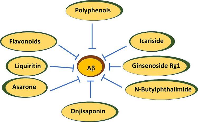
Onjisaponin B (OB) reduces Aβ by cleavage of APP without suppressing the beta-amyloid-cleaving enzyme (BACE1) and γ-secretase activities. In vivo, oral introduction of OB curtails Aβ pathology and behavioral deficits in APP/PS1 mice (Singh et al., 2015; Li et al., 2016a,b). OB can decrease IL-1β, IL-6, TNF-α, and malondialdehyde in the serum and hippocampus of lipopolysaccharide-initiated cognitive-impaired rats. OB induces antioxidant, antiinflammatory, and antiapoptosis protective efficacy in lipopolysaccharide-treated PC12 cells (Li et al., 2018, Fig. 1).
Fig. 1 This image shows the protective efficacy of multifarious plant bioactive compounds against AD.
Asarones
Asarones, the essential ingredients of Acori Tatarinowii Rhizoma, can ameliorate the proliferation and self-restoration of neural progenitor cells. Studies have proved that asarones activated extracellular signal-regulated kinase (ERK), a fragment of a critical kinase cascade for neurogenesis (Mao et al., 2015). Asarones have reduced Aβ accumulation in the cortex and hippocampus of APP/PS1 mice brains (Yang et al., 2016, Fig. 1).
Liquiritin
Liquiritin has been isolated from Glycyrrhiza uralensis. As shown before, oxidative stress is a vital event in the pathogenesis of AD. Aβ peptide is a histopathological hallmark of AD. Aβ can deposit in association with metal ions to catalyze ROS generation (Cheignon et al., 2018). Enhanced oxidative stress instigate pathology. Malondialdehyde (MDA) and 8-hydroxy-20 deoxyguanosine (8-OHdG) are the resultant molecules of lipid peroxidation and are extensively found biomarkers of oxidative stress (Draper and Hadley, 1990). Liquiritin lowers MDA and 8-OHdG in the hippocampus of rats with AD and, by relevant mechanisms, substantially ameliorates spatial learning and memory (Jia et al., 2016).
Tanshinone IIA and cryptotanshinone
Tanshinone IIA (TIIA) and cryptotanshinone (CT) are isolated from Salviae miltiorrhizae as chief lipophilic compounds. In vitro, TIIA and CT markedly mitigated intracellular NO and Ca2 + levels in H9c2 cells (Jin and Li, 2013). The gliosis-linked and neuroinflammatory compounds in the hippocampal tissues showed a significant decrease in the expression of glial fibrillary acidic protein (GFAP), S100β protein, COX-2, inducible nitric oxide synthase (iNOS), and nuclear factor kappa-light-chain-enhancer of activated B cells (NF-kBp65). TIIA and CT exert antiinflammatory and neuroprotective efficacy in a nongenetic mouse model of AD (Maione et al., 2018).
Ginsenoside Rg1
Panax ginseng is the source of Ginsenoside Rg1. It ameliorated hippocampal long-term potentiation and memory in an AD mouse model. It primarily enhances the activity of antioxidant enzymes such as glutathione peroxidase
and superoxide dismutase, curtails activation, and eventually enhances the proliferation of hippocampal neurons (Li et al., 2016a,b; Zhu et al., 2014).
n-Butylidenephthalide
n-Butylidenephthalide (n-BP) is obtained from Angelica sinensis. It is used in the treatment of NDs such as Parkinson’s disease (Fu et al., 2014), amyotrophic lateral sclerosis (Hsueh et al., 2016), and AD (Chang et al., 2015).
n-BP was observed to decrease Aβ, total tau protein and its hyperphosphorylated form, and cellular toxicity in iPSCs-derived neurons stimulated by Down syndrome (Chang et al., 2015).
Green nanotechnology
Plant-based production of NPs is a simple method where a metal salt is mixed with a plant extract and the reaction is carried out in a short time at room temperature. Using this strategy, silver (Ag) and gold (Au) NPs were produced that are robust, corresponding to other metallic NPs (Gour and Jain, 2019). Synthesis of silver nanoparticles (SNPs) with multifarious medicinal plants for pharmaceutical and biological applications has long been in use (Mani et al., 2012). Green-synthesized NPs induce therapeutic applications, e.g., antimicrobial (Rajenran et al., 2015), antioxidant (Mohan et al., 2014), cytotoxic (Shawkey et al., 2013), and antiinflammatory properties (Mani et al., 2015). Nanotechnology has been of significant importance in the diagnosis and treatment of AD currently (Nazem and Mansoori, 2011). Numerous NPs including titanium dioxide, silica dioxide, and silver and zinc oxide showed therapeutic efficacy against neurological diseases (Nazıroğlu et al., 2017), where oxidative NPs can reduce the activities of ROS-scavenging enzymes like glutathione peroxidase (GSH-Px), superoxide dismutase (SOD), and catalase in the brain of rats and mice (Nazıroğlu et al., 2017;Youssif et al., 2019).
Conclusions and future perspectives
Since the discovery of AD by Alois Alzheimer, numerous plant extracts and their bioactive ingredients have been extensively studied for their protective efficacy against it. Plant extracts showed exemplary protection against various pathological events and hallmarks, such as Aβ and tau, through various mechanistic roles. However, low bioavailability and short systemic
circulation impede their success. To overcome these challenges, nanotechnology has been used extensively. More specifically, use of green nanotechnology that comprises only natural compounds play an essential role in AD treatment. Despite the success of numerous plant-based molecules and nanotechnology-based drug delivery systems, intensive further studies are warranted to corroborate these findings.
References
Amini,Y., Saif, N., Greer, C., Hristov, H., Isaacson, R., 2020. The role of nutrition in individualized Alzheimer’s risk reduction. Curr. Nutr. Rep. 9, 55–63.
Ashafaq, M., Raza, S.S., Khan, M.M., Ahmad, A., Javed, H., Ahmad, M.E., et al., 2012. Catechin hydrate ameliorates redox imbalance and limits inflammatory response in focal cerebral ischemia. Neurochem. Res. 37, 1747–1760.
Atlante, A., de Bari, L., Bobba, A., Amadoro, G., 2017. A disease with a sweet tooth: exploring the Warburg effect in Alzheimer’s disease. Biogerontology 18, 301–319.
Atlante, A., Amadoro, G., Bobba, A., Latina,V., 2020. Functional foods: an approach to modulate molecular mechanisms of Alzheimer’s disease. Cell 9, 2347.
Barnard, N.D., Bush, A.I., Ceccarelli, A., Cooper, J., de Jager, C.A., Erickson, K.I., et al., 2014. Dietary and lifestyle guidelines for the prevention of Alzheimer’s disease. Neurobiol. Aging 35, S74–S78.
Bekris, L.M., Yu, C.E., Bird, T.D., Tsuang, D.W., 2010. Genetics of Alzheimer disease. J. Geriatr. Psychiatry Neurol. 23, 213–227.
Bergamini, E., 2010. Nutraceuticals: a valuable aid to be used cautiously. G. Gerontol. 58, 255–258. Bird, T.D., 2008. Genetic aspects of Alzheimer disease. Genet. Med. 10, 231–239.
Brown, G.C., 2015. Living too long: the current focus of medical research on increasing the quantity, rather than the quality, of life is damaging our health and harming the economy. EMBO Rep. 16, 137–141.
Bubber, P., Haroutunian, V., Fisch, G., Blass, J.P., Gibson, G.E., 2005. Mitochondrial abnormalities in Alzheimer brain: mechanistic implications. Ann. Neurol. 57, 695–703.
Butterworth, R.F., Besnard, A.M., 1990. Thiamine-dependent enzyme changes in temporal cortex of patients with Alzheimer’s disease. Metab. Brain Dis. 5, 179–184.
Calissano, P., Ciotti, M.T., Galli, C., Mercanti, D., Dus, L., Canu, N., et al., 1998. The role of IGF-I in cerebellar granule cell survival and terminal differentiation. In: Müller, E.E. (Ed.), IGFs in the Nervous System. 1998. Springer, Berlin, Germany, pp. 60–71.
Caruana, M., Cauchi, R., Vassallo, N., 2016. Putative role of red wine polyphenols against brain pathology in Alzheimer’s and Parkinson’s disease. Front. Nutr. 3, 31.
Cascella, M., Bimonte, S., Muzio, M.R., Schiavone, V., Cuomo, A., 2017. The efficacy of Epigallocatechin-3-gallate (green tea) in the treatment of Alzheimer’s disease: an overview of pre-clinical studies and translational perspectives in clinical practice. Infect. Agent Cancer 12, 36.
Cenini, G., Lloret, A., Cascella, R., 2019. Oxidative stress in neurodegenerative diseases: from a mitochondrial point of view. Oxid. Med. Cell. Longev. 2019, 18.
Chang, C.Y., Chen, S.M., Lu, H.E., Lai, S.M., Lai, P.S., Shen, P.W., Chen, P.Y., Shen, C.I., Harn, H.J., Lin, S.Z., et al., 2015. N-butylidenephthalide attenuates Alzheimer’s disease-like cytopathy in Down syndrome induced pluripotent stem cell-derived neurons. Sci. Rep. 5, 8744.
Cheignon, C., Tomas, M., Bonnefont-Rousselot, D., Faller, P., Hureau, C., Collin, F., 2018. Oxidative stress and the amyloid beta peptide in Alzheimer’s disease. Redox Biol. 14, 450–464.
Clementi, M.E., Marini, S., Coletta, M., Orsini, F., Giardina, B., Misiti, F., 2005. Abeta(31–35) and Abeta(25–35) fragments of amyloid beta-protein induce cellular death through apoptotic signals: role of the redox state of methionine-35. FEBS Lett. 579, 2913–2918.
Cole, J.H., Marioni, R.E., Harris, S.E., Deary, I.J., 2019. Brain age and other bodily ‘ages’: implications for neuropsychiatry. Mol. Psychiatry 24, 266–281.
Colizzi, C., 2018. The protective effects of polyphenols on Alzheimer’s disease: a systematic review. Alzheimers Dement. 5, 184–196.
Cremonini, A.L., Caffa, I., Cea, M., Nencioni, A., Odetti, P., Monacelli, F., 2019. Nutrients in the prevention of Alzheimer’s disease. Oxid. Med. Cell. Longev. 2019, 20. Crimmins, E.M., 2015. Lifespan and healthspan: past, present, and promise. Gerontologist 55, 901–911.
Devi, K.P., Shanmuganathan, B., Manayi, A., Nabavi, S.F., Nabavi, S.M., 2017. Molecular and therapeutic targets of genistein in Alzheimer’s disease. Mol. Neurobiol. 54, 7028–7041. Ding, B.,Yuan, L.,Yu, H., Li, L., Ma, W., Bi, X., et al., 2011. Genistein and folic acid prevent oxidative injury induced by β-amyloid peptide. Basic Clin. Pharmacol. Toxicol. 108, 333–340.
Dos Santos, S.M., Romeiro, C.F.R., Rodrigues, C.A., Cerqueira, A.R.L., Monteiro, M.C., 2019. Mitochondrial dysfunction and alpha-lipoic acid: beneficial or harmful in Alzheimer’s disease? Oxid. Med. Cell. Longev. 2019, 14.
Draper, H.H., Hadley, M., 1990. Malondialdehyde determination as index of lipid peroxidation. Methods Enzymol. 186, 421–431.
Ejaz Ahmed, M., Khan, M.M., Javed, H., Vaibhav, K., Khan, A., Tabassum, R., et al., 2013. Amelioration of cognitive impairment and neurodegeneration by catechin hydrate in rat model of streptozotocin-induced experimental dementia of Alzheimer’s type. Neurochem. Int. 62, 492–501.
Elbaz, A., Carcaillon, L., Kab, S., Moisan, F., 2016. Epidemiology of Parkinson’s disease. Rev. Neurol. 172, 14–26.
Endres, K., Deller, T., 2017. Regulation of alpha-secretase ADAM10 in vitro and in vivo: genetic, epigenetic and protein-based mechanisms. Front. Mol. Neurosci. 10, 56.
Francis, S.T., Head, K., Morris, P.G., Macdonald, I.A., 2006. The effect of flavanol-rich cocoa on the fMRI response to a cognitive task in healthy young people. J. Cardiovasc. Pharmacol. 47 (Suppl. 2), S215–S220.
Fricker, M., Tolkovsky, A.M., Borutaite, V., Coleman, M., Brown, G.C., 2018. Neuronal cell death. Physiol. Rev. 98, 813–880.
Fu, R.H., Harn, H.J., Liu, S.P., Chen, C.S., Chang, W.L., Chen, Y.M., et al., 2014. n-Butylidenephthalide protects against dopaminergic neuron degeneration and alpha-synuclein accumulation in Caenorhabditis elegans models of Parkinson’s disease. PLoS One 9, e85305.
Gomes, A., Pimpão, R.C., Fortalezas, S., Figueira, I., Miguel, C., Aguiar, C., et al., 2015. Chemical characterization and bioactivity of phytochemicals from Iberian endemic Santolina semidentata and strategies for ex situ propagation. Ind. Crop Prod. 74, 505–513.
Gomes, B.A.Q., Silva, J.P.B., Romeiro, C.F.R., Dos Santos, S.M., Rodrigues, C.A., Gonçalves, P.R., et al., 2018. Neuroprotective mechanisms of resveratrol in Alzheimer’s disease: role of SIRT1. Oxid. Med. Cell. Longev. 2018, 15.
Gour, A., Jain, N., 2019. Advances in green synthesis of nanoparticles. Artif. Cells Nanomed. Biotechnol. 47 (1), 844–851.
Hsueh, K.W., Chiou, T.W., Chiang, S.F., Yamashita, T., Abe, K., Borlongan, C.V., et al., 2016. Autophagic down-regulation in motor neurons remarkably prolongs the survival of ALS mice. Neuropharmacology 108, 152–160.
Jacob, K.D., Hooten, N.N., Trzeciak, A.R., Evans, M.K., 2013. Markers of oxidant stress that are clinically relevant in aging and age-related disease. Mech. Ageing Dev. 134, 139–157.
Jia, S.L., Wu, X.L., Li, X.X., Dai, X.L., Gao, Z.L., Lu, Z., et al., 2016. Neuroprotective effects of liquiritin on cognitive deficits induced by soluble amyloid-beta1-42 oligomers injected into the hippocampus. J. Asian Nat. Prod. Res. 18, 1186–1199.
Jin, H.J., Li, C.G., 2013. Tanshinone IIA and cryptotanshinone prevent mitochondrial dysfunction in hypoxia-induced H9c2 cells: association to mitochondrial ROS, intracellular nitric oxide and calcium levels. Evid. Based Complement. Alternat. Med. 2013, 610694. Kennedy, D.O., 2016. B vitamins and the brain: mechanisms, dose and efficacy—a review. Nutrients 8, 68.
Khan, H., Marya, Amin, S., Kamal, M.A., Patel, S., 2018. Flavonoids as acetylcholinesterase inhibitors: current therapeutic standing and future prospects. Biomed. Pharmacother. 101, 860–870.
Kim, D.H., Kim, S., Jeon, S.J., Son, K.H., Lee, S., Yoon, B.H., et al., 2009. Tanshinone I enhances learning and memory, and ameliorates memory impairment in mice via the extracellular signal-regulated kinase signalling pathway. Br. J. Pharmacol. 158, 1131–1142. Kodali, M., Parihar,V.K., Hattiangady, B., Mishra,V., Shuai, B., Shetty, A.K., 2015. Resveratrol prevents age-related memory and mood dysfunction with increased hippocampal neurogenesis and microvasculature and reduced glial activation. Sci. Rep. 28, 8075.
Li, Q., Zhao, H., Zhao, M., Zhang, Z., Li, Y., 2010. Chronic green tea catechins administration prevents oxidative stress-related brain aging in C57BL/6J mice. Brain Res. 1353, 28–35.
Li, X., Cui, J., Yu, Y., Li, W., Hou, Y., Wang, X., et al., 2016a. Traditional Chinese nootropic medicine radix polygalae and its active constituent onjisaponin B reduce beta-amyloid production and improve cognitive impairments. PLoS One 11, e0151147.
Li, F.,Wu, X., Li, J., Niu, Q., 2016b. Ginsenoside Rg1 ameliorates hippocampal long-term potentiation and memory in an Alzheimer’s disease model. Mol. Med. Rep. 13, 4904–4910.
Li, X., Sun, Y., Wei, Y., Zhou, L., Liu, L., Yin, P., et al., 2018. Onjisaponin B (OB) is neuroprotective during cognitive loss through immune-mediated and SIRT1 pathways. Curr. Neurovasc. Res. 15, 94–102.
Lin, L., Ni, B., Lin, H., Zhang, M., Li, X.,Yin, X., et al., 2015. Traditional usages, botany, phytochemistry, pharmacology and toxicology of Polygonum multiflorum Thunb.: a review. J. Ethnopharmacol. 159, 158–183.
Liu, C.C., Kanekiyo, T., Xu, H., Bu, G., 2013. Apolipoprotein E and Alzheimer disease: risk, mechanisms, and therapy. Nat. Rev. Neurol. 9, 106–118.
Liu, C.M., Ma, J.Q., Liu, S.S., Zheng, G.H., Feng, Z.J., Sun, J.M., 2014. Proanthocyanidins improves lead-induced cognitive impairments by blocking endoplasmic reticulum stress and nuclear factor-кB-mediated inflammatory pathways in rats. Food Chem. Toxicol. 72, 295–302.
Lukiw, W.J., 2012. Amyloid beta (Aβ) peptide modulators and other current treatment strategies for Alzheimer’s disease (AD). Expert Opin. Emerg. Drugs. https://doi.org/10.151 7/14728214.2012.672559
Maione, F., Piccolo, M., De Vita, S., Chini, M.G., Cristiano, C., De Caro, C., et al., 2018. Down regulation of pro-inflammatory pathways by tanshinone IIA and cryptotanshinone in a non-genetic mouse model of Alzheimer’s disease. Pharmacol. Res. 129, 482–490.
Mancuso, C., Siciliano, R., Barone, E., Preziosi, P., 2012. Natural substances and Alzheimer’s disease: from preclinical studies to evidence-based medicine. Biochim. Biophys. Acta Mol. basis Dis. 1822, 616–624.
Mani, A., Lakshmi, S., Gopal, V., 2012. Bio-mimetic synthesis of silver nanoparticles and evaluation of its free radical scavenging activity. Int. J. Biol. Pharm. Res. 3 (4), 631–633.
Mani, A., Seethalakshmi, S., Gopal, V., 2015. Evaluation of in vitro anti-inflammatory activity of silver nanoparticles synthesised using piper nigrum extract. J. Nanomedicine Nanotechnol. 6, 2.