No part of this publication may be reproduced or transmitted in any form or by any means, electronic or mechanical, including photocopying, recording, or any information storage and retrieval system, without permission in writing from the publisher. Details on how to seek permission, further information about the Publisher’s permissions policies and our arrangements with organizations such as the Copyright Clearance Center and the Copyright Licensing Agency, can be found at our website: www.elsevier.com/permissions
This book and the individual contributions contained in it are protected under copyright by the Publisher (other than as may be noted herein).
Notice
Knowledge and best practice in this field are constantly changing. As new research and experience broaden our understanding, changes in research methods, professional practices, or medical treatment may become necessary.
Practitioners and researchers must always rely on their own experience and knowledge in evaluating and using any information, methods, compounds or experiments described herein. Because of rapid advances in the medical sciences, in particular, independent verification of diagnoses and drug dosages should be made. To the fullest extent of the law, no responsibility is assumed by Elsevier, authors, editors or contributors for any injury and/or damage to persons or property as a matter of products liability, negligence or otherwise, or from any use or operation of any methods, products, instructions, or ideas contained in the material herein.
Library of Congress Control Number: 2019953760
Content Strategist: Sarah Barth
Content Development Manager: Ellen Wurm-Cutter
Content Development Specialist: Meghan Andress
Publishing Services Manager: Deepthi Unni
Project Manager: Janish Ashwin Paul
Design Direction: Ryan Cook
To all the pediatric transplant recipients, children with cancer, and their families, who are the true inspiration for this textbook and who put their trust in us to help them through their illness. We also dedicate this book to the organ donors and their families, who give the ultimate gift and allow transplantation to be a reality.
To my wife, Dr. Susan Emmett, my children, Amelia and Aidan Steinbach, my parents, Dr. Charles and Katherine Steinbach, and my in-laws, Dr. John and Karen Emmett.
William J. Steinbach, MD
To my children, David, Erin, Molly, and Allison, my granddaughter, Lyla, and my wife, Jenny, who has been my partner and best friend for the past 36 years.
Michael D. Green, MD, MPH
To my parents, Dr. Milton and Lois Michaels, and my mentor, Dr. Ellen Wald, for their continued support and wisdom.
Marian G. Michaels, MD, MPH
To my husband, Ronnen Isakov, my children, Avital and Gavin Isakov, and my parents, Nancy and Nathan Danziger, for their limitless support.
Lara A. Danziger-Isakov, MD, MPH
To my wife, Megan Fisher, for her constant love, continuous support, and limitless encouragement, to my children, Makenzie, Bradan, and Luke, for sacrificing their time with Dad for this effort, and to my parents, Tim and Kathy, for encouraging us to reach our dreams.
Brian T. Fisher, DO, MPH, MSCE
CONTRIBUTORS
Chapter 1
Yeh-Chung Chang, MD, MSCE
Assistant Professor of Pediatrics
Division of Pediatric Infectious Diseases
Duke University School of Medicine Durham, North Carolina
Andrew Barbas, MD
Assistant Professor of Surgery
Division of Abdominal Transplant Surgery
Duke University School of Medicine Durham, North Carolina
Chapter 2
Jasmeen Dara, MD, MS
Assistant Professor of Clinical Pediatrics
Division of Pediatric Blood and Marrow Transplantation
University of California—San Francisco San Francisco, California
Christopher C. Dvorak, MD
Professor of Clinical Pediatrics
Chief, Division of Pediatric Allergy, Immunology, and Blood and Marrow Transplantation
University of California—San Francisco San Francisco, California
Chapter 3
Neil Patel, PharmD, BCOP
Department of Pharmacy
Children’s Hospital of Philadelphia Philadelphia, Pennsylvania
Abby Green, MD
Assistant Professor of Pediatrics
Division of Pediatric Infectious Diseases
Division of Pediatric Hematology/Oncology
St. Louis Children’s Hospital
Washington University in St. Louis St. Louis, Missouri
Director, Pediatric Infectious Diseases Training Program
Division of Pediatric Infectious Diseases
University of Miami Miller School of Medicine
Miami, Florida
Terri Stillwell, MD, MPH
Clinical Assistant Professor
Division of Pediatric Infectious Diseases
University of Michigan Medical School
Ann Arbor, Michigan
Chapter 33
Inci Yildirim, MD, PhD, MSc
Assistant Professor of Pediatrics and Epidemiology
Division of Infectious Diseases
Professor, Department of Epidemiology
Rollins School of Public Health
Emory University
Atlanta, Georgia
Elizabeth Doby Knackstedt, MD
Assistant Professor
Division of Pediatric Infectious Diseases
University of Utah School of Medicine
Medical Director Transplant/ Immunocompromised Infectious Diseases
Primary Children’s Hospital
Salt Lake City, Utah
Chapter 34
Erick F. Mayer Arispe, MD, MSc
Assistant Professor of Pediatrics
State University of New York
Downstate Medical Center and New York City Health + Hospitals/Kings County Hospital
New York, New York
Andi L. Shane, MD, MPH, MSc
Associate Professor of Pediatrics
Interim Chief, Division of Pediatric Infectious Diseases
Marcus Professor of Hospital Epidemiology and Infection Control
Emory University School of Medicine and Children’s Healthcare of Atlanta Atlanta, Georgia
PREFACE
Medicine is changing. The advent of paradigm-shifting advances in both hematopoietic stem cell and solid organ transplantation techniques have allowed a greater number of children than ever before to benefit from these lifesaving procedures. The rapid growth of newer chemotherapeutic classes and enhanced immunomodulation targeting innovative cellular pathways have accelerated cancer care and fostered the rapid overall expanse of the number of immunocompromised patients.
These revolutionary advances in immunosuppression come with a cost. Although the primary concern after transplantation or treatment for a malignancy will likely remain failure/relapse of the underlying indication or rejection of the transplant, severe infections in these immunocompromised patients are now a leading cause of death. The tenuous immunologic balance struck to prevent rejection or halt malignancy is always precariously weighed against the profound level of immunosuppression and increased susceptibility to infection seen in these patients. As medicine continues to push what is possible in new approaches to treatments of disease, this balance will only shift further toward greater concerns for infectious complications and mortality. Furthermore, the presentations of these infectious complications will vary as chemotherapeutic regimens and immunomodulation evolve. This ever-changing field requires transplant and oncology infectious disease physicians to stay at the forefront of knowledge for these therapies so that they can anticipate the infectious presentations that will invariably arise from them.
Pediatric medicine is also changing. Presentations of infectious diseases, diagnostic strategies, and treatment paradigms in adults are not always the same as those in children. Children undergoing transplantation and treatment for cancer often are immunologically naïve to important pathogens associated with infections in these populations and may not be old enough to have received their full complement of protective immunizations by the time that they are receiving care for these conditions. Children are also much more likely than adults to develop community-acquired viral and bacterial infections and are prone to more clinically significant disease. Treatments that are approved for the care of infections in adults may not have been approved or even studied in the pediatric population.
This textbook serves as the first edition dedicated toward the goal of elevating the subfield of pediatric transplant and oncology infectious diseases. The authors of each chapter were deliberately selected from a worldwide cadre of investigators and clinicians actively deciphering the mechanisms of disease and developing the latest approaches to optimal pediatric care.
William J. Steinbach, MD
Michael D. Green, MD, MPH
Marian G. Michaels, MD, MPH
Lara A. Danziger-Isakov, MD, MPH
Brian T. Fisher, DO, MPH, MSCE
Today we recognize the importance of fever in a patient with neutropenia, whether the consequence of cytotoxic chemotherapy for cancer or a transplantation regimen, as the sign of a potentially life-threatening infection that prompts the need for immediate empiric broadspectrum antibiotic therapy. This practice tracks back to the still seminal study that Gerry Bodey and colleagues reported in 1966 associating profound and prolonged neutropenia with the risk of infection.1 During the past several decades, the numbers of patients at risk for fever and neutropenia have continued to increase, first with increasingly intensive combinations of cytotoxic chemotherapy regimens for leukemias and lymphomas and then for the solid tumors. The first bone marrow transplantation was performed in 1968, spawning the field of allogeneic, autologous, and now stem cell–based transplantation regimens. An additional risk group included solid organ transplantations (including kidney transplant and liver, heart, lung, and intestine). These patients also experience neutropenia and joined the ranks of immunocompromised hosts.2 Many of these patients also had other alterations in the cellular and humoral immune system that made them vulnerable to a plethora of viral infections—especially herpesviruses such as HSV, CMV, VZV, EBV and HHV6, as well as respiratory and other infections. Serious infections with opportunistic fungi also emerged as important causes of infection, particularly in patients with prolonged neutropenia.
The treatment of childhood malignancies has improved dramatically over the past several decades, with survival rates approaching 90%.3 That said, episodes of chemotherapy-related fever and neutropenia remain an important complication of these otherwise successful treatment regimens. Despite the overall success of the treatment of childhood cancer, the “holy grail” has always been the hope for more selective and specific cancer treatments that would not result in a compromised immune system and a heightened risk for infection; progress has been made with the development of tyrosine kinase inhibitors as well as other small molecules, monoclonal antibodies, and more recently, an expanding repertoire of immunotherapeutics (including checkpoint inhibitors, CAR-T cells), although some of these also result in a perturbation of the host’s microbiome or other unique risks for infection.4-6
Cytotoxic therapy also results in alterations in humoral and cellular and innate immunity, breaches of the mucosal cutaneous barriers (including those related to IV catheters), and changes in the microbiome, and other changes in the host defense matrix.
Our understanding of the normal and abnormal immune system has become increasingly more sophisticated, aided by knowledge from other compromised hosts, especially those with HIV/AIDS. This was further refined by the elucidation of immune networks, including the delineation of the role of T-helper, suppressor, and regulatory cells, phagocytes, dendritic cells, mast cells and basophils, natural killer cells, and various cytokines and interleukins, interferons, and innate immune function receptors, along with genetically defined alterations that further define the risk for infection.
Nearly 80% of the microorganisms associated with infection in the febrile neutropenic patient arise from the endogenous microbial flora, highlighting the balance between aerobic and anaerobic organisms that presaged the evolving understanding of the microbiome and its role in the risk for infection as well as in modulation of host defenses, including the risk for graft-versus-host disease.7,8 The gut microflora is increasingly recognized as a complex microenvironment, and anaerobes are essential in inhibiting adherence of new aerobes by altering metabolism and nutrient availability and by producing inhibitory
toxins and fatty acids. Studies in germ-free mice also foreshadowed the relationship of the microbial flora with the immune system and prospect of graft-versus-host disease and with the response to various checkpoint inhibitors, awareness that the gut flora and microbiome can be associated with response to immunotherapy. There are also increasing data that the gut microbiome can affect the response to chemotherapy, including stem cell therapy, as well as modulate the immune system.9
Commensal organisms within the lumen of the gut also have profound influences on the immune system at the local level within the gut mucosa, both in draining mesenteric lymph nodes and systemically.10 Some bacterial metabolites can enter the bloodstream directly, further altering the systemic immune system and thus altering granulopoiesis.11 Indeed, dysbiosis in the setting of hematopoietic stem cell transplantation has been associated with differences in long-term survival, whereby individuals with lower diversity in microbiota have shortened survival and higher mortality rates compared with those with higher diversity.
Within this broad context it is also important to be cognizant of the changes in the patterns of infection that have occurred over the past decades. Gram-positive and gram-negative aerobic bacteria continue to play an important role in the infectious complications associated with immunosuppressive therapies, although the predominant organisms have varied over time and can also be institution specific. Although anaerobes remain infrequent causes of primary infection, they can be associated with mixed infections (especially cellulitis and fasciitis and perianal infections), although some, like Clostridium septicum, can also cause serious infections in neutropenic patients, even in the absence of fever. Important nosocomial infections—including C. difficile—occur within hospital settings as a consequence of antibiotics or certain chemotherapy agents that alter the microbial flora in the gastrointestinal tract and that are transmitted as the consequence of poor hand hygiene.
In addition to bacteria, fungi, viruses, and parasites are also causes of infection in immunocompromised hosts. Among the fungi, Candida, Aspergillus, Mucor, Trichosporon, Fusarium, Scedosporium, dematiaceous molds, and others are important, although they are still difficult to diagnose as causes of primary and secondary infections.12-17 Fungal organisms are either endogenous (like Candida) or acquired fungi, like Aspergillus, mucorales, and others.12,13
Viruses are also important causes of infection in immunocompromised patients and have received increased recognition as primary or secondary causes of infection as diagnostic tools have improved. Among these are respiratory viruses, including influenza, parainfluenza, RSV, coronavirus, human metapneumovirus, and rhinovirus. Adenovirus has been a particularly serious cause of infection.18,19 It is also important to note that co-infections with respiratory viruses and invasive fungal infections have been described.20 Also important are the herpesviruses, from herpes simplex to varicella zoster, HSV-6, EBV, and CMV. The latter has been notable in having different presentations in different settings, especially in the early days of allogeneic bone marrow transplantation but also in HIV/AIDS.
Changes in diagnostic tools, from culture and Gram stain to sequencing and molecular diagnosis, along with various potential markers of infection, have been pursued over the years, although reliable predictive tools still require development.
Treatment options have also improved with the availability of new classes of antimicrobials. The principles of empiric, prophylactic, and therapeutic antimicrobial management have also continued to evolve as the result of single- and multiple-institution clinical trials.
The management of infectious complications in cancer and transplant patients requires a broad and deep knowledge of infectious diseases, immunology, chemotherapy, transplantation biology, and more. Thankfully, Bill Steinbach, Mike Green, Marian Michaels, Lara Danziger-Isakov, and Brian Fisher have assembled a comprehensive resource that addresses the rapidly evolving changes in this field in a science-based as well as a practical and accessible resource. Their book, Pediatric Transplant and Oncology Infectious Diseases, is a truly authoritative resource and guide for infectious disease, oncology, and transplantation providers and trainees.
Philip A. Pizzo, MD
David and Susan Heckerman Professor of Pediatrics and of Microbiology and Immunology and Former Dean, Stanford University School of Medicine and Founding Director, Stanford Distinguished Careers Institute Stanford University Palo Alto, California
REFERENCES
1. Bodey GP, Buckley M, Sathe YS, Freireich EJ. Quantitative relationships between circulating leukocytes and infection in patients with acute leukemia. Ann Intern Med. 1966;64(2):328-340.
2. Rubin RH, Schaffner A, Speich R. Introduction to the Immunocompromised Host Society consensus conference on epidemiology, prevention, diagnosis, and management of infections in solid-organ transplant patients. Clin Infect Dis. 2001;33(suppl 1):S1-S4.
3. Hunger SP, Mullighan CG. Acute lymphoblastic leukemia in children. N Engl J Med. 2015;373(16):1541-1552.
4. June CH, Sadelain M. Chimeric antigen receptor therapy. N Engl J Med. 2018;379(1):64-73.
5. Druker BJ, Talpaz M, Resta DJ, et al. Efficacy and safety of a specific inhibitor of the BCR-ABL tyrosine kinase in chronic myeloid leukemia. N Engl J Med. 2001;344(14):1031-1037.
6 Longo DL. Imatinib changed everything. N Engl J Med. 2017;376(10): 982-983.
7. Kramer BS, Pizzo PA, Robichaud KJ, Witesbsky F, Wesley R. Role of serial microbiologic surveillance and clinical evaluation in the management of cancer patients with fever and granulocytopenia. Am J Med. 1982;72(4): 561-568.
8. Schimpff SC, Young VM, Greene WH, Vermeulen GD, Moody MR, Wiernik PH. Origin of infection in acute nonlymphocytic leukemia. Significance of hospital acquisition of potential pathogens. Ann Intern Med. 1972;77(5):707-714.
9. Gopalakrishnan V, Helmink BA, Spencer CN, Reuben A, Wargo JA. The influence of the gut microbiome on cancer, immunity, and cancer immunotherapy. Cancer Cell. 2018;33(4):570-580.
10. Koh AY. The microbiome in hematopoietic stem cell transplant recipients and cancer patients: opportunities for clinical advances that reduce infection. PLoS Pathog. 2017;13(6):e1006342.
11. Salva S, Alvarez S. The role of microbiota and immunobiotics in granulopoiesis of immunocompromised hosts. Front Immunol. 2017;8:507.
12. Arif S, Perfect JR. Emergence of the molds other than Aspergillus in immunocompromised patients. Clin Chest Med. 2017;38(3):555-573.
13. Ascioglu S, Rex JH, de Pauw B, et al. Defining opportunistic invasive fungal infections in immunocompromised patients with cancer and hematopoietic stem cell transplants: an international consensus. Clin Infect Dis. 2002;34(1):7-14.
15. Luplertlop N. Pseudallescheria/Scedosporium complex species: from saprobic to pathogenic fungus. J Mycol Med. 2018;28(2):249-256.
16. Polvi EJ, Li X, O’Meara TR, Leach MD, Cowen LE. Opportunistic yeast pathogens: reservoirs, virulence mechanisms, and therapeutic strategies. Cell Mol Life Sci. 2015;72(12):2261-2287.
17. Walsh TJ, Groll AH. Emerging fungal pathogens: evolving challenges to immunocompromised patients for the twenty-first century. Transpl Infect Dis. 1999;1(4):247-261.
18. Khanal S, Ghimire P, Dhamoon AS. The repertoire of adenovirus in human disease: the innocuous to the deadly. Biomedicines. 2018;6(1). doi:10.3390/biomedicines6010030.
19. Tebruegge M, Curtis N. Adenovirus: an overview for pediatric infectious diseases specialists. Pediatr Infect Dis J. 2012;31(6):626-627.
20. Shah MM, Hsiao EI, Kirsch CM, Gohil A, Narasimhan S, Stevens DA. Invasive pulmonary aspergillosis and influenza co-infection in immunocompetent hosts: case reports and review of the literature. Diagn Microbiol Infect Dis. 2018;91(2):147-152.
SECTION 1 General Immunocompromised Host Infection Principles
1 The Surgical and Immunosuppressive Basis for Infections in the Pediatric Solid Organ Transplant Recipient, 1
Yeh-Chung Chang, MD, MSCE and Andrew Barbas, MD
2 Immunologic Recovery and Basis for Infections in the Pediatric Hematopoietic Stem Cell Transplant Recipient, 10
Jasmeen Dara, MD, MS and Christopher C. Dvorak, MD
3 Cancer and Antineoplastic Therapies and the Risk of Infection in the Pediatric Cancer Patient, 22
Neil Patel, PharmD, BCOP and Abby Green, MD
SECTION 2 Common Immunocompromised Host Infection Situations
4 Infectious Disease Evaluation of Infants and Children Awaiting Solid Organ or Hematopoietic Stem Cell Transplant, 34
Rebecca Pellett Madan, MD, MS and Lara A. Danziger-Isakov, MD, MPH
5 Donor Screening and Donor-Derived Infections, 40
Marian G. Michaels, MD, MPH and Michael D. Green, MD, MPH
6 Prevention of Infections in the Hematopoietic Stem Cell Transplant Recipient, 46
Gabriela M. Marón Alfaro, MD, MS and Hayley A. Gans, MD
7 Prevention of Infections in the Solid Organ Transplantation Recipient, 54
Michele Estabrook, MD and Monica I. Ardura, DO, MSCS
8 Management Principles for Patients With Neutropenia, 56
Brian T. Fisher, DO, MPH, MSCE and Lillian Sung, MD, PhD
9 Vaccination Issues for Transplantation and Chemotherapy, 63
Klara M. Posfay-Barbe, MD, MS and Natasha Halasa, MD, MPH
10 Microbiome Implications in Transplantation and Oncology, 71
Matthew S. Kelly, MD, MPH and Michael A. Silverman, MD, PhD
11 Antimicrobial Stewardship in Immunocompromised Hosts, 78
Joshua Wolf, MBBS, PhD, FRACP, Jeffrey S. Gerber, MD, PhD, and Michael J. Smith, MD, MSCE
12 Hospital Infection Prevention for Pediatric Transplant Recipients and Oncology Patients, 82
Ibukunoluwa C. Akinboyo, MD and Dawn Nolt, MD, MPH
13 Safe Living After Transplantation or Chemotherapy, 90
Blanca E. Gonzalez, MD and Marian G. Michaels, MD, MPH
SECTION 3 Specific Infections in Transplant Recipients and Oncology Patients
14 Multidrug-Resistant Gram-Negative Infections in Transplant and Oncology Patients, 97
Mehreen Arshad, MBBS, Andrew Nowalk, MD, PhD, and Pranita D. Tamma, MD, MHS
15 Bartonella, Legionella, Mycoplasma, and Ureaplasma, 103
Daniel Dulek, MD and Victoria A. Statler, MD, MSc
16 Nontuberculous and Tuberculous Mycobacterium, 109
Flor M. Munoz, MD, MSc and Philana Ling Lin, MD, MSc
17 Cytomegalovirus, 118
Lara A. Danziger-Isakov, MD, MPH and Tanvi Sharma, MD, MPH 18 Epstein-Barr Virus and Posttransplant Lymphoproliferative Disorder, 126
Michael D. Green, MD, MPH, Thomas Gross, Jr., MD, PhD, and Upton D. Allen, MBBS 19 Herpes Simplex and Varicella-Zoster Viruses, 134
William J. Muller, MD, PhD and Betsy C. Herold, MD 20 Human Herpesvirus 6, 7, and 8, 142
Debra J. Lugo, MD and Danielle M. Zerr, MD, MPH 21 Respiratory Viruses, 148
Alpana Waghmare, MD and Janet A. Englund, MD
22 Adenoviruses, 155
Diana F. Florescu, MD and Erica J. Stohs, MD, MPH 23 BK and Other Polyomavirus Associated Diseases in Children, 162
Benjamin L. Laskin, MD, MSc and Hans H. Hirsch, MD, MSc 24 Aspergillosis, 170
William J. Steinbach, MD 25 Mucormycosis, Fusariosis, Scedosporiasis, and Other Invasive Mold Diseases, 181
Rachel L. Wattier, MD, MHS and William J. Steinbach, MD 26 Candidiasis, 195
Jennifer E. Schuster, MD, MSCI and Brian T. Fisher, DO, MPH, MSCE 27 Cryptococcosis and Other Rare Invasive Yeasts Infections, 206
Philip Lee, PharmD and David L. Goldman, MD 28 Histoplasmosis, Blastomycosis, and Coccidioidomycosis, 214
John C. Christenson, MD and Thomas G. Fox, MD 29 Toxoplasma gondii, 227
Sharon F. Chen, MD, MS and Hayley A. Gans, MD 30 Nocardia and Actinomyces, 233
Grant C. Paulsen, MD and Paul K. Sue, MD 31 Pneumocystis Pneumonia, 241
Catherine Burton, MD, MSc, FRCPC and Benjamin Hanisch, MD 32 Strongyloides, Cryptosporidium, and Other Parasitic Infections, 247
Ivan A. Gonzalez, MD, MSc and Terri Stillwell, MD, MPH 33 Gastrointestinal Viruses, 253
Inci Yildirim, MD, PhD, MSc and Elizabeth Doby Knackstedt, MD 34 Clostridioides difficile Infection, 258
Erick F. Mayer Arispe, MD, MSc and Andi L. Shane, MD, MPH, MSc
Index, 263
The Surgical and Immunosuppressive Basis for Infections in the Pediatric Solid Organ Transplant Recipient
Yeh-Chung Chang, MD, MSCE and Andrew Barbas, MD
Balanced immunosuppression is essential to ensure acceptance of a solid organ transplant and an overall successful patient outcome. The fundamental purpose of immunosuppression is to modulate the immune system’s ability to recognize the transplanted organ. However, an overly suppressed immune system increases the risk of certain infections in pediatric solid organ transplant recipients. The goal of balanced immunosuppression is to carefully walk the fine line between too little immunosuppression, which predisposes patients to organ rejection, and too much immunosuppression, which predisposes patients to opportunistic infections.
Although the focus on immunosuppression and its link to infection is warranted, there are other risk factors for infection in pediatric solid organ transplant recipients. Before transplant these may be similar between children and adults. Chronic disease alone is a key risk factor. Potential transplant recipients may undergo multiple rounds of antibiotic treatment for pneumonia, cholangitis, peritonitis, and urinary tract infection, thus increasing their chances of an antibiotic-resistant or opportunistic pathogen. Many potential recipients may also need hospitalization, thus increasing their exposure to multiple types of infections. Transplant candidates are often dependent on the use of central venous catheters, peritoneal dialysis catheters, hemodialysis catheters, ventricular assist devices or extracorporeal membrane oxygenation, all of which increase the risk of systemic invasion by various microorganisms.
Sources of infection after transplant broadly include donor-derived infections, infections acquired perioperatively, reactivation of latent infections, and other infections acquired throughout the patient’s lifetime after transplantation, when there is the added effect of immunosuppressive medications. Postoperatively, poor wound healing is common, and there may be open chests or open abdomens that increase infection risk.
There are also unique issues in pediatric solid organ transplant recipients that contribute to the overall risk of infection. Pediatric recipients are more likely to have malnutrition, which can affect normal immune responses. The actual transplant surgical procedure can involve smaller vascular structures, with higher risk of complications (hematoma, thrombosis). The pediatric solid organ transplant recipient is often naïve to numerous infections, as there is less lifetime exposure to infectious agents. Compounding this is the fact that many
children cannot complete the full primary immunization series before transplant. All of these factors contribute to an underdeveloped protective immunity. The following sections review the important surgical and immunologic risk factors for infection in more detail, with a focus on pediatric considerations when appropriate.
SURGICAL CONSIDERATIONS
Surgical infections in the pediatric solid organ transplant recipient are an important source of morbidity, particularly in the early period after transplantation. Surgical infections are broadly classified as either superficial or deep surgical site infections. The risk and nature of these infections differ by organ type.
Superficial Surgical Site Infections
Superficial surgical site infections refer primarily to wound infections in the skin from incisions made during the transplant procedure. Most commonly, these are caused by gram-positive organisms that colonize the skin. Antimicrobial prophylaxis administered before skin incision has been proven to reduce the incidence of these infections and has been adopted for transplant procedures.1 Treatment of typical superficial surgical site infection includes antibiotic therapy with coverage of gram-positive organisms and local wound care. Local wound care may include exploration of any areas of induration and redness, which may harbor purulent drainage in the subcutaneous space. If such areas are found, treatment consists of reopening the skin and subcutaneous tissue, evacuating the subcutaneous fluid collection, sending any diagnostic samples for microbiologic cultures, and leaving the wound open to heal by secondary intention (granulation from the subcutaneous layer upward). Local wound care thereafter typically includes wet-todry dressing changes or the application of a negative-pressure dressing (wound vacuum-assisted closure).
Necrotizing wound infections represent a rare but severe form of wound infection that must be diagnosed and treated expeditiously, particularly in immunosuppressed individuals. These severe necrotizing infections are commonly polymicrobial, but can also be caused by group A Streptococcus and clostridial organisms. Presentation includes severe pain at the surgical site, high fevers, leukocytosis, and electrolyte abnormalities. These infections are characterized by rapid progression
along soft tissue planes including fascia. Treatment requires intravenous antibiotic therapy and urgent operative debridement of involved tissues, which typically includes skin, subcutaneous tissue, and deeper fascia.2
Deep Surgical Site Infections. Deep surgical site infections occur in body cavities that are exposed during the surgical procedure. Most commonly, these infections are related to the development of fluid collections in these compartments, which are either primarily or secondarily infected. The causes of deep surgical site infections vary by the type of surgical procedure performed and are discussed by organ type. In many cases, catheter-based drainage of these infected fluid collections combined with antimicrobial therapy allows prompt resolution, but in some cases surgical debridement and drainage is required.
Heart transplantation. The most common deep space infection after heart transplantation is mediastinitis, which is characterized by a deep infection of the sternum. The incidence after heart transplantation is 2.5% to 7.5%, and risk factors include younger age (,1 year), epicardial pacing wires, and red blood cell transfusion.3,4 Mediastinitis is typically a monomicrobial infection, with the most common etiology being both methicillin-sensitive Staphylococcus aureus and methicillinresistant S. aureus. Treatment generally requires operative debridement of infected tissues, complex chest closure incorporating soft tissue flaps, and prolonged antimicrobial therapy.
Lung transplantation. After lung transplantation, deep space surgical infections most commonly occur in the pleural space. Fluid and hematoma can accumulate in the pleural space and become secondarily infected, developing into an empyema if the infection progresses. Infected pleural fluid is typically managed with chest tube drainage, but if an empyema develops, surgical debridement and drainage are warranted. The incidence of empyema is approximately 3% to 5% in the lung transplant population and is associated with a significant increase in morbidity and mortality.5,6
Kidney transplantation. Deep space infection after kidney transplantation arises from infected fluid collections in the surgical bed. Kidney grafts can be implanted in either an intraperitoneal or retroperitoneal location, depending on the size of the recipient. In younger/smaller recipients, the graft is typically placed in an intraperitoneal location, using the distal aorta and inferior vena cava as sites for vascular inflow and outflow, respectively. In this setting, fluid collections that arise thereafter are located in the peritoneal cavity. Fluid collections may consist of hematoma, lymphatic fluid, or, less commonly, urine from a urine leak between the transplanted ureter and recipient bladder. Most common among these are hematomas, which can serve as a rich source of nutrient media for microorganisms.
In older/larger pediatric recipients, the kidney graft is usually placed in a retroperitoneal position, using the external iliac artery and vein for inflow and outflow, respectively. The retroperitoneal space is a more confined, limited space and is thus usually easier to manage if fluid collections develop. The same types of fluid collections (hematoma, lymphocele, and urinoma) can arise in this space and are typically well managed with percutaneous catheter drainage.
Liver transplantation. Deep space infections after liver transplantation are common and can arise from multiple sources. The formation of a hematoma in the peritoneal cavity is very common after liver transplantation, owing to the coagulopathy that is common in both the pretransplant and early posttransplant period.
Biliary leakage is a primary source of infected fluid collections after liver transplantation. Owing to the relative scarcity of appropriately sized pediatric donors, many pediatric patients receive partial liver grafts consisting of a portion of an adult donor liver, either from a
living or deceased donor. The most commonly used partial graft consists of the left-lateral section of an adult donor liver. The biliary drainage from this graft is via the left hepatic duct. Bile leaks can occur from the biliary anastomosis between the graft and a roux-en-Y limb of jejunum. More commonly, bile leaks arise from the cut surface of the liver, where the left-lateral section is divided from the remainder of the donor liver. Fortunately, most of these “cut surface” bile leaks are self-limited and well managed with surgical drains left at the time of transplant.
Multivisceral and intestinal transplantation. Infections in multivisceral and intestinal transplantation are common owing to the intensive induction immunosuppression administered and the exposure to enteric organisms related to bowel anastomosis. A multivisceral transplant typically consists of the donor liver, pancreas, and small intestine, retrieved from the donor as a single unit (en bloc). The vascular inflow for the graft is provided by an aortic conduit arising from the recipient infrarenal aorta, and the vascular outflow for the graft is via the inferior vena cava. In an isolated intestinal graft, the donor intestine is supplied by the superior mesenteric artery and the vascular outflow by the superior mesenteric vein, which are anastomosed to the aorta and inferior vena cava of the recipient, respectively.
Deep space infections after multivisceral or intestinal transplants may arise from enteric contamination or leakage at the sites of bowel anastomosis, most commonly involving gram-negative and anaerobic organisms. In general, two separate enteric anastomoses are required: one proximal and one distal. The proximal enteric anastomosis is usually constructed between the recipient stomach/proximal intestine and the graft jejunum. The distal enteric anastomosis is constructed between the graft ileum (or colon, if it is included) and the recipient colon. A diverting ileostomy is typically created to allow endoscopic access for the protocol biopsies necessary to monitor the intestinal graft for rejection.
The other primary sources of deep space infection after multivisceral or intestinal transplantation are infected hematomas that arise in the postoperative setting, similar to the other solid organ transplants discussed previously.
IMMUNOLOGIC OVERVIEW
COMPONENTS OF THE IMMUNE SYSTEM
There is a complex interplay within the diverse components of the immune system that helps protect hosts from infectious threats and foreign substances.7,8 The first main component consists of the members of the innate immune system: neutrophils, macrophages, dendritic cells, natural killer cells, complement, and various signals such as cytokines and Toll-like receptors. The innate immune system provides constant surveillance against external pathogens. The second component consists of the acquired, or adaptive, immune system, including T cells and B cells, which help the immune system fine-tune the elimination of specific threats, and contribute to memory and tolerance. The acquired immune system helps regulate the overall immune response. The focus of this section is on alloactivation of the acquired immune system, specifically T and B cells, and related processes. The immune system is extremely intricate, and other immune mechanisms fall outside the scope of this chapter.
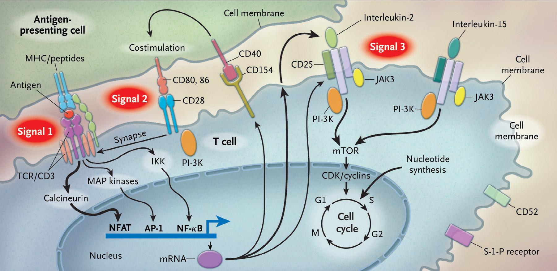
T cells are activated through a complex pathway of signals (Fig. 1.1), and more than one signal is required for full activation.9 The major histocompatibility complex (MHC) on the antigen-presenting cell (APC) brings an antigen that binds to the T-cell receptor, known as signal 1. Additionally, a costimulatory signal, between B7 ligands
Fig. 1.1 This figure demonstrates the required signals for T-cell activation as well as the mechanistic targets for immunosuppressive agents. (From Halloran PF. Immunosuppressive drugs for kidney transplantation.
N Engl J Med. 2004;351:2715-2729.)
(B7-1, or CD80; and B7-2, or CD86) on the APC and CD28 on the T-cell represents signal 2.10 Lastly, an interaction between the cytokine IL-2, and its respective receptor on the T cell is represented by signal 3. The IL-2 receptor is made of three subunits, including alpha (CD25), beta (CD122), and gamma chains.
The normal process by which the host learns to recognize self from nonself includes the use of the MHC. There are two specific types of MHC complexes: class I and class II. Class I MHC is expressed by all nucleated cells and is composed of a polymorphic alpha chain, as defined by human leukocyte antigen (HLA) alleles and a highly conserved monomorphic beta-2 microglobulin chain. Nucleated cells constantly have turnover of their proteins, and the proteasome creates peptides, some of which bind to the MHC complex and are translocated across the cell membrane. The extracellular peptide–MHC is then shown to regulatory CD8 T cells, which are normally able to differentiate peptides that bear the intrinsic signature of the host, versus peptides that would indicate a foreign invader, such as a virus, or a malignant cell. Abnormal cells are then targeted for destruction. HLA alleles associated with the MHC class I complex include A, B, and C. In theory, the rise of polymorphisms in the HLA alleles helps with the immune response to a variety of infections and contributes to fitness on an individual and population level.11
The class II MHC complex is present only on APCs, macrophages, dendritic cells, and B cells. The class II MHC complex is bound to extracellular protein and is presented to CD4 T cells, which help potentiate the response to foreign invaders. HLA alleles most commonly associated with the MHC class II complex include DR, DQ, and DP.
Matching based on HLA alleles has been one of the primary strategies to ensure optimal clinical outcomes. Although a perfect match may not always be feasible because of the limited number of organs available or the shortened time frame for transplant, HLA mismatch can lead to increased risk of rejection and increased use of immunosuppressive drug regimens, which ultimately lead to increased risk for infection.
Other components of the immune system are worth mentioning here as they represent targets of current immunosuppressive therapies. Regulatory T cells are important in suppressing effector T-cell function
through changing the cytokine makeup, competing for the same costimulatory signals, and directing cell-to-cell signals. Cultivating the work of regulatory T cells is necessary in reaching tolerance of the transplanted organ. B cells are also pivotal in their role in both fighting infection and other foreign agents through the secretion of antibodies and facilitation of opsonization. B cells undergo different types of differentiation; a key example is plasma cells that help produce the various immunoglobulin (antibody) types. Immunoglobulins bind to specific foreign antigens and help facilitate phagocytosis and the creation of immune complexes that neutralize pathogens and activate complement. B cells can also function as APCs, in regulatory roles, and as memory cells. They contribute to the development of rejection and are therefore often targets of immunosuppressive regimens.
Lastly, the role of complement cannot be underestimated.12 The classic complement pathway is activated when C1q binds to the Fc portion of IgM or IgG, either in an antibody–antigen complex or on the surface of cells. Other pathways that lead to activation of complement include when the serum protein lectin binds to mannose, present on bacteria or viral-infected cells; and when complement spontaneously binds to cells recognized as foreign. The downstream target is the generation of C3b, which helps facilitate both opsonization (phagocytosis) and the creation of the terminal complement complex, which effectively punches holes in the cell membranes of pathogens and foreign cells.
Evolution of the immune system over time. The immune system of neonates and infants in the first year of life is not well developed. The fetal innate and acquired immune system is regulated in utero to better tolerate maternal antigens that may cross the placenta.13 During the first year of life, although T cells are present, the response skews toward tolerance as the T cells begin to recognize self versus nonself. Although maternal antibodies do provide some immune protection for infants through the first 12 months of life, the weak response of the immune system to external threats leaves neonates and infants at high risk of serious infections. For those in this age group who receive an organ transplant, several considerations have been explored. The benefits of the immature neonatal and infant immune system have led to different strategies. ABO-incompatible liver and heart transplants are now being widely performed in young infants, with comparable
results to ABO-matched transplants.14 Many centers consider lighter immunosuppressive regimens in infants and young children. However, given that immunosuppression is still necessary, when infection occurs in younger patients, it can take longer for acquired immunity mechanisms to recognize foreign invaders and clinical symptoms may be more severe and take longer to resolve. There is also a theoretical risk that the immune system may recognize foreign threats as self during this period, leading to mistaken tolerance.
During the transition from infancy to adulthood, children are constantly bombarded by foreign antigens through inhalation, ingestion, and inoculation. This, in turn, strengthens both innate and acquired immunity. Furthermore, the administration of routine childhood vaccination helps generate robust responses to future potential threats. Children are also exposed to the sharing of diverse antigens in different environments, including day care centers, schools, and at home. During this period children may acquire herpesviruses, such as herpes simplex virus (HSV), cytomegalovirus (CMV), and Epstein-Barr virus (EBV), which stay latent in the host. The adolescent immune system is similar to that of adults and environmental factors continue to play a role in the evolving nature of immune responses in this population.
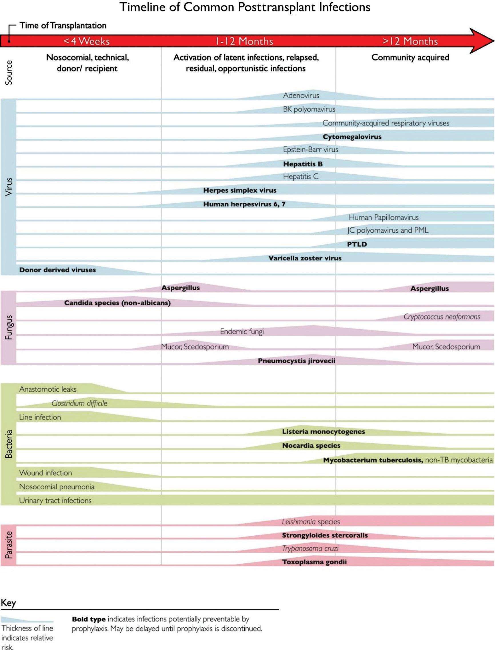
Fig. 1.2 outlines the common time line of posttransplant infections.15 Note that donor-derived, typical bacterial and candidal infections tend to occur earlier (at ,1 month after transplant), whereas most viral and other fungal infections tend to occur later (1 to 12 months after transplant). Patient-specific and regional epidemiologic risk factors are important considerations in the evaluation of every patient. To combat these different threats, most centers have established regimens of antimicrobial prophylaxis against fungal and viral pathogens stratified by specific age group, type of organ transplant, and donor/recipient serostatus.
REJECTION
One of the most common complications after solid organ transplant is organ rejection. There are different time frames of rejection, including hyperacute, acute, and chronic rejection. There are also many different types of organ rejection based on the arms of the immune system: cellmediated and humoral. Both cell-mediated and humoral rejection can occur exclusively or simultaneously.16
Hyperacute Rejection
Hyperacute rejection occurs in vascularized grafts. The mechanism occurs through the action of antidonor antibodies that are already present in the recipient, leading to thrombosis in blood vessels and graft necrosis. These antibodies can occur in pediatric patients if they have been exposed to blood products or have undergone a previous transplant. Hyperacute rejection happens within minutes to hours after the transplant. Current testing methods for these antibodies include conducting flow, complement-dependent cytotoxicity, and virtual crossmatching before transplant.17 In these assays, recipient serum is incubated with donor lymphocytes that carry different HLA antigens, and a positive reaction would indicate that there are donorspecific antibodies. Implementation of these screenings before transplant has led to countermeasures that have significantly decreased the incidence of hyperacute rejection.
Acute Rejection
Acute rejection occurs between 1 week and several months after transplant. Traditionally, this rejection has been separated into two categories: acute cellular rejection and acute humoral rejection. There are cases when one type of rejection is more dominant, but both acute cellular and acute humoral rejection can occur at the same time.
Acute cellular or cell-mediated rejection occurs when recipient T cells recognize donor tissue cells as foreign. As CD81 cytotoxic T cells mature in the host, they begin to recognize foreign antigens in the graft through the class I MHC molecule. The cytotoxic T cells then release perforins and granzyme, which induces apoptosis. Tumor necrosis factor alpha is also secreted by the cytotoxic cells, which then leads to an inflammatory cascade resulting in both upregulated and larger numbers of immune cells at the site of the foreign antigens, leading to injury of the transplanted organ.
Acute humoral rejection is becoming a significant cause of allograft rejection. Some studies have shown that it accounts for 15% to 20% of all rejection during the first year after transplant, especially in sensitized patients.16 In humoral rejection, the role of B cells is important as they produce donor-specific antibodies and other cytokines, which lead to graft injury. Antibodies can be made to HLA class antigens, minor histocompatibility antigens, ABO antigens, and other non-HLA antigens. The complement system is activated and assists in creating further injury, as demonstrated by positive C4d staining in tissue specimens from affected organs.
The Banff criteria were created in 1991 to help diagnose and grade rejection for kidney transplants. For acute cell-mediated rejection, there is an emphasis on pathologic features, including interstitial inflammation, tubulitis, arteritis, and glomerulonephritis. The standardized criteria for diagnosing humoral rejection have focused on morphologic and immunohistochemical staining of tissue, as well as on serologic evidence of donor-specific antibodies. Subsequent revisions of the Banff criteria have incorporated molecular diagnostics into the diagnosis of antibody-mediated rejection.18 There are similar guidelines for the diagnosis of T-cell rejection in liver, heart, and lung transplant patients. However, antibody-mediated rejection and intragraft markers vary according to organ type.
The main approach to the treatment of acute cellular rejection consists of high-dose corticosteroids. If the rejection proves refractory to corticosteroids, biologic agents such as anti-thymocyte globulin (ATG) or alemtuzumab (Campath) are used. Humoral or antibodymediated rejection is difficult to treat. Plasmapheresis, in combination with intravenous immunoglobulin (IVIG), is the first-line treatment and can be followed by other agents, including rituximab, bortezomib, and complement inhibitors such as eculizumab.
Chronic Rejection
Chronic rejection is now one of the leading causes of graft rejection and failure; hyperacute rejection is becoming rarer because of the successful screening of donor-specific antibodies and more sophisticated immunosuppression regimens that have decreased the incidence of acute cellular rejection. A total of 20% of solid organ transplant recipients have graft loss by 5 years, and 50% lose their graft by 10 years after transplant as the result of chronic antibody-mediated rejection.19 Outcomes have improved following the more advanced treatment of acute rejection, but outcomes of chronic rejection have not changed through out the years. This affects pediatric solid organ transplant recipients as these patients have ongoing morbidities, infections, and the adverse effects of immunosuppressive medications. In addition, many pediatric transplant recipients proceed with another transplant as graft dysfunction progresses, which further increases the risk of infection.
The mechanisms of chronic rejection can include either cellular, humoral, or both processes, but is most commonly antibody-mediated. Different rounds of inflammation stimulate the expansion of memory B and T cells, which then begin to develop de novo donorspecific antibodies. Diagnostic descriptions of chronic rejection vary by organ: chronic allograft nephropathy or interstitial fibrosis/tubular atrophy in kidney transplants, cardiac allograft vasculopathy in heart
Fig. 1.2 This figure summarizes the normal timeline of infections in solid organ transplants. Increased thickness of lines denotes higher risk. (From Fishman JA, Avery RK. Chapter 94: Late Infectious Disease After Organ Transplantation in Textbook of Organ Transplantation. 1st edn. New York: John Wiley and Sons; 2014.)
transplants, bronchiolitis obliterans in lung transplants, and obliterative arteriopathy or interstitial fibrosis in liver transplants. It is thought that risk factors for chronic rejection include young age at the time of transplant, frequent infections, trauma, prior episodes of acute rejection, and medication nonadherence.20 Complement is often not involved in this process, and C4d staining is negative in the tissue.21 The long, indolent course of chronic rejection makes timely diagnosis of chronic rejection difficult. Many centers continue to screen transplant patients for donor-specific antibodies, but there is no universally accepted approach. More importantly, better therapies and further research are needed for chronic rejection as it remains difficult to treat.
CURRENT OUTCOMES
There is a fine balance between immunosuppression and infection in pediatric solid organ transplant patients. Overall, graft survival has increased over time as more immunosuppressive agents are now available and regimens are being optimized. There is some variation in clinical outcomes across the different types of organ transplants given distinct practices in immunosuppression. Nevertheless, infections remain a common complication in all pediatric solid organ transplants.
In pediatric kidney transplants, 10-year rates of patient and graft survival have reached 90.5% and 60.2%, respectively. There has been a significant improvement in early outcomes but limited improvement in long-term graft survival. Rates of acute rejection have fallen to 23%, but rates of infection remain at 39.6% within the first 2 years after kidney transplant.22 Patient age younger than 18 years was associated with a higher risk of infection in another study comparing all kidney transplant recipients.23
In pediatric liver transplant recipients, using the latest data from the Studies of Pediatric Liver Transplantation (SPLIT) national consortium, patient survival rates at 3, 12, 24, and 36 months were 90.9%, 86.9%, 84.2% and 83.8%, respectively, whereas graft survival rates at 3, 12, 24, and 36 months were 85.5%, 80.2%, 76.0%, and 75.3%, respectively.24 Rates of rejection at 3, 12, 24, and 36 months were 44.8%, 52.9%, 59.1%, and 60.3%, respectively. In this population, infection accounted for 28.4% of deaths and was a contributing factor in 39% of deaths.24 Bacterial and fungal infections (not further specified) were the major infections causing death in this study.
Data from the International Society for Heart and Lung Transplantation showed that pediatric heart transplant recipients had a median survival of 22.3 years if they received a transplant in the first year of life, 18.4 years for recipients between 1 and 5 years, 14.4 years for recipients between 6 and 10 years, and 13.1 years for recipients between 11 and 17 years.25 Rejection during the first year after heart transplant ranged from 22% to 36%, and 5-year incidence rates of rejection reached 48%.26 Cardiac allograft vasculopathy affected 25% to 34% of patients 10 years after transplant. Infections caused 14% of all deaths in the first year after pediatric heart transplant, with bacterial causes in the early period, transitioning to viral and fungal causes of infection later in the first year after transplant.25
The majority of pediatric lung transplants are performed in children between the ages of 11 to 17 years, and combined heart–lung transplant outcomes are integrated into the lung transplant data. Overall median survival is 5.4 years for children after transplant, but if pediatric lung transplant recipients survive past the first year, overall median survival increases to 8.8 years after transplant.27 Rejection rates at 1 year are 29%, and more than half of patients have bronchiolitis obliterans syndrome by 5 years after transplant. Non-CMV infection was the cause of death of 15.7% of patients at 1 month and 27.6% of patients between 1 and 12 months; this makes non-CMV infection the leading cause of death between 1 and 12 months after transplant in pediatric lung recipients.27
Data on pediatric intestinal transplant patients are limited, but for a population of both pediatric and adult intestinal transplants, 1-, 5-, and 10-year survival rates have been 76%, 56%, and 43%, respectively.28 However, the risk of infection is high in this population, and up to 90% of patients develop a bacterial infection within the first year after transplant, and viral infections such as enteritis, CMV, and EBV infection are extremely common.29
IMMUNOSUPPRESSIVE MEDICATIONS
The next section describes the different classes of immunosuppressive medications (Table 1.1). In general, the approach to immunosuppression
includes an induction regimen around the time of transplant and a maintenance regimen, with the goal of preventing rejection.
Induction Therapies
The purpose of induction therapy is to provide high-dose immunosuppression early in the post-transplant period to prevent acute rejection. There is no single standard approach to induction immunosuppression across different centers. Regimens may also differ according to type or organ and by patient-specific risk factors. Most commonly, biologic agents that deplete or disable T cells are used, and the effect of these agents can be long lasting. In other cases, induction therapy may consist of high-dose steroids alone.
Biologic Agents. The most commonly used agent for induction therapy is ATG. ATG is a polyclonal agent generated in rabbits (rATG or thymoglobulin) or horses (ATGAM). The antibodies are active against various T-cell markers, including CD2, CD3, CD4, CD8, CD11a, CD18, CD44, CD45, HLA-DR, class I heavy chains, and b2-microglobulin. Typically after treatment, T-cell depletion results and effects can last for several weeks to months, greatly increasing the risk of infection. There is also risk of major infusion reactions. Many pediatric centers continue to use ATG for their induction regimens, although many other options are now becoming available.
Another agent that results in T-cell depletion is alemtuzumab (Campath), a monoclonal human antibody to CD52. CD52 is found on all T cells, B cells, and macrophages, dendritic cells, eosinophils, and natural killer cells and alemtuzumab causes profound depletion of these cells.30,31 Effects can be seen up to 1 to 2 years after administration and, similar to ATG, alemtuzumab generates a high and prolonged risk of infection. Use of alemtuzumab shows a trend toward decreasing acute rejection, and reasons to use alemtuzumab include pursuing avoidance or early withdrawal of steroids and reducing calcineurin inhibitor (CNI) use.
The T-cell–depleting biologic agents (ATG and alemtuzumab) all have potential infectious and noninfectious adverse effects. The most common noninfectious adverse effects include early side effects, such as fever, chills, rash, and hypotension. Longer-term effects include the increased risk for infection, as well as potential for malignancy. Intense T-cell depletion significantly increases the risk of viral reactivation, including CMV, EBV, HSV, varicella, and polyomaviruses (such as BK virus), hepatitis B, and hepatitis C. These agents also predispose patients to increased risk of severe and prolonged infection with other viral agents such as respiratory viruses. Lastly, there is also an increased risk of fungal infections, including Candida species, endemic mycoses (Histoplasma capsulatum, Blastomyces dermatitidis, and Coccidioides immitis), and Pneumocystis jirovecii
Basiliximab (Simulect) is a CD25 inhibitor that blocks the costimulatory signal through interleukin (IL)-2. CD25 is expressed only by activated T cells. Thus, use of basiliximab represents a more targeted approach without causing full T-cell depletion. Basiliximab is being used in both pediatric kidney and liver transplant patients. There have been conflicting data regarding its efficacy in pediatric patients, with one small study showing no difference between basiliximab and placebo,32 and another study showing a comparable effect between basiliximab and ATG.33 Basiliximab does not cause as much T-cell depletion as ATG or alemtuzumab and therefore has a more favorable safety profile with decreased infection risk. However, there is still an association with higher risk of herpesvirus infections including CMV and HSV.
Corticosteroids. Corticosteroids have been used in both induction and maintenance therapy and are also heavily used in the treatment of
TABLE 1.1 Summary of Immunosuppressive Medications
rejection. They have both antiinflammatory and immunosuppressive effects. The mechanism of action is mediated through binding to cytoplasmic glucocorticoid receptors, which then translocate to the cell nucleus to affect the transcription of various genes, including the nuclear activating factor family.34 This then leads to decreased production of cytokines, including IL-1, IL-2, interferon gamma, and tumor necrosis factor alpha. Globally, corticosteroids act on the immune system in various ways, including inhibiting lymphocyte proliferation and function and impairing the function of phagocytes. Corticosteroids are thus a powerful weapon and can be used to both prevent and treat acute and chronic rejection.
There are many well-known adverse effects of corticosteroids, including hypertension, weight gain, peptic ulcers, acne, hirsutism, stunting of growth, hyperglycemia, adrenal suppression, muscle breakdown, osteoporosis, behavioral changes, and encephalopathy (posterior reversible encephalopathy syndrome). 35 In addition, long-term use of corticosteroids can predispose transplant recipients to infections, including reactivation of latent viruses and fungal infections, such as Candida and Aspergillus species, endemic mycoses ( Histoplasma , Blastomyces, and Coccidioides ), and Pneumocystis jirovecii . Although these many adverse effects have driven the need to develop corticosteroid-sparing regimens, corticosteroids often remain the mainstay of immunosuppressive regimens after transplant.
Maintenance Therapies
The goal of maintenance immunosuppressive therapy is to prevent both acute and chronic rejection. The effect of maintenance therapy is less pronounced compared with induction therapy, but the effects can be additive over time.
Calcineurin Inhibitors. One of the mainstays of maintenance therapy is the CNIs: tacrolimus and cyclosporine. Tacrolimus, also known as FK506 or fujimycin (Prograf), was discovered in 1987 and is a macrolide that is produced by the fungus Streptomyces tsukubaensis. Cyclosporine, a cyclic undecapeptide, was discovered in 1976 and was extracted from the fungus Tolypocladium inflatum Gams. Tacrolimus binds to intracellular immunophilin proteins called FK-binding proteins, and cyclosporine binds to cyclophilins. These immunophilin-immunosuppressant complexes then bind to calcineurin, inhibiting its enzymatic activity. Normally, calcineurin dephosphorylates and therefore facilitates nuclear transcription of the transcription factor NF-AT, leading to transcription of multiple cytokines, including IL-2. Therefore, CNIs play a large part in inhibiting T-cell activation, although there is evidence to suggest that there is also inhibition of T-cell proliferation and general function.36 The use of CNIs has led to increased length of graft survival.
Although CNIs have become a mainstay of immunosuppressive regimens after transplant, they have important systemic adverse effects. Nephrotoxicity was noted early in the use of cyclosporine and tacrolimus. Both CNIs can cause hypertension independent of their effects on the kidney, and both can also affect the bone marrow, leading to myelosuppression and cytopenias. Tacrolimus has also been implicated in the development of diabetes and tremors, more so than cyclosporine. Cyclosporine is more often associated with gingival hyperplasia and hirsutism. Both peripheral and central nervous systems can be affected during use of CNIs, and symptoms can include headaches, peripheral tremors, and at its worst, seizures, altered mental status, and encephalopathy.
In addition, CNIs are metabolized by the cytochrome P450 system. Natural occurring substances in grapefruit juice and other
medications, including rifampin and azoles, can affect the metabolism of CNIs. Most protocols require frequent monitoring of serum levels of CNIs to ensure that pediatric patients have therapeutic, but not toxic, levels.
Generally, CNIs contribute less to infection risk compared with T-cell–depleting agents (ATG or alemtuzumab). At baseline, there is a small but modest increase in risk of CMV and BK virus infection with CNIs. However, those patients who are receiving tacrolimus maintenance therapy have lower overall incidence rates of CMV infection compared with other maintenance regimens; this may have more to do with lower rates of rejection, and thus, decreased need for corticosteroids and T-cell–depleting agents.37 Lastly, although some CNIs possess some intrinsic in vitro activity against some fungal species, the immunosuppressive properties of these agents are more potent and outweigh their antifungal effectiveness.37
Mammalian Target of Rapamycin Inhibitors. Mammalian target of rapamycin (mTOR) inhibitors include sirolimus (also known as rapamycin or Rapamune), and everolimus. Sirolimus was isolated in 1972 from the fungus Streptomyces hygroscopius. These medications act on the mammalian target of rapamycin or mTOR pathway. mTOR is a serine-threonine kinase and forms complexes mTORC1 and mTORC2, which eventually lead to signals that control protein synthesis, cell cycle progression, cell growth, and proliferation.38 The mTOR inhibitors lead to inhibition of T-cell proliferation and a deadened response to cytokines, such as IL-2.
Major adverse effects include stomatitis, diarrhea, cytopenias, lymphocele, poor wound healing, hypertension, rash, and interstitial lung disease. Like CNIs, mTOR inhibitors are also metabolized by the cytochrome system and drug levels are affected by other medications including azoles and rifampin. Therapeutic drug monitoring is usually advised for this class of immunosuppression.
It is often difficult to isolate the infectious risks of mTOR inhibitors as they are often used with other forms of immunosuppression. There is a trend toward a higher risk of HSV infection. Although some studies show that mTOR inhibitors are associated with a decreased risk of CMV or EBV infection, other studies do not show a significant difference.39
Antimetabolites. Major antimetabolite agents include mycophenolate mofetil (MMF or CellCept) and azathioprine (Imuran). MMF is the prodrug for mycophenolic acid, which inhibits inosine monophosphate dehydrogenase. This affects de novo guanosine synthesis. B and T lymphocytes are more affected than other cells because activated lymphocytes rely on a special isoform of inosine monophosphate dehydrogenase that has an increased affinity for mycophenolic acid. This leads to a cytostatic effect. The most common adverse effects are located in the gastrointestinal tract (up to 40% to 50%), but there can be leukopenia and neutropenia as well.40 Azathioprine is one of the oldest immunosuppressive agents still in use in solid organ transplantation today. It was synthesized in 1957 as a 6-mercaptopurine prodrug. Once activated, azathioprine then terminates DNA synthesis by incorporating itself into actively replicating DNA strands, leading to breakage of the helix. It can also masquerade as inosine monophosphate and inhibit de novo purine synthesis. Major adverse effects of azathioprine include nausea, vomiting, diarrhea, cytopenias, rashes, and hair loss.41 There is also some concern that both MMF and azathioprine increase risk of malignancy (lymphomas, skin cancer).
MMF has been linked to decreased rates of rejection and increased rates of graft survival compared with azathioprine. However, MMF has also been associated with increased rates of CMV, varicella, and BK virus infection. The association with BK virus infection is less clear as MMF is often used in concert with tacrolimus in the kidney transplant
population, and it is difficult to distinguish the role of MMF versus the role of tacrolimus in BK infection.42
Biologic Agents. Belatacept (Nulojix) is composed of a recombinant cytotoxic T-lymphocyte antigen-4 (CTLA-4) linked to a modified Fc portion of human immunglobulin (IG) G1. The CTLA-4 then binds to CD80 and CD86, which prevents the interaction of CD80 and CD86 with CD28 on T cells, thus inhibiting one of the costimulatory signals. Most of the available data for belatacept have been derived from adults. One study showed similar efficacy between belatacept and current CNI regimens in terms of patient and graft survival, and there were improved cardiovascular and metabolic outcomes.43 However, in this study, there was also a higher incidence of posttransplant lymphoproliferative disorder (PTLD), especially in EBV-seronegative patients. Other combinations of belatacept have been used, including in conjunction with alemtuzumab and sirolimus to use both a steroid and CNI-sparing regimen, and results have been comparable to current standard regimens.44 This is a promising medication for adolescents as it can be used only in EBV IgG–positive patients and is associated with increased adherence to CNI regimens. The goal of belatacept use is to try to preserve renal function over time. Additional benefits include reduction of donor-specific antibodies and minimization/avoidance of steroids.
Standard Approach to Maintenance Therapy. Most current maintenance immunosuppressive regimens immediately after transplant include a CNI paired with an antimetabolite. A corticosteroid is often used initially and is tapered off slowly. As the patient becomes further removed from transplant and if there are no episodes of rejection, immunosuppression is slowly decreased over time. There have been some rare reports of achieving tolerance and lifting of all immunosuppressive agents, but currently most pediatric solid organ transplant recipients continue their immunosuppression into adulthood.
Rejection Therapies
Therapies used in rejection include corticosteroids, ATG, alemtuzumab as previous mentioned, as well as rituximab, bortezomib, eculizumab, and plasmapheresis in conjunction with intravenous immunoglobulin.
Biologic Agents. Rituximab (Rituxan) is an anti-CD20 chimeric monoclonal antibody that results in depletion of B lymphocytes. The goal of this therapy is to decrease the production of donor-specific antibodies. The main adverse effects of rituximab include infusion reactions (fevers, chills, hypotension, bronchospasm) and the loss of humoral immunity. Rituximab has been shown to help treat acute antibody-mediated rejection, and PTLD, but data in the treatment of chronic rejection in pediatric patients are lacking.45 A myriad of infections have been seen in transplant recipients who have received rituximab, including a variety of bacterial infections, viral infections (hepatitis B, BK virus), and Pneumocystis pneumonia.
Bortezomib is a proteasome inhibitor that was originally approved for treatment of multiple myeloma and has a targeted effect on B lymphocytes. Initial studies seemed promising, but one recent trial did not show any improvement in late antibody mediated rejection.46 Bortezomib has been associated with increased risk of HSV and varicella infection.
Eculizumab is a human monoclonal antibody against C5, which helps temper the complement cascade. Eculizumab has been used to treat refractory antibody-mediated rejection in both kidney and pediatric liver transplant patients.47,48 It has also been used to prevent
acute rejection in highly sensitized or ABO-incompatible transplants, but there have been a few breakthrough episodes of rejection. Lastly, eculizumab is helpful for treatment of thrombotic microangiopathy or atypical hemolytic uremic syndrome, which can occur after transplant. Given its effect on the complement system, use of eculizumab increases the risk of meningococcal infection. It is highly recommended that patients should be vaccinated with both meningococcal ACWY and meningococcal B vaccines before eculizumab administration. Because of reports of breakthrough meningococcal infection despite administration of all available meningococcal vaccinations, many centers also provide antibacterial prophylaxis (amoxicillin) in addition to vaccination when patients receive eculizumab, although this practice is not standardized.
Other Therapies. Other therapies include plasmapheresis and IVIG. The goal of plasmapheresis, or plasma exchange, is to draw off donorspecific antibodies, immune complexes, and activated complement factors, which helps limit the ongoing inflammatory cascade of injury. Multiple treatments are often required. IVIG provides antiinflammatory and immunosuppressive effects and also provides some humoral protection for the patient.
Standard Approach to Rejection Therapy. Before treatment of rejection, it is best to diagnose the type of rejection, which is best done through tissue biopsy. The standard approach to therapy is to start with high-dose corticosteroids. Other therapies (ATG, alemtuzumab, rituximab, plasmapheresis, IVIG) are added as necessary according to the type and severity of rejection and whether there is a lack of response to corticosteroid monotherapy. The amount of immunosuppression and cumulative effect of rejection therapy should prompt intensified monitoring of latent infections and consideration of antiviral and anti-Pneumocystis prophylaxis.
New Therapies. There have been many other therapies in various stages of development and trials.49 FK778 (manitimus) is a derivative
of a leflunomide metabolite and acts to inhibit de novo pyrimidine synthesis by acting on tyrosine kinase. However, trials have not been convincing enough to move its development forward. Tofacitinib is an oral Janus kinase 3 inhibitor that could prevent acute rejection, but it had a high rate of adverse events, including CMV infection, PTLD, anemia, and neutropenia. FTY720 (fingolimod) is a sphingosine receptor antagonist that traps lymphocytes in lymphoid tissues, not allowing them to exit. Adverse events with FTY720 use included bradycardia, gastrointestinal side effects, macular edema, and increased airway resistance. There are no ongoing plans to move forward with FTY720 in solid organ transplantation at this time. Tocilizumab is an anti–IL-6 monoclonal antibody that has shown promising results in treatment of chronic rejection, decreasing donor-specific antibody levels and stabilizing renal function.50 Further studies are needed for these medications, especially in pediatric solid organ transplant populations.
CONCLUSION
The immune system plays an essential role in protecting human hosts from infection. Once a pediatric patient receives a solid organ transplant, modulation of the immune system is essential in preventing rejection. The field has advanced significantly since the early days of solid organ transplant. Multiple targets of the immune system and T-cell alloactivation have been discovered, resulting in more targeted immunosuppression and improved early outcomes. Yet improvements in chronic rejection have remained elusive and infection continues to affect a high number of pediatric solid organ transplant patients. In addition, long-term immunosuppression can lead to various adverse effects. Further research in the pediatric solid organ transplant population is needed, including newer drugs, protocols, and regimens. The ultimate goal in pediatric transplantation would be to balance the necessity of immunosuppression with the mitigation of adverse effects and infections, which would optimize outcomes in this patient population.
Abstract: Pediatric solid organ transplant changes the lives of thousands of patients every year. Balanced immunosuppression is essential in ensuring acceptance of the organ transplant and successful outcomes. The purpose of immunosuppression is to modulate the immune system’s ability to recognize the transplanted organ, otherwise known as rejection. However, an overly suppressed immune system increases the risk of certain infections in pediatric solid organ transplant patients. The goal of balanced immunosuppression is to walk the line between organ rejection and infection. Risk factors other than immunosuppression also contribute to the risk of infection in pediatric solid organ transplants. They are more likely to have chronic disease and malnutrition, which can affect normal immune responses. Many pediatric patients are also dependent on the use of central lines, peritoneal, or hemodialysis catheters, all of which increase the risk of invasive infections. The actual transplant surgery can be complicated
by working with smaller vascular and other types of structures, and patients often have poor wound healing after surgery. Frequently, the pediatric solid organ transplant patient is also naïve to many different infections, as there is less lifetime exposure to infectious agents. Many children cannot complete the full immunization schedule before transplant. These factors contribute to underdeveloped protective immunity. As children have not had the chance to acquire immunity to a variety of infections, this can elevate the risk of severe infections after transplant. Sources of infection after transplant include donor-derived infections, infections acquired around the time of surgery, reactivation of latent infections, and other infections acquired through the lifetime of patients after transplant.
Keywords: Pediatric solid organ transplant, surgical risks, rejection, immunosuppression