Activate the eBook version of this title at no additional charge.
Elsevier eBooks for Practicing Clinicians gives you the power to browse and search content, view enhanced images, highlight and take notes—both online and offline.
Unlock your eBook today.
1. Visit expertconsult.inkling.com/redeem
2. Scratch box below to reveal your code
3. Type code into “Enter Code” box
4. Click “Redeem”
5. Log in or Sign up
6. Go to “My Library” It’s that easy!
Place Peel Off Sticker Here
For technical assistance: email expertconsult.help@elsevier.com call 1-800-401-9962 (inside the US) call +1-314-447-8300 (outside the US)
PEDIATRIC DERMATOLOGY
PEDIATRIC DERMATOLOGY
FIFTH EDITION
Bernard A. Cohen, MD Professor of Pediatrics and Dermatology
No part of this publication may be reproduced or transmitted in any form or by any means, electronic or mechanical, including photocopying, recording, or any information storage and retrieval system, without permission in writing from the publisher. Details on how to seek permission, further information about the Publisher’s permissions policies and our arrangements with organizations such as the Copyright Clearance Center and the Copyright Licensing Agency, can be found at our website: www.elsevier.com/permissions
This book and the individual contributions contained in it are protected under copyright by the Publisher (other than as may be noted herein).
Notice
Practitioners and researchers must always rely on their own experience and knowledge in evaluating and using any information, methods, compounds or experiments described herein. Because of rapid advances in the medical sciences, in particular, independent verification of diagnoses and drug dosages should be made. To the fullest extent of the law, no responsibility is assumed by Elsevier, authors, editors or contributors for any injury and/or damage to persons or property as a matter of products liability, negligence or otherwise, or from any use or operation of any methods, products, instructions, or ideas contained in the material herein.
Previous editions copyrighted 2013, 2005, 1999, and 1993
Internation Standard Book Number: 978-0-7020-7963-4
Content Strategist: Charlotta Kryhl
Content Development Specialist: Laura Klein
Publishing Services Manager: Shereen Jameel
Project Manager: Aparna Venkatachalam
Design Direction: Brian Salisbury
Printed in India
Since I began taking clinical photographs during my residency training over 30 years ago, I have been impressed by the virtually unlimited variation in the expression of skin disease. However, with careful observation, clinical patterns that permit the development of a reasonable differential diagnosis emerge. In collaboration with my colleagues in the fifth edition, we have been able to use over 600 images, a third of which are new, to demonstrate the diverse variations and common patterns that are fundamental to an understanding of skin eruptions in children. The algorithm at the end of each chapter is designed as a practical approach to evaluating pediatric patients.
Pediatric Dermatology is designed for the pediatric and primary care provider with an interest in dermatology and the dermatology practitioner who cares for children. The text is organized around practical clinical problems. This book should not be considered an encyclopedic text of pediatric dermatology; it should be used in conjunction with the further reading list suggested at the end of Chapter 1. Classic papers and more recent literature are included in the further reading lists at the end of each chapter.
I have been fortunate to work with oral pathologists on the dermatology faculty in the roles of teacher and consultant. With their help, the importance of recognizing oral lesions in the care of children is reflected in Chapter 9, which is devoted to oral pathology.
Although the focus of this chapter is on primary lesions of the oral mucosa, a discussion of clues of systemic disease is included. I am also excited to introduce the new Chapter 10 that focuses on urologic, gynecologic, and anogenital findings in children. Chapter 2, which is devoted to dermatologic disorders of newborns and infants, remains the longest chapter in the book as a result of the continued blossoming of neonatology as a respected pediatric discipline. I never cease to be amazed by how human beings manipulate their skin accidentally, deliberately, secretly, and/or therapeutically. With this in mind, Chapter 11, Psychodermatology, focuses on psychodermatoses and concludes with disorders that are triggered, exacerbated, or caused primarily by external factors.
Finally, the format of the text should be user friendly. The pages and legends have been numbered in a standard textbook fashion, and the index was again revised to include all of the disorders listed in the text and legends. The text and images incorporate advances made in diagnosis, evaluation, and treatment during the last eight years, since the publication of the fourth edition. I only hope that students of pediatric dermatology will enjoy reading the book as much as I enjoyed working with my colleagues in pediatric dermatology completing this new edition.
Bernard A. Cohen 2021
A C K N O W L E D G M E N T S
This book would not have been possible without the help of the children and parents who allowed me to photograph their skin eruptions and the practitioners who referred them to me. I am particularly indebted to the faculty, especially my colleague Annie Grossberg; residents, nurse practitioners; nurses; physicians assistants; and students at the Johns Hopkins Children’s Center and the Departments of Pediatrics and Dermatology at the Johns Hopkins University School of Medicine for their inspiration and support. I would again like to thank my friends at the Children’s Hospital of Pittsburgh where this book was first conceived.
Although we have been involved with online dermatology for over a decade, the craziness associated with the COVID pandemic has allowed for a dramatic expansion of virtual visits and high-quality clinical imaging. Many primary care providers, patients, and parents have learned how to organize online consultation, which will undoubtedly revolutionize the acquisition of data for clinical evaluation and teaching. This is all reflected in the fifth edition.
I am also indebted to the oral pathology faculty who call dermatology their home. They have taught me to seek clues for dermatologic and systemic disease from evaluation of the mucous membranes, and to respect oral pathology in its own right. Without them, the conception of Chapter 9 and the most recent updates would not have been possible.
I am also excited to thank Drs. Tina Ho and Kalyani S. Marathe who encouraged us to include a new chapter focused on urologic and anogenital lesions, which are often misdiagnosed in this age group.
I continue to be grateful for the persistent prodding and sensitive guidance of the editors at Elsevier who are responsible for completion of this book in a timely fashion. I would also like to thank Tracy Shuford for keeping the lines of communication open between the publisher and my office, despite the 6-hour time difference.
I will be forever indebted to the coauthors of the chapters in the fifth edition including Katherine Brown Püttgen for Chapter 2 Neonatal Dermatology, Jessica L. Feig for Chapter 3 Papulosquamous Eruptions, A. Yasmine Kirkorian and Nidhi Shah for Chapter 4 Vesiculopustular Eruptions, Kaiane Anoush Habeshian for Chapter 5 Nodules and Tumors, Daren J. Simkin for Chapter 6 Pigmentary Disorders, George O. Denny for Chapter 7 Reactive Erythema, Saleh Rachidi for Chapter 8 Disorders of the Hair and Nails, Nikhil Shyam for Chapter 9 Oral Cavity, and Sherry Guralnick Cohen for Chapter 11 Psychodermatology.
I would like to thank the residents in dermatology and pediatrics, who by their questions and consultations, have helped me prioritize topics for inclusion in this book.
Finally, I would like to again acknowledge Dr. Nancy Esterly who contributed the foreword to the second edition (reprinted in the subsequent editions). I think of her often and would like to honor her by using her foreword in this edition as well. Dr. Esterly taught me that pediatric dermatology could be exciting and academically challenging. As a role model and one of the mothers of pediatric dermatology, her memory continues to guide all of us in pediatric dermatology. I would also like to acknowledge Dr. Frank Oski who brought me home to Baltimore, where he incorporated pediatric dermatology into the pediatric training program. Hopefully, we can continue to live up to the high standards that he demanded.
FIGURE CREDITS
The following figures have been reprinted from Zitelli BJ, Davis HW (eds). Atlas of pediatric physical diagnosis, 3rd edn. Mosby, St Louis, 1997: 4.10, 7.8, 7.9, 8.1, 8.15, 8.49, 11.7, 11.9, 11.10, 11.13, 11.15.
I am grateful for the use of images contributed by Dr. Russ Corio and Dr. Gary Warnock for contributing additional images to the chapter on the Oral Cavity (Chapter 9).
To Sherry for her continued patience, love, understanding, and contributions to this edition, which took longer than I thought!
To Michael, Jared, and Jennie for keeping me young and laughing. It has been exciting to see them mature into young adults who now contribute to the care of children and adults in their own ways.
To Zeke and Thea who keep me honest!
To all of the children who made this project possible.
F O R E W O R D
NOTE FROM DR. COHEN
I have asked the managing editor to reprint the Foreword from the second edition (also reprinted in subsequent editions) written by Dr. Nancy Esterly to honor her for her contributions to pediatric dermatology, the training of many practitioners of the specialty, and my own career. In the spring of 1983 when I was desperately searching for a mentor in pediatric dermatology, Nan adopted me during my elective month at Children’s Memorial Hospital in Chicago.
Dr. Esterly has been the quintessential practitioner of pediatric dermatology since her pediatric and dermatology training in Baltimore over 40 years ago. She was one of the founders of the Society for Pediatric Dermatology and embodies the tripartite mission of pediatric dermatology of patient care, resident teaching, and clinical research.
FOREWORD TO THE SECOND EDITION
It is not often that one encounters a single-author textbook that is outstanding in both text and illustrations. But, once again, Bernard Cohen has crafted an exceptional basic pediatric dermatology text
liberally illustrated with photographs depicting a wide range of skin problems in infants and children.
In this fourth edition of Pediatric D ermatology, the text has been expanded to include a 20-page chapter devoted entirely to mucosal lesions and accompanied by more than 50 new photographs of patients with problems ranging from the common herpes simplex infection to the uncommon ectodermal dysplasias. In keeping with the very successful style of previous editions, the requisite algorithm, diagrams of the oral cavity and up-to-date references are included in this chapter. In addition, new photographs have been added and some old ones replaced throughout the book.
For beginners in this discipline, Dr. Cohen’s text is an excellent place to start. For those of us who practice pediatric dermatology, there is still much to be learned from a well-put-together text such as this one.
Nancy B. Esterly, MD Professor Emeritus Medical College
of Wisconsin Milwaukee, Wisconsin
Anna M. Bender, MD
Assistant Professor of Dermatology
Department of Dermatology
Weill Cornell Medical College
New York, New York
Bernard A. Cohen, MD
Professor of Pediatrics and Dermatology
Johns Hopkins Children’s Center
Johns Hopkins University School of Medicine Baltimore, Maryland
Skin Care Specialty Physicians Lutherville, Maryland
George O. Denny, MD, MS Physician—Fellow
Dermatology
Johns Hopkins University School of Medicine Baltimore, Maryland
Jessica L. Feig, MD, PhD
Attending Physician
Dermatology
Dermatology Laser and Surgery
New York, New York
Kaiane Anoush Habeshian, MD
Assistant Professor of Dermatology and Pediatrics
Children’s National Hospital
George Washington University School of Medicine and Health Sciences
Washington, District of Columbia
Tina Ho, MD, PhD
Resident Physician Department of Dermatology University of Cincinnati Cincinnati, Ohio
A. Yasmine Kirkorian, MD
Associate Professor of Dermatology and Pediatrics
Children’s National Hospital
George Washington University School of Medicine and Health Sciences
Washington, District of Columbia
Kalyani S. Marathe, MD
Division Director Division of Dermatology
Associate Professor Department of Pediatrics University of Cincinnati Cincinnati, Ohio
John C. Mavropoulos, MD MPH PhD
Resident in Dermatology Department of Dermatology
Johns Hopkins University School of Medicine Baltimore, Maryland
Katherine Brow n Püttgen, MD
Director, Pediatric Dermatology Dermatology
Intermountain Healthcare
Salt Lake City, Utah
Adjunct Associate Professor Dermatology
Johns Hopkins University School of Medicine Baltimore, Maryland
Saleh Rachidi, MD, PhD
Resident Physician Dermatology
Johns Hopkins University School of Medicine Baltimore, Maryland
Nidhi Shah, BS
Medical Student Department of Dermatology
George Washington University School of Medicine and Health Sciences
Washington, District of Columbia
Dr. Nikhil Shyam, MD, FAAD Board-certified Dermatologist Private Practice Wayne, PA
Daren J. Simkin, MD
Assistant Professor Department of Dermatology
Johns Hopkins University School of Medicine Baltimore, Maryland
Preface, v Acknowledgments, vi Dedication, vii Foreword, viii Contributors, ix
1. Introduction to Pediatric Dermatology, 1 Bernard A. Cohen
2. Neonatal Dermatology, 14
Katherine Brown Püttgen and Bernard A. Cohen
3. Papulosquamous Eruptions, 68 Jessica L. Feig and Bernard A. Cohen
4. Vesiculopustular Eruptions, 108
Nidhi Shah and A. Yasmine Kirkorian
5. Nodules and Tumors, 133
Kaiane Anoush Habeshian and Bernard A. Cohen
6. Pigmentary Disorders, 157 Daren J. Simkin, John C. Mavropoulos, and Bernard A. Cohen
7. Reactive Erythema, 180 George O. Denny and Bernard A. Cohen
8. Disorders of the Hair and Nails, 227 Saleh Rachidi, Anna M. Bender, and Bernard A. Cohen
9. Oral Cavity, 260 Nikhil Shyam and Bernard A. Cohen
10. Genital Disorders, 286
Tina Ho and Kalyani S. Marathe
11. Psychodermatology, 296
Sherry Guralnick Cohen and Bernard A. Cohen
Subject Index, 313
Introduction to Pediatric Dermatology
Bernard A. Cohen
ANATOMY AND FUNCTION OF THE SKIN
Most of us think of skin as a simple, durable covering for the skeleton and internal organs. Yet skin is actually a very complex and dynamic organ consisting of many parts and appendages (Fig. 1.1). The outermost layer of the epidermis, the stratum corneum, is an effective barrier to the penetration of irritants, toxins, and organisms, as well as a membrane that holds in body fluids. The remainder of the epidermis, the stratum granulosum, stratum spinosum, and stratum basale, manufactures this protective layer. Melanocytes within the epidermis are important for protection against the harmful effects of ultraviolet light, and the Langerhans cells and other dendritic cells are one of the body’s first lines of immunologic defense and play a key role in systemic and cutaneous diseases such as drug reactions and infections. The dermis, consisting largely of fibroblasts and collagen, is a tough, leathery, mechanical barrier against cuts, bites, and bruises. Its collagenous matrix also provides structural support for a number of cutaneous appendages. Hair, which grows from follicles deep within the dermis, is important for cosmesis, as well as protection from sunlight and particulate matter. Sebaceous glands arise as an outgrowth of the hair follicles. Oil produced by these glands helps to lubricate the skin and contributes to the protective function of the epidermal barrier. The nails are specialized organs of manipulation that also protect sensitive digits. Thermoregulation of the skin is accomplished by eccrine sweat glands and changes in the cutaneous blood flow regulated by glomus cells. The skin also contains specialized receptors for heat, pain, touch, and pressure. Sensory input from these structures helps to protect the skin surface against environmental trauma. Beneath the dermis, in the subcutaneous tissue, fat is stored as a source of energy and also acts as a soft protective cushion.
EXAMINATION AND ASSESSMENT OF THE SKIN
The skin is the largest and most accessible and easily examined organ of the body, and it is often the organ of most frequent concern and quality of life for the patient. Therefore all practitioners should be able to recognize basic skin diseases and dermatologic clues to systemic disease.
Optimal examination of the skin is best achieved in a well-lit room. The clinician should inspect the entire skin surface,
including the hair, nails, scalp, and mucous membranes. This may present particular problems in infants and teenagers, because it may be necessary to examine the skin in small segments to prevent cooling or embarrassment, respectively. Although no special equipment is required, a hand lens and side lighting are useful aids in the assessment of skin texture and small, discrete lesions. In many offices, the otoscope can be adapted for this purpose by removing the plastic speculum.
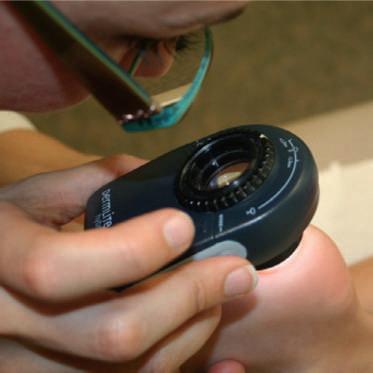
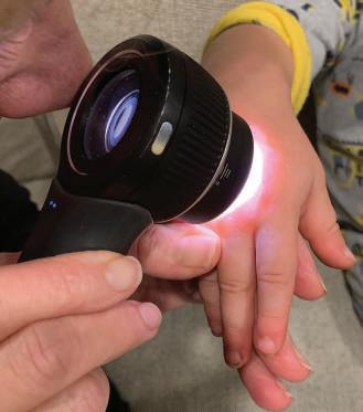
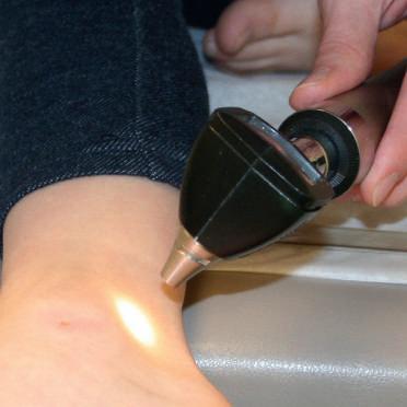
There are also a number of relatively inexpensive portable dermatoscopic devices, which can also be used to enhance the examination (also known as epiluminescence microscopy). These instruments have traditionally provided a magnified (310) view of the skin with a nonpolarized light source, a transparent plate, and a liquid medium between the dermatoscope and the skin. This allows for a view of the superficial structures in the skin without interference from surface reflections. Dermatoscopic heads can be purchased for otoscope/ ophthalmoscope handpieces, and mineral oil or alcohol gel can be applied directly to the skin lesion. Newer devices allow for toggling between nonpolarized and polarized light, which provides visualization of deeper structures in the dermis. (Fig. 1.2).
Despite the myriad of conditions affecting skin, a systematic approach to the evaluation of a rash facilitates and simplifies the process of developing a manageable differential diagnosis. After assessing the general health of a child, the practitioner should obtain a detailed history of the cutaneous symptoms, including the date of onset, inciting factors, evolution of lesions, and presence or absence of pruritus. Recent immunizations, infections, drugs, and allergies may be directly related to new rashes. The family history may suggest a hereditary or contagious process, and the clinician may need to examine other members of the family. A review of nursery records and images provided by parents and in the electronic record will help document the presence and evolution of congenital and acquired lesions.
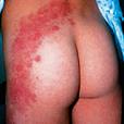
Attention should then turn to the distribution and pattern of the rash. The distribution refers to the location of the skin findings, while the pattern defines a specific anatomic or physiologic arrangement. For example, the distribution of a rash may include the extremities, face, or trunk, while the pattern could be flexural or intertriginous areas (Fig. 1.3a). Other common patterns include sun-exposed sites, acrodermatitis (predilection for the distal extremities), pityriasis rosea (truncal, following the skin cleavage lines), clothing-protected sites, acneiform rashes, lines of Blaschko, and segmental lesions (Fig. 1.3b–h).
Next, the clinician should consider the local organization and configuration of the lesions, defining the relationship of primary and secondary lesions to one another in a given location ( Table 1.1) and the shape of the lesions. Are the lesions diffusely scattered or clustered (herpetiform)? Are they dermatomal, linear, serpiginous, circular, annular, or reticulated?
The depth of the lesions in the skin, as noted by both observation and palpation, may also give further clues ( Table 1.2).
Disruption of the normal skin markings by scale, papules, vesicles, or pustules points to the involvement of the epidermis. Alterations in skin color alone can occur in epidermal and dermal processes. In disorders of pigmentation, the color of the pigment may suggest the anatomic depth of the lesion. Shades of brown are present in flat junctional nevi, lentigines,
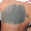
and café-au-lait spots, where the increased pigment resides in the epidermis or superficial dermis. In Mongolian spots and nevus of Ota, the Tyndall effect results in bluish-green to gray macules from melanin uniformly distributed in the middermis. If the epidermal markings are normal but the lesion is elevated, the disorder usually involves the dermis. Dermal lesions have well-demarcated firm borders. Nodules and tumors deep in the dermis or subcutaneous tissue can distort the surface markings, which are otherwise intact. Some deep-seated lesions can only be appreciated by careful palpation.
Lesion color can provide important clues for diagnosis and the pathophysiology of the underlying process ( Table 1.3). Brown, blue, gray, bronze, and black lesions are associated with disorders that alter normal pigmentation, while white lesions
Hair follicle
Epidermis
Sebaceous gland
Eccrine gland
Fat
Dermis
Capillary
Sebaceous gland
Hair shaft/follicle
Pilar smooth muscle
Eccrine sweat gland
Epidermis
Dermis
Subcutaneous fat
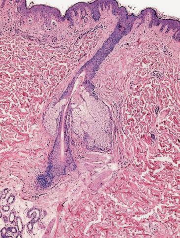
Fig. 1.1 (a) Skin photomicrograph and (b) schematic diagram of normal skin anatomy.
Fig. 1.2 Examination of pigmented nevi with (a) handheld DermLite dermatoscope by 3Gen, (b) DermLite DL4W dermatoscope by 3Gen, and (c) Welch Allyn otoscope.
may be associated with loss of normal pigmentation or the accumulation of scale, crust, or exudates. Red and blue lesions are associated with inflammatory and vascular processes. Nonblanching blue or purple lesions should suggest the presence of purpura. Yellow lesions occur when the skin is infiltrated with inflammatory or tumor cells containing lipid. Other pigments from topical agents (e.g. silver, gold), oral medications (e.g. minocycline, amiodarone), foreign bodies (e.g. asphalt, tattoo
pigments), and infectious agents (e.g. Pseudomonas species, Corynebacterium species) may impart specific colors to cutaneous lesions.
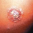
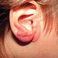
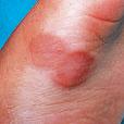
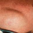
Finally, the clinician may develop a differential diagnosis using the morphology of the cutaneous lesions. Primary lesions (macules, papules, plaques, vesicles, bullae, pustules, wheals, nodules, and tumors) arise de novo in the skin (Fig. 1.4). Secondary lesions (scale, crust, erosions, ulcers, scars with atrophy
TABLE 1.3 Lesion Color
Inflammatory disorders, such as eczema, psoriasis, urticaria, erythema chronicum migrans, and other figurate erythemas
and/or fibrosis, excoriations, and fissures) evolve from primary lesions or result from scratching of primary lesions by the patient (Fig. 1.5).
The practitioner who becomes comfortable with dermatology will integrate all of these approaches into their evaluation of a child with a skin problem. This will be reflected in the clinically focused format of this text.
Each chapter will finish with an algorithm that summarizes the material in a differential diagnostic flow pattern. The limited bibliography includes comprehensive, historically significant, and/or well-organized reviews of the subject. Readers may also find some of the texts and online further reading listed at the end of this chapter useful.
DIAGNOSTIC TECHNIQUES
Potassium Hydroxide Preparation
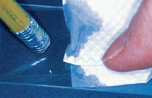
There are a number of rapid, bedside diagnostic procedures in dermatology. One of the most useful techniques is a wet mount of skin scrapings for microscopic examination (Fig. 1.6). Potassium hydroxide (KOH) 20% is used to change the optic properties of skin samples and make scales more transparent. The technique requires practice and patience.
The first step is to obtain the material by scraping loose scales at the margin of a lesion, nail parings, subungual debris, or the small, pearly globules from a molluscum body. Short residual hair stubs (black dots in tinea capitis) may also be painlessly shaved off the scalp with a #15 blade. Scale is placed on the slide and moved to the center with a cover slip. One or two drops of KOH are added and gently warmed with a match or the microscope light. Boiling the specimen will introduce artifacts and should be avoided, so sitting the specimen aside for 5 min is an alternative to gentle heating. Excess KOH can be removed with a paper towel applied to the edge of the cover slip. Thick specimens may be more easily viewed after gentle but firm pressure is applied to the cover slip with a pencil eraser. Thick scale will also dissolve after being set aside for 15–20 min.
View the preparation under a microscope, with the condenser and light at low levels to maximize contrast, and with the
Tattoo, vascular malformation, hemangiomas, blue nevus, Mongolian spot
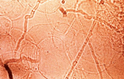
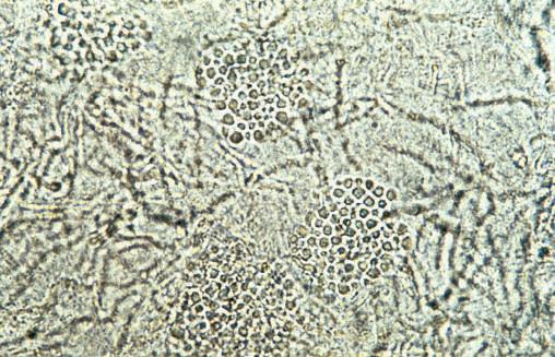
objective at 310. Focus up and down as the entire slide is rapidly scanned. True hyphae are long branching green hyaline rods of uniform width that cross the borders of epidermal cells. They often contain septae. False positives may be vegetative fibers, cell borders, or other artifacts. Yeast infections show budding yeast and pseudohyphae. Molluscum bodies are oval discs that have homogeneous cytoplasm and are slightly larger than keratinocytes. In hair fragments, the fungi appear as small, round spores packed within or surrounding the hair shaft (see Fig. 8.19e in Chapter 8). Hyphae are only rarely seen on the hair.
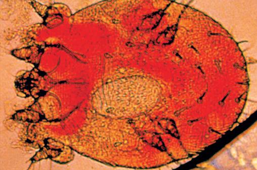
Scabies Preparation
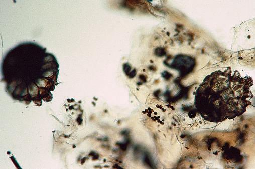
A skin scraping showing a mite, its egg, or feces is necessary to diagnose infestation with Acarus scabiei because many other skin rashes resemble scabies clinically (Fig. 1.7). The most important factor for obtaining a successful scraping is the choice of site. Burrows and papules, which are most likely to harbor the mite, are commonly located on the wrists, fingers, and elbows. In infants, primary lesions may also be found on the trunk, palms, and soles. A fresh burrow can be identified as a 5–10 mm elongated papule, with a vesicle or pustule at one end. A small, dark spot resembling a fleck of pepper may be seen in the vesicle. This spot is the mite, and it can be lifted out of its burrow with a needle or the point of a scalpel. Usually, it is best to hold the skin taut between the thumb and index finger while vigorously scraping the burrow. Although this may induce a small amount of bleeding, if performed with multiple, short, rapid strokes, it is usually painless. A drop of mineral oil should be applied to the skin before scraping to ensure adherence of the scrapings to the blade. The scrapings are then placed on the slide, another drop of mineral oil is added, and a cover slip is applied. Gentle pressure with a pencil eraser may be used to flatten thick specimens.
Mites are eight-legged arachnids easily identified with the scanning power of the microscope. Care must be taken to focus through thick areas of skin scrapings so as not to miss any camouflaged mites. The presence of eggs (smooth ovals, approximately one half the size of an adult mite) or feces (brown pellets, often seen in clusters) are also diagnostic. If eggs or feces
Red Purple Brown Gray Blue Bronze Green Yellow
Macule/patch
Fig. 1.4 Primary skin lesions. Macule: a small (usually 1 cm), flat lesion showing an alteration in color or tone. Large macule is a patch. Papule: a small (1 cm), sharply circumscribed, elevated lesion. An elevated lesion over 1 cm is referred to as a plaque. Nodule: a soft or solid mass in the dermis or subcutaneous fat. Tumor: a large nodule, localized and palpable, of varied size and consistency. Vesicle: a blister containing transparent fluid. Bulla: a large blister. Wheal: an evanescent, edematous, circumscribed, elevated lesion that appears and disappears quickly. (Adapted from CIBA.)
are found first, perusal of the entire slide usually reveals the adult mite.
The dermatoscope can also be used to visualize the female mite whose mouth parts appear as an elongated triangleshaped spot referred to as a delta sign.
Fig. 1.5 Secondary skin lesions. Scale: dry and/or greasy fragments of adherent epidermis. Pustule: a sharply circumscribed lesion containing free pus. Crust: a dry mass of exudate from erosions or ruptured vesicles/pustules, consisting of serum, dried blood, scales, and pus. Erosion: well-defined partial-thickness loss of epidermis. Ulcer: a clearly defined, full-thickness loss of epidermis that may extend into the subcutis. Scar: a permanent skin change resulting from new formation of connective tissue after destruction of the epidermis and cutis. When the loss of dermis and/or fat is prominent, the lesion may be atrophic. Fibrosis may result in firm, thickened papules or plaques. Excoriation: any scratch mark on the surface of the skin. Fissure: any linear crack in the skin, usually accompanied by inflammation and pain. (Adapted from CIBA.)
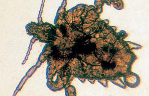
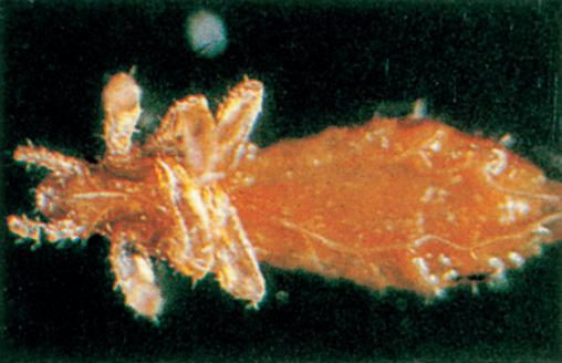
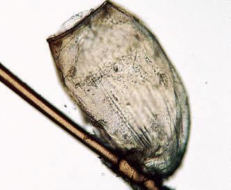
Lice Preparation
Lice are six-legged insects visible to the unaided eye that are commonly found on the scalp (Fig. 1.8), eyelashes, and pubic areas. Pubic lice are short and broad, with claws spaced far apart for grasping the sparse hairs on the trunk, pubic area, and
Papule/plaque
Tumor
Nodule
Vesicle
Bulla
Wheal
Scale
Pustule
Ulcer
Erosion
Fibrotic scar
Atrophic scar
Excoriation Fissure
Fig. 1.6 Potassium hydroxide (KOH) preparation. (a) Small scales are scraped from the edge of the lesion onto a microscopic slide. (b) The scales are crushed to form a thin layer of cells in order to visualize the fungus easily. (c) In this positive KOH preparation of skin scrapings, fungal hyphae are seen as long septate, branching rods at the margins and center of the scales. (d) Pseudohyphae and spores typical of tinea versicolor give the appearance of spaghetti and meatballs.
Fig. 1.7 (a) Microscopic appearance of the adult scabies mite. Note the small oval egg within the body. (b) Scraping from an adolescent with crusted scabies shows two mites and multiple fecal pellets.
eyelashes, whereas scalp lice are long and thin, with claws closer together to grasp the denser hairs found on the head. The lice are best identified close to the skin, where their eggs are more numerous and more obvious. Diagnosis can be made by identifying the louse, or by plucking hairs and confirming the presence of its eggs or “nits” by microscopic examination.
A dermatoscope or magnifying glass can also be used for confirmation of lice infestation.
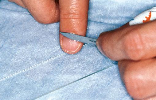
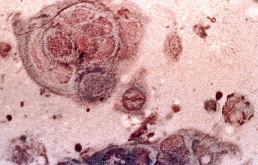
Tzanck Smear
The Tzanck smear is an important diagnostic tool in the evaluation of blistering diseases. It is most commonly used to
distinguish viral diseases, such as herpes simplex, varicella, and herpes zoster, from nonviral disorders (Fig. 1.9). It is important to note that Tzanck smears from vesicles of vaccinia and smallpox do not demonstrate multinucleated giant cells. The smear is obtained by removing the “roof” of the blister with a curved scalpel blade or scissors, and scraping the base to obtain the moist, cloudy debris. The material is then spread onto a glass slide, air dried, and stained with Giemsa or Wright stain. The diagnostic finding of viral blisters is the multinucleated giant cell. The giant cell is a syncytium of epidermal cells,
1.9 Tzanck Smear. Note the multinucleated giant cells characteristic of viral infection with herpes simplex and varicella/zoster.
with multiple overlapping nuclei; it is much larger than other inflammatory cells. A giant cell may be mistaken for multiple epidermal cells piled on top of each other. If a microscope and stain are available, the Tzanck smear can be used for rapid confirmation of the clinical suspicion of infection, while more sensitive studies like polymerase chain reaction are pending.
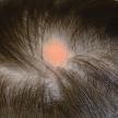
Wood Light
Wood light is an ultraviolet source that emits at a wavelength of 365 nm. Formerly, its most common use was in screening patients with alopecia for tinea capitis, as the most common causative organisms, Microsporum audouinii and other Microsporum species, were easily identified by blue-green fluorescence under Wood light. However, today in North America, Trichophyton species are the most common fungi associated with tinea capitis, but it does not fluoresce. In the United States, fewer than 10% of cases are caused by M. canis and other Microsporum species. In Europe, Africa, and Asia, organisms that cause ectothrix scalp infection and which fluoresce include M. ferrugineum, M. audouinii, and M. canis.
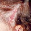
Wood light is still of value in diagnosing a number of other diseases. Erythrasma is a superficial bacterial infection of moist skin in the groin, axilla, and toe webs. It appears as a brown or red flat plaque and is caused by a Corynebacterium that excretes a pigment that contains a porphyrin. This pigment fluoresces coral red or pink under Wood light. Tinea versicolor, a superficial fungal infection with hypopigmented macules and plaques on the trunk, also fluoresces under Wood light with a greenyellow color. Pseudomonas infection of the toe web space and colonization of the skin in burn patients will fluoresce yellowgreen. Patients with porphyria cutanea tarda excrete uroporphyrins in their urine, and examination of a urine specimen will show an orange-yellow fluorescence. Adequate blood levels of tetracycline produce yellow fluorescence in the opening of hair follicles, while lack of fluorescence indicates poor intestinal absorption or poor patient compliance. Positive fluorescence in the skin is diagnostically useful, but many of the pigments that fluoresce are water soluble and readily removed by swimming or bathing.
Fig. 1.8 Microscopic appearance of lice. (a) The crab louse has a short, broad body, with claws spaced far apart. (b) The head louse has a long, thin body, with claws closer together. (c) A hatched nit is tightly cemented to the hair shaft.
Fig.
Wood light also emits purple light in the visible spectrum. This wavelength can be used to accentuate subtle changes in pigmentation. The purple light is absorbed by melanin in the epidermis, and variably reflected by patches of hypopigmentation and depigmentation. It can be helpful to distinguish increased pigmentation in the epidermis that will be enhanced by the purple light from increased pigmentation in the dermis that does not enhance. Purple light may be particularly useful in evaluating light-pigmented individuals with vitiligo or ash leaf macules (congenital hypopigmented macules).
DERMATOLOGIC THERAPEUTICS
General Principles
Single component generic preparations are often effective and inexpensive. Fixed multiple component preparations are occasionally useful and may increase adherence to the treatment regimen, but the practitioner must be aware of all the constituent agents and the increased risk of adverse drug reactions. Specially formulated medications are often prohibitively expensive and seldom indicated in general practice. Fortunately, there are now some special formulating pharmacies that follow Food and Drug Administration guidelines, prepare safe and cost effective products, and obtain insurance preauthorization. The practitioner must calculate the quantity of medication required for the patient to comply with instructions. In a child, 15–30 g of an ointment is needed to cover the entire skin surface once. This quantity will vary with the vehicle used and the experience of the individual applying the preparation.
Topical Vehicles
Two variables are particularly important in the selection of effective topical therapy: the active medication and the vehicle. No matter how effective the active medication, adherence to the recommended regimen will require that the clinician consider the selection of a vehicle carefully. In infants and young children, ointments tend to be better tolerated than other vehicles, while in older children, adolescents, and young adults, more elegant vehicles (e.g. creams, foams, sprays, solutions) that are free of lingering odors, color residues, or tackiness, will encourage adherence.
Ointments
In general, ointments are occlusive and allow for high transcutaneous penetration of the active drug. Ointments are stable for long periods and require few preservatives and bacteriostatic additives. As a consequence, they are least likely to cause contact allergy or irritation. These vehicles are well tolerated when the skin is cracked or fissured, particularly in young children with chronic skin disease (e.g. atopic dermatitis, psoriasis). Unfortunately, ointments tend to be messy and may stain clothing, so they are not often welcome by older children and adolescents.
Open Wet Dressings
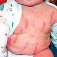
Open wet dressings, using tap water or normal saline, provide symptomatic relief by cooling and drying acute inflammatory
lesions. They cleanse the skin by loosening exudates and crusts that can be painlessly removed before the dressing dries. Various astringents and antiseptics, such as vinegar or 5% aluminum acetate solution (e.g. Burrow solution), may be added to compression solutions in a 1:20–40 dilution. Bleach baths and chlorine swimming pools can also be used as gentle antiseptics and anti-inflammatory agents in patients prone to chronic or recurrent infection such as atopic dermatitis and epidermolysis bullosa. The concentration of chlorine in bleach baths (1/4 cup of household bleach or 59 mL to a 40-gallon bathtub to give a concentration of sodium hypochlorite of 6–8.25%) is designed to reproduce the concentration of chlorine recommended for supervised swimming pools.
Powders and Lotions
Powders promote drying and are especially useful in the intertriginous areas. Lotions are powders suspended in water (e.g. calamine lotion). When these preparations dry, they cool the skin and provide a uniform covering of the suspended agent. The clinician should warn patients and parents against the use of combination products that might result in irritation or percutaneous absorption of the active ingredients (e.g. calamine and diphenhydramine).
Gels
Gels are aqueous preparations that liquefy on contact with the skin and leave a uniform film on drying. Gels are well tolerated in hair-bearing areas. Water-based gels are best tolerated by children, while alcohol-containing products are more likely to cause burning or irritation. Gels are well tolerated in hair-bearing areas.
Aerosols
Aerosols and sprays act in a manner similar to lotions and gels. Active ingredients are incorporated into an aqueous phase. A convenient delivery system usually allows for easy dispersion over the skin surface. Aerosols are also particularly useful on the scalp.
Creams
Traditional creams are suspensions of oil in water. As the proportion of oil increases, the preparation approaches the consistency of an ointment, which is the most lubricating vehicle. Creams are water washable and hygroscopic. They may be drying and occasionally sensitizing.
Pastes
Pastes, which are mixtures of powder in ointment, are messy and may be difficult to remove from the skin. They are used to protect areas prone to irritation, such as the diaper area. Pastes can be removed with mineral oil.
Foams
Foams represent a novel vehicle, which enhances percutaneous absorption of medication in a cosmetically acceptable elegant
preparation. Foams remain stable until applied to the skin, where warming from natural body heat results in volatilization of inert contents with deposition of the active medication on the skin surface. Because foams contain minimal solid ingredients, there is little residue, making them particularly attractive vehicles for products designed for the scalp and intertriginous areas. A number of topical steroid foams have been approved for the treatment of atopic dermatitis and psoriasis, while other agents have been approved for seborrheic dermatitis and ichthyosis.
Shampoos and Washes
Short contact therapy with medicated shampoos and washes may also enhance adherence, particularly in adolescents with busy schedules and little time for topical therapy. These formulations contain insoluble particulate drugs such as benzoyl peroxide, salicylic acid, corticosteroids, and antifungal agents, some of which remain after showering or washing. Shampoos
TABLE 1.4 Topical Corticosteroids
Generic Name
Super high-potency topical steroids—class 1
High-potency topical steroids—class 2
Betamethasone dipropionate augmented 0.05%
Clobetasol propionate 0.05%
Fluocinonide 0.1%
Halobetasol propionate 0.05%
Amcinonide 0.1%
Desoximetasone 0.25%, 0.05%
Diflorasone diacetate 0.05%
Halcinonide 0.1%
Fluocinonide 0.05%
Mometasone furoate 0.1%
Topical steroids—class 3 Amcinonide 0.1%
Betamethasone dipropionate 0.05%
Betamethasone valerate 0.1%
Clobetasone butyrate 0.05%
Fluocinonide 0.05%
Fluticasone propionate 0.05%
Topical steroids—class 4
Betamethasone valerate 0.1%
Clocortolone pivalate 0.1%
Desoximetasone 0.05%
Fluocinolone acetonide 0.025
Flurandrenolide 0.05%
Hydrocortisone probutate 0.1%
Hydrocortisone valerate 0.2%
Prednicarbate 0.1%
Triamcinolone acetonide 0.1%, 0.025%
and washes may also be particularly useful when longer periods of contact are likely to result in burning or irritation.
Topical Corticosteroids
Topical steroids are available in every type of vehicle. A good approach is to become familiar with one or two products in each of the potency ranges ( Table 1.4). A check of local pharmacies is useful in determining the availability and cost of medications.
Most childhood skin eruptions requiring topical steroids can be readily managed with twice-daily applications of lowor medium-potency preparations. Moreover, a number of studies have shown that twice-daily applications of midpotency agents can be applied to most areas of the skin in children for long periods of time safely. With few exceptions, low-potency medications should be used on the face and intertriginous areas because more potent preparations may produce atrophy, telangiectasias, and hypopigmentation. Regardless of
Diprolene ointment 0.05%
Clobex lotion, spray, shampoo 0.05%
Olux E foam 0.05%, Olux foam 0.05%
Temovate cream, ointment, solution 0.05%
Vanos cream 0.01%
Ultravate cream, ointment 0.05%
Topicort cream, ointment 0.25%, gel 0.05%
ApexiCon E cream 0.05%
Maxiflor ointment 0.05%
Halog, Halog E ointment, cream 0.1%
Lidex cream, gel, ointment 0.05%
Elocon ointment 0.1%
Cyclocort cream, lotion 0.01%
Diprosone cream 0.05%
Valisone ointment 0.1%
Betacap 0.1% (UK)
Eumovate ointment, cream 0.05% (UK)
Lidex ointment, cream, gel 0.05%
Cutivate 0.05%, cream, lotion
Luxiq foam 0.1% ointment, cream, lotion
Cloderm cream 0.1%
Topicort LP cream 0.05%
Synalar ointment 0.025%
Cordran ointment, lotion 0.05%
Pandel cream 0.1%
Westcort ointment 0.2%
Dermatop ointment 0.1%
Kenalog ointment 0.1%, 0.025%
TABLE 1.4 Topical Corticosteroids—cont’d
Topical steroids— class 5
Topical steroids—class 6
Generic Name
Betamethasone dipropionate 0.05%
Betamethasone valerate 0.1%
Fluocinolone acetonide 0.025%
Fluticasone propionate 0.05%
Hydrocortisone butyrate 0.1%
Hydrocortisone valerate 0.2%
Prednicarbate 0.1%
Triamcinolone acetonide 0.1%
Alclometasone dipropionate 0.05%
Desonide 0.05%
Fluocinolone acetonide 0.01%
Topical steroids—class 7 Hydrocortisone 2.5%
Dexamethasone 0.04%
Methylprednisolone acetate 0.25%
Prednisolone 0.5%
Topical steroids—class 8 Hydrocortisone 0.5%, 1%
the potency of a medication, patients should be followed carefully for steroid-induced changes, even though they are only rarely produced by therapy restricted to 2–4 weeks. Patients receiving chronic therapy to sensitive areas should take frequent “time-outs” from their topical steroids (e.g. 1 week per month) and should taper them when possible. Tapering may be achieved by decreasing the frequency of application, as well as by mixing the active preparation with a bland emollient such as petrolatum.
Steroids may mask infections and suppress local and systemic immune responses. Consequently, they are contraindicated in most patients with viral, fungal, bacterial, or mycobacterial infections.
Topical Calcineurin Inhibitors
The topical nonsteroidal anti-inflammatory agents, pimecrolimus (Elidel) and tacrolimus (Protopic), provide an alternative for the treatment of atopic dermatitis. These calcineurin inhibitors selectively suppress the release of inflammatory mediators from lymphocytes without compromising the function of melanocytes, fibroblasts, or endothelial cells. As a consequence, they are not associated with the development of pigment alteration, atrophy, or telangiectasias. They can be applied at any site including the genital skin, breasts, and face. However,
they are contraindicated in erythrodermic conditions, where significant percutaneous absorption may occur.
The nonsteroidal agents are approved for use in children over 2 years of age, but recent studies on a large number of patients from 3 months to 2 years old demonstrate safety and efficacy similar to older children.
A black box warning cautions prescribers and patients against using these agents in children under 2 years of age and long term in any patient. However, when used judiciously, they offer a safe alternative to topical steroids particularly in sensitive areas of the skin. Before using these agents, it is imperative that the practitioner discuss the black box warning and the rationale for prescribing them.
Emollients (Lubricants)
Another topical non-steroidal agent crisaborole ointment 2%, which is anti-inflammatory phosphodiesterase-4 inhibitor, is safe and approved for children with eczema down to 3 months of age. Any preparation that reduces friction and leaves a smooth, occlusive film that prevents drying is classified as a lubricant ( Table 1.5). In patients with chronic dermatitis, ointments (or water-in-oil–based products) provide the best lubrication, especially during the dry winter months. Less oily preparations (oil-in-water creams, lotions, foams, aerosols) are
TABLE 1.5 Emollients
Types of Skin Moisturizing
1. Extremely dry skin
Base Type Product Name
Ointment or oil-based Bag Balm Blue Star ointment
Elta Swiss skin cream
Johnson’s Baby Oil
Palmer’s Cocoa Butter
Theraplex Emollient
Vaseline Petroleum Jelly
Mineral Oil
A1D ointment
Alpha Keri Moisture Rich Baby Oil
Aquaphor Healing ointment
2. Dry Water-in-oil emulsion
A1D ointment with zinc oxide
Acid Mantle cream
Elta Light moisturizing cream
Eucerin Original moisturizing cream/lotion
Jergens All-Purpose Face Cream
Olay Body lotion
Restoraderm lotion
St. Ives Swiss Formula products
Sween Cream
Theraplex Clear lotion
Vanicream
Vaseline Intensive Care lotion
3. Normal to dry Oil-in-water Alpha hydroxy cream/lotion
Aqua Care cream
Biore Balancing Moisturizer Normal to Dry
Caress Body Silkening lotion
Carmol 40 cream
Complex 15 lotion
Curél Moisturizing lotion
Cutemol cream
Gold Bond Moisturizing Body Lotion
Jergens Original Scent lotion
Keri lotion
LactiCare lotion
Lubriderm Skin Therapy
Moisturel cream/lotion
Neutrogena lotion
Nivea Body Creamy Conditioning Oil
Nutraderm lotion
Olay Active Hydrating Original Cream
Pacquin Plus skin cream
Pond’s Age Defying lotion/cream
Purpose Alpha Hydroxy Moisture cream/lotion
Sarna lotion
4. Normal to oily Oil-free Carmol 10 Deep Moisturizing lotion
CeraVe lotion or cream
Cetaphil lotion or cream
Corn Huskers lotion
Epilyt lotion
Gerber Baby Lotion
Johnson’s Baby Lotion
Lubriderm Skin Therapy
Neutrogena Combination Skin Moisture
Olay Regenerist Facial Moisturizer
Walgreens Glycerin and Rosewater
often preferred by patients during the spring and summer. More elegant products should be considered in older children and young adults, especially for use on the scalp or intertriginous areas. Cultural preferences should also be taken into account when selecting a lubricant. Preparations containing topical sensitizers such as fragrance, neomycin, and benzocaine should be avoided, particularly in patients with inflamed skin.
Sun Protective Agents
These agents ( Table 1.6) include sunscreens (light-absorbing compounds) and sunblocks (inert compounds that reflect light). Although the long-awaited guidelines from the Food and Drug Administration have not yet been finalized, in 2019 the Food and Drug Administration recently described two sun-blocking ingredients, titanium dioxide and zinc oxide, as generally recognized as safe and effective (GRASE). The Food and Drug Administration also listed two other ingredients para-aminobenzoic acid (PABA) and trolamine salicylate as not GRASE and 12 other ingredients as potentially safe ingredients pending the presentation of more data on safety and efficacy (see Table 1.6 for listing of GRASE and potentially GRASE ingredients). Most experts recommend the use of broad-spectrum sun protective agents (protective against both ultraviolet A and B light) with an SPF of at least 30. Parents should also be counseled to purchase products that are water resistant. See Chapter 7 Photodermatoses for further discussion of these agents.
Dioxybenzone
Titanium
Zinc oxide
Avobenzone
Octocrylene
Avobenzone
Meradimate
Cinoxate
Zinc oxide
Oxybenzone
Sulisobenzone
Octocrylene
Cinoxate
Padimate
Homosalate
Octisalate
Ensulizole
Filtered out by ozone, does not reach the Earth’s surface UVA, Ultraviolet
Schachner LA, Hansen RC. Pediatric dermatology, 4th edn. CV Mosby, New York, 2011.
USEFUL WEBSITES
A good resource for skin diseases, conditions, and treatments: https://www.dermnetnz.org
General medical reference with data from a number of medical texts: https://www.emedicine.com
National Library of Medicine: https://www.nlm.nih.gov
PubMed: National Library of Medicine reference journal database: https://www.ncbi.nlm.nih.gov/pubmed
Online Mendelian Inheritance in Man: https://www.ncbi.nlm.nih. gov/omim
A good resource for pediatricians and families: https://www.aad.org › public › kids
See section for patients and families: https://pedsderm.net
Neonatal Dermatology
Katherine Brown Püttgen and Bernard A. Cohen
INTRODUCTION
Newborn skin differs from adult skin in several important ways (Table 2.1). It has less hair and fewer sweat and sebaceous gland secretions, is thinner, has fewer intercellular attachments, and has fewer melanosomes. These differences are magnified in the preterm neonate. As a consequence, newborns are not as well equipped to handle thermal stress and sunlight, have increased transepidermal water loss (TEWL) and penetration of toxic substances and medications, and are more likely to develop blisters or erosions in response to heat, chemical irritants, mechanical trauma, and inflammatory skin conditions. An algorithmic approach to diagnosis for neonatal dermatology is summarized at the end of the chapter (see Fig. 2.95).
BARRIER PROPERTIES AND USE OF TOPICAL AGENTS
The barrier properties of the skin reside primarily in the stratum corneum, the compact layer of flattened keratinocytes that cover the surface. Although keratinization begins at 24 weeks, it is not complete until close to term. TEWL and drug absorption through the epidermis at term are similar to those in older children and adults. Skin barrier properties in babies of gestational age 36 weeks approximate those of term infants within several days but may be delayed by 14–21 days in children of gestational age less than 32–34 weeks. Barrier maturation may be further delayed when epidermal injury, inflammation, or hyperemia are present. Sepsis, ischemia, and acidosis in the severely ill newborn may also compromise barrier function. Moreover, even in healthy term infants, the increased surface-to-volume ratio compared with older children and adults may result in relatively high transcutaneous penetration of topical agents.
Percutaneous absorption of toxic substances in newborns, particularly preterm infants and term infants with disruption of barrier function, has been well documented. Aniline dyes used to mark diapers have caused methemoglobinemia and death. Topical corticosteroids may produce adrenal suppression and systemic effects. Vacuolar encephalopathy has been demonstrated in infants bathed with hexachlorophene, particularly premature infants exposed repeatedly.
Pentachlorophenol poisoning occurred in 20 infants who were accidentally exposed to this chemical in nursery linens. Topical application of povidone-iodine to the perineum before delivery and the umbilical cord after delivery has resulted in
elevated plasma iodine levels and thyroid dysfunction in the neonate. Other substances, which include isopropyl alcohol, ethyl and methyl alcohol, and chlorhexidine, are readily absorbed and may produce toxic reactions.
In general, topical agents should be used in newborns and infants only if systemic administration of the agent is not associated with toxicity. Antiseptic agents must be used only with great caution and to limited areas of the skin, particularly in premature infants under 30 weeks’ gestational age during the first several weeks of life. Injuries to the skin induced by tape, monitors, adhesives, and cleansing agents must be kept to a minimum, as these tend to compromise barrier function. When lubrication is necessary, small amounts of petrolatum or other fragrance-free, bland emollients are adequate. Some investigators advocate the routine use of barrier products (e.g. petrolatum, Aquaphor) in low birth weight, premature infants to reduce heat and insensible water loss. Bubble blankets and humidified air also help minimize energy requirements. There is evidence that the early institution of daily use of petrolatumbased emollients in infants at high risk for atopic dermatitis prevents atopic dermatitis development in these children.
At birth, the skin is covered with vernix caseosa, a greasy white material of pH 6.7–7.4 (Fig. 2.1). It contains lipids, protein, lanugo hairs, shed skin cells, and water. It also helps to minimize TEWL and has antibacterial and antioxidant properties. Beneath the vernix, the skin has a pH of 5.5–6.0. Overwashing of the baby, particularly with harsh soaps, may result in irritation, alkaline pH, and a decrease in normal barrier function of the stratum corneum. As a consequence, bathing is done gently a few times a week with tap water. Mild, soap-free syndet (synthetic detergent) cleansers with neutral to slightly acidic pH (5.5–7.0) are restricted to areas in which cutaneous bacteria are most numerous, such as the umbilicus, diaper area, neck, and axillae. In ill newborns, bathing may be limited to saline compresses of areas of irritated, macerated skin, which occur commonly in intertriginous areas.
Thermoregulation
Cold stress is the major risk to naked preterm infants nursed in a dry incubator. Decreased epidermal and dermal thicknesses result in increased heat loss from radiation and conduction. Minimal subcutaneous fat and an immature nervous system also decrease the premature infant’s ability to respond to cooling. Although heat loss may be minimized by increasing ambient