No part of this publication may be reproduced or transmitted in any form or by any means, electronic or mechanical, including photocopying, recording, or any information storage and retrieval system, without permission in writing from the publisher. Details on how to seek permission, further information about the Publisher’s permissions policies and our arrangements with organizations such as the Copyright Clearance Center and the Copyright Licensing Agency, can be found at our website: www.elsevier.com/permissions
This book and the individual contributions contained in it are protected under copyright by the Publisher (other than as may be noted herein).
Notice
Practitioners and researchers must always rely on their own experience and knowledge in evaluating and using any information, methods, compounds or experiments described herein. Because of rapid advances in the medical sciences, in particular, independent verification of diagnoses and drug dosages should be made. To the fullest extent of the law, no responsibility is assumed by Elsevier, authors, editors or contributors for any injury and/or damage to persons or property as a matter of products liability, negligence or otherwise, or from any use or operation of any methods, products, instructions, or ideas contained in the material herein.
Previous editions copyrighted 2014, 2008, 2002, 1996, 1988, and 1984
International Standard Book Number: 978-0-323-68107-0
Content Strategist: Sarah Barth
Content Development Specialist: Ann Ruzycka Anderson
Publishing Services Manager: Shereen Jameel
Project Manager: Aparna Venkatachalam
Designer: Ryan Cook
To my wife Issun, our sons (Douglas, Christopher, and Warren), daughters-in-law (Bomin and Soyeon), and grandchildren (Natalie, Audrey, and Madeleine).
Myung K. Park, MD
In memory of my father, whose love for reading was contagious, whose love for education will remain eternal.
Mehrdad Salamat, MD
PREFACE
Since the publication of the sixth edition of Park’s Pediatric Cardiology for Practitioners in 2014, important advances have been made not only in the diagnosis but also in the medical and surgical management of children with congenital and acquired heart diseases. These advances make it necessary to update this book. Extensive updating and revisions have been made throughout the book at the level that is appropriate for cardiology fellows, primary care physicians, residents, medical students, and other health care providers. This comprehensive book will also serve as a quick review for practicing cardiologists. Any health care provider who is interested in learning about pediatric cardiology topics will also find this book very informative. Despite extensive revision, the book maintains its original goal of providing practitioners with fundamental and practical information for the management of children with cardiac problems. Thus, the general layout of the book has been preserved to serve as a small reference book, avoiding excessive theoretical and sometimes controversial discussions or detailed surgical descriptions commonly found in subspecialty textbooks.
Although every topic and chapter has been updated, certain topics were given more revision than others. Moderate revisions were made to the following topics: noninvasive imaging techniques, device management of certain heart conditions, management of selected congenital heart defects, infective endocarditis, acute rheumatic fever, cardiomyopathies, cardiac arrhythmias, congestive heart failure, pulmonary hypertension, ambulatory blood pressure (BP) monitoring, and syncope in children. Major revisions were made to the following topics: pulse oximetry, Kawasaki disease, long and short QT syndromes, pediatric preventive cardiology, and athletes’ sports participation. The neonatal pulse oximetry screening was extensively revised with inclusion of scientific background of the technique along with recommended algorithm by the American Academy of Pediatrics. Kawasaki disease was extensively updated, including evaluation of suspected incomplete Kawasaki disease with a recommended algorithm. Cardiac arrhythmias, especially supraventricular tachycardia, long QT syndrome, and short QT syndrome, received significant amount of revision. A
new chapter on pediatric preventive cardiology was added to discuss metabolic syndrome, cardiovascular risk factors, dyslipidemia screening, obesity, and smoking to emphasize the need for practitioners’ efforts in the prevention of heart disease during childhood. Eligibility recommendations for athletic participation have been updated based on the new 14-point evaluation, new classification of sports, and recommendations for specific heart conditions based on a recent American Heart Association and American College of Cardiology Joint Scientific Statement.
Sections dealing with BP and systemic hypertension have been expanded because the normal BP standards based on the age and height percentile published by the National High Blood Pressure Education Program (HBPEP) are not only scientifically and logically unsound but also impractical for busy practitioners to follow. This is an important issue because the consequences of the diagnosis and management of hypertension based on scientifically unsound BP standards are immense. As such, this important topic is discussed in depth, and borderline blood pressure levels that require physicians’ attention are presented with data from the San Antonio Children’s Blood Pressure Study. The data by the HBPEP are presented in the Appendix only for the sake of completeness. In addition, often neglected basic knowledge of BP measurement in clinical practice has been reviewed in some detail. The new 2017 classification of BP levels for children and adults are presented along with the revised recommendations.
I am very pleased to report that my long-time friend, Dr. Mehrdad Salamat, has accepted my invitation to become a coauthor of this book and to become the primary author of the future editions of the book. Dr. Salamat is an ardent teacher of pediatric cardiology, beloved by medical students, residents, pediatricians, nursing staff, and his peers. His participation as the coauthor has made this revision more objective and balanced. I am certain that Dr. Salamat will continue to maintain the original goal of this book in the future. I wish him the best in carrying out the paramount responsibility.
As in the evaluation of any other system, history taking is a basic step in cardiac evaluation. Maternal history during pregnancy is often helpful in the diagnosis of congenital heart disease (CHD) because certain prenatal events are known to be teratogenic. Past history, including the immediate postnatal period, provides more direct information relevant to the cardiac evaluation. Family history also helps link a cardiac problem to other medical problems that may be prevalent in the family. Box 1.1 lists important aspects of history taking for children with potential cardiac problems.
GESTATIONAL AND NATAL HISTORY
Infections, medications, and excessive alcohol intake may cause CHD, especially if they occur early in pregnancy.
Infections
1. Maternal rubella infection during the first trimester of pregnancy commonly results in multiple anomalies, including cardiac defects.
2. Infections by cytomegalovirus, herpesvirus, and coxsackievirus B are suspected to be teratogenic if they occur in early pregnancy. Infections by these viruses later in pregnancy may cause myocarditis.
3. Human immunodeficiency virus infection has been associated with infantile cardiomyopathy.
Medications, Alcohol, and Smoking
1. Several medications are suspected teratogens.
Chest Pain, 4
Syncope, 4
Palpitation, 4
Joint Symptoms, 4
Neurologic Symptoms, 5
Medications, 5
Family History, 5
Hereditary Disease, 5
Congenital Heart Disease, 5
Rheumatic Fever, 5
Hypertension and Atherosclerosis, 5
b. Anticonvulsants are suspected of causing CHD. Phenytoin (Dilantin) has been associated with pulmonary stenosis (PS), aortic stenosis (AS), coarctation of the aorta (COA), and PDA. Trimethadione (Tridione) has been associated with TGA, tetralogy of Fallot (TOF), and hypoplastic left heart syndrome (HLHS).
c. Angiotensin-converting enzyme (ACE) inhibitors (captopril, enalapril, lisinopril) and angiotensin II receptor antagonists (losartan) taken during the first trimester have been reported to cause congenital malformations of multiple systems, including cardiac defects (e.g., ASD, VSD, PDA, and PS).
d. Lithium has been associated with Ebstein’s anomaly.
e. Retinoic acid may cause conotruncal anomalies.
f. Valproic acid may be associated with various heart defects such as ASD, VSD, aortic stenosis, pulmonary atresia with intact ventricular septum, and COA.
g. Other medications suspected of causing CHD (VSD, TOF, TGA) include progesterone and estrogen.
2. Excessive alcohol intake during pregnancy has been associated with VSD, PDA, ASD, and TOF (fetal alcohol syndrome).
3. Although cigarette smoking has not been proved to be teratogenic, it does cause intrauterine growth retardation.
Maternal Conditions
1. There is a high incidence of cardiomyopathy in infants born to mothers with diabetes. In addition, these babies have a higher incidence of structural heart defects (e.g., TGA, VSD, PDA, ECD, COA, HLHS).
a. Amphetamines have been associated with ventricular septal defect (VSD), patent ductus arteriosus (PDA), atrial septal defect (ASD), and transposition of the great arteries (TGA).
BOX 1.1 Selected Aspects of History
Gestational and Natal History
Infections, medications, excessive smoking, or alcohol intake during pregnancy
Birth weight
Postnatal, Past, and Present History
Weight gain, development, and feeding pattern
Cyanosis, “cyanotic spells,” and squatting
Tachypnea, dyspnea, puffy eyelids
Frequency of respiratory infection
Exercise intolerance
Heart murmur
Chest pain
Syncope
Palpitation
Joint symptoms
Neurologic symptoms
Medications
Family History
Hereditary disease
Congenital heart defect
Rheumatic fever
Sudden unexpected death
Diabetes mellitus, arteriosclerotic heart disease, hypertension, dyslipidemia, and so on
2. Maternal lupus erythematosus and mixed connective tissue disease have been associated with a high incidence of congenital heart block in offspring secondary to placental crossing of maternal anti-Ro and anti-La antibodies.
3. The incidence of CHD increases from about 1% in the general population to as much as 15% if the mother has CHD, even if it is postoperative (see Table A.2 in Appendix A).
Birth Weight
Birth weight provides important information about the nature of the cardiac problem.
1. If an infant is small for gestational age, it may indicate intrauterine infections or use of chemicals or drugs by the mother. Rubella syndrome and fetal alcohol syndrome are typical examples.
2. Infants with high birth weight, often seen in offspring of mothers with diabetes, show a higher incidence of cardiac anomalies. Infants with TGA often have birth weights higher than average; these infants have cyanosis.
POSTNATAL HISTORY
Weight Gain, Development, and Feeding Pattern
Weight gain and general development may be delayed in infants and children with congestive heart failure (CHF) or severe cyanosis. Weight is affected more significantly than height. If weight is severely affected, physicians should suspect a more general dysmorphic condition. Poor feeding of
recent onset may be an early sign of CHF in infants, especially if the poor feeding is the result of fatigue and dyspnea.
Cyanosis, “Cyanotic Spells,” and Squatting
The presence of cyanosis should be assessed. If the parents think that their child has cyanosis, the physician should ask them about the onset (e.g., at birth, several days after birth), severity of cyanosis, permanent or paroxysmal nature, parts of the body that were cyanotic (e.g., fingers, toes, lips), and whether the cyanosis becomes worse after feeding. Evanescent acrocyanosis is normal in neonates.
A “cyanotic spell” is seen most frequently in infants with TOF and requires immediate attention, although it has become less frequent because most surgical repairs are done in early infancy. Physicians should ask about the time of its appearance (e.g., in the morning on awakening, after feeding), duration of the spells, and frequency of the spells. Most important is whether infants were breathing fast and deep during the spell or were holding their breath. This helps differentiate between a true cyanotic spell and a breath-holding spell.
The physician should ask whether the child squats when tired or has a favorite body position (e.g., knee–chest position) when tired. Squatting strongly suggests cyanotic heart disease, particularly TOF. Fortunately, squatting is extremely rare now with early surgical repair of cyanotic CHDs.
Tachypnea, Dyspnea, and Puffy Eyelids
Tachypnea, dyspnea, and puffy eyelids are signs of CHF. Leftsided heart failure produces tachypnea with or without dyspnea. Tachypnea becomes worse with feeding and eventually results in poor feeding and poor weight gain. A sleeping respiratory rate of more than 40 breaths/min is noteworthy. A rate of more than 60 breaths/min is abnormal, even in a newborn.
Wheezing or persistent cough at night may be an early sign of CHF. Puffy eyelids and sacral edema are signs of systemic venous congestion. Ankle edema, which is commonly seen in adults, is not found in infants.
Frequency of Respiratory Infections
Congenital heart diseases with large left-to-right shunt and increased pulmonary blood flow predispose to lower respiratory tract infections. Frequent upper respiratory tract infections are not related to CHD, although children with vascular rings may sound as if they have a chronic upper respiratory tract infection.
Exercise Intolerance
Decreased exercise tolerance may result from any significant heart disease, including large left-to-right shunt lesions, cyanotic defects, valvular stenosis or regurgitation, and arrhythmias. Obese children may be inactive and have decreased exercise tolerance in the absence of heart disease. A good assessment of exercise tolerance can be obtained by asking the following questions: Does the child keep up with other children? How many blocks can the child walk or run? How many flights of stairs can the child climb without fatigue? Does the weather or the time of day influence the child’s exercise tolerance?
With infants who do not walk or run, an estimate of exercise tolerance can be gained from the infant’s history of feeding pattern. Parents often report that the child takes naps; however, many normal children nap regularly.
Heart Murmur
If a heart murmur is the chief complaint, the physician should obtain information about the time of its first appearance and the circumstances of its discovery. A heart murmur heard within a few hours of birth usually indicates a stenotic lesion (AS, PS), atrioventricular (AV) valve regurgitation, or small left-to-right shunt lesions (VSD, PDA). The murmur of large left-to-right shunt lesions, such as VSD or PDA, may be delayed because of slow regression of pulmonary vascular resistance. In the case of stenotic lesion, the onset of the murmur is not affected by the pulmonary vascular resistance, and the murmur is usually heard shortly after birth. A heart murmur that is first noticed on a routine examination of a healthy-looking child is more likely to be innocent, especially if the same physician has been following the child’s progress. A febrile illness is often associated with the discovery of a heart murmur.
Chest Pain
Chest pain is a common reason for referral and parental anxiety. If chest pain is the primary complaint, the physician asks whether the pain is activity related (e.g., Do you have chest pain only when you are active, or does it come even when you watch television?). The physician also asks about the duration (e.g., seconds, minutes, hours) and nature of the pain (e.g., sharp, stabbing, squeezing) and radiation to other parts of the body (e.g., neck, left shoulder, left arm). Chest pain of cardiac origin is not sharp; it manifests as a deep, heavy pressure or the feeling of choking or a squeezing sensation, and it is usually triggered by exercise. The physician should ask whether deep breathing improves or worsens the pain. Pain of cardiac origin, except for pericarditis, is not affected by respiration.
Cardiac conditions that may cause chest pain include severe AS (usually associated with activity), pulmonary hypertension or pulmonary vascular obstructive disease, and mitral valve prolapse (MVP). Chest pain in MVP is not necessarily associated with activity, but there may be a history of palpitation. There is increasing doubt about the relationship between chest pain and MVP in children. Less common cardiac conditions that can cause chest pain include severe PS, pericarditis of various causes, and current or history of Kawasaki’s disease (in which stenosis or aneurysm of the coronary artery is common).
Most children complaining of chest pain do not have a cardiac condition (see Chapter 30); cardiac causes of chest pain are rare in children and adolescents. The three most common noncardiac causes of chest pain in children are costochondritis, trauma to the chest wall or muscle strain, and respiratory diseases with cough (e.g., bronchitis, asthma, pneumonia, pleuritis). The physician should ask whether the patient has experienced recent trauma to the chest or has engaged in activity that may have resulted in pectoralis muscle soreness.
Gastroesophageal reflux and exercise-induced asthma are other recognizable causes of noncardiac chest pain in children. Exercise-induced asthma (or bronchospasm) typically occurs 5 to 10 minutes into vigorous physical activities in a child with asthma or with inadequately treated asthma. It can occur in a child previously undiagnosed with asthma. A psychogenic cause of chest pain is also possible; parents should be asked whether there has been a recent cardiac death in the family.
Syncope
Syncope is a transient loss of consciousness and muscle tone that result from inadequate cerebral perfusion. Dizziness is the most common prodromal symptom of syncope. These complaints could represent a serious cardiac condition that may result in sudden death. It may also be due to noncardiac causes, such as benign vasovagal syncope, dehydration, metabolic abnormalities, or neuropsychiatric disorders. Dehydration or inadequate hydration is an important contributing factor.
A history of exertional syncope may suggest arrhythmias (particularly ventricular arrhythmias, such as seen in long QT syndrome, hypertrophic cardiomyopathy, or severe obstructive lesions, e.g., severe AS). Syncope provoked by exercise, that is accompanied by chest pain, or with a history of unoperated or operated heart disease suggests potential cardiac cause of syncope. Syncope while sitting down suggests arrhythmias or seizure disorders. Syncope while standing for a long time suggests vasovagal syncope (often in association with dehydration) without an underlying cardiac disease; this is the most common syncope in children (see Chapter 31 for further discussion). Hypoglycemia is a very rare cause of syncope occurring in the morning. Syncopal duration less than 1 minute suggests vasovagal syncope, hyperventilation, or syncope caused by another orthostatic mechanism. A longer duration of syncope suggests convulsive disorders, migraine, or cardiac arrhythmias.
Family history should include coronary heart disease risk factors, including history of myocardial infarction in family members younger than 30 years of age, cardiac arrhythmia, CHD, cardiomyopathies, long QT syndrome, seizures, metabolic and psychological disorders. A detailed discussion of this topic is presented in Chapter 31
Palpitation
Palpitation is a subjective feeling of rapid heartbeats. Some parents and children report sinus tachycardia as palpitation. Paroxysms of tachycardia (e.g., supraventricular tachycardia) or single premature beats commonly cause palpitation (see Chapter 32). Children with hyperthyroidism or MVP may first be taken to the physician because of complaints of palpitation.
Joint Symptoms
When joint pain is the primary complaint, acute rheumatic arthritis or rheumatoid arthritis is a possibility, although the incidence of the former has dramatically decreased in recent
years in the United States. The number of joints involved, duration of the symptom, and migratory or stationary nature of the pain are important. Arthritis of acute rheumatic fever typically involves large joints, either simultaneously or in succession, with a characteristic migratory nature. Pain in rheumatic joint is so severe that children refuse to walk. A history of recent sore throat (and throat culture results) or rashes suggestive of scarlet fever may be helpful. The physician also asks whether the joint was swollen, red, hot, or tender (see Chapter 20 for further discussion).
Neurologic Symptoms
A history of stroke suggests thromboembolism secondary to cyanotic CHD with polycythemia or infective endocarditis. In the absence of cyanosis, stroke can rarely be caused by paradoxical embolism of a venous thrombus through an ASD. Although very rare, primary hypercoagulable states should also be considered which include such conditions as factor
V Leiden thrombophilia, antithrombin III deficiency, protein C deficiency, protein S deficiency, disorders of fibrinolytic system (e.g., hypoplasminogenemia, abnormal plasminogen, plasminogen activator deficiency), dysfibrinogenemia, factor XII deficiency, and lupus anticoagulant (Barger, 2000). A host of other conditions cause secondary hypercoagulable states. A history of headache may be a manifestation of cerebral hypoxia with cyanotic heart disease, severe polycythemia, or brain abscess in cyanotic children. Although it is claimed to occur in adults, hypertension with or without COA rarely causes headaches in children. Choreic movement strongly suggests rheumatic fever.
Medications
Physicians should note the name, dosage, timing, and duration of cardiac and noncardiac medications. Medications may be responsible for the chief complaint of the visit or certain physical findings. Tachycardia and palpitation may be caused by cold medications or antiasthmatic drugs. A history of tobacco and illicit drug use, which could be the cause of chief complaints, should be obtained, preferably through a private interview with the child.
FAMILY HISTORY
Hereditary Disease
Some hereditary diseases may be associated with certain forms of CHD. For example, Marfan’s syndrome is frequently associated with aortic aneurysm or with aortic or mitral insufficiency. Holt-Oram syndrome (ASD and limb abnormalities), long-QT syndrome (sudden death caused by ventricular arrhythmias), and idiopathic sudden death in the family should be inquired about. PS secondary to a dysplastic pulmonary valve is common in Noonan’s syndrome. Lentiginous skin lesion (Noonan syndrome with multiple
lentigines, formerly called LEOPARD syndrome) is often associated with PS and cardiomyopathy. Selected hereditary diseases in which cardiovascular disease is frequently found are listed in Table 2.1 along with other nonhereditary syndromes.
Congenital Heart Disease
The incidence of CHD in the general population is about 1% or, more precisely, 8 to 12 of 1000 live births. This does not include PDA in premature infants. The recurrence risk of CHD associated with inherited diseases or chromosomal abnormalities is related to the recurrent risk of the syndrome. A history of CHD in close relatives increases the chance of CHD in a child. In general, when one child is affected, the risk of recurrence in siblings is about 3%, which is a threefold increase. Having a child with hypoplastic left heart syndrome (HLHS) increases the risk of CHD in subsequent child (to approximately 10%) and most centers perform fetal echocardiography. The risk of recurrence is related to the prevalence of particular defects. Whereas lesions with a higher prevalence (e.g., VSD) tend to have a higher risk of recurrence, lesions with a lower prevalence (e.g., tricuspid atresia, persistent truncus arteriosus) have a lower risk of recurrence. Table A.1 in Appendix A lists the recurrence risk figures for various CHDs, which can be used for counseling. The importance of cytoplasmic inheritance has recently been shown in some families based on the observation that the recurrence risk is substantially higher if the mother is the affected parent (see Table A.2 in Appendix A).
Rheumatic Fever
Rheumatic fever frequently occurs in more than one family member. There is a higher incidence of the condition among relatives of children with rheumatic fever. Although the knowledge of genetic factors involved in rheumatic fever is incomplete, it is generally agreed that there is an inherited susceptibility to acquiring rheumatic fever.
Hypertension and Atherosclerosis
Essential hypertension and coronary artery disease show a strong familial pattern. Therefore, when a physician suspects hypertension in a young person, it is important to obtain family history of hypertension. Atherosclerosis results from a complex process in which hereditary and environmental factors interact. The most important risk factor for atherosclerosis is the positive family history with coronary heart disease occurring before age 55 years in one’s father or grandfather and before age 65 years in one’s mother or grandmother. Clustering of cardiovascular risk factors occur frequently in the same individual (metabolic syndrome), which calls for investigation for other risk factors when one risk factor is found. Detailed discussion of cardiovascular risk factors is presented in Chapter 33
Physical Examination 2
OUTLINE
Growth Pattern, 6
Inspection, 6
General Appearance and Nutritional State, 7
Chromosomal Syndromes, 7
Hereditary and Nonhereditary Syndromes and Other Systems Malformations, 7
Color, 7
Clubbing, 7
Respiratory Rate, Dyspnea, and Retraction, 7
Sweat on the Forehead, 7
Acanthosis Nigricans, 12
Inspection of the Chest, 12
Palpation, 12
Peripheral Pulses, 12
Chest, 13
Blood Pressure Measurement, 13
Auscultation, 18
Heart Sounds, 19
Gallop Rhythm, 20
As with the examination of any child, the order and extent of the physical examination of infants and children with potential cardiac problems should be individualized. The more innocuous procedures, such as inspection, should be done first, and the more frightening or uncomfortable parts should be delayed until later in the examination.
Supine is the preferred position for examining patients in any age group. However, if older infants and young children between 1 and 3 years of age refuse to lie down, they can be examined initially while sitting on their mothers’ laps.
GROWTH PATTERN
Growth impairment is frequently observed in infants with congenital heart diseases (CHDs). The growth chart should reflect height and weight in terms of absolute values and in percentiles. Accurate plotting and following of the growth curve are essential parts of the initial and follow-up evaluations of a child with significant heart problems. In overweight children, acanthosis nigricans should be checked in the neck, armpits, and abdomen.
Different patterns of growth impairment are seen in different types of CHD.
Systolic and Diastolic Sounds, 20
Extracardiac Sounds, 21
Heart Murmurs, 21
Classification of Heart Murmurs, 21
Systolic Murmurs, 21
Types of Systolic Murmurs, 22
Location of Systolic Murmurs, 22
Transmission of Systolic Murmurs, 22
Quality of Systolic Murmurs, 22
Differential Diagnosis of Systolic Murmurs at Various Locations, 25
Diastolic Murmurs, 26
Continuous Murmurs, 27
Innocent Heart Murmurs, 27
Some Special Features of the Cardiac Examination of Neonates, 29
Normal Physical Findings of Neonates, 29
Abnormal Physical Findings in Neonates, 30
Role of Pulse Oximetry in Newborn Examination, 30
1. Cyanotic patients have disturbances in both height and weight.
2. Acyanotic patients, particularly those with large left-toright shunts, tend to have more problems with weight gain than with linear growth. The degree of growth impairment is proportional to the size of the shunt.
3. Acyanotic patients with pressure overload lesions without intracardiac shunt grow normally.
Poor growth in a child with mild cardiac anomaly or failure of catch-up weight gain after repair of the defect may be caused by failure to recognize certain syndromes, inadequate calorie intake, or the underlying genetic predisposition.
INSPECTION
Much information can be gained by simple inspection without disturbing a sleeping infant or frightening a child with a stethoscope. Inspection should include the following: general appearance and nutritional state; any obvious syndrome or chromosomal abnormalities; color (i.e., cyanosis, pallor, jaundice); clubbing; respiratory rate, dyspnea, and retraction; sweat on the forehead; and chest inspection.
General Appearance and Nutritional State
The physician should note whether the child is in distress, well-nourished or undernourished, and happy or cranky. Obesity should also be noted; besides being associated with other cardiovascular risk factors such as dyslipidemia, hypertension, and hyperinsulinemia, obesity is also an independent risk factor for coronary artery disease.
Chromosomal Syndromes
Obvious chromosomal abnormalities known to be associated with certain congenital heart defects should be noted by the physician. For example, about 40% to 50% of children with Down syndrome have a congenital heart defect; the two most common defects are endocardial cushion defect (ECD) and ventricular septal defect (VSD). A newborn with trisomy 18 syndrome usually has a congenital heart defect. Table 2.1 shows cardiac defects associated with selected chromosomal abnormalities along with other hereditary and nonhereditary syndromes.
Hereditary and Nonhereditary Syndromes and Other Systems Malformations
Congenital cardiovascular anomalies are associated with a number of hereditary or nonhereditary syndromes and malformations of other systems. For example, a child with a missing thumb or deformities of a forearm may have an atrial septal defect (ASD) or VSD (e.g., Holt-Oram syndrome [cardio-limb syndrome]). Newborns with CHARGE (coloboma, heart defects, choanal atresia, growth or mental retardation, genitourinary anomalies, ear anomalies) association show a high prevalence of conotruncal abnormalities (e.g., tetralogy of Fallot [TOF], double-outlet right ventricle [RV], persistent truncus arteriosus). A list of cardiac anomalies in selected hereditary and nonhereditary syndromes is given in Table 2.1. Certain congenital malformations of other organ systems are associated with an increased prevalence of congenital heart defects (Table 2.2).
Color
The physician should note whether the child is cyanotic, pale, or jaundiced. In cases of cyanosis, the degree and distribution should be noted (e.g., throughout the body, only on the lower or upper half of the body). Mild cyanosis is difficult to detect. The arterial saturation is usually 85% or lower before cyanosis is detectable in patients with normal hemoglobin levels (see Chapter 11).
Cyanosis is more noticeable in natural light than in artificial light. Cyanosis of the lips may be misleading, particularly in children who have deep pigmentation. The physician should also check the tongue, nail beds, and conjunctiva. When in doubt, the use of pulse oximetry is confirmatory. Children with cyanosis do not always have cyanotic congenital heart defects. Cyanosis may result from respiratory diseases or central nervous system disorders. Cyanosis that is associated with arterial desaturation is called central cyanosis. Cyanosis associated with normal arterial saturation is called peripheral cyanosis. Even mild cyanosis in a newborn requires thorough investigation (see Chapter 14).
Peripheral cyanosis may be noticeable in newborns who are exposed to cold and those with congestive heart failure (CHF) because, in both conditions, peripheral blood flow is sluggish, losing more oxygen to peripheral tissues. Cyanosis is also seen in polycythemic patients with normal O2 saturation (see Chapter 11 for the relationship between cyanosis and hemoglobin levels). Circumoral cyanosis, cyanosis around the mouth, is found in normal children with fair skin. Isolated circumoral cyanosis is not significant. Acrocyanosis is a bluish or red discoloration of the fingers and toes of normal newborns in the presence of normal arterial oxygen saturation.
Pallor may be seen in infants with vasoconstriction from CHF or circulatory shock or in severely anemic infants. Newborns with severe CHF and those with congenital hypothyroidism may have prolonged physiologic jaundice. Patent ductus arteriosus (PDA) and pulmonary stenosis (PS) are common in newborns with congenital hypothyroidism. Hepatic disease with jaundice may cause arterial desaturation because of the development of pulmonary arteriovenous fistula (e.g., arteriohepatic dysplasia).
Clubbing
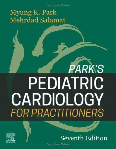
Long-standing arterial desaturation (usually longer than 6 months’ duration), even if too mild to be detected by an inexperienced person, results in clubbing of the fingernails and toenails. Clubbing from cyanotic CHD is almost not seen in the United States because of early surgical repairs of the defect. When fully developed, clubbing is characterized by a widening and thickening of the ends of the fingers and toes, as well as by convex fingernails and loss of the angle between the nail and nail bed (Fig. 2.1). Reddening and shininess of the terminal phalanges are seen in the early stages of clubbing. Clubbing appears earliest and most noticeably in the thumb. Clubbing may also be associated with lung disease (e.g., abscess), cirrhosis of the liver, and subacute bacterial endocarditis. Occasionally, clubbing occurs in normal people, such as in familial clubbing.
Respiratory Rate, Dyspnea, and Retraction
The physician should note the respiratory rate of every infant and child. If the infant breathes irregularly, the physician should count for a whole minute. The respiratory rate is faster in children who are crying, upset, eating, or feverish. The most reliable respiratory rate is that taken during sleep. After finishing a bottle of formula, an infant may breathe faster than normal for 5 to 10 minutes. A resting respiratory rate more than 40 breaths/min is unusual, and more than 60 breaths/min is abnormal at any age. Tachypnea, along with tachycardia, is the earliest sign of left-sided heart failure. If the child has dyspnea or retraction, this may be a sign of a more severe degree of left-sided heart failure or significant lung pathology.
Sweat on the Forehead
Infants with CHF often have a cold sweat on their foreheads. This is an expression of heightened sympathetic activity as a compensatory mechanism for decreased cardiac output.
TABLE 2.1 Major Syndromes Associated with Cardiovascular Abnormalities
Disorder
22q11.2 deletion syndrome (overlaps with and includes DiGeorge syndrome and velocardiofacial syndrome)
Facial asymmetry and hypoplasia; hypoplasia or aplasia of the pinna with blind or absent external ear canal (microtia); ear tags; cleft lip or palate; epitubular dermoid; hypoplastic vertebrae
Etiology
AR; mutation in 4p16.2
Ethanol or its byproducts
Exposure to trimethadione
Exposure to warfarin
Late-onset ataxia, skeletal deformities AR; mutation in 9q21.11
Facial asymmetry and hypoplasia, microtia, ear tag, cleft lip or palate, hypoplastic vertebrae
Very common; cardiomyopathy Large tongue and flabby muscles, cardiomegaly; LVH and short PR interval on ECG, severe ventricular hypertrophy on echocardiogram; normal FBS and GTT
Frequent; ASD, VSD
Homocystinuria Frequent; medial degeneration of aorta and carotids, atrial or venous thrombosis
Infant of diabetic mother
Kartagener syndrome (primary ciliary dyskinesia)
LEOPARD syndrome (Noonan syndrome with multiple lentigines)
Long QT syndrome: Jervell and Lange-Nielsen syndrome (1) and Romano-Ward syndrome (2)
Congenital deafness (not in Romano-Ward syndrome), syncope resulting from ventricular arrhythmias, family history of sudden death (±)
Arachnodactyly with hyperextensibility, subluxation of lens; pectus deformity, myopia
Coarse features, large tongue, depressed nasal bridge, kyphosis, retarded growth, hepatomegaly, corneal opacity (not in Hunter’s syndrome), mental retardation; most patients die by 10 to 20 yr of age
AR; mostly mutation in 21q22.3
Fetal exposure to high glucose levels
AR; different genes
AD; 85% mutation in 12q24.13
Multiple mutations; AR (1), AD (2)
AD; mutation in 15q21.1
AR (I) 4p16.3
XR (II) Xq28
AR (IV)
16q24.3 (A) 3p22.3 (B)
TABLE 2.1 Major Syndromes Associated with Cardiovascular Abnormalities—cont’d
Disorder
Muscular dystrophy (Duchenne type)
Neurofibromatosis (von Recklinghausen disease; NF type 1)
Waddling gait, “pseudohypertrophy” of calf muscle XR; Xp21.2-p21.1
Cafe-au-lait spots, multiple neurofibroma, acoustic neuroma (type 2), variety of bone lesions
Similar to Turner syndrome but may occur both in males and females, without chromosomal abnormality
Micrognathia, glossoptosis, cleft soft palate
Hepatic involvement, telangiectases, hemangioma or fibrosis
Excessive bone fragility with deformities of skeleton, blue sclera, hyperlaxity of joints
Alopecia, atrophy of subcutaneous fat, skeletal hypoplasia and dysplasia
Triad of the syndrome: deafness, cataract, and CHDs; others include intrauterine growth retardation, microcephaly, microphthalmia, hepatitis, neonatal thrombocytopenic purpura
Broad thumbs or toes; hypoplastic maxilla with narrow palate; beaked nose; short stature; mental retardation
Broad nasal tip with anteverted nostrils; ptosis of eyelids; syndactyly of second and third toes; short stature; mental retardation
Thrombocytopenia, absent or hypoplastic radius, normal thumb; “leukemoid” granulocytosis and eosinophilia
Underdeveloped lower jaw and zygomatic bone; defects of lower eyelids with downslanting palpebral fissure; malformation of auricle or ear canal defect; cleft palate
AD; 30%–50% new mutations; 17q11.2
Usually sporadic; AD 12q24.13 (∼50%)
Usually sporadic
AD
AD or AR
Mutations in 1q22; AD
Maternal rubella infection during the first trimester
Sporadic; 16p13.3 deletion
AR; mutations in 11q13.4
AR; deletion of 1q21.1
60% new mutation; AD
Trisomy 13 syndrome (Patau’s syndrome)
Very common (80%); VSD, PDA, dextrocardia
Trisomy 18 syndrome (Edward syndrome) Very common (90%); VSD, PDA, PS
Low birth weight, central facial anomalies, polydactyly, chronic hemangiomas, lowset ears, visceral and genital anomalies
Tuberous sclerosis Frequent; rhabdomyoma Triad of adenoma sebaceum (2–5 yr of age), seizures, and mental defect; cystlike lesions in phalanges and elsewhere; fibrous-angiomatosus lesions (83%) with varying colors in nasolabial fold, cheeks, and elsewhere
Turner syndrome (XO syndrome) Frequent (35%); COA, bicuspid aortic valve, AS; hypertension, aortic dissection later in life
VATER association (VATER or VACTERL syndrome)
Short female; broad chest with widely spaced nipples; congenital lymphedema with residual puffiness over the dorsum of fingers and toes (80%)
TABLE 2.1 Major Syndromes Associated with Cardiovascular Abnormalities—cont’d
Disorder
Velocardiofacial syndrome (Sprintzen syndrome; part of 22q11.2 deletion syndrome)
Williams syndrome
Cardiovascular Abnormalities:
Frequency
Very common (85%); truncus arteriosus, TOF, pulmonary atresia with VSD, interrupted aortic arch type B, VSD, and TGA
Frequent; supravalvular AS, PA stenosis, renal artery stenosis
Structural or functional palatal abnormalities, unique facial characteristics (“elfin facies” with auricular abnormalities, prominent nose with squared nasal root and narrow alar base, vertical maxillary excess with long face), hypernasal speech, conductive hearing loss, hypotonia, developmental delay and learning disability
Varying degree of mental retardation, so-called “elfin” facies (consisting of some of the following: upturned nose, flat nasal bridge, long philtrum, flat malar area, wide mouth, full lips, widely spaced teeth, periorbital fullness); hypercalcemia of infancy?
AD; microdeletion of 22q11.2
Sporadic; 7q23 deletion
Zellweger syndrome (cerebro-hepato-renal syndrome) Frequent; PDA, VSD or ASD Hypotonia, high forehead with flat facies, hepatomegaly, albuminemia AR; multiple genes
±, May or may not be present; AD, autosomal dominant; AR, autosomal recessive; AS, aortic stenosis; ASD, atrial septal defect; CHD, congenital heart disease; COA, coarctation of the aorta; ECD, endocardial cushion defect; ECG, electrocardiogram; FBS, fasting blood sugar; GTT, glucose tolerance test; HOCM, hypertrophic obstructive cardiomyopathy; LVH, left ventricular hypertrophy; MVP, mitral valve prolapse; PA, pulmonary artery; PDA, patent ductus arteriosus; PPHN, persistent pulmonary hypertension of newborn; PS, pulmonary stenosis; TGA, transposition of the great arteries; TOF, tetralogy of Fallot; VSD, ventricular septal defect; XR, sex-linked recessive.
TABLE 2.2 Prevalence of Associated Congenital Heart Defects in Patients with Other System
Malformation
Organ System and Malformation
Frequency (Range) (%)
CENTRAL NERVOUS SYSTEM
Hydrocephalus 6 (4.5–14.9)
Specific Cardiac Defects
VSD, ECD, TOF
Dandy-Walker syndrome 3 (2.5–4.3) VSD
Agenesis of corpus callosum 15 No specific defects
Meckel-Gruber syndrome 14
TE fistula or esophageal atresia 21 (15–39)
Diaphragmatic hernia 11 (9.6–22.9)
THORACIC CAVITY
GASTROINTESTINAL
Duodenal atresia 17
No specific defects
VSD, ASD, TOF
No specific defects
No specific defects
Jejunal atresia 5 No specific defects
Anorectal anomalies 22 No specific defects
Imperforate anus 12 TOF, VSD
VENTRAL WALL
Omphalocele 21 (19–32)
Gastroschisis 3 (0–7.7)
Renal agenesis
Bilateral 43
GENITOURINARY
No specific defects
No specific defects
No specific defects
Unilateral 17 No specific defects
Horseshoe kidney 39 No specific defects
Renal dysplasia 5 No specific defects
ASD, Atrial septal defect; ECD, endocardial cushion defect; TE, trachea-esophageal; TOF, tetralogy of Fallot; VSD, ventricular septal defect. Modified from Copel JA, Kleinman CS: Congenital heart disease and extracardiac anomalies: association and indications for fetal echocardiography, Am J Obstet Gynecol 154:1121, 1986.
NORMAL
CLUBBING
Widening Convex nail Thickening
Acanthosis Nigricans
Acanthosis nigricans is a dark pigmentation of skin creases most commonly seen on the neck in the majority of obese children and those with type 2 diabetes. It is also found in the axillae, groins, and inner thighs and on the belt line of the abdomen. The affected skin can become thickened. This condition is associated with insulin resistance and it eventually causes type 2 diabetes. Rarely, acanthosis occurs in patients with Addison disease, Cushing syndrome, polycystic ovary syndrome (Stein-Leventhal syndrome), hypothyroidism, and hyperthyroidism. This condition is associated with insulin resistance and a higher risk of developing type 2 diabetes.
Inspection of the Chest
Precordial bulge, with or without actively visible cardiac activity, suggests chronic cardiac enlargement. Acute dilatation of the heart does not cause precordial bulge. Pigeon chest (pectus carinatum), in which the sternum protrudes on the midline, is usually not a result of cardiomegaly.
Pectus excavatum (undue depression of the sternum) rarely, if ever, causes significant cardiac embarrassment. Occasionally, it may be a cause of a pulmonary systolic murmur or a large cardiac silhouette on a posteroanterior view of a chest radiograph, which compensates for the diminished anteroposterior diameter of the chest. As a group, children with a significant pectus excavatum have a shorter endurance time than normal children.
Harrison’s groove, a line of depression in the bottom of the rib cage along the attachment of the diaphragm, indicates poor lung compliance of long duration, such as that seen in large left-to-right shunt lesions.
PALPATION
Palpation should include the peripheral pulses (their presence or absence, the pulse rate, the volume of the pulses) and the precordium (the presence of a thrill, the point of maximal impulse [PMI], precordial hyperactivity). Although ordinarily palpation follows inspection, auscultation may be more fruitful on a sleeping infant who might wake up and become uncooperative.
Peripheral Pulses
1. The physician should count the pulse rate and note any irregularities in the rate and volume. The normal pulse rate varies with the patient’s age and status. The younger the patient, the faster the pulse rate. Increased pulse rate may indicate excitement, fever, CHF, or arrhythmia. Bradycardia may mean heart block, effects of drugs, and so on. Irregularity of the pulse suggests arrhythmias, but sinus arrhythmia (an acceleration with inspiration) is normal.
2. The right and left arm and an arm and a leg should be compared for the volume of the pulse. Every patient should have palpable pedal pulses of the dorsalis pedis, tibialis posterior, or both. It is often easier to feel pedal pulses than femoral pulses. Attempts at palpating a femoral pulse often wake up a sleeping infant or upset a toddler. If a good pedal pulse is felt, coarctation of the aorta (COA) is effectively ruled out, especially if the blood pressure (BP) in the arm is normal.
3. Weak leg pulses and strong arm pulses suggest COA. If the right brachial pulse is stronger than the left brachial pulse, the cause may be COA occurring near the origin of the left subclavian artery or supravalvular aortic stenosis (AS). A weaker right brachial pulse than the left suggests aberrant right subclavian artery arising distal to the coarctation.
4. Bounding pulses are found in aortic run-off lesions such as PDA, aortic regurgitation (AR), large systemic arteriovenous fistula, or persistent truncus arteriosus (rarely). Pulses are bounding in premature infants because of the lack of subcutaneous tissue and because many have PDA.
5. Weak, thready pulses are found in cardiac failure or circulatory shock or in the leg of a patient with COA. A systemic-to-pulmonary artery (PA) shunt (either classic Blalock-Taussig shunt or modified Gore-Tex shunt) or subclavian flap angioplasty for repair of COA may result in an absent or weak pulse in the arm affected by surgery. Arterial injuries resulting from previous cardiac catheterization may cause a weak pulse in the affected limb.
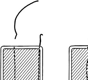
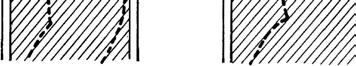
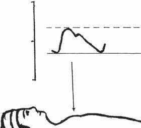
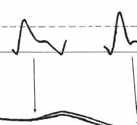
6. Pulsus paradoxus (paradoxical pulse) is suspected when there is marked variation in the volume of arterial pulses with the respiratory cycle. The term pulsus paradoxus does not indicate a phase reversal; rather, it is an exaggeration of normal reduction of systolic pressure during inspiration. When arterial BP is being monitored through an indwelling arterial catheter, the presence of pulsus paradoxus is easily detected by a wide swing (>10 mm Hg) in arterial pressure. In a child without arterial pressure monitoring, accurate evaluation requires sphygmomanometry (Fig. 2.2). Pulsus paradoxus may be associated with cardiac tamponade secondary to pericardial effusion or constrictive pericarditis or to severe respiratory difficulties seen with asthma or pneumonia. It is also seen in patients who are on ventilators with high pressure settings, but in these cases, the BP increases with inflation.
The presence of pulsus paradoxus is confirmed by the use of a sphygmomanometer as described below.
a. The cuff pressure is raised about 20 mm Hg above the systolic pressure.
Fig. 2.1 Diagram of normal and clubbed fingers.
Hg
Fig. 2.2 Diagram of pulsus paradoxus. Note the reduction in systolic pressure of more than 10 mm Hg during inspiration. EXP, Expiration; INSP, inspiration.
b. The pressure is lowered slowly until Korotkoff sound 1 is heard for some but not all cardiac cycles, and the reading is noted (line A on Fig. 2.2).
c. The pressure is lowered further until systolic sounds are heard for all cardiac cycles, and the reading is noted (line B on Fig. 2.2).
d. If the difference between readings A and B is greater than 10 mm Hg, pulsus paradoxus is present.
Chest
One should palpate the following on the chest: apical impulse, PMI, hyperactivity of the precordium, and palpable thrill.
Apical Impulse
Palpation of the apical impulse is usually superior to percussion in the detection of cardiomegaly. Its location and diffuseness should be noted. Percussion in infants and children is inaccurate and adds little. The apical impulse is normally at the fifth intercostal space in the midclavicular line after age 7 years. Before this age, the apical impulse is in the fourth intercostal space just to the left of the midclavicular line. An apical impulse displaced laterally or downward suggests cardiac enlargement.
Point of Maximal Impulse
The PMI is helpful in determining whether the RV or left ventricle (LV) is dominant. With RV dominance, the impulse is maximal at the lower left sternal border or over the xiphoid process; with LV dominance, the impulse is maximal at the apex. Normal newborns and infants have RV dominance and therefore more RV impulse than older children. If the impulse is more diffuse and slow rising, it is called a heave. If it is well localized and sharp rising, it is called a tap. Heaves are often associated with volume overload. Taps are associated with pressure overload.
Hyperactive Precordium
The presence of a hyperactive precordium characterizes heart disease with volume overload, such as that seen in defects with large left-to-right shunts (e.g., PDA, VSD) or heart
disease with severe valvular regurgitation (e.g., AR, mitral regurgitation [MR]).
Thrills
Thrills are vibratory sensations that represent palpable manifestations of loud, harsh murmurs. Palpation for thrills is often of diagnostic value. A thrill on the chest is felt better with the palm of the hand than with the tips of the fingers. However, the fingers are used to feel a thrill in the suprasternal notch and over the carotid arteries.
1. Thrills in the upper left sternal border originate from the pulmonary valve or PA and therefore are present in PS, PA stenosis, or PDA (rarely).
2. Thrills in the upper right sternal border are usually of aortic origin and are seen in AS.
3. Thrills in the lower left sternal border are characteristic of a VSD.
4. Thrills in the suprasternal notch suggest AS but may be found in PS, PDA, or COA.
5. The presence of a thrill over the carotid artery or arteries accompanied by a thrill in the suprasternal notch suggests diseases of the aorta or aortic valve (e.g., COA, AS). An isolated thrill in one of the carotid arteries without a thrill in the suprasternal notch may be a carotid bruit.
6. Thrills in the intercostal spaces are found in older children with severe COA and extensive intercostal collaterals.
BLOOD PRESSURE MEASUREMENT
Whenever possible, every child should have his or her BP measured as part of the physical examination. The status of the child at the time of BP measurement, such as moving, crying, or fighting, should be considered in the interpretation of obtained BP values before making any decision about the normalcy of the measurement. When BP is measured in a reasonably quiet situation, an average value of 2 or more BP values is compared with a set of normative BP standards to see if obtained BP values are normal or abnormal. Unfortunately, there have been multiples problems regarding the proper method of measuring BP as well as the normative BP standards for children.
Scientifically unsound methods of BP measurement using arm length-based BP cuffs, recommended by two previous National Institute of Health (NIH) Task Forces (of 1977 and 1987), have dominated the field and they have been the source of confusion for several decades. At this time, both the methodology and the BP standards recommended by the NIH Task Forces have been abandoned. An unfortunate part of these wrong recommendations was that most children’s BP studies were carried out using wrong BP cuffs selected based on the length of the arm. In addition, the new sets of normative BP standards recommended by the Working Group of the National High Blood Pressure Education Program (NHBPEP) have major flaws scientifically and statistically and their usefulness has become debatable.
In this subsection, the following important issues in children’s BP measurement will be discussed.
a. What is the currently recommended BP measuring method?
b. How good are BP standards recommended by the NHBPEP?
c. Which normal BP standards should be used and why?
d. How accurate are oscillometric BP measurements?
e. Comparison of BP readings by oscillometric device and those by the auscultatory method
f. How to interpret arm and leg BP values in children
g. BP levels in neonates and small children
h. How important is the concept of peripheral amplification of systolic pressure?
i. Need for simplified BP tables
1. What is the currently recommended BP measuring method?
In the recent past, two Task Forces (1977 and 1987) of the NIH have recommended BP cuff selection based on the length of the arm, initially recommending the cuff width to be two thirds of the length of the arm and later changing it to three fourths of the length of the arm. The BP cuff selection based on the length of the arm is scientifically unsound and violates the physical principles underlying indirect BP measurement. The NIH Task Forces have provided normal BP standards based on these unscientific methods.
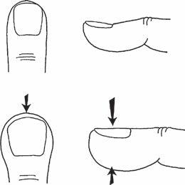
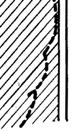
As early as 1901, von Recklinghausen recognized the importance of the width of the BP cuff in relation to the arm circumference. For adults, the correct BP cuff width has been settled as 40% of the circumference of the arm since 1951 by the recommendations of the American Heart Association (AHA) (Fig. 2.3). It made no sense to choose the BP cuff based on the arm length in children. Park and Guntheroth (1970) compared directly measured brachial artery pressure during cardiac catheterization and the arm BP measured using the cuff width 40% of the arm circumference and concluded that the same 40% arm circumference criterion was good for children. In 1988, the AHA’s Special Task Force accepted the 40% cuff selection method for children, but the two past NIH Task Forces did not change their recommendations. In 2004, however, the Working Group of the NHBPEP has accepted the correct BP cuff selection method based on the arm circumference.
The following summarizes current views on BP measurement techniques recommended by the AHA and the NHBPEP.
a. Both the AHA and the NHBPEP recommend the BP cuff width to be 40% of the circumference (equivalent to 125% of the diameter) of the arm with the cuff long enough to completely or nearly completely encircle the extremity. The cuff should be at least 40% of the arm circumference. When a 40% cuff is not available, it is better to choose one size bigger than the ideal one. One size smaller than the ideal cuff (i.e., 31%–35% arm circumference) gave readings much higher than the reference values (15 mm Hg higher for systolic pressure and 11 mm Hg higher for diastolic pressure). Conversely, one size larger cuff (i.e., 43%–48%) resulted in relatively smaller changes in BP readings (6 mm Hg lower for systolic pressure and 3 mm Hg lower for diastolic pressure) (Park et al, 1976).
b. The NHBPEP recommends Korotkoff phase 5 (K5) as the diastolic pressure, but the AHA recommends K4 as the diastolic pressure; some earlier studies showed better agreement between K4 and direct intraarterial diastolic pressure in children.
c. Both the AHA and the NHBPEP recommend averaging of 2 or more readings (because the averaged values are closer to the basal BP level and are more reproducible).
d. Both the AHA and the NHBPEP recommend sitting position with the arm at the heart level.
2. How good are BP standards recommended by the NHBPEP?
Normative BP standards recommended by the Working Group of the NHBPEP are not as good as it was made to believe; they have major flaws as described below.
a. Although the Working Group recommended the BP cuff to be 40% of the circumference of the arm, the BP data comprising their standards were not obtained by the currently recommended method. The majority of the original data were obtained by the arm length–based cuff selection method. These values are also from single measurement, rather than the averages of 2 or more readings, as currently recommended. In other words, the original source of the elaborate BP standards of the Working Group is one that has been abandoned by the program itself.
b. Expressing children’s BP levels by age and height percentiles is statistically unsound. Height has no statistically important role in children’s BP levels (Park, et al, 2001). Partial correlation analysis used in the San Antonio Children’s Blood Pressure Study (the San Antonio Study) shows that when auscultatory BP levels were adjusted for age and weight, the correlation coefficient of systolic pressure with height was very small (r = 0.068 for boys; r = 0.072 for girls), but when adjusted for age and height, the correlation of systolic pressure with weight remained high (r = 0.343 for boys; r = 0.294 for girls). These findings indicate that the contribution of height to BP levels is negligible. The apparent
Fig. 2.3 Diagram showing a method of selecting an appropriate-sized blood pressure cuff based on the arm diameter. The cuff should be at least 40% (to 45%) of the arm circumference but less than 50%. The cuff width 125% of the arm diameter equals to 40% of the arm circumference: the cuff width approximately 140% of the arm diameter equals to 45% of the circumference. Therefore, one should aim for a cuff width between 125% and 140% of the arm diameter as shown.
correlation of height to BP levels may all be secondary to a close correlation that exists between height and weight (r = 0.86). A similar conclusion was reached with oscillometric BP levels in the San Antonio study. Although weight is a very important contributor to BP, weight cannot be used as a second variable because this would interfere with detection of high BP in obese children. Thus, we found no rationale to use anything other than age and gender to express children’s normative BP standards.
c. Requiring additional computations to find the height percentile of a child, especially on such highly variable office BP readings, is not only unreasonable and but also unjustified. Height has no statistical importance in children’s BP readings.
d. The Working Group does not point out that the auscultatory and oscillometric BP readings are not interchangeable. The San Antonio Study, in which both auscultatory and oscillometric methods were used, found that oscillometric systolic pressures are significantly higher than auscultatory BP readings and thus they are not interchangeable (see later for further details). This finding is important in view of popular use of oscillometric device in BP measurements in pediatric practice.
3. Which normal BP standards should be used and why?
a. The BP standards of the NHBPEP are not acceptable standards because they are riddled with major flaws as discussed earlier. However, the NHBPEP’s normative BP standards are presented in Appendix B for the sake of completeness (Tables B.1 and B.2, Appendix B).
b. Normative BP percentile values from the San Antonio Study are recommended as a better alternative to BP standards of the NHBPEP. The BP data from the San
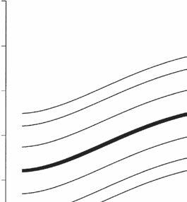
Fig. 2.4 Age-specific percentile curves of auscultatory systolic and diastolic (K5) pressures in boys 5 to 17 years of age. Blood pressure values are the average of three readings. The width of the blood pressure cuff was 40% (up to 50%) of the circumference of the arm. Percentile values for the figure are shown in Table B.3, Appendix B. (From Park MK, Menard SW, Yuan C: Comparison of blood pressure in children from three ethnic groups, Am J Cardiol 87:1305–1308, 2001.)
Antonio Study are the only available BP standards that have been obtained according to the currently recommended method. In the San Antonio Study, BP readings were obtained in more than 7000 school children of three ethnic groups (African American, Mexican American, and non-Hispanic white), enrolled in kindergarten through the 12th grade in the San Antonio, Texas, area. Both the auscultatory and oscillometric (model Dinamap 8100) methods were used in the study, and the data were the averages of three readings. No consistent ethnic difference was found among the three ethnic groups, but there were important gender differences. Therefore, auscultatory BP data were expressed according to age and gender (Park et al, 2001) (Figs. 2.4 and 2.5). Percentile BP values for these figures are presented in Appendix B (Tables B.3 and B.4).
c. When BP is measured using an oscillometric device, one should use a device-specific normative BP standards. The San Antonio Study found that the readings by auscultatory method and by Dinamap 8100 are
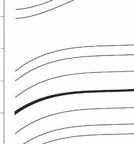
Fig. 2.5 Age-specific percentile curves of auscultatory systolic and diastolic (K5) pressures in girls 5 to 17 years of age. Blood pressure values are the average of three readings. The width of the blood pressure cuff was 40% (up to 50%) of the circumference of the arm. Percentile values for the figure are shown in Table B.4, Appendix B. (From Park MK, Menard SW, Yuan C: Comparison of blood pressure in children from three ethnic groups, Am J Cardiol 87:1305–1308, 2001.)
significantly different and thus are not interchangeable (Park et al, 2005) as will be described further. The San Antonio Study provides oscillometric specific BP standards for children. Percentile BP values by an oscillometric method (Dinamap 8100) are presented in Appendix B (Tables B.6 and B.7).
4. How accurate are oscillometric BP measurements?
The accuracy of indirect BP measurement by an oscillometric method (Dinamap Model 1846) has been demonstrated. In fact, oscillometric BP levels correlated better with intra-arterial pressures than the auscultatory method (Park et al, 1987). The oscillometric method also provides some advantages over auscultation; it eliminates observer-related variations, and it can be successfully used in infants and small children. Auscultatory BP measurement in small infants is not only difficult to obtain but also has not been shown to be accurate. Percentile values of normative oscillometric BPs in neonates and children up to 5 years of age are presented in Appendix B (Table B.5). Aside from the issue of accuracy, the oscillometric method is widely used in large pediatric and
pediatric cardiology practices and in the setting of emergency departments. One caution is that not all oscillometric devices in clinical use have been validated for their accuracy.
5. Comparison of BP readings by oscillometric device and those by the auscultatory method
In the San Antonio Study, we used both the auscultatory and oscillometric methods to obtain BP readings in children, alternating the device used, under exactly the same protocol. We found that BP levels obtained by the Dinamap (Model 8100) were on average 10 mm Hg higher than the auscultatory method for the systolic pressure and 5 mm Hg higher for the diastolic pressure (Park et al, 2001). This study, however, does not show superiority of any one of the devices but simply indicates that the indirect BP readings are different according to the sensitivity of the detection device used. It may imply that the detection of oscillation by the machine occurs earlier (at a higher level of systolic pressure) than sounds that human ears can hear. The gold standard remains an intraarterial BP reading. It is clear from this study that the auscultatory and the Dinamap BPs are not interchangeable. When oscillometric method is used, one must use oscillometric specific normative BP standards (Park et al, 2005). Dinamap 8100 specific BP standards are presented in Appendix B (Tables B.6 and B.7).
6. How to interpret arm and leg BP values in children
Four-extremity BP measurements are often obtained to rule out COA. The same cuff selection criterion (i.e., 40% of the circumference) applies for calf or thigh pressure determination, often requiring the use of a larger cuff for the lower extremity. The patient should be in supine position for BP measurements in the arm and leg. When using the auscultatory method, the thigh pressure is obtained with the stethoscope place in the popliteal fossa (over the popliteal artery) with the legs bent and in supine position. Calf BP is difficult to obtain by the auscultatory method.
How do BP levels in the arm and leg compare in normal children? Even when a considerably wider cuff is selected for the thigh, the Dinamap systolic pressure in the thigh or calf is about 5 to 10 mm Hg higher than that in the arm (Park et al, 1993). This partly reflects the peripheral amplification of systolic pressure (see later for further discussion). In newborns, however, the systolic pressures in the arm and the calf are the same (Park et al, 1989). The absence of a higher systolic pressure in the leg in newborns may be related to the presence of normally narrow segment of the aortic isthmus. Thus, the systolic pressure in the thigh (or calf) should be higher than or at least equal to that in the arm, except in newborns. If the systolic pressure is lower in the leg, COA may be present. Leg BP determinations are mandatory in a child with hypertension in the arm to rule out COA. The presence of a femoral pulse does not rule out a coarctation.
7. BP levels in neonates and small children
Blood pressure measurement is important in newborns and small children to diagnose COA, hypertension, or hypotension. In contrast to the recommendations of the NHBEP, the auscultatory method is difficult to apply in newborns and small children because of weak Korotkoff sounds in
TABLE 2.3 Normative Blood Pressure Levels by Dinamap Monitor in Children Age 5 Years and Youngera
1–3 days
1 mo–2 yr
2–5 yr
(50)
(72)
(74)
(50)
(83)
(82)
aDinamap Model 1846SX was used. Blood pressure (BP) levels are systolic/diastolic, with the mean in parentheses.
(62)
(86)
(85)
Modified from Park MK, Menard SM: Normative oscillometric blood pressure values in the first 5 years in an office setting, Am J Dis Child 143:860, 1989.
these age groups, and thus normative standards are not reliable. Therefore, the oscillometric method is frequently used instead. Abbreviated normative Dinamap BP standards for newborns and small children (5 years of age and younger) are presented in Table 2.3. Full percentile values are presented in Appendix B (Table B.5).
8. How important is the concept of peripheral amplification of systolic pressure?
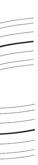
Many physicians incorrectly assume that peripherally measured BPs, such as those measured in the arm, reflect central aortic pressure. Some physicians also incorrectly think that the systolic pressure in the central aorta is higher than that in the brachial, radial, and pedal arteries. As shown schematically in Figure 2.6, systolic pressure becomes higher and higher as one moves farther peripherally, although the diastolic and mean pressures remain the same or decrease slightly (O’Rourke, 1968). This phenomenon is known as peripheral amplification of systolic pressure. If this is correct, how does blood flow distally? There is a change in the arterial pressure wave form with systolic peaking as one moves peripherally as shown in Figure 2.6, but the area under the curve decreases slightly in the peripheral sites, so that the blood flows peripherally.
The main purpose of measuring indirect BP is to estimate the pressure in the central aorta, the perfusing pressure for the brain and the heart. Systolic pressures measured, either by direct or indirect methods, do not always reflect the central aortic pressure. Peripherally obtained systolic pressures are
usually higher than the central aortic pressure but the magnitude of difference is not easy to predict. Physicians should be aware of some clinically important situations, in which the peripheral amplification becomes more marked.
The following are some key points of the peripheral amplification of clinical significance.
1. The systolic amplification is greater in children (with more reactive arteries) than in older adults (who may have degenerative arterial disease).
b. The amplification is more marked in vasoconstricted states, and many of them are clinically important, such as those seen (a) with impending circulatory shock from hemorrhage or dehydration, (b) in a child in CHF (in which generalized peripheral vasoconstriction is present), and (c) in patients receiving catecholamine infusion or other vasoconstrictors in the setting of critical care units.
c. Reduced level of peripheral amplification of systolic pressure is noted in vasodilated states, such as those seen in subjects receiving vasodilators and after receiving a contrast dye (which has vasodilating effects) during cardiac catheterization.
At sites other than the upper arm, systolic pressure may be significantly higher than that in the central aorta. Although the systolic pressure in the arm tends to be slightly higher than that in the central aorta, the upper arm is the only place where the systolic amplification is smallest. Radial artery pressure is likely to be significantly higher than the brachial artery pressure. The following lists two clinically important examples.
Fig. 2.6 Schematic diagram of pulse wave changes at different levels of the systemic arteries. AO, Aorta; FA, femoral artery; PA, pedal artery. (Modified from Geddes LA: Handbook of Blood Pressure Measurement, Clifton, NJ, 1991, Humana Press.)
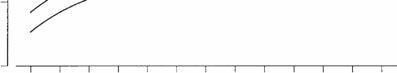
a. Arm systolic pressure in subjects running on treadmill can be markedly higher than the central aortic pressure. A dramatic illustration of this phenomenon is shown in Figure 6.1 (in the section on exercise stress testing) in a young adult running on treadmill with catheters in the ascending aorta and the radial artery. At rest, the difference in systolic pressure between the two sites is about 30 mm Hg. At the last stage of the stress test, the difference increases further to 76 mm Hg, with the radial artery pressure reaching a state known as hypertensive emergency. Even in the setting of an intensive care unit, one should avoid using peripheral lines such as the radial artery if the pressure monitoring is the main purpose of the line; the mean pressure may be better than the systolic pressure in such a situation.
b. Nice-looking wrist BP devices are on the market. These devices are likely to give higher BP readings than the upper arm device, depending on the condition of the patient. Therefore, BP should not be obtained in the wrist.
9. Need for simplified BP tables
The main reason for measuring BP routinely is to identify those children with hypertension or those at risk of developing hypertension. Even though the BP standards from the San Antonio Study are simple enough to use, it is still time consuming to check the complete table in busy practice.
In 2009, Kaelber and Picket proposed a simple table of BP levels, without height percentiles, that may require attention or additional investigation being at “elevated blood pressure levels” (formerly called prehypertensive levels). Elevated BP is defined as BP levels between the 90th and 95th percentiles for children and 120 to 129 systolic and 80 mm Hg diastolic
for adolescents (older than 13 years of age) and adults. We followed suit and developed similar tables of the 90th percentile values for both auscultatory and oscillometric BP for children based on the San Antonio Study (Table 2.4). It is interesting to find that the values suggested by Kaelber and Picket agree very closely with the 90th percentile values in the San Antonio Study. All one needs is this single page table posted on the wall of the BP room.
AUSCULTATION
Although auscultation of the heart requires more skill, it also provides more valuable information than other methods of physical examination. Whereas the bell-type chest piece is better suited for detecting low-frequency events, the diaphragm selectively picks up high-frequency events. When the bell is firmly pressed against the chest wall, it acts like the diaphragm by filtering out low-frequency sounds or murmurs and picking up high-frequency events. Physicians should ordinarily use both the bell and the diaphragm, although using the bell both lightly and firmly pressed against the chest may be equally effective, especially in sleeping infants. Using only the diaphragm may result in missing some important low-frequency murmurs or sounds, such as mid-diastolic rumble, pulmonary regurgitation (PR) murmur, and faint Still’s innocent heart murmurs. One should not limit examination to the four traditional auscultatory areas. The entire precordium, as well as the sides and back of the chest, should be explored with the stethoscope. Systematic attention should be given to the following aspects:
1. Heart rate and regularity: Heart rate and regularity should be noted on every child. Extremely fast or slow heart rates
aThe 90th percentiles of systolic pressure for males 14 years of age and older are higher than 120 mm Hg in the San Antonio Study, but 120 mm Hg is listed to be consistent with the definition of elevated blood pressure in adults. DP, Diastolic pressure; SP, systolic pressure. Values are from Park MK, Menard SW, Yuan C: Comparison of blood pressure in children from three ethnic groups, Am J Cardiol 87:1305–1308, 2001.
TABLE 2.4 The 90th Percentiles of Blood Pressure by Auscultatory and Oscillometric
Insp.
Fig. 2.7
or irregularity in the rhythm should be evaluated by an electrocardiogram (ECG) and a long rhythm strip.
2. Heart sounds: Intensity and quality of the heart sounds, especially the second heart sound (S2), should be evaluated. Abnormalities of the first heart sound (S1) and the third heart sound (S3) and the presence of a gallop rhythm or the fourth sound (S4) should be noted. Muffled heart sounds should also be noted.
3. Systolic and diastolic sounds: An ejection click in early systole provides a clue to aortic or pulmonary valve stenosis. A midsystolic click provides important clues to the diagnosis of mitral valve prolapse (MVP). An opening snap in diastole (present in mitral stenosis [MS]) should be noted, but it is extremely rare in children.
4. Heart murmurs: Heart murmurs should be evaluated in terms of intensity, timing (systolic or diastolic), location, transmission, and quality.
Heart Sounds
The heart sound should be identified and analyzed before the analysis of heart murmurs (Fig. 2.7). Muffled and distant heart sounds are present in pericardial effusion and heart failure.
First Heart Sound
The S1 is associated with closure of the mitral and tricuspid valves. It is best heard at the apex or lower left sternal border. Splitting of the S1 may be found in normal children, but it is infrequent. Abnormally wide splitting of S1 may be found in right bundle branch block (RBBB) or Ebstein’s anomaly. Splitting of S1 should be differentiated from ejection click or S4.
1. Ejection click is found in PS and bicuspid aortic valve. In patients with PS, it is easily audible at the upper left sternal border in PS. In bicuspid aortic valve, the click may be louder at the lower left sternal border or apex than at the upper right sternal border.
2. S4 is rare in children.
Second Heart Sound
The S2 in the upper left sternal border (i.e., pulmonary valve area) is of critical importance in pediatric cardiology. The S2 must be evaluated in terms of the degree of splitting and the intensity of the pulmonary closure component of the second
AS, Aortic stenosis; ASD, atrial septal defect; LBBB, left bundle branch block; MR, mitral regurgitation; PAPVR, partial anomalous pulmonary venous return; PS, pulmonary stenosis; RBBB, right bundle branch block; TGA, transposition of the great arteries; TOF, tetralogy of Fallot; TS, tricuspid stenosis; WPW, Wolff-Parkinson-White.
heart sound (P2) in relation to the intensity of the aortic closure component of the second heart sound (A2). Although best heard with the diaphragm of a stethoscope, both components are readily audible with the bell. Abnormalities of splitting of the S2 and the intensity of the P2 are summarized in Box 2.1.
Splitting of the S2. In every normal child, with the exception of occasional newborns, two components of the S2 should be audible in the upper left sternal border. The first is the A2; the second is the P2. Both components are best heard at the pulmonary valve area (upper left sterna border). The A2 is not the second heart sound at the aortic area; rather, it is the first (or aortic closure) component of the second heart sound at the pulmonary area.
Normal Splitting of the S2. The degree of splitting of the S2 varies with respiration, increasing with inspiration and decreasing or becoming single with expiration (see Fig. 2.7). During inspiration, because of a greater negative pressure in the thoracic cavity, there is an increase in systemic venous return to the right side of the heart. This increased volume of blood in the RV prolongs the duration of RV ejection time, which delays the closure of the pulmonary valve, resulting in a wide splitting of the S2. The absence of splitting (i.e., single S2) or a widely split S2 usually indicates an abnormality.
Abnormal Splitting of the S2. Abnormal splitting may be in the form of wide splitting, narrow splitting, a single S2, or paradoxical splitting of the S2 (rarely).
Diagram showing the relative intensity of A2 and P2 and the respiratory variation in the degree of splitting of the S2 at the upper left sternal border (pulmonary area). Exp, Expiration; Insp, inspiration.
1. A widely split and fixed S2 is found in conditions that prolong the RV ejection time or that shorten the LV ejection. Therefore, it is found in:
a. ASD or partial anomalous pulmonary venous return (PAPVR) (conditions in which the amount of blood ejected by the RV is increased; volume overload)
b. PS (the valve stenosis prolongs the RV ejection time; pressure overload)
c. RBBB (a delay in electrical activation of the RV) delays the completion of the RV ejection
d. MR (a decreased forward output seen in this condition shortens the LV ejection time, causing aortic closure to occur earlier than normal)
e. An occasional normal child, including “prolonged hangout time” seen in children with dilated PA (a condition called idiopathic dilatation of the PA). In dilated PA, the increased capacity of the artery produces less recoil to close the pulmonary valve, which delays closure.
2. A narrowly split S2 is found in conditions in which the pulmonary valve closes early (e.g., pulmonary hypertension) or the aortic valve closure is delayed (e.g., AS). This is occasionally found in a normal child.
3. A single S2 is found in the following situations:
a. When only one semilunar valve is present (e.g., aortic or pulmonary atresia, persistent truncus arteriosus)
b. When the P2 is not audible (e.g., transposition of the great arteries [TGA], TOF, severe PS)
c. When aortic closure is delayed (e.g., severe AS)
d. When the P2 occurs early (e.g., severe pulmonary hypertension)
e. In an occasional normal child
4. A paradoxically split S2 is found when the aortic closure (A2) follows the pulmonary closure (P2) and therefore is seen when the LV ejection is greatly delayed (e.g., severe AS, left bundle branch block [LBBB], sometimes Wolff-Parkinson-White [WPW] pre-excitation).
Intensity of the P2 The relative intensity of the P2 compared with the A2 must be assessed in every child. In the pulmonary area, the A2 is usually louder than the P2 (see Fig. 2.7). Judgment as to normal intensity of the P2 is based on experience. There is no substitute for listening to the hearts of many normal children. Abnormal intensity of the P2 may suggest a pathologic condition. Increased intensity of the P2, compared with that of the A2, is found in pulmonary hypertension. Decreased intensity of the P2 is found in conditions with decreased diastolic pressure of the PA (e.g., severe PS, TOF, tricuspid atresia).
Third Heart Sound
The S3 is a somewhat low-frequency sound in early diastole and is related to rapid filling of the ventricle (Fig. 2.8). It is best heard at the apex or lower left sternal border. It is commonly heard in normal children and young adults. A loud S3 is abnormal and is audible in conditions with dilated ventricles and decreased ventricular compliance (e.g., largeshunt VSD or CHF). When tachycardia is present, it forms a “Kentucky” gallop.
Fourth Heart Sound or Atrial Sound
The S4 is a relatively low-frequency sound of late diastole (i.e., presystole) and is rare in infants and children (see Fig. 2.8). When present, it is always pathologic and is seen in conditions with decreased ventricular compliance or CHF. With tachycardia, it forms a “Tennessee” gallop.
Gallop Rhythm
A gallop rhythm is a rapid triple rhythm resulting from the combination of a loud S3, with or without an S4, and tachycardia. It generally implies a pathologic condition and is commonly present in CHF. A summation gallop represents tachycardia and a superimposed S3 and S4.
Systolic and Diastolic Sounds
1. An ejection click (or ejection sound) is usually caused by thickened semilunar valve leaflets and rarely dilated great arteries. It follows the S1 very closely and occurs at the time of the ventricular ejection’s onset. Therefore, it sounds like a splitting of the S1. However, whereas it is usually audible at the base (either side of the upper sternal border), the split S1 is usually audible at the lower left sternal border (exception with an aortic click, discussed in a later section). If the physician hears what sounds like a split S1 at the upper sternal border, it may be an ejection click (Fig. 2.9). The pulmonary click is heard at the second and third left intercostal spaces and changes in intensity with respiration, being louder on expiration. The aortic click is best heard at the second right intercostal space but may be louder at the apex or mid-left sternal border. It usually does not change its intensity with respiration. The ejection click is most often associated with:
Fig. 2.8 Diagram showing the relative relationship of the heart sounds. The filled bar shows an abnormal sound.
Fig. 2.9 Diagram showing the relative position of ejection click (EC), midsystolic click (MC), and diastolic opening snap (OS). Filled bars are abnormal sounds.
a. Stenosis of semilunar valves (e.g., PS or AS)
b. Dilated great arteries, which are seen in systemic or pulmonary hypertension, idiopathic dilatation of the PA, TOF (in which the aorta is dilated), and persistent truncus arteriosus
2. Midsystolic click with or without a late systolic murmur is heard at the apex in mitral valve prolapse (MVP) (see Fig. 2.9 and Chapter 21).
3. Diastolic opening snap is rare in children and is audible at the apex or lower left sternal border. It occurs somewhat earlier than the S3 during diastole and originates from a stenosis of the atrioventricular (AV) valve, such as MS (see Fig. 2.9).
Extracardiac Sounds
1. A pericardial friction rub is a grating, to-and-fro sound produced by friction of the heart against the pericardium. This sounds similar to sandpaper rubbed on wood. Such a sound usually indicates pericarditis. The intensity of the rub varies with the phase of the cardiac cycle rather than the respiratory cycle. It may become louder when the patient leans forward. Large accumulation of fluid (pericardial effusion) may result in disappearance of the rub.
2. A pericardial knock is an adventitious sound associated with chronic (i.e., constrictive) pericarditis. It rarely occurs in children.
Heart Murmurs
Each heart murmur must be analyzed in terms of intensity (grade 1 to 6), timing (systolic or diastolic), location, transmission, and quality (musical, vibratory, blowing, and so on).
Intensity
Intensity of the murmur is customarily graded from 1 to 6.
Grade 1: barely audible
Grade 2: soft but easily audible
Grade 3: moderately loud but not accompanied by a thrill
Grade 4: louder and associated with a thrill
Grade 5: audible with the stethoscope barely on the chest
Grade 6: audible with the stethoscope off the chest
The difference between grades 2 and 3 or grades 5 and 6 may be somewhat subjective. The intensity of the murmur may be influenced by the status of cardiac output. Thus, any factor that increases the cardiac output (e.g., fever, anemia, anxiety, exercise) intensifies any existing murmur or may even produce a murmur that is not audible at basal conditions.
Classification of Heart Murmurs
Based on the timing of the heart murmur in relation to the S1 and S2, the heart murmur is classified into systolic, diastolic, and AV.
Systolic Murmurs
Most heart murmurs are systolic in timing in that they occur between the S1 and S2. Systolic murmurs were classified by Aubrey Leatham in 1958 into two subtypes according to the time of onset: (1) ejection type and (2) regurgitant type (Fig. 2.10). Recently, Joseph Perloff classified systolic murmurs according to their time of onset and termination into four subtypes: (1) midsystolic (or ejection), (2) holosystolic, (3) early systolic, or (4) late systolic (Fig. 2.11). The holosystolic and early systolic murmurs of Perloff belong to the regurgitant murmur of Leatham. The types of murmurs (described earlier) and the location of the maximum intensity of the murmur are very important in the assessment of a systolic murmur. Transmission of the murmur to a particular direction and the quality of the murmur also help in deciding the cause of the murmur. These aspects are discussed in detail later.
Fig. 2.10 Diagram of Leatham’s classification of systolic murmurs. This classification is based primarily on the relationship of the S1 to the onset of the murmur. A, Ejection systolic murmurs. Short ejection-type murmur with the apex of the “diamond” in the early part of systole is found with mild stenosis of semilunar valves (top). With increasing severity of stenosis, the murmur becomes louder and longer, and its apex moves toward the S2 (middle). In severe pulmonary stenosis, the murmur is louder and may go beyond the A2 (bottom). B, Regurgitant systolic murmurs. A regurgitant systolic murmur is most often caused by ventricular septal defect (VSD) and is usually holosystolic, extending all the way to the S2 (top). The regurgitant murmur may end in middle or early systole (not holosystolic) in some children, especially in those with smallshunt VSD and in some neonates with VSD (middle and bottom). Regardless of the length or intensity of the murmur, all regurgitant systolic murmurs are pathologic.