Activate the eBook version of this title at no additional charge.
Elsevier eBooks for Practicing Clinicians gives you the power to browse and search content, view enhanced images, highlight and take notes—both online and offline.
Unlock your eBook today.
1. Visit expertconsult.inkling.com/redeem
2. Scratch box below to reveal your code
3. Type code into “Enter Code” box
4. Click “Redeem”
5. Log in or Sign up
6. Go to “My Library” It’s that easy!
Place Peel Off Sticker Here
For technical assistance: email expertconsult.help@elsevier.com call 1-800-401-9962 (inside the US) call +1-314-447-8300 (outside the US)
Paller and Mancini - Hurwitz
Clinical Pediatric Dermatology
Paller and ManciniHurwitz Clinical Pediatric Dermatology
A Textbook of Skin Disorders of Childhood and Adolescence
SIXTH EDITION
Amy S. Paller, MD
Walter J. Hamlin Professor and Chair of Dermatology
Director, Skin Biology and Disease Resource-Based Center
Northwestern University Feinberg School of Medicine
Professor of Pediatrics
Ann & Robert H. Lurie Children’s Hospital of Chicago Chicago, Illinois USA
Anthony J. Mancini, MD
Professor of Pediatrics and Dermatology
Northwestern University Feinberg School of Medicine
Head, Division of Pediatric Dermatology Director, Fellowship Training Program
Ann & Robert H. Lurie Children’s Hospital of Chicago
Chicago, Illinois USA
3251 Riverport Lane
St. Louis, Missouri 63043
PALLER AND MANCINI – HURWITZ CLINICAL PEDIATRIC DERMATOLOGY, SIXTH EDITION
No part of this publication may be reproduced or transmitted in any form or by any means, electronic or mechanical, including photocopying, recording, or any information storage and retrieval system, without permission in writing from the publisher. Details on how to seek permission, further information about the Publisher’s permissions policies and our arrangements with organizations such as the Copyright Clearance Center and the Copyright Licensing Agency, can be found at our website: www.elsevier.com/permissions.
This book and the individual contributions contained in it are protected under copyright by the Publisher (other than as may be noted herein).
Notices
Knowledge and best practice in this field are constantly changing. As new research and experience broaden our understanding, changes in research methods, professional practices, or medical treatment may become necessary.
Practitioners and researchers must always rely on their own experience and knowledge in evaluating and using any information, methods, compounds or experiments described herein. Because of rapid advances in the medical sciences, in particular, independent verification of diagnoses and drug dosages should be made. To the fullest extent of the law, no responsibility is assumed by Elsevier, authors, editors or contributors for any injury and/or damage to persons or property as a matter of products liability, negligence or otherwise, or from any use or operation of any methods, products, instructions, or ideas contained in the material herein.
Previous editions copyrighted 2016, 2011, 2006, 1993 and 1981.
Library of Congress Control Number: 2020948302
Content Strategist: Charlotta Kryhl
Content Development Specialist: Kathleen Nahm
Content Coordinator: Reina Carmellite F. Cabusao
Project Manager: Janish Paul
Designer: Renee Duenow
Marketing Manager: Aly Abrams
We again dedicate the text to our families, whose ongoing patience, encouragement, support, and personal sacrifice enabled us to complete this project: To Etahn, Josh, Max, Laura, and Ben, and to Nicki, Mallory, Christopher, Mackenzie, and Alexander: thank you.
We also dedicate this sixth edition to Sidney Hurwitz, MD, the epitome of a role model in academic pediatric dermatology.
Dedication
Preface
We were truly honored when initially asked to consider updating Hurwitz Clinical Pediatric Dermatology. Dr. Hurwitz was a true icon of our specialty and one of its founding fathers. We have advanced this text to be evidence based but full of our tips for the diagnosis and treatment of pediatric skin diseases in the four editions since Dr. Hurwitz “passed the baton” after writing the first and second editions. In this Paller/Mancini text, we have maintained the focus on educating and enlightening pediatricians, dermatologists, family practitioners, medical students, residents, fellows, nurses, and other allied pediatric care providers with a book that is practical, relevant, and user friendly in both its online and paper formats.
What lies between these covers will be familiar but with many additions. The field of pediatric dermatology has continued to expand since our last edition. The molecular bases for many established skin diseases continue to be elucidated, and several new disease and syndrome associations have been described. The therapeutic armamentarium for cutaneous disease has broadened, with many new forms of pathogenesis-based therapy available for patients, including through new classes of drugs. We have strived to maintain a book that represents a balance between narrative text, useful tables, and vivid clinical photographs.
Several new features have been added to this sixth edition, including a downloadable e-book with the printed edition and more than 350 new clinical images. We have updated the sections on atopic dermatitis, psoriasis, connective tissue diseases, and autoinflammatory disorders to reflect our growing armamentarium of new medications. Our sections on the ichthyoses, hair and ectodermal dysplasias, genetic blistering disorders, and mosaic gene disorders have grown based on so many new discoveries of underlying causes. New directions, such as the use of stem cell and cell therapy, as well as recombinant protein, for treating epidermolysis bullosa are also mentioned.
Our discussion of treatment for pediatric head lice reflects the multitude of new therapy options, and we have expanded the discussion of viral exanthematous diseases, including the resurgence in measles infections and the broadened scope of manifestations related to infection with human herpesviruses and enteroviruses. There are
expanded discussions of mycoplasma-associated disorders and updated sections on congenital syphilis, toxoplasmosis, and pediatric melanoma. A discussion of congenital Zika virus infections has been added, as have expanded discussions of therapy for infantile hemangiomas, newer approaches for other vascular tumors and malformations, and the expanding spectrum of genetic mutations and clinical presentations of vascular disorders and syndromes. We have expanded the discussions of pediatric vasculitides, as well as factitial disorders and delusional infestation in children.
Several new tables have been added or updated in this edition, including the evaluation of suspected neonatal infection with herpes simplex virus, a discussion of monitoring for comorbidities in psoriasis, new classifications for ectodermal dysplasias and epidermolysis bullosa that focus on a molecular basis, fixed-dose combination therapies for acne vulgaris, contraindications to combined oral contraceptives, clinical features of polycystic ovary syndrome, the RASopathies, gene polymorphisms and pigmentation, the epidermal necrolysis classification, diagnostic criteria for hemophagocytic lymphohistiocytosis, an update on the contemporary classification of pediatric histiocytoses, consensus-derived PHACES syndrome criteria, the updated ISSVA classification of vascular anomalies, updated treatment recommendations for herpes simplex virus infections, revised Jones criteria for acute rheumatic fever, and features of juvenile dermatomyositis.
As with our last edition, the references have again been extensively updated in our companion online edition, leaving only some excellent reviews and landmark articles to allow for more complete textual content for our readers of the print version.
We continue to be indebted to several individuals, without whom this work would not have been possible. First and foremost, we thank Dr. Sidney Hurwitz, whose vision, dedication, and enthusiasm for the specialty of pediatric dermatology lives on as a legacy in this text, initially published in 1981. We are indebted to Teddy Hurwitz, his wife, who entrusted to us the ongoing tradition of this awesome project; to Dr. Alvin Jacobs, a “father” of pediatric dermatology, who kindled the flame of the specialty in both of us through his teaching at Stanford; to Dr. Nancy B. Esterly, the “mother” of pediatric dermatology, whose
Amy S. Paller, MD
Anthony J. Mancini, MD
superb clinical acumen and patient care made her the perfect role model for another female physician who yearned to follow in her footsteps; and to Dr. Alfred T. Lane, who believed in a young pediatric intern and mentored him through the process of becoming a mentor himself.
We are also indebted to the staff at Elsevier, most notably Charlotta Kryhl, Melissa Rawe, Kathleen Nahm, and Janish Paul, who worked tirelessly through this edition to again meet the many demands of two finicky academicians; to our patients, who continue to educate us on
a daily basis and place their trust in us to provide them care; to the clinicians who referred many of the patients seen in these pages; to our pediatric dermatology fellows (especially Dr. Ayelet Ollech), nurses, and office staff (especially Melissa Jones and Giuliana Wells), who contributed enormously through assistance with literature searches and the taking and archiving of our many clinical photographs; and to our families, whose understanding, sacrifice, support, and unconditional love made this entire endeavor possible.
Dedication, v Preface, vi
1 An Overview of Dermatologic Diagnosis and Procedures, 1
2 Cutaneous Disorders of the Newborn, 11
3 Eczematous Eruptions in Childhood, 42
4 Papulosquamous and Related Disorders, 82
5 Hereditary Disorders of Cornification (the Ichthyoses and Palmoplantar Keratodermas), 108
6 Hereditary Disorders of the Dermis, 139
7 Disorders of Hair and Nails, 160
8 Acne Vulgaris and Other Disorders of the Sebaceous and Sweat Glands, 205
9 Cutaneous Tumors and Tumor Syndromes, 227
10 Histiocytoses and Malignant Skin Diseases, 268
11 Disorders of Pigmentation, 287
12 Infantile Hemangiomas, Vascular Malformations, and Other Vascular Disorders of Infancy and Childhood, 326
13 Bullous Disorders of Childhood, 369
14 Bacterial, Mycobacterial, and Protozoal Infections of the Skin, 391
15 Viral Diseases of the Skin, 420
16 Exanthematous Diseases of Childhood, 444
17 Skin Disorders Caused by Fungi, 467
18 Infestations, Bites, and Stings, 497
19 Photosensitivity and Photoreactions, 520
20 Urticarias and Other Hypersensitivity Disorders, 541
21 Vasculitic Disorders, 572
22 Collagen Vascular Disorders, 588
23 Endocrine Disorders and the Skin, 623
24 Inborn Errors of Metabolism, 644
25 Skin Signs of Other Systemic Diseases, 662
26 Abuse and Factitial Disorders, 690
Index, 705
1 An Overview of Dermatologic Diagnosis and Procedures
CHAPTER OUTLINE Configuration of Lesions
Distribution and Morphologic Patterns of Common Skin Disorders
Changes in Skin Color
Accurate diagnosis of cutaneous disease in infants and children is a systematic process that requires careful inspection, evaluation, and some knowledge of dermatologic terminology and morphology to develop a prioritized differential diagnosis. The manifestations of skin disorders in infants and young children often vary from those of the same diseases in older children and adults. The diagnosis may be obscured, for example, by different reaction patterns or a tendency toward easier blister formation. In addition, therapeutic dosages and regimens often differ from those of adults, with medications prescribed on a “per kilogram” (/kg) basis and with liquid formulations.
Nevertheless, the same basic principles that are used to detect disorders affecting viscera apply to the detection of skin disorders. An adequate history should be obtained, a thorough physical examination performed, and, whenever possible, the clinical impression verified by appropriate laboratory studies. The easy visibility of skin lesions all too often results in a cursory examination and hasty diagnosis. Instead, the entire skin should be examined routinely and carefully, including the hair, scalp, nails, oral mucosa, anogenital regions, palms, and soles, because visible findings often hold clues to the final diagnosis.
The examination should be conducted in a well-lit room. Initial viewing of the patient at a distance establishes the overall status of the patient and allows recognition of distribution patterns and clues to the appropriate final diagnosis. This initial evaluation is followed by careful scrutiny of primary and subsequent secondary lesions in an effort to discern the characteristic features of the disorder.
Although not always diagnostic, the morphology and configuration of cutaneous lesions are of considerable importance to the classification and diagnosis of cutaneous disease. A lack of understanding of dermatologic terminology commonly poses a barrier to the description of cutaneous disorders by clinicians who are not dermatologists. Accordingly, a review of dermatologic terms is included here (Table 1.1). The many examples to show primary and secondary skin lesions refer to specific figures in the text that follows.
Configuration of Lesions
A number of dermatologic entities assume annular, circinate, or ring shapes and are interpreted as ringworm or superficial fungal infections. Although tinea is a common annular dermatosis of childhood, there are multiple other disorders that must be included in the differential diagnosis of ringed lesions, including pityriasis rosea, seborrheic dermatitis, nummular eczema, lupus erythematosus, granuloma annulare, annular psoriasis, erythema multiforme, erythema annulare centrifugum, erythema migrans, secondary syphilis, sarcoidosis, urticaria, pityriasis alba, tinea versicolor, lupus vulgaris, drug eruptions, and cutaneous T-cell lymphoma.
The terms arciform and arcuate refer to lesions that assume arc-like configurations. Arciform lesions may be seen in erythema multiforme, urticaria, pityriasis rosea, bullous dermatosis of childhood, and sometimes epidermolysis bullosa simplex.
Racial Variations in the Skin and Hair Procedures to Aid in Diagnosis
Therapeutic Procedures
Lesions that tend to merge are said to be confluent. Confluence of lesions is seen, for example, in childhood exanthems, Rhus dermatitis, erythema multiforme, tinea versicolor, and urticaria.
Lesions localized to a dermatome supplied by one or more dorsal ganglia are referred to as dermatomal. Herpes zoster classically occurs in a dermatomal distribution.
Discoid is used to describe lesions that are solid, moderately raised, and disc shaped. The term has largely been applied to discoid lupus erythematosus, in which the discoid lesions usually show atrophy and dyspigmentation.
Discrete lesions are individual lesions that tend to remain separated and distinct. Eczematoid and eczematous are adjectives relating to inflamed, dry skin with a tendency to thickening, oozing, vesiculation, and/or crusting; although atopic dermatitis is a classic eczematous disorder, other examples of eczema are contact, nummular, and dyshidrotic forms.
Grouping and clustering are characteristic of vesicles of herpes simplex or herpes zoster, insect bites, lymphangioma circumscriptum, contact dermatitis, and bullous dermatosis of childhood.
Guttate or drop-like lesions are characteristic of flares of psoriasis in children and adolescents that follow an acute upper respiratory tract infection, usually streptococcal.
Gyrate refers to twisted, coiled, or spiral-like lesions, as may be seen in patients with urticaria and erythema annulare centrifugum.
Iris or target-like lesions are concentric ringed lesions characteristic of erythema multiforme. The classic “targets” in this condition are composed of a central dusky erythematous papule or vesicle, a peripheral ring of pallor, and then an outer bright red ring.
Keratosis refers to circumscribed patches of horny thickening, as seen in seborrheic or actinic keratoses, keratosis pilaris, and keratosis follicularis (Darier disease). Keratotic is an adjective pertaining to keratosis and commonly refers to the epidermal thickening seen in chronic dermatitis and callus formation.
The Koebner phenomenon or isomorphic response refers to the appearance of lesions along a site of injury. The linear lesions of warts and molluscum contagiosum, for example, occur from autoinoculation of virus from scratching; those of Rhus dermatitis (poison ivy) result from the spread of the plant’s oleoresin. Other examples of disorders that show a Koebner phenomenon are psoriasis, lichen planus, lichen nitidus, pityriasis rubra pilaris, and keratosis follicularis (Darier disease).
Lesions in a linear or band-like configuration appear in the form of a line or stripe and may be seen in epidermal nevi, Conradi syndrome, linear morphea, lichen striatus, striae, Rhus dermatitis, deep mycoses (sporotrichosis or coccidioidomycosis), incontinentia pigmenti, pigment mosaicism, porokeratosis of Mibelli, or factitial dermatitis. In certain genetic and inflammatory disorders, such linear configurations represent the lines of Blaschko, which trace clones of embryonic epidermal cells and, as such, represent a form of cutaneous mosaicism. This configuration presents as a linear pattern on the extremities, wavy or S-shaped on the lateral trunk, V-shaped on the central trunk, and varied patterns on the face and scalp.
Text continued on p. 7
Table 1.1 Glossary of Dermatologic Terms
Lesion Description
PRIMARY LESIONS
Illustration
Examples
The term primary refers to the most representative, but not necessarily the earliest, lesions; it is distinguished from the cutaneous features of secondary changes such as excoriation, eczematization, infection, or results of previous therapy.
Macule Flat, circumscribed change of the skin. It may be of any size, although this term is often used for lesions ,1 cm. A macule may appear as an area of hypopigmentation or as an area of increased coloration, most commonly brown (hyperpigmented) or red (usually a vascular abnormality). It is usually round but may be oval or irregular; it may be distinct or may fade into the surrounding area.
Patch Flat, circumscribed lesion with color change that is .1 cm in size.
Ephelides; lentigo (see Fig. 11.45); flat nevus (see Fig. 9.1); and tinea versicolor (see Fig. 17.40)
Congenital dermal melanocytosis (see Fig. 11.65); port wine stain (see Fig. 12.63); nevus depigmentosus (see Fig. 11.24); larger café-au-lait spot (see Fig. 11.49); and areas of vitiligo (see Figs. 11.2–11.10)
Papule Circumscribed, nonvesicular, nonpustular, elevated lesion that measures ,1 cm in diameter. The greatest mass is above the surface of the skin. When viewed in profile, it may be flat topped, dome shaped, acuminate (tapering to a point), digitate (fingerlike), smooth, eroded, or ulcerated. It may be covered by scales, crusts, or a combination of secondary features.
Plaque Broad, elevated, disk-shaped lesion that occupies an area of .1 cm. It is commonly formed by a confluence of papules.
Elevated nevus (see Fig. 9.4); verruca (see Fig. 15.20); molluscum contagiosum (see Fig. 15.40); perioral dermatitis (see Fig. 8.23); and individual lesions of lichen planus (see Fig. 4.54)
Psoriasis (see Fig. 4.6); lichen simplex chronicus (neurodermatitis, see Fig. 3.43); granuloma annulare (see Fig. 9.62); nevus sebaceus (see 9.43–9.46); and lesions of lichen planus (see Fig. 4.55)
w
Table 1.1 Glossary of Dermatologic Terms (Continued)
Lesion Description
Nodule Circumscribed, elevated, usually solid lesion that measures 0.5–2 cm in diameter. It involves the dermis and may extend into the subcutaneous tissue with its greatest mass below the surface of the skin.
Tumor Deeper circumscribed solid lesion of the skin or subcutaneous tissue that measures .2 cm in diameter. It may be benign or malignant.
Illustration
Examples
Erythema nodosum (see Fig. 20.45); pilomatricoma (see Figs. 9.50 and 9.51); subcutaneous granuloma annulare (see Fig. 9.64); and nodular scabies (see Figs. 18.9 and 18.10)
Deep hemangioma (see Fig. 12.8) and plexiform neurofibroma (see Fig. 11.56)
Wheal Distinctive type of elevated lesion characterized by local, superficial, transient edema. White to pink or pale red, compressible, and evanescent, they often disappear within a period of hours. They vary in size and shape.
Darier sign of mastocytosis (see Fig. 9.59); urticarial vasculitis (see Fig. 21.15); and various forms of urticaria (see Figs. 20.2–20.7)
Vesicle Sharply circumscribed, elevated, fluidcontaining lesion that measures #1 cm in diameter.
Herpes simplex (see Figs. 15.8 and 15.12); hand-foot-andmouth disease (see Fig. 16.32); pompholyx (see Fig. 3.47); varicella (see Fig. 16.1); and contact dermatitis (see Fig. 3.65, B)
Continued on following page
Table 1.1 Glossary of Dermatologic Terms (Continued)
Lesion Description
Bulla Larger, circumscribed, elevated, fluidcontaining lesion that measures .1 cm in diameter.
Pustule Circumscribed elevation ,1 cm in diameter that contains a purulent exudate. It may be infectious or sterile.
Illustration
Abscess Circumscribed, elevated lesion .1 cm in diameter, often with a deeper component and filled with purulent material.
OTHER PRIMARY LESIONS
Comedone Plugged secretion of horny material retained within a pilosebaceous follicle. It may be flesh colored (as in closed comedone or whitehead) or slightly raised brown or black (as in open comedone or blackhead). Closed comedones, in contrast to open comedones, may be difficult to visualize. They appear as pale, slightly elevated, small papules without a clinically visible orifice.
Burrow Linear lesion produced by tunneling of an animal parasite in the stratum corneum.
Telangiectasia Persistent dilation of superficial venules, capillaries, or arterioles of the skin.
Examples
Blistering distal dactylitis (see Fig. 14.22); bullous pemphigoid (see Fig. 13.33); chronic bullous disease of childhood (see Fig. 13.37); bullous flea bite reaction (see Figs. 18.33 and 18.34); and epidermolysis bullosa (see Fig. 13.4)
Folliculitis (see Fig. 14.11); transient neonatal pustular melanosis (see Fig. 2.22); pustular psoriasis (see Fig. 4.30); and neonatal cephalic pustulosis (see Fig. 2.16), neonatal S. aureus pustulosis (see Fig. 2.21), and infantile acropustulosis (see Fig. 2.24)
Staphylococcal abscess (in a neonate, see Fig. 2.5; in a patient with hyperimmunoglobulinemia E, see Fig. 3.41)
Acne comedones (see Figs. 8.3 and 8.4) and nevus comedonicus (see Fig. 9.47)
Scabies (see Fig. 18.3) and cutaneous larva migrans (creeping eruption, see Fig. 18.41)
Spider angioma (see Fig. 12.97); periungual lesion of dermatomyositis (see Figs. 22.26 and 22.29); CREST syndrome (see Fig. 22.45); and focal dermal hypoplasia (see Fig. 6.18)
Table 1.1 Glossary of Dermatologic Terms (Continued)
Lesion Description
SECONDARY LESIONS
Illustration
Examples
Secondary lesions represent evolutionary changes that occur later in the course of the cutaneous disorder. Although helpful in dermatologic diagnosis, they do not offer the same degree of diagnostic aid as that afforded by primary lesions of a cutaneous disorder.
Crust Dried remains of serum, blood, pus, or exudate overlying areas of lost or damaged epidermis. Crust is yellow when formed by dried serum, green or yellowish-green when formed by purulent exudate, and dark red or brown when formed by bloody exudative serum.
Scale Formed by an accumulation of compact desquamating layers of stratum corneum as a result of abnormal keratinization and exfoliation of cornified keratinocytes.
Herpes simplex (see Fig. 15.5); weeping eczematous dermatitis (see Fig. 3.1); and dried honey-colored lesions of impetigo (see Fig. 14.1)
Seborrheic dermatitis (greasy and yellowish, see Fig. 3.3); psoriasis (silvery and mica-like, see Fig. 4.1); lichen striatus (fine and adherent, see Fig. 3.52); and lamellar ichthyosis (large and adherent, see Fig. 5.16)
Fissure Dry or moist, linear, often painful cleavage in the cutaneous surface that results from marked drying and longstanding inflammation, thickening, and loss of elasticity of the integument.
Angular cheilitis (see Fig. 17.46) and dermatitis on the palmar aspect of the fingers (see Fig. 3.58) or the plantar aspect of the foot (see Fig. 3.69)
Erosion Moist, slightly depressed vesicular or bullous lesions in which part or all of the epidermis has been lost. Because erosions do not extend into the underlying dermis or subcutaneous tissue, healing occurs without subsequent scar formation.
Herpes simplex (see Figs. 3.31 and 15.3); epidermolytic ichthyosis in a neonate (see Fig. 5.6); and superficial forms of epidermolysis bullosa (see Fig. 13.5)
Continued on following page
Table 1.1 Glossary of Dermatologic Terms (Continued)
Lesion Description
Excoriation Traumatized or abraded (usually selfinduced) superficial loss of skin caused by scratching, rubbing, or scrubbing of the cutaneous surface.
Ulcer Necrosis of the epidermis and part or all of the dermis and/or the underlying subcutaneous tissue.
Illustration
Examples
Atopic dermatitis (see Fig. 3.9) and acne excoriée (see Fig. 8.22)
Pyoderma gangrenosum (see Fig. 25.18) and ulcerated hemangioma of infancy (see Figs. 12.15 and 12.26)
Atrophy Cutaneous changes that result in depression of the epidermis, dermis, or both. Epidermal atrophy is characterized by thin, almost translucent epidermis, a loss of the normal skin markings, and wrinkling when subjected to lateral pressure or pinching of the affected area. In dermal atrophy, the skin is depressed.
Lichenification Thickening of the epidermis with associated exaggeration of skin markings. Lichenification results from chronic scratching or rubbing of a pruritic lesion.
Anetoderma (see Fig. 22.63); morphea (see 22.54 and 22.56); steroid-induced atrophy (see Fig. 3.36); and focal dermal hypoplasia (see Fig. 6.19)
Atopic dermatitis (see Fig. 3.8); chronic contact dermatitis (see Fig. 3.61, B); and lichen simplex chronicus (see Fig. 3.43)
Table 1.1 Glossary of Dermatologic Terms (Continued)
Lesion Description
Scar A permanent fibrotic skin change that develops after damage to the dermis. Initially pink or violaceous, scars are permanent, white, shiny, and sclerotic as the color fades. Although fresh scars often are hypertrophic, they usually contract during the subsequent 6–12 months and become less apparent. Hypertrophic scars must be differentiated from keloids, which represent an exaggerated response to skin injury. Keloids are pink, smooth, and rubbery and are often traversed by telangiectatic vessels. They tend to increase in size long after healing has taken place and can be differentiated from hypertrophic scars by the fact that the surface of a keloidal scar tends to extend beyond the area of the original wound.
Illustration
Moniliform refers to a banded or necklace-like appearance. This is seen in monilethrix, a hair deformity characterized by beaded nodularities along the hair shaft.
Multiform refers to disorders in which more than one variety or shape of cutaneous lesions occurs. This configuration is seen in patients with erythema multiforme, urticaria multiforme, early Henoch–Schönlein purpura, and polymorphous light eruption.
Nummular means coin shaped and is usually used to describe nummular dermatitis.
Polycyclic refers to oval lesions containing more than one ring, as commonly is seen in patients with urticaria.
A reticulated or net-like pattern may be seen in erythema ab igne, livedo reticularis, cutis marmorata, cutis marmorata telangiectatica congenita, and lesions of confluent and reticulated papillomatosis.
Serpiginous describes the shape or spread of lesions in a serpentine or snake-like configuration, particularly those of cutaneous larva migrans (creeping eruption) and elastosis perforans serpiginosa.
Umbilicated lesions are centrally depressed or shaped like an umbilicus or navel. Examples include lesions of molluscum contagiosum, varicella, vaccinia, variola, herpes zoster, and Kaposi varicelliform eruption.
Universal (universalis) implies widespread disorders affecting the entire skin, as in alopecia universalis.
Zosteriform describes a linear arrangement along a nerve, as typified by lesions of herpes zoster, although herpes simplex infection can also manifest in a zosteriform distribution.
Distribution and Morphologic Patterns of Common Skin Disorders
The regional distribution and morphologic configuration of cutaneous lesions are often helpful in dermatologic diagnosis.
Acneiform lesions are those having the form of acne, and an acneiform distribution refers to lesions primarily seen on the face, neck, chest, upper arms, shoulders, and back (see Figs. 8.3 through 8.8).
Sites of predilection of atopic dermatitis include the face, trunk, and extremities in young children; the antecubital and popliteal fossae are the most common sites in older children and adolescents (see Figs. 3.1 through 3.11).
The lesions of erythema multiforme may be widespread but have a distinct predilection for the hands and feet (particularly the palms and soles) (see Figs. 20.35 through 20.37).
Lesions of herpes simplex may appear anywhere on the body but have a distinct predisposition for the areas about the lips, face, and genitalia (see Figs. 15.1 through 15.14). Herpes zoster generally has a dermatomal or nerve-like distribution and is usually but not necessarily unilateral (see Figs. 15.15 through 15.17). More than 75% of cases occur between the second thoracic and second lumbar
Examples
Keloid (see Fig. 9.93); healed areas of recessive dystrophic epidermolysis bullosa (see Fig. 13.26); post-hemangioma scarring (see Fig. 12.21); congenital erosive and vesicular dermatosis with reticulated supple scarring (see Fig. 2.21, B); and amniocentesis scars (see Fig. 2.4)
vertebrae. The fifth cranial nerve commonly is involved, and only rarely are lesions seen below the elbows or knees.
Lichen planus often affects the limbs (see Figs. 4.54, 4.55, and 4.58). Favorite sites include the lower extremities, the flexor surface of the wrists, the buccal mucosa, the trunk, and the genitalia.
The lesions of lupus erythematosus most commonly localize to the bridge of the nose, malar eminences, scalp, and ears, although they may be widespread (see Figs. 22.3 and 22.4). Patches tend to spread at the border and clear in the center with atrophy, scarring, dyspigmentation, and telangiectases. The malar or butterfly rash is neither specific for nor the most common sign of lupus erythematosus; telangiectasia without the accompanying features of erythema, scaling, or atrophy is never a marker of this disorder other than in neonatal lupus.
Molluscum contagiosum is a common viral disorder characterized by dome-shaped, skin-colored to erythematous papules, often with a central white core or umbilication (see Figs. 15.41 through 15.47). These papules most often localize to the trunk and axillary areas. Although molluscum lesions can be found anywhere, the scalp, palms, and soles are rare sites of involvement.
Photodermatoses are cutaneous disorders caused or precipitated by exposure to light. Areas of predilection include the face, ears, anterior V of the neck and upper chest, dorsal aspect of the forearms and hands, and exposed areas of the legs. The shaded regions of the upper eyelids, subnasal, and submental regions tend to be spared. The major photosensitivity disorders are lupus erythematosus, dermatomyositis, polymorphous light eruption, drug photosensitization, phototoxic reactions, and porphyria (see Chapter 19).
Photosensitive reactions cannot be distinguished on a clinical basis from lesions of photocontact allergic conditions. They may reflect internal as well as external photoallergens and may simulate contact dermatitis from airborne sensitizers. Lupus erythematosus can be differentiated by the presence of atrophy, scarring, hyperpigmentation or hypopigmentation, and periungual telangiectases. Dermatomyositis with swelling and erythema of the cheeks and eyelids should be differentiated from allergic contact dermatitis by the heliotrope hue and other associated changes, particularly those of the fingers (periungual telangiectases and Gottron papules).
Pityriasis rosea begins as a solitary round or oval scaling lesion known as the herald patch in 70% to 80% of cases, which may be annular and is often misdiagnosed as tinea corporis (see Fig. 4.49). After an interval of days to 2 weeks, affected individuals develop a generalized symmetric eruption that involves mainly the trunk and proximal limbs. The clue to diagnosis is the distribution of lesions, with the long axis of these oval lesions parallel to the lines of cleavage in what has been termed a Christmastree pattern (see Figs. 4.50, 4.51, and 4.53). A common variant, inverse pityriasis rosea, often localizes in the inguinal region (see Fig. 4.52), but the parallel nature of the long axis of lesions remains characteristic.
Psoriasis classically consists of round, erythematous, well-marginated plaques with a rich red hue covered by a characteristic grayish or silvery-white mica-like (micaceous) scale that on removal may result in pinpoint bleeding (Auspitz sign) (see Figs. 4.1 through 4.16). Although exceptions occur, lesions generally are seen in a bilaterally symmetric pattern with a predilection for the elbows, knees, scalp, and lumbosacral, perianal, and genital regions. Nail involvement, a valuable diagnostic sign, is characterized by pitting of the nail plate, discoloration, separation of the nail from the nailbed (onycholysis), and an accumulation of subungual scale (subungual hyperkeratosis). A characteristic feature of this disorder is the Koebner or isomorphic response in which new lesions appear at sites of local injury.
Scabies is an itchy disorder in which lesions are characteristically distributed on the wrists and hands (particularly the interdigital webs), forearms, genitalia, areolae, and buttocks in older children and adolescents (see Figs. 18.1 through 18.12). Other family members may be similarly affected or complain of itching. In infants and young children, the diagnosis is often overlooked because the distribution typically involves the palms, soles, and often the head and neck. Obliteration of demonstrable primary lesions (burrows) because of vigorous hygienic measures, excoriation, crusting, eczematization, and secondary infection is particularly common in infants.
Seborrheic dermatitis is an erythematous, scaly or crusting eruption that characteristically occurs on the scalp, face, and postauricular, presternal, and intertriginous areas (see Figs. 3.3, 3.44, and 3.45). The classic lesions are dull, pinkish-yellow, or salmon colored with fairly sharp borders and overlying yellowish greasy scale. Morphologic and topographic variants occur in many combinations and with varying degrees of severity from mild involvement of the scalp with occasional blepharitis to generalized, occasionally severe erythematous scaling eruptions. The differential diagnosis may include atopic dermatitis, psoriasis, various forms of diaper dermatitis, Langerhans cell histiocytosis, scabies, tinea corporis or capitis, pityriasis alba, contact dermatitis, Darier disease, and lupus erythematosus.
Warts are common viral cutaneous lesions characterized by the appearance of skin-colored small papules of several morphologic types (see Figs. 15.19 through 15.37). They may be elevated or flat lesions and tend to appear in areas of trauma, particularly the dorsal surface of the face, hands, periungual areas, elbows, knees, feet, and genital or perianal areas. Close examination may reveal capillaries appearing as punctate dots scattered on the surface.
Changes in Skin Color
The color of skin lesions commonly assists in making the diagnosis (see Chapter 11). Common disorders of brown hyperpigmentation include postinflammatory hyperpigmentation, pigmented and epidermal nevi, café-au-lait spots, lentigines, incontinentia pigmenti, fixed drug eruption, photodermatitis and phytophotodermatitis, melasma, acanthosis nigricans, and Addison disease. Blue coloration is seen in congenital dermal melanocytosis, blue nevi, nevus of Ito and nevus of Ota, and cutaneous neuroblastomas. Cysts, deep hemangiomas, and pilomatricomas often show a subtle blue color, whereas the blue of venous malformations and glomuvenous malformations is often a more intense, dark blue. Yellowish discoloration of the skin is common in infants, related to the presence of carotene derived from excessive ingestion of foods, particularly yellow vegetables containing carotenoid pigments. Jaundice may be distinguished from carotenemia by scleral icterus. Localized yellow lesions may represent juvenile xanthogranulomas, nevus sebaceous, xanthomas, or mastocytomas. Red lesions are usually vascular in origin, such as superficial hemangiomas, spider telangiectases, and nevus flammeus (capillary malformations), or inflammatory, such as the scaling lesions of atopic dermatitis or psoriasis.
Localized lesions with decreased pigmentation may be hypopigmented (decreased pigmentation) or depigmented (totally devoid of pigmentation); Wood lamp examination may help to differentiate depigmented lesions, which fluoresce a bright white, from hypopigmented lesions. Localized depigmented lesions may be seen in vitiligo, Vogt–Koyanagi syndrome, halo nevi, chemical depigmentation, piebaldism, and Waardenburg syndrome. Hypopigmented lesions are
more typical of postinflammatory hypopigmentation, pityriasis alba, tinea versicolor, leprosy, nevus achromicus, tuberous sclerosis, and the hypopigmented streaks of pigment mosaicism. A generalized decrease in pigmentation can be seen in patients with albinism, untreated phenylketonuria, and Menkes syndrome. The skin of patients with Chédiak–Higashi and Griscelli syndromes takes on a dull silvery sheen and may show decreased pigmentation.
Racial Variations in the Skin and Hair
The skin of Black and other darker-skinned children varies in several ways from that of lighter-skinned children based on genetic background and customs (see Chapter 11). The erythema of inflamed black skin may be difficult to see and likely accounts for the purportedly decreased incidence of macular viral exanthems such as erythema infectiosum. Erythema in Black children commonly has a purplish tinge that can be confusing to unwary observers. The skin lesions in several inflammatory disorders such as in atopic dermatitis, pityriasis rosea, and syphilis commonly show a follicular pattern in Black children.
Postinflammatory hypopigmentation and hyperpigmentation occur readily and are more obvious in darker-skinned persons, regardless of racial origin. Pityriasis alba and tinea versicolor are more commonly reported in darker skin types, perhaps because of the easy visibility of the hypopigmented lesions in marked contrast to uninvolved surrounding skin. Lichen nitidus is more apparent and reportedly more common in Black individuals; lichen planus is reported to be more severe, leaving dark postinflammatory hyperpigmentation. Vitiligo is particularly distressing to patients with darker skin types, whether Black or Asian, because of the easy visibility in contrast with surrounding skin.
Although darker skin may burn, sunburn and chronic sun-induced diseases of adults such as actinic keratosis and carcinomas of the skin induced by ultraviolet light exposure (e.g., squamous cell carcinoma, keratoacanthoma, basal cell carcinoma, and melanoma) generally have an extremely low incidence in Blacks and Hispanics. Congenital melanocytic nevi also tend to have a lower tendency to transform to malignancy in darker-skinned individuals. Café-au-lait spots are more numerous and seen more often in Blacks, although the presence of six or more should still raise suspicion about neurofibromatosis. Dermatosis papulosa nigra commonly develop in adolescents, especially those who are female, of African descent. Mongolian spots occur more often in persons of African or Asian descent. Physiologic variants in children with darker skin include increased pigmentation of the gums and tongue, pigmented streaks in the nails, and Voigt–Futcher lines, lines of pigmentary demarcation between the posterolateral and lighter anteromedial skin on the extremities.
Qualities of hair may also differ among individuals of different races. Black hair tends to tangle when dry and becomes matted when wet. As a result of its naturally curly or spiral nature, pseudofolliculitis barbae is more common in Blacks than in other groups. Tinea capitis is particularly common in prepubertal Blacks; the tendency to use oils because of hair dryness and poor manageability may obscure the scaling of tinea capitis. Pediculosis capitis, in contrast, is relatively uncommon in this population, possibly related to the diameter and shape of the hair shaft. Prolonged continuous traction on hairs may result in traction alopecia, particularly with the common practice of making tight cornrow braids. The use of other hair grooming techniques such as chemical straighteners, application of hot oils, and hot combs increases the risk of hair breakage and permanent alopecia. Frequent and liberal use of greasy lubricants and pomades produces a comedonal and sometimes papulopustular form of acne (pomade acne).
Keloids form more often in individuals of African descent, often as a complication of a form of inflammatory acne, including nodulocystic acne and acne keloidalis nuchae. Other skin disorders reportedly seen more commonly are transient neonatal pustular melanosis, infantile acropustulosis, impetigo, papular urticaria, sickle cell ulcers, sarcoidosis, and dissecting cellulitis of the scalp. Atopic dermatitis and Kawasaki disease have both been reported most often in children of Asian descent.
Procedures to Aid in Diagnosis
BETTER VISUALIZATION
Although most lesions are diagnosed by clinical inspection, several techniques are used to aid in diagnosis. The Wood lamp (black light) is an ultraviolet A (UVA)-emitting device with a peak emission of 365 nm. With the room completely dark and the light held approximately 10 cm from the skin, the examiner can see (1) more subtle differences in pigmentation and the bright whiteness of vitiligo lesions based on the strong absorbance of the light by melanin, and (2) characteristic fluorescence of organisms such as the pink-orange fluorescence of urine in porphyria (see Chapter 19), the coral red fluorescence of erythrasma, the yellow-orange fluorescence of tinea versicolor, the green fluorescence of ectothrix types of tinea capitis (e.g., Microsporum) (see Chapter 17), and sometimes Pseudomonas infection. False-positive assessments can result from detection of other fluorescent objects, such as lint, threads, scales, and ointments.
Diascopy involves pressing a glass microscope slide firmly over a lesion and watching for changes in appearance. Purpura, which does not blanch with diascopy because the erythrocytes have leaked into tissue, can be distinguished from erythema caused by vasodilation, which blanches because the pressure from the slides forces the erythrocytes to move out of the compressed vessels. The yellow-brown (“apple jelly”) color of granulomatous lesions (e.g., granuloma annulare, sarcoidosis) persists during diascopy, and the constricted blood vessels of nevus anemicus blend in with surrounding skin during diascopy and do not refill when the slide is lifted after diascopy (as do the surrounding normal areas).
Magnification using a lens or lighted devices such as the otoscope, ophthalmoscope, or dermatoscope can be used to more easily visualize lesions such as nailfold capillaries, especially after swabbing the skin with alcohol or applying a drop of oil.1 2 Indeed, dermoscopy (also known as dermatoscopy or epiluminescence microscopy) refers to examination of the skin with a dermatoscope, a handheld magnifier with an embedded light source. Dermoscopy provides more than just magnification, because it allows the viewer to visualize dermal diagnostic clues. In pediatric patients, dermoscopy can be particularly useful for reassurance regarding the benign nature of pigmented nevi,3 visualization of vascular lesions, and hair disorders ranging from shaft defects to alopecia areata.4–8 Reflectance confocal microscopy9–11 is a noninvasive technology for in vivo imaging of skin at nearly histologic resolution. Although largely used currently to distinguish benign lesions from skin cancer, it has also been used to identify skin accumulation of cysteine for diagnosis of infantile nephropathic cystinosis.12
Several diagnostic techniques involve procedures to obtain scales or discharge (by scraping or swabbing) for analysis. Scraping can be performed with a sterile surgical blade, Fomon blade, or even less costly small metal spatula. An endocervical brush (Cytobrush)13 or moistened swab can be used for obtaining scales and broken hairs for fungal cultures and may be less frightening for young children (see Chapter 17). Vesicular lesions can be scraped for Tzanck smears and obtaining epidermal material for direct fluorescent analysis and viral (primarily herpes) cultures or to show the cellular content such as eosinophils in the vesicular lesions of incontinentia pigmenti. Potential scabies lesions, especially burrows, can be dotted with mineral oil and scraped vigorously for microscopic analysis, which may reveal live mites, eggs, or feces (see Chapter 18). When looking for superficial fungi, both potassium hydroxide (KOH) wet-mount preparations and cultures are often performed, although KOH examination should be performed in a Clinical Laboratory Improvement Amendments (CLIA)-approved setting (see Chapter 17). For skin lesions, the blade or Cytobrush should scrape the active lesional border. For possible tinea capitis, it is important to obtain broken (infected) hairs and scales. The Cytobrush technique has been shown to be more effective than scraping,13 and vigorously rubbing with a moistened cotton swab (either with tap water or the Culturette transport medium) before inoculation into fungal culture medium is well tolerated, easy, and reliable. Nail scrapings and subungual debris can also be obtained for evaluation; nail clippings can be sent for histopathologic evaluation with special stains to demonstrate fungal elements.
Hair plucks tend to be traumatic for children and often cause hair shaft distortion, but gentle-traction hair pulling yields hair that is appropriate for determining whether alopecia areata is still active (hair-pull test) and for microscopic evaluation of the telogen bulbs of telogen effluvium and the distorted bulb and ruffled cuticle of loose anagen syndrome (see Chapter 7). Cutting the hair shafts may suffice for seeking hair shaft abnormalities via a microscopic trichogram (which may require polarizing light such as to detect trichothiodystrophy) and detecting nits of pediculosis versus hair casts (see Chapter 18).
Patch testing is key to determining or confirming triggers of delayedtype hypersensitivity reactions in children with allergic contact dermatitis (see Chapter 3). Round aluminum (Finn) chambers are taped to the back for 48 hours, and reactions are detected immediately after removal and generally twice thereafter to capture late reactivity. Although a ready-to-apply system is available (TRUE test), many important potential allergens are missing; thus expanded testing is often necessary to comprehensively evaluate possible triggers and is usually best performed by dermatologists who have expertise in patch testing more comprehensively.
Although swabs of mucosae and of purulent skin material are appropriate for microbial cultures, obtaining biopsy material for special stains and cultures of suspected deep fungal or mycobacterial infections is better for pathogen detection (see Therapeutic Procedures section). Biopsies are also important for making a diagnosis based on routine histopathologic, immunofluorescent, and/or immunohistochemical evaluation. For example, immunofluorescent testing is used to delineate the level of cleavage and absent skin proteins in epidermolysis bullosa (see Chapter 13), as well as to define the immune deposits and patterning in immunobullous disorders (see Chapter 13) and Henoch–Schönlein purpura (see Chapter 21); in contrast, immunohistochemistry is important for confirming the diagnosis of Langerhans cells in histiocytosis (see Chapter 10) and a variety of cutaneous lymphoproliferative disorders. Clinicopathologic correlation is important, however, and the pathologic result should be questioned (or repeated) if not consistent with clinical findings.
Therapeutic Procedures
The most common therapeutic procedures in pediatric dermatology are (1) treatment of warts with cryotherapy, (2) treatment of molluscum with cantharidin or curettage, (3) lesional biopsy or excision, and (4) laser therapy. These techniques should only be performed by trained, experienced practitioners. Phototherapy with ultraviolet B (UVB) light, narrow-band UVB, and UVA/UVA1 light is used occasionally in children and is discussed in Chapter 19.
Cryotherapy involves the application of liquid nitrogen to lesional skin, which causes direct injury. It is most commonly used for warts (see Chapter 15) but can be selectively applied to keloids and molluscum contagiosum. Although spray delivery is possible, application with a cotton swab that is adapted with extra cotton to fit the size of the lesion allows better retention of the liquid nitrogen, provides better avoidance of nonlesional skin, and is less frightening for young children. More pedunculated lesions (or filiform warts) can be treated by grasping the lesion with a forceps and freezing the forceps close to the lesion rather than the lesion directly. Generally, freezing is performed until there is a white ring around the lesion, often with two to three freeze-thaw cycles. Cryotherapy is painful and as a result is generally reserved for children 8 years of age and older. Alternative cryotherapy agents that contain dimethyl ether or chlorodifluoromethane achieve temperatures considerably warmer than liquid nitrogen and are not as effective. Potential complications include hypopigmentation and atrophic scarring.
Cantharidin is an extract from the blister beetle, Cantharis vesicatoria, that leads to epidermal vesiculation after application to molluscum contagiosum lesions (see Chapter 15). Although traditionally requiring compounding and administration using a wooden applicator, a single-use applicator with cantharidin is in development. Because the extent of blistering cannot be controlled (with some children developing extensive blisters and others virtually none), lesions near the eyes, on mucosae, and near other sensitive areas should not be treated with
cantharidin. In addition, lesions should not be occluded. Blistering occurs in 24 to 48 hours, and crusting clears within about a week. Associated discomfort is variable, but application is painless.
Curettage is a scraping technique used most commonly after topical anesthetic application for physical removal of molluscum contagiosum, especially for larger lesions for which cantharidin is less effective. Curettage can also be used after electrodesiccation (with a hyfrecator) to remove the desiccated tissue, most commonly for removal of a pyogenic granuloma (see Chapter 12). Many pediatric dermatologists avoid the use of curettage in younger patients, given the associated discomfort.
Biopsies and excisions are performed in pediatric patients as intervention, not just for diagnosis. The decision to remove a lesion therapeutically should be based on the indication and urgency for removal, the age and maturity of the pediatric patient, the location, and the expected cosmetic result. Careful explanation of the procedure to the parents and child is important to allay concerns and manage expectations. If possible, the area to be biopsied, excised, or injected with anesthesia can be treated initially with a topical anesthetic cream (e.g., 4% lidocaine or 2.5% lidocaine/2.5% prilocaine) under a clear occlusive film (e.g., waterproof transparent dressing [Tegaderm] or Glad Press’n Seal wrap, which adheres more effectively than traditional plastic cling wrap). Buffering the lidocaine with sodium bicarbonate and use of a 30-gauge needle also help to decrease the pain of injection; once buffered, lidocaine with epinephrine must either be kept refrigerated or discarded after a week because of accelerated epinephrine degradation. Regional nerve blocks can be used selectively for larger excisions or cryotherapy. Distraction techniques such as conversation, listening to music, watching a video, or applying vibrations to a contiguous area can also allay fear at almost any age. Punch biopsy is most useful for removing lesions smaller than 6 mm in diameter. For larger lesions and in cosmetically sensitive areas, an elliptical excision is preferred. Elliptical excisions ideally have their long axis following skin lines to minimize tension on the wound and to optimize the ultimate cosmetic appearance of the scar. Shave biopsies are appropriate for the superficial removal of skin tags (acrochordons) and more protuberant small nevi that are cosmetically problematic; however, shave removal can be followed by lesional regrowth and should not be performed if there is any concern about lesional atypia or malignancy. Surgical wounds of 4 mm or larger in diameter should be closed with suture; wounds that are 3 mm or less can be left to heal via secondary intention after hemostasis (including with insertion of Gelfoam), although suturing of any lesion often gives a better cosmetic result. Although octylcyanoacrylates such as Dermabond are appropriate for closure of lacerations, the cosmetic result of their use in elective procedures may be suboptimal and they are generally not recommended. Deep sutures are often required to close the deeper space of larger or deeper wounds (e.g., .6 mm in diameter) using buried absorbable suture materials. Although a variety of methods are available for closing at the surface, interrupted or running subcuticular suturing with nonabsorbable suture material is most often used. Adhesive wound closure strips such as Steri-Strips are often used to further protect the wound from dehiscence.
The most common complications of biopsies and surgical excisions are wound infection, dehiscence, postoperative bleeding, and hematoma (especially on the scalp). Contact dermatitis may also occur, especially in reaction to adhesives or topical antibiotics; many surgeons use petrolatum alone after skin surgery to avoid the risk of contact dermatitis. Parents should be given clear, written postoperative instructions about keeping dressings in place (and the wound completely dry) for the first 48 hours, appropriate wound care thereafter, limitation in physical activity (generally 4 weeks without sports or gym if an excision), managing potential complications, and when to have sutures removed (typically 7 days for the face and 10 to 14 days on the body and extremities).
Light amplifications by stimulated emission of radiation (lasers) produce intense light energy at a specific wavelength that can be emitted as a pulse or continuous wave to target tissue components for destruction. After absorption of the light, heat is generated and the target tissue is selectively destroyed. This process of selective destruction has been called selective photothermolysis and carries the benefit of destruction of the target chromophores (substances that absorb specific wavelengths of light) with minimal damage to surrounding tissues.13
By far the most common laser used in children is the pulsed-dye laser (PDL; wavelength 585 to 595 nm), which targets hemoglobin and is used for a variety of vascular lesions, including capillary malformations (port wine stains, salmon patches), macular (flat) infantile hemangiomas, ulcerated hemangiomas (in which case it helps speed reepithelialization), spider telangiectasias, and even small pyogenic granulomas. PDL has also been used (with more variable response) for inflammatory linear verrucous epidermal nevus, erythematous striae, warts14,15, acne,16 and even some inflammatory dermatoses such as psoriasis and eczema.
The response of a port wine stain to PDL therapy is variable and may depend on the depth of the dermal capillaries, location of the stain (e.g., central facial stains classically respond less to PDL therapy than lesions on the forehead or peripheral face, in part likely related to the more superficial depth of laterally located dilated capillaries),17 size of the stain, and age at the time treatment is initiated. Sequential treatment sessions are often necessary (generally at 4- to 8-week intervals), and multiple treatments may be necessary to achieve significant improvement.18 Port wine stains located on the extremities tend to require more treatments than those located elsewhere, and those associated with early-onset hypertrophy have a poorer response and higher complication rate after PDL treatment.19 Recent studies in adults with facial port wine stains suggest that the effect of the combination of topical rapamycin and PDL may be greater and more persistent than PDL alone.20
Other lasers used in pediatric patients include neodymium:yttriumaluminum-garnet (Nd:YAG; 1064 nm),21 alexandrite (755 nm),22 diode (810 nm), Q-switched ruby (694 nm), and intense pulsed light (555 to 950 nm) lasers, which have shown variable benefit in port wine stains, venous malformations, deeper hemangiomas, and pigmented lesions (mongolian spots, nevus of Ota, Becker melanosis).13,23 The xenon-chloride excimer laser (308 nm) provides a wavelength similar to narrow-band UVB therapy, with the advantage of being able to selectively treat a more targeted area of the skin. It has been demonstrated to be useful in psoriasis, vitiligo, nevus depigmentosus, pityriasis alba, and postinflammatory hypopigmentation.24–28
The complete list of 28 references for this chapter is available online at http://expertconsult.inkling.com.
Key References
Haliasos EC, Kerner M, Jaimes-Lopez N, et al. Dermoscopy for the pediatric dermatologist. Part I. Dermoscopy of pediatric infectious and inflammatory skin lesions and hair disorders. Pediatr Dermatol 2013;30:163–71.
Haliasos EC, Kerner M, Jaimes N, et al. Dermoscopy for the pediatric dermatologist. Part II. Dermoscopy of genetic syndromes with cutaneous manifestations and pediatric vascular lesions. Pediatr Dermatol 2013;30:172–81.
Haliasos EC, Kerner M, Jaimes N, et al. Dermoscopy for the pediatric dermatologist. Part III. Dermoscopy of melanocytic lesions. Pediatr Dermatol 2013;30:281–93.
Rudnicka L, Olszewska M, Waskiel A, et al. Trichoscopy in hair shaft disorders. Dermatol Clin 2018;36(4):421–30.
Bonifaz A, Isa-Isa R, Araiza J, et al. Cytobrush-culture method to diagnose tinea capitis. Mycopathologia 2007;163(6):309–13.
Cordisco MR. An update on lasers in children. Curr Opin Pediatr 2009;21:499–504. Craig LM, Alster TS. Vascular skin lesions in children: a review of laser surgical and medical treatments. Dermatol Surg 2013;39:1137–46.
Dinulos JGH. Cosmetic procedures in children. Curr Opin Pediatr 2011;23:395–8.
Franca K, Chacon A, Ledon J, et al. Lasers for cutaneous congenital vascular lesions: a comprehensive overview and update. Lasers Med Sci 2013;28:1197–204.
References
1. Piotto DP, Sekiyama J, Kayser C, et al. Nailfold videocapillaroscopy in healthy children and adolescents: description of normal patterns. Clin Exp Rheumatol 2016;34 Suppl. 100(5):193–9.
2. Bar th Z, Schwartz T, Flato B, et al. Association between nailfold capillary density and pulmonary and cardiac involvement in medium to longstanding juvenile dermatomyositis. Arthritis Care Res (Hoboken) 2019;71(4): 492–7.
3. Guitera P, Menzies SW, Argenziano G, et al. Dermoscopy and in vivo confocal microscopy are complementary techniques for diagnosis of difficult amelanotic and light-coloured skin lesions. Br J Dermatol 2016;175(6):1311–19.
4. Haliasos EC, Kerner M, Jaimes-Lopez N, et al. Dermoscopy for the pediatric dermatologist. Part I. Dermoscopy of pediatric infectious and inflammatory skin lesions and hair disorders. Pediatr Dermatol 2013;30:163–71.
5. Haliasos EC, Kerner M, Jaimes N, et al. Dermoscopy for the pediatric dermatologist. Part II. Dermoscopy of genetic syndromes with cutaneous manifestations and pediatric vascular lesions. Pediatr Dermatol 2013;30:172–81.
6 Haliasos EC, Kerner M, Jaimes N, et al. Dermoscopy for the pediatric dermatologist. Part III. Dermoscopy of melanocytic lesions. Pediatr Dermatol 2013;30:281–93.
7. Lencastre A, Tosti A. Role of trichoscopy in children’s scalp and hair disorders. Pediatr Dermatol 2013;30:674–82.
8. Rudnicka L, Olszewska M, Waskiel A, et al. Trichoscopy in hair shaft disorders. Dermatol Clin 2018;36(4):421–30.
9. Chiaverini C, Le Duff F, Deville A, et al. Reflectance confocal microscopy for the diagnosis of Langerhans cell histiocytosis. Br J Dermatol 2018;179(1): 186–7.
10. Iriar te C, Rao B, Haroon A, et al. Acral pigmented Spitz nevus in a child with transepidermal migration of melanocytes: dermoscopic and reflectance confocal microscopic features. Pediatr Dermatol 2018;35(2):e99–102.
11. Levine A, Markowitz O. Introduction to reflectance confocal microscopy and its use in clinical practice. JAAD Case Rep 2018;4(10):1014–23.
12. Chiaverini C, Kang HY, Sillard L, et al. In vivo reflectance confocal microscopy of the skin: a noninvasive means of assessing body cystine accumulation in infantile cystinosis. J Am Acad Dermatol 2013;68(4):e111–6.
13. Bonifaz A, Isa-Isa R, Araiza J, et al. Cytobrush-culture method to diagnose tinea capitis. Mycopathologia 2007;163(6):309–13.
14. Veitch D, Kravvas G, Al-Niaimi F. Pulsed dye laser therapy in the treatment of warts: a review of the literature. Dermatol Surg 2017;43(4):485–93.
15. Shin YS, Cho EB, Park EJ, et al. A comparative study of pulsed dye laser versus long pulsed Nd:YAG laser treatment in recalcitrant viral warts. J Dermatolog Treat 2017;28(5):411–16.
16. Voravutinon N, Rojanamatin J, Sadhwani D, et al. A comparative split-face study using different mild purpuric and subpurpuric fluence level of 595-nm pulsed-dye laser for treatment of moderate to severe acne vulgaris. Dermatol Surg 2016;42(3):403–9.
17. Yu W, Ma G, Qiu Y, et al. Why do port-wine stains (PWS) on the lateral face respond better to pulsed dye laser (PDL) than those located on the central face? J Am Acad Dermatol 2016;74(3):527–35.
18. Craig LM, Alster TS. Vascular skin lesions in children: a review of laser surgical and medical treatments. Dermatol Surg 2013;39:1137–46.
19. Passeron T, Salhi A, Mazer JM, et al. Prognosis and response to laser treatment of earlyonset hypertrophic port-wine stains (PWS). J Am Acad Dermatol 2016;75(1):64–8.
20. Marques L, Nunez-Cordoba JM, Aguado L, et al. Topical rapamycin combined with pulsed dye laser in the treatment of capillary vascular malformations in Sturge-Weber syndrome: phase II, randomized, double-blind, intraindividual placebo-controlled clinical trial. J Am Acad Dermatol 2015;72(1):151–8.e1.
21. Mur thy AS, Dawson A, Gupta D, et al. Utility and tolerability of the long-pulsed 1064-nm neodymium:yttrium-aluminum-garnet (LP Nd:YAG) laser for treatment of symptomatic or disfiguring vascular malformations in children and adolescents. J Am Acad Dermatol 2017;77(3):473–9.
22. Carlsen BC, Wenande E, Erlendsson AM, et al. A randomized side-by-side study comparing alexandrite laser at different pulse durations for port wine stains. Lasers Surg Med 2017;49(1):97–103.
24. Franca K, Chacon A, Ledon J, et al. Lasers for cutaneous congenital vascular lesions: a comprehensive overview and update. Lasers Med Sci 2013;28:1197–204.
25. Mudigonda T, Dabade TS, Feldman SR. A review of protocols for 308 nm excimer laser phototherapy in psoriasis. J Drugs Dermatol 2012;11(1):92–7.
26. Cho S, Zheng Z, Park YK, Roh MR. The 308-nm excimer laser: a promising device for the treatment of childhood vitiligo. Photodermatol Photoimmunol Photomed 2011;27(1):24–9.
27. Bae JM, Jung HM, Chang HS, et al. Treatment of nevus depigmentosus using the 308-nm excimer laser: a retrospective study of 14 patients. J Am Acad Dermatol 2016;75(3):626–7.
28. Bae JM, Choo JY, Chang HS, et al. Effectiveness of the 308-nm excimer laser on hypopigmentation after lichen striatus: a retrospective study of 12 patients. J Am Acad Dermatol 2016;75(3):637–9.
Cutaneous Disorders of the Newborn 2
CHAPTER OUTLINE
Neonatal Skin
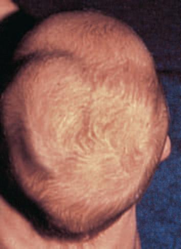
Physiologic Phenomena of the Newborn Cephalohematoma
Caput Succedaneum
Complications from Fetal and Neonatal
Diagnostic Procedures
Neonatal Skin
The skin of the infant differs from that of an adult in that it is thinner (40% to 60%), is less hairy, and has a weaker attachment between the epidermis and dermis.1 In addition, the body surface area–to–weight ratio of an infant is up to five times that of an adult. The infant is therefore at a significantly increased risk for skin injury, percutaneous absorption, and skin-associated infection. Premature infants born before 32 to 34 weeks’ estimated gestational age may have problems associated with an immature stratum corneum (the most superficial cell layer in the epidermis), including an increase in transepidermal water loss (TEWL). This increased TEWL may result in morbidity because of dehydration, electrolyte imbalance, and thermal instability. Interestingly, in the majority of premature infants an acceleration of skin maturation occurs after birth such that most develop intact barrier function by 2 to 3 weeks of life.2 However, in extremely lowbirthweight infants, this process may take up to 4 to 8 weeks.3 In light of the elevated TEWL levels seen in premature infants, a variety of studies have evaluated the use of occlusive dressings or topical emollients in an effort to improve compromised barrier function.4–7
The risk of percutaneous toxicity from topically applied substances is increased in infants, especially those born prematurely.8,9 Percutaneous absorption is known to occur through two major pathways: (1) through the cells of the stratum corneum and the epidermal malpighian layer (the transepidermal route) and (2) through the hair follicle–sebaceous gland component (the transappendageal route). Increased neonatal percutaneous absorption may be the result of the increased skin surface area–to–weight ratio as well as the stratum corneum immaturity seen in premature neonates. Although transdermal delivery methods may be distinctly advantageous in certain settings, extreme caution must be exercised in the application of topical substances to the skin of infants, given the risk of systemic absorption and potential toxicity. Table 2.1 lists some compounds reported in association with percutaneous toxicity in infants and children.
SKIN CARE OF THE NEWBORN
The skin of the newborn is covered with a grayish-white, greasy material termed vernix caseosa. The vernix represents a physiologic protective covering derived partially from secretions of the sebaceous glands and in part as a decomposition product of the infant’s epidermis. Vernix contains protein, lipids, and water and provides water-binding free amino acids that facilitate the adaptation from amniotic fluid immersion in utero to the dry ambient postnatal state.10 Although its function is not completely understood, it may act as a natural protectant cream to “waterproof” the fetus in utero, where it is submerged in the amniotic fluid.11 Some studies suggest that vernix be left on as a protective coating for the newborn skin and that it be allowed to come off
Abnormalities of Subcutaneous Tissue
Miscellaneous Cutaneous Disorders
Developmental Abnormalities of the Newborn
Congenital Infections of the Newborn
by itself with successive changes of clothing (generally within the first few weeks of life). It has been suggested that vernix-based topical creams may be effective in augmenting stratum corneum repair and maturation in infants and could play a role in the treatment of epidermal wounds.12
The skin acts as a protective organ. Any break in its integrity therefore affords an opportunity for initiation of infection. The importance of skin care in the newborn is compounded by several factors:
1. The infant does not have protective skin flora at birth.
2. The infant has at least one and possibly two open surgical wounds (the umbilicus and circumcision site).
3. The infant is exposed to fomites and personnel that potentially harbor a variety of infectious agents.
Skin care should involve gentle cleansing with a nontoxic, nonabrasive neutral material. During the 1950s, the use of hexachlorophenecontaining compounds became routine for the skin care of newborns as prophylaxis against Staphylococcus aureus infection. In 1971 and 1972, however, the use of hexachlorophene preparations as skin cleansers for newborns was restricted because of studies demonstrating vacuolization in the central nervous system (CNS) of infants and laboratory animals after prolonged application of these preparations.13 At the minimum, neonatal skin care should include gentle removal of blood from the face and head, and meconium from the perianal area, by gentle rinsing with water. Ideally, vernix caseosa should be removed from the face only, allowing the remaining vernix to come off by itself. However, the common standard of care is for gentle drying and wiping of the newborn’s entire skin surface, which is most desirable from a thermoregulatory standpoint. Recommendations for “first bathing” of the newborn suggest timing it according to local culture, performing it only after the newborn is physiologically stable and able to appropriately thermoregulate, considering the use of water alone or water and an appropriately formulated pH neutral (or acidic) liquid cleanser such as a synthetic detergent (syndet) or liquid baby cleanser, and considering the use of gloves by healthcare workers if performing the initial bath.14–16
There is no single method of umbilical-cord care that has been proven to limit bacterial colonization and disease, and cord care practices are quite variable in relation to cultural beliefs and healthcare disparities. Several methods have been reported, including local application of isopropyl alcohol, triple dye (an aqueous solution of brilliant green, proflavine, and gentian violet), and antimicrobial agents such as bacitracin or silver sulfadiazine cream. The routine use of povidone-iodine should be discouraged, given the risk of iodine absorption and transient hypothyroxinemia or hypothyroidism. A safer alternative is a chlorhexidine-containing product.17 A recent clinical review suggests consideration of application of antimicrobial agents
Table 2.1 Reported Hazards of Percutaneous Absorption in Infants and Children
Elevated blood levels of immunosuppressive medication
Ulceration of mucous membranes, skin necrosis, vomiting, diarrhea
Uremia
Reprinted with permission from Bree AF, Siegfried EC. Neonatal skin care and toxicology. In: Eichenfield LF, Frieden IJ, Esterly NB, editors. Textbook of neonatal dermatology. 2nd ed. London: Saunders Elsevier; 2008:59–72.
for infants born at home or otherwise outside of birthing centers or hospitals in resource-limited settings but highlights that their use in high-resource countries or the setting of in-hospital delivery has not been found to provide clear benefit.18
Physiologic Phenomena of the Newborn
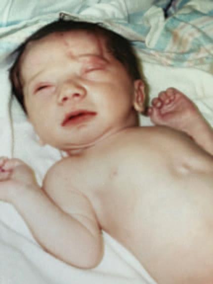
Neonatal dermatology, by definition, encompasses the spectrum of cutaneous disorders that arise during the first 4 weeks of life. Many such conditions are transient, appearing in the first few days to weeks of life only to disappear shortly thereafter. The appreciation of normal phenomena and their differentiation from the more significant cutaneous disorders of the newborn is critical for the general physician, obstetrician, and pediatrician, as well as for the pediatric dermatologist. At birth, the skin of the full-term infant is normally soft, smooth, and velvety. Desquamation of neonatal skin generally takes place 24 to 36 hours after delivery and may not be complete until the third week of life. Desquamation at birth is an abnormal phenomenon and is indicative of postmaturity, intrauterine anoxia, or congenital ichthyosis.
The skin at birth has a purplish-red color that is most pronounced over the extremities. Except for the hands, feet, and lips, where the transition is gradual, this quickly changes to a pink hue. In many infants, a purplish discoloration of the hands, feet, and lips occurs
during periods of crying, breath holding, or chilling. This normal phenomenon, termed acrocyanosis, appears to be associated with an increased tone of peripheral arterioles, which in turn creates vasospasm, secondary dilation, and pooling of blood in the venous plexuses, resulting in a cyanotic appearance to the involved areas of the skin. The intensity of cyanosis depends on the degree of oxygen loss and the depth, size, and fullness of the involved venous plexus. Acrocyanosis, a normal physiologic phenomenon, should not be confused with true cyanosis.
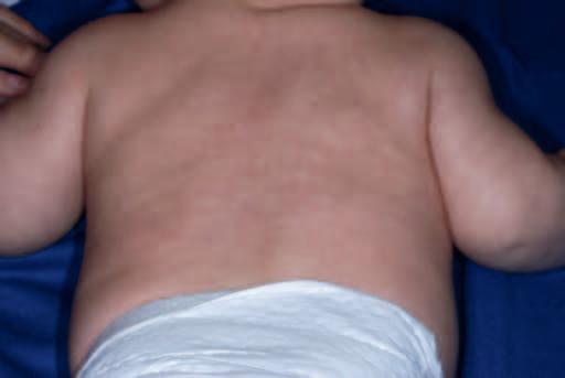
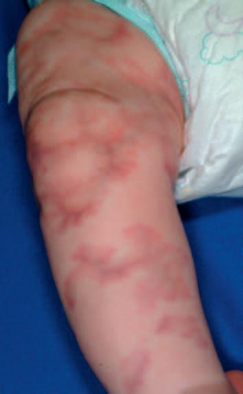
CUTIS MARMORATA
Cutis marmorata is a normal reticulated bluish mottling of the skin seen on the trunk and extremities of infants and young children (Fig. 2.1). This phenomenon, a physiologic response to chilling with resultant dilation of capillaries and small venules, usually disappears as the infant is rewarmed. Although a tendency for cutis marmorata may persist for several weeks or months, this disorder bears no medical significance and treatment generally is unnecessary. In some children cutis marmorata may tend to recur until early childhood, and in patients with Down syndrome, trisomy 18, and Cornelia de Lange syndrome, this reticulated marbling pattern may be persistent. When the changes are persistent (even with rewarming) and are deep violaceous in color, cutis marmorata telangiectatica congenita (Fig. 2.2; see also Chapter 12) should be considered. In some infants a white negative pattern of cutis marmorata (cutis marmorata alba)
may be created by a transient hypertonia of the deep vasculature. Cutis marmorata alba is also a transitory disorder and appears to have no clinical significance.
HARLEQUIN COLOR CHANGE
Harlequin color change, a form of vascular autonomic dysregulation not to be confused with harlequin ichthyosis (see Chapter 5), is occasionally observed in full-term infants but classically described in premature infants. It occurs when the infant is lying on his or her side and consists of reddening of one-half of the body with simultaneous blanching of the other half. Attacks develop suddenly and may persist for 30 seconds to 20 minutes. The side that lies uppermost is paler, and a clear line of demarcation runs along the midline of the body. At
times, this line of demarcation may be incomplete; when attacks are mild, areas of the face and genitalia may not be involved.
This phenomenon appears to be related to immaturity of hypothalamic centers that control the tone of peripheral blood vessels and has been observed in infants with severe intracranial injury as well as in infants who appear to be otherwise perfectly normal. Some medications, including anesthetics and prostaglandin E, may exacerbate the condition.19 Although the peak frequency of attacks of harlequin color change generally occurs between the second and fifth days of life, attacks may occur anywhere from the first few hours to as late as the second or the third week of life.20
INFANTILE TRANSIENT SMOOTH MUSCLE CONTRACTION OF THE SKIN
Believed to be a primitive reflex or autonomic phenomenon, infantile transient smooth muscle contraction of the skin refers to a transient rippling of the skin of otherwise-healthy infants, often triggered by cold or blowing air exposure. In a series of nine full-term newborns with this phenomenon, the authors noted several asymptomatic episodes occurring daily for brief periods (usually 1 minute), with completely normal–appearing skin in between episodes. Histologic examination was unremarkable in the skin biopsy specimens from three patients, and the episodes spontaneously resolved over 18 to 24 months. This condition appears to be related to transient arrector pili smooth muscle contraction, without features to suggest smooth muscle hamartoma.21
BRONZE BABY SYNDROME
Bronze baby syndrome (BBS) is a term used to describe infants who develop a grayish-brown discoloration of the skin, serum, and urine while undergoing phototherapy for hyperbilirubinemia. Although the exact source of the pigment causing the discoloration is not clear, the syndrome usually begins 1 to 7 days after the initiation of phototherapy, resolves gradually over a period of several weeks after phototherapy is discontinued, and appears to be related to a combination of photoisomers of bilirubin or biliverdin or a photoproduct of copperporphyrin metabolism.22–24 Infants who develop BBS usually have modified liver function, particularly cholestasis, of various origins.25 Although very few babies with cholestasis develop BBS during phototherapy, those who do should be investigated for underlying liver disease.26 The disorder should be differentiated from neonatal jaundice, cyanosis associated with neonatal pulmonary disorders or congenital heart disease, an unusual progressive hyperpigmentation (universalacquired melanosis, or “carbon baby” syndrome),27 and chloramphenicol intoxication (“gray baby” syndrome).28 Although early recognition of BBS suggests further evaluation for associated disorders, it does not appear to be a contraindication to ongoing phototherapy for hyperbilirubinemia.29 A distinctive purpuric eruption on exposed skin has also been described in newborns receiving phototherapy and is possibly related to a transient increase in circulating porphyrins.30 This condition, however, is unlikely to be confused with BBS.
Cephalohematoma
A cephalohematoma is a subperiosteal hematoma overlying the calvarium. These lesions are more common after prolonged labor, instrument-assisted deliveries, and abnormal presentations. They usually develop over the first hours of life and present as subcutaneous swellings in the scalp. They do not cross the midline (Fig. 2.3) because they are limited to one cranial bone, which helps distinguish them from caput succedaneum (see the next section). Occasionally, a cephalohematoma may occur over a linear skull fracture. Other potentially associated complications include calcification (which may persist radiographically for years), osteomyelitis, hyperbilirubinemia, and infection. Signs of an infected cephalohematoma, usually caused by Staphylococcus aureus or Escherichia coli, include erythema, increasing size, and fluctuance.31Although infected lesions (which are rare) may require aspiration,32 most lesions require no therapy, with spontaneous resorption and resolution occurring over several weeks to months.
Fig. 2.1 Cutis marmorata. Reticulate bluish mottling that resolves with rewarming.
Fig. 2.2 Cutis marmorata telangiectatica congenita. Violaceous, reticulate patches with subtle atrophy. These changes did not resolve with rewarming and were associated with mild ipsilateral limb hypoplasia.
Caput Succedaneum
Caput succedaneum is a localized edema of the newborn scalp related to the mechanical forces involved in parturition. It is probably related to venous congestion and edema secondary to cervical and uterine pressure and as such is more common with prolonged parturition and seen most often in primigravidas. Caput succedaneum presents as a boggy scalp mass and may result in varying degrees of bruising and necrosis in addition to the edema, at times with tissue loss. In distinction to cephalohematoma, caput succedaneum lesions often cross the midline. These lesions tend to resolve spontaneously over 48 hours, and treatment is generally unnecessary. One possible complication in cases of severe caput succedaneum is permanent alopecia. Halo scalp ring refers to an annular alopecia that presents in a circumferential ring around the scalp in infants with a history of caput.33 It represents a pressure necrosis phenomenon, and the hair loss may be transient or occasionally permanent.
Complications from Fetal and Neonatal Diagnostic Procedures
Fetal complications associated with invasive prenatal diagnostic procedures include cutaneous puncture marks, scars or lacerations, exsanguination, ocular trauma, blindness, subdural hemorrhage, pneumothorax, cardiac tamponade, splenic laceration, porencephalic cysts, arteriovenous or ileocutaneous fistulas, digital loss (in 1.7% of newborns whose mothers had undergone early chorionic villus sampling), musculoskeletal trauma, disruption of tendons or ligaments, and occasionally gangrene. Cutaneous puncture marks, which occur in 1% to 3% of newborns whose mothers have undergone amniocentesis, may be seen as single or multiple 1- to 6-mm pits, dimples, or round depressed scars on any cutaneous surface of the newborn (Fig. 2.4).34–36
Fetal scalp monitoring can result in infection, bleeding, or fontanel puncture, and prenatal vacuum extraction can produce a localized area of edema, ecchymosis, or localized alopecia. The incidence of scalp electrode infection varies from 0.3% to 5%, and although local sterile abscesses account for the majority of adverse sequelae, S. aureus or Gram-negative infections, cellulitis, tissue necrosis, subgaleal abscess, osteomyelitis, necrotizing fasciitis, and neonatal herpes simplex infections may also occur as complications of this procedure (Fig. 2.5).37–39 It is not unusual for new parents to be under the false impression that
fetal scalp electrodes are the cause of aplasia cutis congenita (ACC; see later in this chapter).
Scalp injuries sustained during the birth process tend to be minor and include lacerations, erosions, and ecchymoses. Injuries of the scalp and face occur in approximately 16% of vacuum-assisted deliveries and in 17% of forceps-assisted deliveries (Fig. 2.6).40 In a large series of vacuum-assisted deliveries, occurrence of head injury did not correlate with duration of vacuum application or the number of popoffs or pulls.41
Transcutaneous oxygen monitoring (application of heated electrodes to the skin for continuous detection of tissue oxygenation) and pulse oximetry may also result in erythema, tissue necrosis, and firstor second-degree burns. Although lesions associated with transcutaneous oxygen monitoring generally resolve within 48 to 60 hours, persistent atrophic hyperpigmented craters may at times be seen as a complication. Frequent (every 2 to 4 hours) changing of electrode sites and reduction of the temperature of the electrodes to 43° C, however, can lessen the likelihood of this complication.42 43
Anetoderma of prematurity refers to macular depressions or outpouchings of skin associated with loss of dermal elastic tissue seen in premature infants. Reports suggest that these cutaneous lesions may correlate with placement of electrocardiographic or other monitoring electrodes or leads.44,45
Fig. 2.3 Cephalohematoma. Note the sharp demarcation at the midline.
Fig. 2.4 Amniocentesis scars. Multiple depressed scars on the thigh of an infant born to a mother who had an amniocentesis during pregnancy. (Courtesy Lester Schwartz, MD.)
Fig. 2.5 Staphylococcal scalp abscess. Fluctuant, erythematous nodule on the scalp of this 9-day-old infant as a complication of intrauterine fetal monitoring.
Calcinosis cutis may occur on the scalp or chest of infants or children at sites of electroencephalograph or electrocardiograph electrode placement, as a result of diagnostic heel sticks performed during the neonatal period, or after intramuscular or intravenous administration of calcium chloride or calcium gluconate for the treatment of neonatal hypocalcemia. Seen primarily in high-risk infants who receive repeated heel sticks for blood chemistry determinations, heel-stick calcinosis refers to calcified nodules that usually begin as small depressions on the heels. It is considered a form of dystrophic calcinosis cutis (see Chapter 9). With time, generally after 4 to 12 months, tiny yellow or white papules appear (Fig. 2.7, A), gradually enlarge to form nodular deposits, migrate to the cutaneous surface, extrude their contents (“transepidermal elimination”) (Fig. 2.7, B), and generally disappear spontaneously by the time the child reaches 18 to 30 months of age.
Although calcified heel nodules are usually asymptomatic, children may at times show signs of discomfort with standing or wearing shoes. In such instances, gentle cryosurgery and curettage can be both diagnostic and therapeutic. Rarely, persistent or symptomatic cases may require surgical excision.46 Calcinosis cutis after electroencephalography or electrocardiography is more likely to be seen in infants and young children or individuals where the skin has been abraded and usually disappears spontaneously within 2 to 6 months. It can be avoided by the use of an electrode paste that does not contain calcium chloride, and like calcified heel sticks, they may be treated by gentle cryosurgery and curettage.47 48
Abnormalities of Subcutaneous Tissue
Skin turgor is generally normal during the first few hours of life. As normal physiologic dehydration occurs during the first 3 or 4 days of life (up to 10% of birthweight), the skin generally becomes loose and wrinkled. Subcutaneous fat is normally quite adequate at birth and increases until about 9 months of age, thus accounting for the traditional chubby appearance of the healthy newborn. A decrease or absence of this normal panniculus is abnormal and suggests the possibility of prematurity, postmaturity, or placental insufficiency.
Sclerema neonatorum and subcutaneous fat necrosis (SCFN) are two disorders that affect the subcutaneous fat of the newborn. Although there is considerable diagnostic confusion between these two entities, there are several distinguishing features that enable a clinical differentiation (Table 2.2). Sclerema neonatorum seems to occur significantly less often than SCFN.
SCLEREMA NEONATORUM
Sclerema neonatorum is a diffuse, rapidly spreading, wax-like hardening of the skin and subcutaneous tissue that occurs in premature or debilitated infants during the first few weeks of life and may rarely present after several months of life.49 The disorder, usually associated with a serious underlying condition such as sepsis or other infection, congenital heart disease, respiratory distress, intracranial hemorrhage, diarrhea, or dehydration, is characterized by a diffuse nonpitting woody induration of the involved tissues. The process is symmetric, usually starting on the legs and buttocks, and may progress to involve all areas except the palms, soles, and genitalia.50 As the disorder spreads, the skin becomes cold, yellowish-white, mottled, stony hard, and cadaver-like. The limbs become immobile, and the face acquires a fixed mask-like expression. Infants with this disorder become sluggish, feed poorly, show clinical signs of shock, and, in a large percentage of cases, die.
Although the cause of this disorder is unknown, it appears to represent a nonspecific sign of severe illness rather than a primary
Fig. 2.6 Forceps-induced ecchymoses. This newborn boy was delivered vaginally with forceps assistance and developed this bruising, which conforms to the shape of the forceps blade.
Fig. 2.7 Heel-stick calcinosis. (A) Firm, yellow-white papule on the lateral plantar heel of an infant who had multiple heel sticks as a newborn. (B) Hyperkeratotic firm papule on the heel of another infant, with early transepidermal elimination before resolution.