University of Missouri-Kansas City School of Medicine
Preface
Hypnopedia: the art or process of learning while asleep by means of lessons recorded on disk or tapes
As a child, I was always fascinated by the advertisements on the back of the comic books that my brother Howard and I avidly read. Among the many ads for a myriad of amazing items and services was one featuring a picture of a white-bearded Russian scientist standing next to a sleeping woman, touting that for just $19.95 you could purchase lessons that could teach you to Learn While You Sleep. Given that the Russians had just launched Sputnik and had supposedly detonated a hydrogen bomb, I was completely convinced that this was something I could not live without. I must admit that part of my desire to buy Learn While You Sleep was that I hated school and was always looking for an easier way to complete my lessons.
While I was never able to con my parents into spending the $19.95 for the Learn While You Sleep lessons, they did buy me a pair of the x-ray vision glasses for the then princely sum of $1.99. Needless to say, they didn’t work nearly as well as I had hoped, and I began to wonder if the other things advertised on the back pages of my comics were as bogus. I didn’t have to wonder too long as the full-size replica of a Sherman tank that my brother had ordered off the back of a Superman comic turned out to be little more than a big orange cardboard box. So much for Learn While You Sleep!
At this point, the reader might ask, ‘‘What does an old comic book ad for Learn While You Sleep have to do with a review text for pain management?’’
Well, as my brother Howard, with whom I have practiced pain management for the past 26 years, will tell you, I am still and always looking for an easier way to do things. When I started studying for my American Board of Anesthesiology recertification examination in pain management, there were no texts written to specifically help one review pain management in an organized and time-efficient manner, and I approached my publishers with the concept of creating such a review text. The result of our efforts is Pain Review.
In writing Pain Review, it was my goal to create a text that not only contained all of the material needed to review the specialty of pain management but also to organize that material into small, concise, easy-to-read chapters.
I believe that by breaking up the overwhelming amount of knowledge related to pain management into smaller and more manageable packets of information, the task of reviewing the entire specialty becomes much less daunting. I have also made liberal use of illustrations, as in many chapters a picture is the best way to convey a concept or technique.
Whether you are getting ready to take your certification or recertification examination in pain management or simply want to learn more about the specialty, I hope that Pain Review will serve your needs and help with your studies.
Steven D. Waldman, MD, JD
SECTION 1 ANATOMY
1.
5.
6.
7.
8.
239. Vagus Nerve Block
240. Spinal Accessory Nerve Block
241. Phrenic Nerve Block
242. Facial Nerve Block 391
243. Superficial Cervical Plexus Block 392
244. Deep Cervical Plexus Block 393
245. Recurrent Laryngeal Nerve Block 395
246. Stellate Ganglion Block 396
247. Radiofrequency Lesioning of the Stellate Ganglion 400
248. Cervical Facet Block 401
249. Radiofrequency Lesioning of the Cervical Medial Branch 404
288. Injection Technique for Lumbar Myofascial Pain Syndrome 464
289. Splanchnic Nerve Block
290. Celiac Plexus Block
291. Ilioinguinal Nerve Block
292. Iliohypogastric Nerve Block
293. Genitofemoral Nerve Block
Lumbar Sympathetic Ganglion
Ganglion of Walther (Impar) Block
306. Sacroiliac Joint Injection
307. Intra-articular Injection of the Hip Joint
308. Injection Technique for Ischial Bursitis
309. Injection Technique for Gluteal Bursitis 512
310. Injection Technique for Psoas Bursitis 513
311. Injection Technique for Iliopectineal Bursitis 514
312. Injection Technique for Trochanteric Bursitis 515
313. Injection Technique for Meralgia Paresthetica 517
314. Injection Technique for Piriformis Syndrome 518
315. Lumbar Plexus Block 520
316. Femoral Nerve Block 526
317. Obturator Nerve Block 528
318. Sciatic Nerve Block 531
319. Tibial Nerve Block at the Knee 534
320. Tibial Nerve Block at the Ankle 536
321. Saphenous Nerve Block at the Knee 537
Overview of the Cranial Nerves
Abnormal cranial nerve examination should alert the clinician to the possibility of not only central nervous system disease but also significant systemic illness. For this reason, a careful examination of the cranial nerves should be carried out in all patients suffering from unexplained pain. Abnormalities of the cranial nerves may affect one or more of the cranial nerves, and identification of these abnormalities may aid in the localization of a central nervous system lesion or may suggest a more diffuse process such as meningitis, pseudotumor cerebri, or the presence of systemic disease such as diabetes, sarcoidosis, botulism, myasthenia gravis, Guillain-Barré, vasculitis, and others. Common causes of specific cranial nerve abnormalities are listed in respective chapters that discuss each of the 12 cranial nerves. The 12 cranial nerves are listed here in Table 1-1. The classic acrostic, On Old Olympia’s Towering Top A Finn And German Vault And Hop, has been augmented by the use of a novel clockface-based paradigm to help learners memorize the names and functions of the cranial nerves (Fig. 1-1). This clockface paradigm will be presented in each chapter describing the individual cranial nerves.
To best understand cranial nerve abnormalities, it is useful to think about them in the context of their anatomy. Although the anatomy of the specific cranial nerves will be discussed in the individual chapters covering each cranial nerve, the following schema may be applied to all of the 12 cranial nerves. The efferent fibers of the cranial nerves arise deep within the brain in localized anatomic areas called the nuclei of origin. These nerves exit the brain and brainstem at points known as the superficial origins (Fig. 1-2). The afferent fibers of the cranial nerves arise outside the brain and may take the form of either specialized fibers that are grouped together in a sense organ (e.g., the eye or nose) or grouped together within the trunk of the nerve to form ganglia. The fibers enter the brain to coalesce to form the nuclei of termination. Lesions that affect the peripheral portion or trunks of the cranial nerves are called infranuclear lesions. Lesions that affect the nuclei of the cranial nerves are called nuclear lesions. Lesions that affect the central connections of the cranial nerves are called supranuclear lesions. When evaluating a patient presenting with a cranial nerve abnormality, it is also helpful for the clinician to remember that the first two cranial nerves, the olfactory and the optic, are intimately associated with the quite specialized anatomic structures of the nose and eye and are subject to myriad diseases that may present as a cranial nerve lesion. The remaining 10 cranial nerves are much more analogous in structure and function to the spinal nerves and thus more subject to entrapment and/or compression from extrinsic processes such as a tumor, an aneurysm, or an aberrant blood vessel rather than primary disease processes.
Table
1-1 The Cranial Nerves
• 1st—Olfactory
• 2nd—Optic
• 3rd—Oculomotor
• 4th—Trochlear
• 5th—Trigeminal
• 6th—Abducens
• 7th—Facial
• 8th—Acoustic/auditory/vestibulocochlear
• 9th—Glossopharyngeal
• 10th—Vagus
• 11th—Spinal accessory
• 12th—Hypoglossal
Fig. 1-1 The clockface paradigm for the twelve cranial nerves. (Modified from Weiss, KL, Eldevik, OP Bieliauskas, L, et al: Cranial nerve clock: Part I. A declarative memory paradigm. Acad Radiol 2001; 8[12]: 1215–1222.)
Olfactory nerve
Optic chiasm
Oculomotor nerve
Intermediary nerve of Wrisberg (nervus intermedius)
Glossopharyngeal nerve
Vagus nerve
Accessory nerve
Hypoglossal nerve
Spinal nerve
Suggested Readings
Trochlear nerve
Trigeminal nerve
Abducent nerve
Facial nerve
Vestibulocochlear nerve
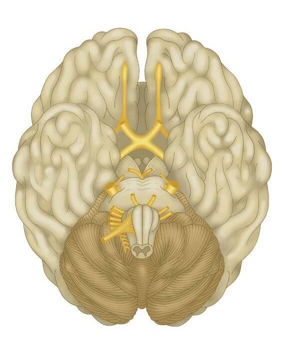
Fig. 1-2 The superficial origin of the cranial nerves. (From Barral JP, Croibier A: Anatomical organization of cranial nerves. In: Barral JP, Croibier A (eds): Manual Therapy for the Cranial Nerves, Edinburgh, Churchill Livingstone, 2009, pp. 15-18.)
Fisch A: Clinical examination of the cranial nerves. In Tubbs RS, Rizk E, Shoja MM, et al (eds): Nerves and Nerve Injuries, San Diego, Academic Press, 2015, pp 195–225. Weiss K, Eldevik OP, Bieliauskas L, et al: Cranial nerve clock: Part I. A declarative memory paradigm, Academic Radiology 8:1215–1222, 2001.
CHAPTER 2
The Olfactory Nerve—Cranial Nerve I
The first cranial nerve is known as the olfactory nerve and is denoted by the Roman numeral I. It is composed of special afferent nerve fibers that are responsible for our sense of smell (Fig. 2-1). The olfactory nerve and associated structures include the chemoreceptors known as the olfactory receptor cells, which are located in the epithelium covering the roof, septum, and superior conchae of the nasal cavity (Fig. 2-2). Inhaled substances dissolve in the moist atmosphere of the nasal cavity and stimulate its chemoreceptors. If a firing threshold is reached, these chemoreceptors initiate action potentials that fire in proportion to the intensity of the stimulus. These stimuli are transmitted via fibers of the olfactory nerve that traverse the cribriform plate to impinge on the olfactory bulb, which contains the cell bodies of the secondary sensory neurons that make up the olfactory tract.
The olfactory tract projects into the cerebral cortex to areas known as the lateral, intermediate, and medial olfactory areas. The lateral olfactory area
is most important to humans’ sense of smell, with the intermediate area less so. The medial olfactory area, via its interconnections with the limbic system, serves to help mediate humans’ emotional response to smell. Collectively, the olfactory receptor cells, epithelium, and bulb tracts and areas are known as the rhinencephalon (Fig. 2-3).
All three olfactory areas interact with a number of autonomic centers via a network of interconnected fibers. The medial forebrain bundle carries information from all three olfactory areas to the hypothalamus, while the stria terminalis carries olfactory information from the amygdala to the preoptic region of the cerebral cortex. The stria medullaris carries olfactory information to the habenular nucleus, which along with the hypothalamus interfaces with a number of cranial nerves to mediate humans’ visceral responses associated with smell. Examples of such visceral responses include the dorsal motor nuclei of the vagus nerve (10th cranial nerve), which can modulate
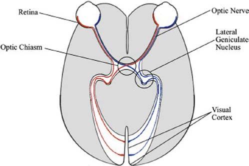
The bipolar cells synapse with and either stimulate or inhibit the ganglion cells that are the secondary sensory neurons of the visual pathway. The axons of the ganglion cells converge at the optic disk near the center of the retina. These axons then exit the posterior aspect of the eye as the optic nerve (cranial nerve II) (Fig. 3-2). Exiting the orbit via the optic canal, the optic nerve enters the middle cranial fossa to join the ipsilateral optic nerve to form the optic chiasm. Fibers from each optic nerve cross the midline to exit the chiasm together as the opposite optic tract (Fig. 3-3).
The optic tracts containing fibers from both optic nerves travel posteriorly, passing around the cerebral peduncles of the midbrain. Most of the fibers of the optic tracts synapse with the tertiary sensory neurons of the lateral geniculate nucleus within their contralateral thalamus (see Fig. 3-3). A few optic tract fibers travel to the pretectal region of the midbrain and provide necessary information for the pupillary light reflex. Via the optic radiations, the tertiary sensory neurons of the lateral geniculate nuclei project to the primary visual cortex, which is located in the occipital lobe (Fig. 3-4).
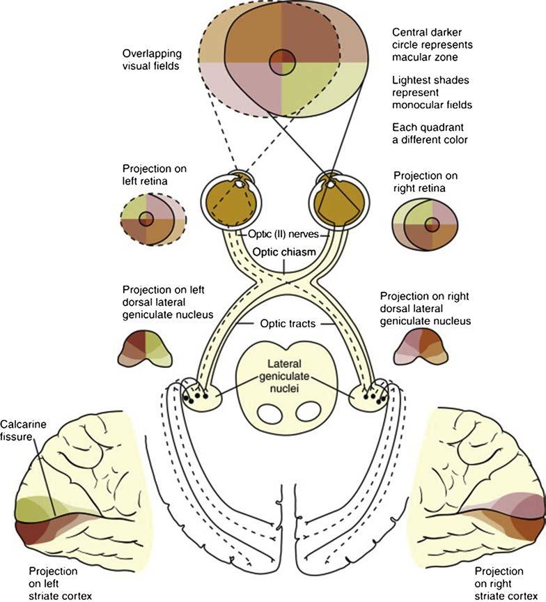
The Visual Field Pathways
The entire area that is seen by the eye when it is focused on a central point is called the visual field of that eye. It must be remembered that the photons entering the cornea converge and pass through the narrow pupil, with the entire visual field being projected on the retina in a reversed and upside down orientation (see Fig. 3-3). This means that the upper half of the retina is stimulated with photons from the lower half of the visual field and the lower half of the retina is stimulated with photons from the upper half of the visual field. Furthermore, the right half of the retina receives stimuli from the left visual field, and the left half of the retina receives stimuli from the right half of the visual field.
Given the consistent way that the ganglion cells from the retina group together to form the optic nerve and carry information to the primary visual cortex, the clinician may find it useful to divide the visual field of each eye into four quadrants: (1) the nasal hemiretina, which lies medial to the fovea; (2) the temporal hemiretina, which lies lateral to the fovea; (3) the superior hemiretina, which lies superior to the fovea; and (4) the inferior hemiretina, which lies inferior to the fovea (see Fig. 3-3). The axons of the ganglion cells of the nasal hemiretina decussate at the optic chiasm and travel on to project onto the contralateral lateral geniculate nucleus and midbrain. The axons of the ganglion cells of the temporal hemiretina remain ipsilateral through their course and project onto the ipsilateral lateral geniculate nucleus and midbrain (see Fig. 3-4). The axons of the ganglion cells of the
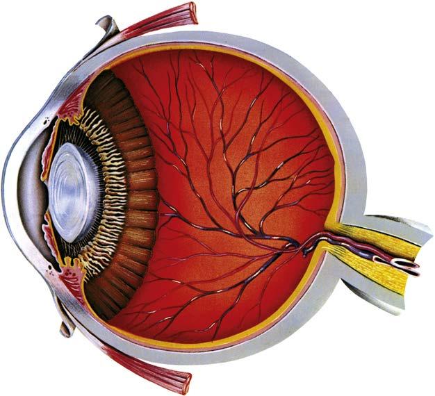
Conjunctiva
Ora serrata
Schlemm's canal
Anterior chamber
Lens
Ciliary body Iris Cornea
Posterior chamber
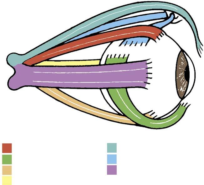
Fig. 3-1 The second cranial nerve is known as the optic nerve and is denoted by the Roman numeral II.
Fig. 3-2 The path of light though the eye. (From Aaron M, Solley WA, Broocker G: Chapter 1 - General Eye Examination. In: Palay DA, Krachmer JH [eds]: Primary Care Ophthalmology, ed 2. Philadelphia, Mosby, 2005, pp 1-23.)
Fig. 3-3 The visual pathway. (From Remington LA: Chapter 13 - Visual Pathway. In: Remington LA (ed): Clinical Anatomy and Physiology of the Visual System, ed 3. St. Louis, Butterworth-Heinemann, 2012, pp 233-252.)
superior hemiretina carrying images from the inferior visual field project via the parietal lobe portion of the optic radiations to the portion of the primary visual cortex located above the calcarine fissure (see Figs. 3-4 and 3-5). The axons of the ganglion cells of the inferior hemiretina carrying images from the superior visual field project via the temporal lobe portion of the optic radiations to the portion of the primary visual cortex located below the calcarine fissure (see Figs. 3-4 and 3-5). Axons of the ganglion cells from the center of the retina or fovea project onto the tip of the occipital pole. Armed with the above knowledge of the functional anatomy of the visual pathway and the optic nerve, based on the patient’s symptoms and visual abnormalities, the clinician can reliably predict what portion of the visual pathway is affected.
Fig. 3-4 Visual field pathways. The visual pathway begins with the retinas in both eyes and depart from the eyes through the optic nerves. All information from the left of the visual field travels through the optic chiasm and continues to the right lateral geniculate nucleus (LGN). The converse occurs for information from the right side of the visual field. This necessitates that information from both eyes crosses at the optic chiasm. From the LGNs, visual information proceeds to the visual cortex of the respective cerebral hemisphere. (From Escobar A: Qualia as the fundamental nature of visual awareness. J Theor Biol 2011; 279[1]:172-176.)
Clinical Evaluation of the Optic Nerve and Visual Pathway
Evaluation of optic nerve function also by necessity includes evaluation of retinal function. The clinician examines each of the patient’s eyes individually and begins the examination with an assessment of visual acuity. Distant vision is tested using a standard Snellen test chart, and near vision is tested by having the patient read the smallest type possible from a Jaeger reading test card placed 14 inches from the eye being tested. Color blindness, which
Table 3-1 Common Diseases That Result in Visual Impairment
Systemic
Diseases
• Diabetes mellitus
• Hypertension
• Vitamin A deficiency
• Vitamin B12 deficiency
• Lead poisoning
• Migraine with aura
• Graves’ disease
• Sarcoidosis
• Collagen vascular diseases
• Atherosclerosis and stroke
• Sickle cell disease
• Multiple sclerosis
• Refsum’s disease
• Tay-Sachs disease
Infection
• HIV-associated infections including cytomegalovirus
• Trachoma
• Bacterial infections including gonococcal infections
• Parasitic infections including onchocerciasis
• Spirochete infections including syphilis
• Viral infections
• Leprosy
Eye Diseases
• Macular degeneration
• Glaucoma
• Cataracts
• Retinitis pigmentosa
• Rod and cone dystrophy
• Best disease, also known as vitelliform macular dystrophy
Trauma
• Burns
• Projectile injuries
• Side effects of medications
• Bungee cord and rubber band injuries
• Fish hook injuries
• Fireworks injuries
• Sports injuries
• Complications of eye surgery
Neoplasms
• Optic gliomas
• Melanoma
• Pituitary adenoma
Suggested Readings
Fingeret M, Medeiros FA, Susanna Jr R, Weinreb RN: Five rules to evaluate the optic disk and retinal nerve fiber layer for glaucoma, Optometry - Journal of the American Optometric Association 76(11):661–668, 2005 Nov.
Fisch A: Clinical examination of the cranial nerves. In: Tubbs RS, et al (eds): Nerves and Nerve Injuries, San Diego, Academic Press, 2015,pp 195–225.
Sadun AA, Wang MY: Optic nerve (cranial nerve II). In: Aminoff M (ed): Encyclopedia of the Neurological Sciences, ed 2. San Diego, Academic Press, 2014, pp 672–674.
Waldman SD: Migraine headache. In: Waldman SD (ed): Atlas of Common Pain Syndromes, ed 3. Philadelphia, Saunders, 2015.
CHAPTER 4
The Oculomotor Nerve—Cranial Nerve III
The oculomotor nerve is the third cranial nerve and is denoted by the Roman numeral III. It is made up of both general somatic efferent and general visceral efferent fibers, which serve two distinct functions. The general somatic efferent fibers of the oculomotor nerve provide motor innervation to four of the six extraocular muscles: (1) the ipsilateral inferior rectus muscle, (2) the ipsilateral inferior oblique muscle, (3) the ipsilateral medial rectus muscle, and (4) the contralateral superior rectus muscle (Fig. 4-1). The superior oblique muscles are innervated by the trochlear nerve (cranial nerve IV), and the lateral rectus muscles are innervated by the abducens nerve (cranial nerve VI) (see Chapters 5 and 7). The actions of the six extraocular muscles are summarized in Table 4-1. The general somatic efferent fibers of the oculomotor nerve also provide motor in-
Fig. 4-1 The extraocular muscles. (From Wojno TH: Orbital Disease. In: Palay DA, Krachmer JH [eds], Primary Care Ophthalmology, ed 2. Philadelphia, Mosby, 2005, pp 275-292.)
Superior rectus CN III Elevation Intorsion Adduction
Medial rectus CN III Adduction
Inferior rectus CN III Depression
Extorsion Adduction
Inferior oblique CN III Extorsion Elevation Abduction
Superior oblique CN IV Intorsion Depression Abduction
Lateral rectus CN VI Abduction
CN, cranial nerve.
nervation to levator palpebrae superioris muscles bilaterally, which elevate the upper eyelids (Fig. 4-2).
The general somatic efferent fibers of the oculomotor nerve that provide motor innervation to four of the six extraocular muscles originate from the oculomotor nucleus located near the midline just ventral to the cerebral aqueduct in the rostral midbrain at the level of the superior colliculus. The oculomotor nucleus is bordered medially by the Edinger-Westphal nucleus (see later). Efferent general somatic fibers exit the oculomotor nucleus and pass ventrally in the tegmentum of the midbrain, passing through the red nucleus and medial portion of the cerebral peduncle to emerge in the interpeduncular fossa at the junction of the midbrain and pons.
Exiting the brainstem, the oculomotor nerve (cranial nerve III) passes between the posterior cerebral and superior cerebellar arteries and then passes through the dura mater to enter the cavernous sinus. The nerve runs along the lateral wall of the cavernous sinus just superior to the trochlear nerve (cranial nerve IV) and enters the orbit via the superior orbital fissure
(Fig. 4-3). After entering the orbit, the oculomotor nerve passes through the tendinous ring of the extraocular muscles and then divides into the superior and inferior divisions. The superior division travels superiorly just lateral to the optic nerve to innervate both the superior rectus and levator palpebrae superioris muscles. The inferior division of oculomotor nerve divides into three branches to innervate the medial rectus, inferior rectus, and inferior oblique muscles (see Fig. 4-2).
The general visceral efferent motor fibers of the oculomotor nerve mediate the eye’s accommodation and pupillary light reflexes by providing parasympathetic innervation of the constrictor pupillae and ciliary muscles of the eye (see Fig. 4-2). After entering the orbit, preganglionic parasympathetic fibers leave the inferior division of the oculomotor nerve to synapse in the ciliary ganglion, which lies deep to the superior rectus muscle near the tendinous ring of the extraocular muscles (see Fig. 4-2). Postganglionic fibers exit the ciliary ganglion via the short ciliary nerves, which enter the posterior aspect of the globe at a point near the spot where the optic nerve exits the eye. Traveling anteriorly