Professor of Paediatric Dentistry and Research Lead, School of Dentistry, University of Central Lancashire, Preston, UK
Honorary Senior Research Fellow, University of Glasgow, UK
Monty S. Duggal
Professor of Paediatric Dentistry, Faculty of Dentistry, National University of Singapore, Singapore
Honorary Professor of Child Dental Health, University of Leeds, UK
Marie Thérèse Hosey
Professor and Head of Paediatric Dentistry, King’s College
London Dental Institute, King’s College London, London, UK
Great Clarendon Street, Oxford, OX2 6DP, United Kingdom
Oxford University Press is a department of the University of Oxford. It furthers the University’s objective of excellence in research, scholarship, and education by publishing worldwide. Oxford is a registered trade mark of Oxford University Press in the UK and in certain other countries
The moral rights of the authors have been asserted
Second edition 2001
Third edition 2005
Fourth edition 2012
Impression: 1
All rights reserved. No part of this publication may be reproduced, stored in a retrieval system, or transmitted, in any form or by any means, without the prior permission in writing of Oxford University Press, or as expressly permitted by law, by licence or under terms agreed with the appropriate reprographics rights organization. Enquiries concerning reproduction outside the scope of the above should be sent to the Rights Department, Oxford University Press, at the address above
You must not circulate this work in any other form and you must impose this same condition on any acquirer
Published in the United States of America by Oxford University Press
198 Madison Avenue, New York, NY 10016, United States of America
British Library Cataloguing in Publication Data
Data available
Library of Congress Control Number: 2017943295
ISBN 978–0–19–250626–9
Printed and bound by Replika Press Pvt Ltd, India
Oxford University Press makes no representation, express or implied, that the drug dosages in this book are correct. Readers must therefore always check the product information and clinical procedures with the most up-to-date published product information and data sheets provided by the manufacturers and the most recent codes of conduct and safety regulations. The authors and the publishers do not accept responsibility or legal liability for any errors in the text or for the misuse or misapplication of material in this work. Except where otherwise stated, drug dosages and recommendations are for the non-pregnant adult who is not breast-feeding
Links to third party websites are provided by Oxford in good faith and for information only. Oxford disclaims any responsibility for the materials contained in any third party website referenced in this work.
Preface to the first edition
The child and adolescent deserves the best of care in all the different disciplines of dentistry. I was delighted to be given the opportunity of trying to draw together all the different aspects of paediatric dentistry and am most grateful to my colleagues for agreeing to contribute the various chapters which make up the book. We have tried to cover as much ground as possible within the obvious publishing restrictions and the finished product will inevitably reflect the editor’s perceptions of where current needs and deficiencies exist. The sudden increase in both erosive tooth surface loss and cosmetic awareness in our younger patients made their inclusion in Chapter 8 important, where previously they have not achieved such prominence in paediatric texts. Similarly, periodontal disease (Chapter 10), oral pathology and oral surgery (Chapter 14), and disability (Chapter 16) are as deserving of detailed inclusion in a ‘paediatric’ as much as in any ‘general’ text.
The book was written with undergraduate dental students in mind, but we hope it will also be useful to those engaged in postgraduate studies and to general dental practitioners. Exhaustive references are deliberately not given but suggested ‘Further reading’ lists are included to help expedite further enquiry and learning.
I hope we have shown in Paediatric Dentistry that the early years of life are the time to get it right for the child and adolescent and there is no reason why our young patients should be denied correct and appropriate care.
R.R.W.
Newcastle upon Tyne April 1996
Preface to the second edition
I am delighted to be given the opportunity to edit the second edition of this popular textbook and am grateful to my colleagues for their continuing contribution. The reviews of the first edition identified the need for a chapter on the treatment of caries in the preschool child and I am grateful to Stephen Fayle for undertaking this task.
There have been small modifications and updates to most chapters, which should keep the reader abreast of current theory and practice. Greater use has also been made of ‘Key points’ for revision purposes.
I hope the second edition will continue to help both undergraduates, postgraduates, and general dental practitioners in their practice of paediatric dentistry.
R.R.W.
Newcastle upon Tyne January 2001
Preface to the third edition
I was very pleased when my younger colleagues Marie Thérèse Hosey and Monty Duggal accepted my offer to join me in editing this third edition. Our book has now sold four and a half thousand copies since its launch in 1997 and it is essential that we maintain a contemporary outlook and publish changes in techniques and philosophies as soon as they have an evidence base.
Since 2001 and the second edition, there have been a significant number of changes of authorship, as well as a change of chapters for some existing authors.
Gerry Winter died in December 2002. He was a wise colleague and friend who was a mentor to many of us. I continue to miss his expertise and availability for consultation, by post or telephone, which he freely gave even after his retirement.
John Murray, Andrew Rugg-Gunn, and Linda Shaw have now retired from clinical practice. I am indebted to them all for their support, both in my own personal career and in the production of out textbook. I am grateful to them for allowing the new chapter authors to use their texts and figures.
The restorative section of the book has been remodelled. The endodontics chapter in the previous editions has now been incorporated into either Chapter 8 or Chapter 12, and there are separate chapters relating to the operative care of the primary and the permanent dentitions. Without the help and friendship of Jim Page the original ‘Operative care of dental caries’ chapter would not have been possible. I am grateful to Jim for allowing us to continue to use his original illustrations from that chapter.
Although designed for the undergraduate we hope the new edition will continue to be used by undergraduate, postgraduate, and general dental practitioner alike, and that their practice of paediatric dentistry will be both fulfilling and enjoyable.
R.R.W. Glasgow January 2005
Preface to the fourth edition
It is difficult to believe that 17 years have passed since work began on the first edition. Our Portuguese edition was marketed in 2007 and this has broadened our market significantly in Brazil.
This edition contains some new contributors, Liege Lourenço-Matharu, Lucy Burbridge, Jenny Harris, and Toby Gillgrass, and their enthusiasm and insight have been invaluable. We are grateful to the comments from reviewers and OUP staff which have resulted in revision of a number of chapters, and so we hope that the end product will be as well received as previous editions.
The excitement of a new edition is tinged with sadness at the loss of Nigel Carter at such a young age. Nigel always gave very generously of his time to paediatric colleagues. On a happier note, Peter Gordon is now enjoying a well-earned retirement and we would all like to thank him for his contribution to the three previous editions.
In looking at the author list for the fourth edition we are struck by the number of contributors who have gained promotion to professorial positions since they originally contributed to Paediatric Dentistry. We must be doing something right!
Richard Welbury Marie Thérèse Hosey Monty Duggal
April 2012
Preface to the fifth edition
It is now 21 years since the first edition of Paediatric Dentistry was published. We are privileged to be a recommended text in all UK and Ireland Dental Schools and also in many Schools across the world, especially in Brazil where Paediatric Dentistry is now published in Portuguese.
We welcome many new contributors to the fifth edition: Barbara Chadwick, Fiona Gilchrist, Guy Jackson, Anjali Kandiah, Sondos Albadri, Simon Stone, Susan Parekh, Kathryn Harley, Agnes Bloch-Zupan, Alex Keightley, Alexander Crighton, and Graeme Wright. It has been a pleasure to have you working with us.
We would like to thank colleagues who contributed previously but who have now retired from clinical practice: Lindsay Hunter, Peter Crawford, and Michael Aldred. Thank you for your time and dedication and best wishes for the future.
As always, as clinical teachers we are especially proud of our former students who are now colleagues and contributors to Paediatric Dentistry.
Finally, thank you to our patients and their parents who have put their trust in us.
Richard Welbury Marie Thérèse Hosey Monty Duggal August 2017
Abbreviations xiii
Contributors to the fifth edition
1 Craniofacial growth and development 1
T.J. Gillgrass and R. Welbury
2 Introduction to the dental surgery 16
A.S. Blinkhorn and B.L. Chadwick
3 History, examination, risk assessment, and treatment planning 31
F. Gilchrist and H.D. Rodd
4 Safeguarding children 49
J.C. Harris and R. Welbury
5 Management of pain and anxiety 67
M.T. Hosey, L. Lourenço-Matharu, and G.J. Roberts
6 Local anaesthesia for children 84
J.G. Meechan and G. Jackson
7 Diagnosis and prevention of dental caries 97
C. Deery and K.J. Toumba
8 Treatment of dental caries in the preschool child 117
S.A. Fayle and P. Kandiah
9 Operative treatment of dental caries in the primary dentition 129
M.S. Duggal and P.F. Day
10 Operative treatment of dental caries in the young permanent dentition 155
J.A. Smallridge and S. Albadri
11 Advanced restorative dentistry 183
N.M. Kilpatrick and L.A.L. Burbridge
12 Periodontal diseases in children 208
P.A. Heasman and P.J. Waterhouse
13 Traumatic injuries to the teeth 227
R. Welbury, J.M. Whitworth, S.J. Stone, and M.S. Duggal
14 Anomalies of tooth formation and eruption 257
S. Parekh, K. Harley, and A. Bloch-Zupan
15 The paedodontic–orthodontic interface 277
T J. Gillgrass and A.J. Keightley
A. Crighton and J.G. Meechan
M.T. Hosey and R. Welbury
J.H. Nunn and G. Wright
Abbreviations
AAGBI Association of Anaesthetists of Great Britain and Ireland
ABH angina bullosa haemorrhagica
AC alveolar crest
AD autosomal dominant
ADHD attention-deficit hyperactivity disorder
ADJ amelodentinal junction
AI amelogenesis imperfecta
AIDS acquired immunodeficiency syndrome
ALL acute lymphocytic leukaemia
ALOSS attachment loss
ALP alkaline phosphatase
AML acute myeloid leukaemia
AMSA anterior middle superior alveolar nerve block
AP anteroposterior
APAGBI Association of Paediatric Anaesthetists of Great Britain and Ireland
S. Albadri, Reader in Paediatric Dentistry, University of Liverpool, Liverpool, UK
A.S. Blinkhorn, NSW Health Chair of Population Oral Health, Faculty of Dentistry, University of Sydney, New South Wales, Australia
A. Bloch-Zupan, Université de Strasbourg, Faculty of Dental Surgery, Institute for Advanced Studies USIAS, Strasbourg, France
Hôpitaux Universitaires de Strasbourg, Pôle de Médecine et Chirurgie Bucco-Dentaires, Reference Center for rare oral and dental diseases, CRMR O-Rares, Strasbourg, France
Institute of Genetics and Cellular and Molecular Biology, CERBM, Université de Strasbourg, CNRS
UMR7104, INSERM U964, Illkirch, France
L.A.L. Burbridge, Consultant in Paediatric Dentistry, Newcastle Dental Hospital/Northern and Yorkshire Cleft Service, UK
B.L. Chadwick, Professor of Paediatric Dentistry, School of Dentistry, Cardiff University, Cardiff, UK
A. Crighton, Honorary Clinical Senior Lecturer, Glasgow Dental Hospital and School, Glasgow, UK
P.F. Day, Associate Professor and Consultant in Paediatric Dentistry, School of Dentistry, University of Leeds, and Bradford District Care Foundation Trust, Salaried Dental Service, Leeds, UK
C. Deery, Professor of Paediatric Dentistry and Dean of the School of Clinical Dentistry, University of Sheffield, Sheffield, UK
M.S. Duggal, Professor of Paediatric Dentistry, Faculty of Dentistry, National University of Singapore, Singapore, and Honorary Professor of Child Dental Health, University of Leeds, UK
S.A. Fayle, Consultant and Senior Clinical Lecturer in Paediatric Dentistry, Leeds Dental Institute and University of Leeds, Leeds, UK
F. Gilchrist, Senior Lecturer/Honorary Consultant in Paediatric Dentistry, School of Clinical Dentistry, University of Sheffield, UK
T.J. Gillgrass, Consultant and Honorary Clinical Senior Lecturer, Glasgow Dental Hospital and School, Glasgow, UK
K. Harley, Consultant and Honorary Clinical Lecturer in Paediatric Dentistry, Eastman Dental Hospital, University College Hospitals Trust, London, UK
J.C. Harris, Consultant in Paediatric Dentistry, Sheffield Teaching Hospitals NHS Foundation Trust, Sheffield, UK
P.A. Heasman, Professor of Periodontology, Department of Restorative Dentistry, Newcastle University, Newcastle, UK
M.T. Hosey, Professor and Head of Paediatric Dentistry, King’s College London Dental Institute, King’s College London, London, UK
G. Jackson, Senior Dentist, NHS Highland, Inverness, UK
P. Kandiah, Consultant in Paediatric Dentistry, Paediatric Dental Department, Manchester Dental Hospital, Manchester, UK
A.J. Keightley, Consultant and Honorary Senior Clinical Lecturer in Paediatric Dentistry, Edinburgh Dental Institute, Edinburgh, UK
N.M. Kilpatrick, Associate Professor and Research Fellow, Department of Paediatrics, University of Melbourne and Murdoch Childrens Research Institute, Melbourne, Australia
L. Lourenço-Matharu, Associate Specialist and Lead Clinician in Dental Paediatric Sedation, King’s College Hospital Dental Institute, London UK
J.G. Meechan, Former Senior Lecturer at Newcastle University and Honorary Consultant Oral Surgeon for Newcastle Hospitals Foundation Trust, UK
J.H. Nunn, Professor Emeritus, School of Dental Science, Trinity College Dublin/Dublin Dental University Hospital, Dublin, Ireland
S. Parekh, Senior Lecturer, Honorary Consultant in Paediatric Dentistry, Eastman Dental Institute, University College London, London, UK
G.J. Roberts, Visiting Professor, King’s College London Dental Institute, London, UK
H.D. Rodd, Professor/Honorary Consultant in Paediatric Dentistry, School of Clinical Dentistry, University of Sheffield, UK
J.A. Smallridge, Consultant in Paediatric Dentistry, Addenbrooke’s Hospital, Cambridge, UK
S.J. Stone, Clinical Lecturer in Restorative Dentistry, Honorary Specialty Registrar Endodontology, Newcastle University, Newcastle upon Tyne, UK
K.J. Toumba, Professor of Paediatric and Preventive Dentistry, School of Dentistry, University of Leeds, Leeds, UK
P.J. Waterhouse, Senior Clinical Lecturer in Child Dental Health, School of Dental Sciences, Newcastle University, UK
R. Welbury, Professor of Paediatric Dentistry and Research Lead, School of Dentistry, University of Central Lancashire, Preston, UK Honorary Senior Research Fellow, University of Glasgow, Glasgow, UK
J.M. Whitworth, Professor of Endodontology/Honorary Consultant in Restorative Dentistry, School of Dental Sciences, Newcastle University, Newcastle, UK
G. Wright, Consultant and Honorary Senior Clinical Lecturer in Paediatric Dentistry, Edinburgh Dental Institute, Edinburgh, UK
1 Craniofacial growth and development
T.J. Gillgrass and R. Welbury
1.1 Introduction
This chapter describes, in general terms, the prenatal development and postnatal growth of the craniofacial skeleton, and the occlusal development of the primary and permanent dentitions.
1.2 Prenatal development
Understanding of embryological development is essential for the dental practitioner who may frequently face patients with common craniofacial anomalies such as cleft lip and/or palate. For routine care, an understanding of their development and aetiology will bring insight to their likely presenting signs and symptoms.
This section will include a brief summary of the development of the face, including the neural crest and pharyngeal arches. It is not the intention of this summary to be in any way a complete or thorough description but simply to describe some of the key cells/interactions and structures.
1.2.1
Neural crest
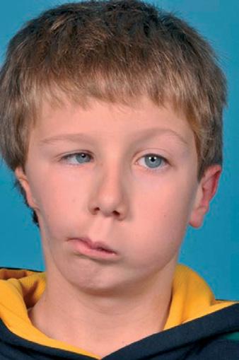
Neural crest cells are derived from the neural fold, and are highly migratory and specialized cells capable of predetermined differentiation. The differentiation occurs after their migration and is essential for the normal development of face and teeth (Fig. 1.1).
1.2.2 Branchial arches
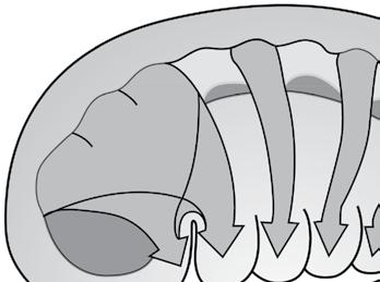
By week 4 the primitive mouth or stomatodeum is bordered laterally and from the developing heart inferiorly by the pharyngeal or branchial arches (Fig. 1.2). These are six bilateral cylindrical thickenings (although the fifth and sixth are small) which form in the pharyngeal wall and into which the neural crest cells migrate. They are separated externally by the branchial grooves and internally by the pharyngeal pouches. The first groove and pouches are involved in the formation of the auditory apparatus and the Eustachian tube.
Each arch has a derived cartilage rod, muscular, nervous, and vascular component. The first two arches and their associated components are central to the development of the facial structures.
This period is also characterized by the development of the organs for hearing, sight, and smell, namely the otic, optic, and nasal placodes.
(a)
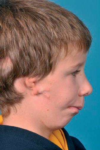
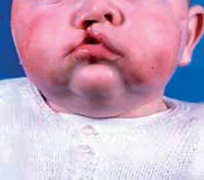
Fig. 1.1 A child with the hemifacial microsomia part of the oculoauricular-vertebral spectrum. The unilateral inhibition of neural crest migration and bronchial arch development results in (a) marked asymmetry and (b) ear defects. (b)
1.2.3 Facial development
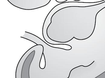
By the end of week 4, thickenings start to develop in the frontal process. The medial and lateral frontonasal processes develop from these, together with the nasal placodes.
The maxillary process develops from the first pharyngeal arch and grows forward to meet the medial and nasal processes, from which it is separated by distinct grooves at week 7 (Fig. 1.3). Its eventual fusion with them creates the upper lip and, from the two medial nasal processes, the incisor teeth and the primary palate. Where this fusion is disturbed a cleft of the lip may form (Fig. 1.4).
The lower lip is formed by fusion of the mandibular process from the first arch.
By week 8 the odontogenic epithelium, which will differentiate into tooth-forming cells, can be determined on the inferior border of the maxillary process, the lateral aspect of the medial process, and the superior border of the mandibular processes.
1.2.4 Secondary palate
Development of the secondary palate starts around week 7. It is formed from three processes: the nasal septum develops in the midline from the frontonasal process and the two palatine shelves develop from
1.3 Postnatal craniofacial growth
There is a great deal of individual variation in the process of postnatal growth and the final form of the craniofacial structures. This section presents a simplified and rather idealized account of bone growth in general and as part of craniofacial growth. Occlusal development is then
Fig. 1.2 Week 6–7 embryo showing branchial arches and migration of neural crest cells into the branchial arch system. Reprinted from Ten Cate’s Oral Histology, 6th edition, Antonio Nanci, copyright (2003) with permission from Elsevier.
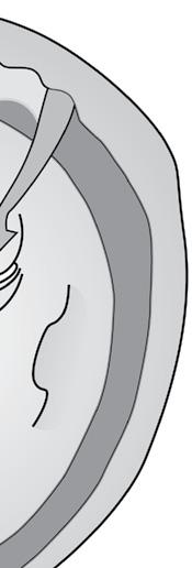
the maxillary processes. At this stage the palatal shelves are directed downwards on either side of the tongue. Between weeks 7 and 8 they elevate to meet the primary palate and nasal septum, to which they fuse (Fig. 1.5).
The trigger for this elevation is still unclear, although high concentrations of glycosaminoglycans which attract water and increase turgidity in the shelves, contractile fibroblasts, and the position of the tongue have all been implicated. Once in contact, the epithelial covering of the shelves must disappear to allow the fusion. Various methods including cell death (‘apoptosis’) and cell transformation have been suggested as methods by which this epithelial covering is lost.
If the shelve fusion fails, this is likely to result in clefts of the palate. The extent of these clefts varies clinically from submucous clefts, affecting the bony structure of the palate and underlying muscular attachment and clefts of the soft palate which may or may not have significant effects on speech, to those including the hard palate producing communication between the nasal and oral cavities (Fig. 1.6).There appear to be distinctive differences between clefts of the palate and those of the lip and palate within different geographical and sexual distributions. This also suggests different disruptive mechanisms and timings, as lip closure occurs earlier in development than palatal fusion. However, as clefts of the palate alone and lip with palate can occur in certain families, it suggests that the distinction may not be complete.
described, before going on to discuss the effect of individual variation in producing departures from this idealized pattern.
An individual’s stature can be charted on standard growth charts during growth. This will present an overall view of the process of growth and
Midbrain
Frontonasal process
Maxillary processes
Mandibular processes
Nasal placode
Stomodeum
representation of early facial development from 4 to 10 weeks i.u.: (a) 4th week in utero (i.u.); (b) 28 days i.u.; (c) 32 days i.u.; (d) 35 days i.u.; (e) 48 days i.u.; (f) 10 weeks i.u. Reproduced from Mitchell L., Introduction to Orthodontics, 4th edition, fig 4.2, page 37, 2013 with permission from Mitchell L, Oxford University Press.
help to detect instances where growth is not proceeding in the usual manner. However, it disguises the fact that the various tissues of the body grow at different rates at different ages (Fig. 1.7).
In order to maintain harmonious facial growth, bone growth must synchronize with that of other tissues. For example, growth of the calvarium is linked to growth of the brain. The cranial vault initially grows much more rapidly than the facial bones in order to keep pace with the developing brain, 90% of which is complete by 5 years of age.
1.3.1 Assessment of postnatal craniofacial growth
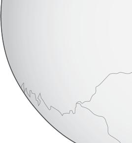
One way of assessing the changes that take place during craniofacial growth is to superimpose tracings of two lateral skull radiographs taken of the same person at different ages. The two radiographs can be compared, as shown in Fig. 1.8, and the changes that have taken place during growth can be examined. A potential difficulty with this approach is that the various bones of the skull grow at different rates at different ages, and there is no single central point about which growth occurs in a radial fashion, i.e. there is no valid fixed radiographic landmark on which to superimpose the films. One convention is to superimpose the tracings of the radiographs on the outline of the sella turcica, using the line from the sella to the frontonasal suture to orientate the films. If this method of superimposition is used, it appears that the cranium expands in a more or less radial fashion to accommodate the brain and the facial skeleton then grows downwards and forwards, away from the cranial base. Another difficulty is that radiographs only produce a two-dimensional representation of what is a three-dimensional structure. Newer
Lateral nasal process
Medial nasal process
(b)
(a)
(d)
(c)
(f)
(e)
Fig. 1.3 Diagrammatic
Fig. 1.4 Failure of fusion resulting in cleft lip and primary palate.
Fig. 1.5 Diagrammatic representation of palatal shelf elevation and subsequent fusion. (a) During week 7 i.u. the palatal shelves begin to develop and lie on either side of the tongue. (b) During week 8 i.u. the palatine shelves elevate rapidly owing to the internal shelf-elevating force and developmental changes in the face. (c) During week 9 i.u. the shelves fuse with each other, the primary palate, and the nasal septum. MC, Meckel’s cartilage; asterisks, palatal shelves. Reproduced from Mitchell L., Introduction to Orthodontics, 4th edition, fig 4.3, page 37, 2013 with permission from Mitchell L, Oxford University Press.
Fig. 1.6 Diagrammatic representations of some of the different types of clefts of the lip and palate: (a) normal; (b) unilateral cleft lip; (c) unilateral cleft lip and anterior palate; (d) bilateral cleft lip and anterior alveolus; (e) cleft of posterior palate (hard and soft); (f) unilateral cleft of the lip and anterior and posterior palate. Reproduced from Johnson D.R and Moore W.J, Anatomy for Dental Students, third edition, fig 2.25, page 52, 1997 with permission from Oxford University Press.
(b) (c)
nasal septum
uvula
incisive foramen
10 years 20 years
Fig. 1.7 Postnatal growth patterns for neural, lymphoid, somatic, and genital tissues shown as percentages of the total increase. The patterns for the maxilla and mandible are shown in blue. Reproduced from Scammon R.D., The Measurement of the Body in Childhood. In Harris J.A. (Ed.) The Measurement of Man. 1930. With permission the University of Minnesota Press.
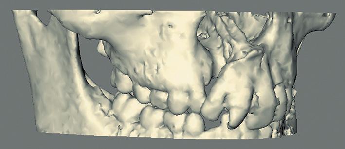
methods of radiological assessment using computed tomography or lower-radiation-dose cone-beam computed tomography (Fig. 1.9) are capable of producing three-dimensional volumetric images of the facial skeleton (Benington et al. 2010).
Soft tissue growth and facial changes are also important for understanding the effects of underlying bony changes. Although computed tomography will capture the soft tissue, it does not accurately depict the colour and texture, and, particularly in the case of conventional computed tomography, produces a significantly higher radiation dose than plain radiographs.
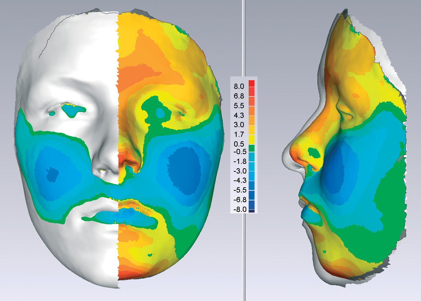
Non-invasive laser scanning or stereophotogrammetry is capable of producing photo-realistic topography of the facial soft tissues without exposure to radiation. Sequential capture is being used for longitudinal growth studies where colour mapping can help illustrate areas of maximum growth (Fig. 1.10) over a substantial time period.
1.3.2 Bone growth
Mineralized bone is formed through a process known as ‘ossification’. This occurs in two ways, intramembranous ossification and endochondral ossification. Intramembranous ossification occurs
Fig. 1.8 Superimpositions on the cranial base showing overall downward and forward direction of facial growth: solid line, 8 years of age; broken line, 18 years of age. Reproduced from Mitchell L., Introduction to Orthodontics, 4th edition, fig 4.7, page 39, 2013 with permission from Mitchell L, Oxford University Press.
Fig. 1.9 Cone-beam computed tomography of a child with a cleft of the alveolus, manipulated to produce a three-dimensional image.
by membrane activity and is seen in the bones of the calvarium, the facial bones, and the mandible. Endochondral ossification occurs by replacement of a cartilage framework. Classically, endochondral ossification is described in long bones but it also occurs in the craniofacial region, most notably in the cranial base.
Growth in bones formed by endochondral ossification occurs at growth centres known as epiphyseal plates in long bones and synchondroses in the cranial base. These are primary growth centres within which the chondroblasts are aligned and clear zones of cell division, hypertrophy, and calcification occur. The most notable of the three synchondroses within the cranial base is the sphenooccipital synchondrosis. The condylar cartilage has a different histological appearance to that of the epiphyseal plates and synchondroses. Although capable of producing bone, its stimuli appear more reactionary to growth around it rather than the primary growth sites which react to both internal and external stimuli.
The apparent growth of the facial bones is a function of remodelling and displacement or translation. Remodelling results in an alteration in the size and shape of bones by deposition and resorption of material on the external and internal surfaces of the bone and suture systems. It is a function of the ‘periosteum’ or ‘osteogenic membrane’. Deposition and resorption go hand in hand; one seldom occurs without the other. Deposition of bone on one aspect of a cortical plate of bone is accompanied by resorption on the other aspect. Displacement or translation occurs when one bone is moved relative to another, primarily due to another area of growth; for example, the maxilla is translated downwards and forwards by growth of the spheno-occipital synchondroses and nasal septum. Such translation will be accompanied by a degree of remodelling.
The suture systems form bone when subjected to traction. In the case of the calvarial bones, the suture systems form new bone and enable the bones to stay in contact with each other when the expansion of the growing brain would otherwise move them apart.
The suture systems allow the bones to respond to growth in neighbouring soft tissue. The suture systems lying between the maxilla and the cranial base allow the downward and forward translation of the maxilla in response to the growth of the soft tissues of the face. It is not proliferation of the vascular connective tissue in the sutures that pushes the bones apart; the whole arrangement of the connective tissue in a suture seems to be designed to enable the suture to respond to a tensile force.
1.3.3 Soft tissue growth
The effects of bony growth can be masked or accentuated by the overlying soft tissues. Notably, this is shown intra-orally in the positions of the dental arches in the so-called neutral zone between the effects of
the tongue, lips, and cheeks. The soft tissues are also responsible for dento-alveolar compensation where the position of the teeth attempts to compensate for skeletal jaw discrepancies.
The growth of the soft tissues, particularly the nose and the length and thickness of the lips, has a profound effect on the appearance of the face. Soft tissue growth shows sexual dimorphism, with changes occurring later and for longer in boys. Changes in the the nose continue into adulthood.
1.3.4 Mechanisms of growth
The mechanisms controlling the process of facial growth are not completely understood. In the post-genomic era it is becoming apparent that genetically encoded factors have a major effect on craniofacial growth; after all, children tend to resemble their parents in facial appearance (Carlson 2005). This may be particularly noted in class III patients and those with a class II division 2 malocclusion. The alternative school of thought is that growth is only loosely under genetic control; rather, the final shape is under the control of its soft tissue environment. This is known as the ‘functional matrix theory’. This is best shown in the cranial vault where the bone growth is reactionary to neural expansion. However, it fails to explain mid-facial growth through the synchondroses.
Therefore it is likely that both mechanisms come into play, The genetically encoded factors can be affected by factors outwith the DNA ‘epigenetic’ that are able to switch them off or on. If this is the case, it should be theoretically possible to influence them (e.g. the use of functional appliances to encourage mandibular growth). At present, however, although a positive response is possible, it appears extremely variable and unpredictable between individuals.
Fig. 1.10 Colour mapping after sequential laser scanning showing facial growth in a forward direction (red) and a negative direction (blue) in the AP plane. Courtesy of Professor Steve Richmond.
1.3.5 Cranial growth
At birth, the cranium is some 60–65% of its adult longitudinal dimensions, and this increases to about 90% by the age of 5 years. The calvarial bones are carried away from each other by the expanding brain and respond by forming new bone in the sutures that separate the bones of the vault of the skull (Fig. 1.11). The six fontanelles that are present at birth reduce in size. The largest (the anterior fontanelle) closes at about 1 year of age and the last to close (the posterolateral fontanelle) closes at about 18 months. The calvarial bones undergo a process of remodelling, with areas of bone deposition and resorption altering the contour of the bones as the volume of the brain cavity increases.
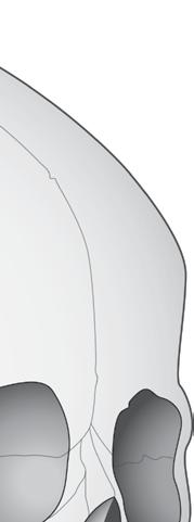
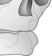
Early fusion of the cranial sutures, or ‘craniostenosis’, results in compensatory growth from the other sutures. This can result in unusual head shapes, and may produce detrimental effects on brain growth and development as it may be accompanied by increased intracranial pressure. The most common craniostenosis involves the sagittal suture. Compensatory growth results in a head shape that is increased in the anteroposterior direction and narrow laterally—‘dolichocephaly’. If the suture fusion is asymmetric, the deformation is also asymmetric— ‘plagiocephaly’ (Fig. 1.12).
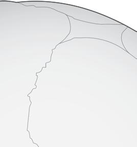
The cranial base also grows to accommodate the changes in the size and shape of the brain, but the process is different to that seen in the calvarial bones. There is considerable lateral growth of the cranial base
1.11 The skull at birth showing the sagittal, coronal, frontal, and lamboid sutures. Reproduced from Johnson D.R and Moore W.J, Anatomy for Dental Students, third edition, fig 2.46, page 71, 1997 with permission from Oxford University Press.
(a) NORMOCEPHALY
(b) DOLICHOCEPHALY (c) ANTERIOR PLAGIOCEPHALY
Fig. 1.12 Skull morphology: (a) normal; (b), (c) abnormal due to early fusion of cranial sutures.