Oxford Handbook of Clinical and Healthcare Research
Oxford Handbook of Clinical and Laboratory Investigation 3e
Oxford Handbook of Clinical Dentistry 6e
Oxford Handbook of Clinical Diagnosis 3e
Oxford Handbook of Clinical Examination and Practical Skills 2e
Oxford Handbook of Clinical Haematology 4e
Oxford Handbook of Clinical Immunology and Allergy 3e
Oxford Handbook of Clinical Medicine—Mini Edition 9e
Oxford Handbook of Clinical Medicine 10e
Oxford Handbook of Clinical Pathology
Oxford Handbook of Clinical Pharmacy 3e
Oxford Handbook of Clinical Rehabilitation 2e
Oxford Handbook of Clinical Specialties 10e
Oxford Handbook of Clinical Surgery 4e
Oxford Handbook of Complementary Medicine
Oxford Handbook of Critical Care 3e
Oxford Handbook of Dental Patient Care
Oxford Handbook of Dialysis 4e
Oxford Handbook of Emergency Medicine 4e
Oxford Handbook of Endocrinology and Diabetes 3e
Oxford Handbook of ENT and Head and Neck Surgery 2e
Oxford Handbook of Epidemiology for Clinicians
Oxford Handbook of Expedition and Wilderness Medicine 2e
Oxford Handbook of Forensic Medicine
Oxford Handbook of Gastroenterology & Hepatology 2e
Oxford Handbook of General Practice 4e
Oxford Handbook of Genetics
Oxford Handbook of Genitourinary Medicine, HIV, and Sexual Health 2e
Oxford Handbook of Geriatric Medicine 2e
Oxford Handbook of Infectious Diseases and Microbiology 2e
Oxford Handbook of Key Clinical Evidence 2e
Oxford Handbook of Medical Dermatology 2e
Oxford Handbook of Medical Imaging
Oxford Handbook of Medical Sciences 2e
Oxford Handbook of Medical Statistics
Oxford Handbook of Neonatology 2e
Oxford Handbook of Nephrology and Hypertension 2e
Oxford Handbook of Neurology 2e
Oxford Handbook of Nutrition and Dietetics 2e
Oxford Handbook of Obstetrics and Gynaecology 3e
Oxford Handbook of Occupational Health 2e
Oxford Handbook of Oncology 3e
Oxford Handbook of Operative Surgery 3e
Oxford Handbook of Ophthalmology 3e
Oxford Handbook of Oral and Maxillofacial Surgery
Oxford Handbook of Orthopaedics and Trauma
Oxford Handbook of Paediatrics 2e
Oxford Handbook of Pain Management
Oxford Handbook of Palliative Care 2e
Oxford Handbook of Practical Drug Therapy 2e
Oxford Handbook of Pre-Hospital Care
Oxford Handbook of Psychiatry 3e
Oxford Handbook of Public Health Practice 3e
Oxford Handbook of Reproductive Medicine & Family Planning 2e
Oxford Handbook of Respiratory Medicine 3e
Oxford Handbook of Rheumatology 3e
Oxford Handbook of Sport and Exercise Medicine 2e
Handbook of Surgical Consent
Oxford Handbook of Tropical Medicine 4e
Oxford Handbook of Urology 3e
Oxford Handbook of Integrated Dental Biosciences
Second Edition
Professor Hugh Devlin
Professor in Restorative Dentistry, University of Manchester, UK
Dr Rebecca Craven
Senior Lecturer in Dental Health, University of Manchester, UK
Great Clarendon Street, Oxford, OX2 6DP, United Kingdom
Oxford University Press is a department of the University of Oxford. It furthers the University’s objective of excellence in research, scholarship, and education by publishing worldwide. Oxford is a registered trade mark of Oxford University Press in the UK and in certain other countries
The moral rights of the authors have been asserted
First Edition published in 2002
Impression: 1
All rights reserved. No part of this publication may be reproduced, stored in a retrieval system, or transmitted, in any form or by any means, without the prior permission in writing of Oxford University Press, or as expressly permitted by law, by licence or under terms agreed with the appropriate reprographics rights organization. Enquiries concerning reproduction outside the scope of the above should be sent to the Rights Department, Oxford University Press, at the address above
You must not circulate this work in any other form and you must impose this same condition on any acquirer
Published in the United States of America by Oxford University Press 198 Madison Avenue, New York, NY 10016, United States of America
British Library Cataloguing in Publication Data
Data available
Library of Congress Control Number: 2017955323
ISBN 978–0–19–875978–2
Printed and bound in China by C&C Offset Printing Co., Ltd.
Oxford University Press makes no representation, express or implied, that the drug dosages in this book are correct. Readers must therefore always check the product information and clinical procedures with the most up-to-date published product information and data sheets provided by the manufacturers and the most recent codes of conduct and safety regulations. The authors and the publishers do not accept responsibility or legal liability for any errors in the text or for the misuse or misapplication of material in this work. Except where otherwise stated, drug dosages and recommendations are for the non-pregnant adult who is not breast-feeding
Links to third party websites are provided by Oxford in good faith and for information only. Oxford disclaims any responsibility for the materials contained in any third party website referenced in this work.
Foreword
Contemporary clinical teaching and learning in dentistry and other health professions now integrates the biosciences with clinical scenarios. This Oxford Handbook of Applied Dental Sciences is, however, the first that provides a format to support this style of learning.
Hugh Devlin and Rebecca Craven have a vast experience of teaching and learning in dentistry and have been at the forefront of advances in this integration. Understanding the relevance of the non-clinical science to the clinical practice that it underpins is now known to provide much more effective learning. This integration enables deep learning. It stimulates interest in the biosciences for the clinical learner and its application becomes more meaningful.
Hugh Devlin is a successful teaching and research enthusiast. He has taught undergraduates and postgraduates at the University of Manchester for over 35 years and his depth and breadth of experience could not be more appropriate for the development of this book. He is a respected clinical academic with over one hundred scientific publications many of which explore the link between basic dental science and their clinical application. Hugh received the IADR Distinguished Scientist Award (2011) for Research in Prosthodontics and Implants.
Rebecca Craven worked in general dental practice and community and hospital dentistry before spending over 25 years in successful university research and teaching. Rebecca, like Hugh, has vast experience in and a passion for teaching and learning and could not be better placed to produce this book. Her studying for the award of ‘Fellowship in Dental Surgery’ required returning to bioscience study after several years of clinical practice and this sparked a lifelong passion to integrate the two disciplines. Rebecca now leads postgraduate Masters programmes in both research and Dental Public Health, a discipline in which she is an NHS Consultant. She also leads the first year of the undergraduate dental programme. Integrating bioscience with clinical care and seeking to apply, appropriately, the best evidence, is at the heart of the University of Manchester Dentistry programmes and of this book.
This book is truly ‘applied’ and presents the bioscience as clinically relevant as possible, with a separate sections detailing the relevant clinical application and putting the science into context. This book therefore supports contemporary methods of teaching where students study a subject based on a clinical scenario. Hugh Devlin and Rebecca Craven have presented a ‘systems’ approach by describing how the different major organs work, what happens with disease, and how this affects the patient’s dental treatment. Rather than discuss sections on pharmacology, histology, epidemiology, and public health, these subjects are woven throughout the text and referred to where their use is most relevant.
The first edition of this book was edited by the late Professor Crispian Scully and had separate sections on physiology, pathology, anatomy, and the
rest. This second edition integrates the topics and focuses more closely on their clinical relevance so that readers will want to supplement their reading of this handbook from other texts, especially for detailed anatomy and cell biology. This book will be an invaluable resource for dental students and dentists studying for higher qualifications after graduation.
Paul Coulthard
BDS MFGDP(UK) MDS FDSRCS FDSRCS(OS) PhD
Dean of the School of Dentistry at the University of Manchester and Professor of Oral and Maxillofacial Surgery
Preface
Our aim is to provide a pocket book of bioscience which is tailored to the needs of dental students. Our approach has been to provide knowledge that is relevant to clinical dental practice and is up-to-date. We want clinicians to reflect on the biological principles and mechanisms, which we hope will encourage deep learning. Science is essential to understand the signs and symptoms of any patient’s disease and the clinician-scientist will be able to offer clear explanations when proposing treatment options to a patient.
Most undergraduate programmes now include some early introduction to dentistry alongside biosciences, but until now that change has not been reflected in textbooks. We aim is to bridge this gap and show directly the relevance of the biology to the clinician.
The previous handbook on this subject, the Oxford Handbook of Applied Dental Sciences, edited by the late Professor Crispian Scully, had separate sections on physiology, pathology, anatomy, and the rest. This new handbook seeks to integrate the topics and focus more closely on their clinical relevance so readers will want to supplement their reading of this handbook from other texts, especially for detailed anatomy and cell biology. The thirteen chapters lead the reader through the major body systems. In most cases, a one page opening is a succinct summary of a topic, enriched by diagrams and illustrations.
The authors have many decades of experience in clinical dentistry and learning and teaching where we have aspired to an integrated and evidencebased approach. The Oxford Handbook format is ideally suited to the dental undergraduate and has proved very popular and practical for students. It is also hoped that clinicians who are seeking to revise their understanding of biosciences or are preparing for higher clinical examinations may find this a useful starting point.
Hugh Devlin Rebecca Craven 2017
Acknowledgements
The authors would like to acknowledge the help of Mr Daniel Wand at the University of Manchester who gave much assistance with many of the diagrams.
Symbols and abbreviations
& and = equal to
multiply
alpha
beta
gamma
kappa i increased
® registered trademark
ACE angiotensin converting enzyme
ACH acetaldehyde
ACS acute coronary syndrome
ACTH adrenocorticotrophic hormone
APTT activated partial thromboplastin time
ADCC antibody dependent cellular cytotoxicity
ADJ amelodentinal junction
ADP adenosine diphosphate
ADS anatomic dead space
AGE advanced glycation end product
ALL acute lymphoblastic leukaemia
ALP alkaline phosphatase
ALT alanine aminotransferase
AML acute myeloid leukaemia
ACE angiotensin converting enzyme
ANP atrial natriuretic hormone
APC antigen presenting cell
ARB angiotensin receptor blockers
ASA American Society of Anaesthesiologists
AST aspartate aminotransferase
ATP adenosine triphosphate
ATP adenosine triphosphate
AV atrioventricular
BMI body mass index
BP blood pressure
BSE bovine spongiform encephalitis
CCK cholecystokinin
CPG central pattern generator
CHD coronary heart disease
CIED cardiovascular implantable electronic device
CKD chronic kidney disease
CKD-MBD CKD-mineral and bone disorder
CLL chronic lymphocytic leukaemia
CML chronic myeloid leukaemia
CNS central nervous system
COMT catechol-O-methyltransferase
COPD chronic obstructive pulmonary disease
CPAP continuous positive airway pressure
CRF chronic renal failure
CSF colony stimulating factor
CT computed tomography
CVA cerebrovascular accident
DCCT Diabetes Control and Complications Trial
DPG diphosphoglycerate
ECG electrocardiogram
e.g. exempli gratia (for example)
eGFR estimated glomerular filtration rate
EGFR epidermal growth factor receptor
EPS extracellular polymeric substance
BFU-E erythroid burst-forming unit
CFU-E erythroid colony forming unit ,
ESR erythrocyte sedimentation rate
ESV end systolic volume
FAP fluorapatite
FEV1 forced expiratory volume
FRC functional residual capacity
FSH follicle stimulating hormone
FVC forced vital capacity
GA general anaesthesia
GABA gamma-aminobutyric acid
GORD gastro-oesophageal reflux disease
GCF gingival crevicular fluid
GHRH growth hormone releasing hormone
GI gastrointestinal
GLP-1 glucagon like peptide-1
GTN glyceryl trinitrate
HAP hydroxyapatite
HbA1c glycated haemoglobin
HERS Hertwig’s epithelial root sheath
HIV human immunodeficiency virus
HLA human leukocyte antigen
HPA axis hypothalamic–pituitary–adrenal
HPV human papilloma virus
HSC haematopoietic stem cell
HSP human heat shock protein
HPV human papilloma virus
HIF-1 hypoxia inducible factor-1
i.e. id est (that is)
IEE internal enamel epithelium
Ig immunoglobin
IL-1 interleukin 1
INR international normalized ratio
KD Kawasaki disease
LDL Lipids
L-DOPA levodopa
LH luteinizing hormone
MHC major histocompatibility complex
MMPs matrix metalloproteinases
MDMA methylenedioxymethamphetamine
MRI magnetic resonance imaging
MSC mesenchymal stem cells
MI myocardial infarction
MIP maximal Intercuspal position
MOA monoamine oxidase
MS multiple sclerosis
MSA multiple system atrophy
MNG multinodular goitre
MROJ medication-related osteonecrosis of the jaw
NK natural killer
NICE National Institute for Health and Care Excellence
NOAC novel oral anticoagulant
NS necrotizing sialometaplasia
NSAIDs non-steroidal anti-inflammatory drugs
RANK nuclear factor kappa-B
OSA obstructive sleep apnoea
OPG osteoprotegerin
PAF platelet activating factor
PAMP pathogen associated molecular pattern
PDL periodontal ligament
PEP post-exposure prophylaxis
PG prostaglandins
PMN polymorphonuclear leucocyte
PPE personal protective equipment
PPI proton pump inhibitor
PRR pattern recognition receptor
PT prothrombin time
PTC papillary thyroid carcinoma
PTH parathyroid hormone
PVS Plummer–Vinson or Paterson–Brown–Kelly syndrome
The early embryo starts as a ball of undifferentiated cells, then organizes into a 3-layered disc:
• Ectoderm—is the origin of skin and infolds to form the nervous system, sensory cells (of eyes, nose, etc), and dental enamel
• Mesoderm—is the origin of connective tissue, bone, muscle, kidneys, gonads, and spleen
• Endoderm—is the origin of epithelial lining of gut and respiratory systems, the parenchyma of liver and pancreas.
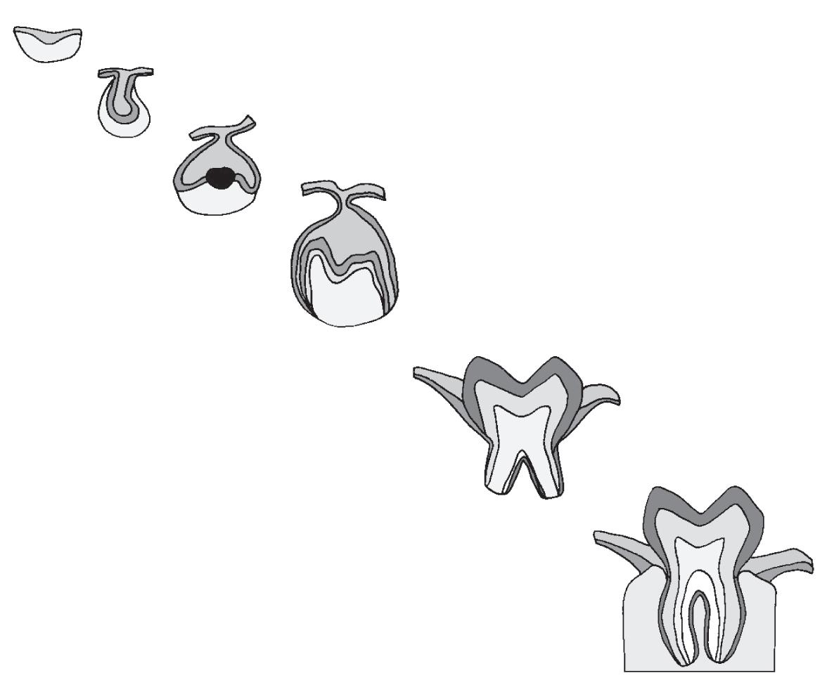
Some ectoderm cells that are involved in infolding to form the neural tube (and subsequently the central nervous system [CNS]) become specialized neural crest cells. These migrate throughout the embryo for specialized purposes and can differentiate into a wide range of cell types and tissues: neurons, glial cells, melanocytes, dermis, tendons, cartilage, bone, and dentine. It is thought that some neural crest derived cells retain some of this potential and may hold promise for regeneration of dental tissue in the adult. The process of tooth formation starts at 5–6 weeks in utero with the formation of the dental lamina. This is a thickening of the ectoderm extending from the lining of the primitive oral cavity down into the underlying ectomesenchyme. Within this dental lamina, focal bud-like thickenings map out the sites of the future teeth, 20 for the primary dentition and later 32 for the permanent. These ectoderm buds together with a surrounding aggregation of ectomesenchymal cells form the earliest stage of the tooth germ. There are 6 stages in which the crown of the tooth is formed (E See Table 1.1). The process progresses from the cusp tip apically towards the root with the outer shape of the crown fully formed before root development starts. (E See Fig. 1.1 and 1.2) The tooth germ comprises:
• Enamel organ develops into the enamel (ectodermal origin)
• Dental papilla develops into the dentine and pulp (neuro-ectodermal origin)
• Dental follicle (sac) develops into the periodontal ligament (neuro-ectodermal origin).
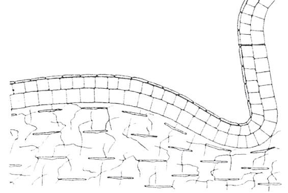
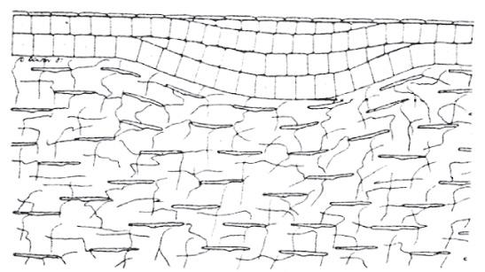
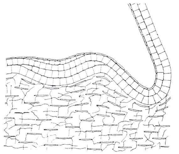
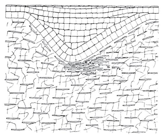
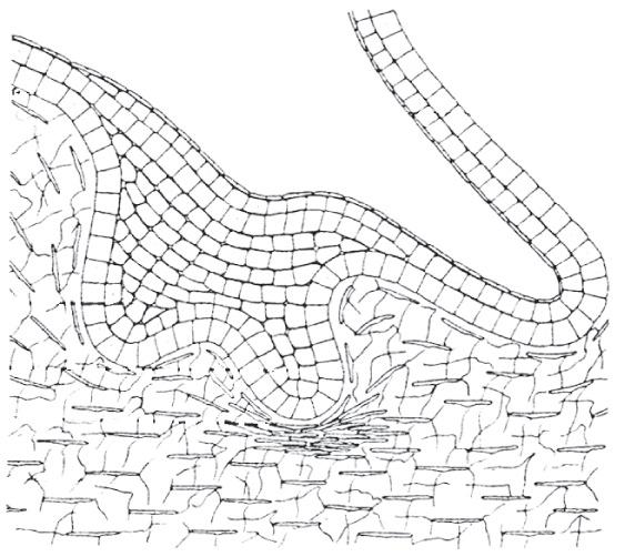
Fig. 1.1 Initiation stage of tooth development. reproduced from Scully, C. Oxford Handbook of Applied Dental Sciences (2003), with permission from oxford University Press.
Fig. 1.2 Diagram showing developmental stages of teeth from initiation to eruption. reproduced from Scully, C. Oxford Handbook of Applied Dental Sciences (2003), with permission from oxford University Press.
Table 1.1 Stages of tooth development
Timing in utero Stage
6 weeks Initiation
8 weeks Bud
9–10 weeks Cap
11–12 weeks Bell
Ectoderm of the primitive stomodeum thickens locally to form dental lamina which maps the later dental arch
Mesenchyme forms neural crest which migrates to the tooth germ
Each dental lamina develops 10 buds
The enamel organ maps out the shape of the crown
Dental papilla is surrounded by follicle and sac
Differentiation of 4 layers
Stratum intermedium
Inner enamel epithelium outer dental papilla
Central cells of dental papilla appositional Dentine and enamel are laid down. Internal enamel epithelium (IEE) cells become preameloblasts which induce odontoblasts to produce predentine. The basement membrane disintegrates, allowing preodontoblasts and preameloblasts to come into contact. Predentine mineralizes and induces enamel matrix production from Tome’s process of ameloblasts. Enamel matrix mineralizes very quickly (dentine facilitates nucleation). odontoblast processes are left as the odontoblast cell bodies continue to lay down more dentine. Centres of calcification (calcospherites) fuse. at the odontoblast layer, there is always a layer of predentine throughout life. Stellate reticulum collapses and nutrients are then supplied from blood supply outside the outer enamel epithelium.
oligodontia (partial anodontia)
Supernumeraries
Macro or microdontia
Enamel/dentine dysplasia
(Continued)
Oral cavity and gut
Table 1.1 (Contd.)
Timing in utero
Stage Activity
Enamel maturation
root formation
Mineral ion uptake increases and crystals grow wider and thicken. Water and organic content are removed from the enamel. at the end of this stage the enamel epithelium degenerates.
once the crown has formed, at the cervical loop, outer and inner enamel epithelium become adjacent (stellate reticulum and stellate intermedium having collapsed). These 2 layers form the hertwig’s epithelial root sheath (hErS) which induces dentine formation between dental papilla and follicle. When complete the basement membrane and hErS disintegrate and some cells remain in a mesh of strands and islands (rests of Malassez). Undifferentiated cells in the dental papilla contact the root dentine and become cementoblasts which secrete cementoid matrix which mineralizes to cementum. Fibroblasts are induced to form periodontal ligament. hyaline layer of cementum is next to the root dentine, a 10 micron highly mineralized layer which allows the first attachment of periodontal ligament fibres. acellular cementum is found near the cervical area. Cementoblasts become encased in their own cementum as cementocytes in cellular cement. root formation takes around 1.5 years from eruption for primary teeth and 2–3 years for permanent teeth.
Potential problems
Enamel pearls (blob of enamel formed on the root by misplaced ameloblasts. Dilaceration— bend in the root due to trauma. accessory roots Concrescence— teeth joined by cementum
Oral cavity and gut
Tooth eruption
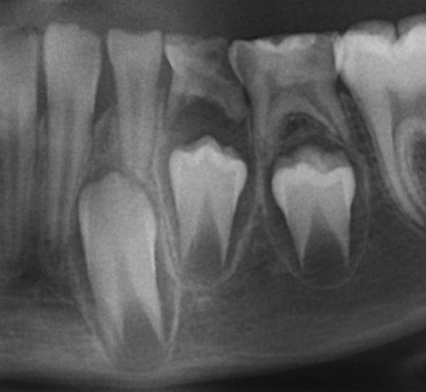
Eruption brings the tooth from its developmental position into its functional position. The mechanism is not fully understood. The dental follicle is crucial (it later becomes the periodontal ligament). It seems the tooth is pushed rather than pulled—there does not seem to be a traction force. The dental follicle evolves to produce a complete crown then transforming growth hormone is released which attracts osteoclasts and macrophages which cause bone remodelling around the crown. Next, the overlying soft tissue breaks down releasing enamel matrix protein which may be part of teething—rhinitis, fever, and inflammation of soft tissue around the erupting crown. Growth and thyroid hormones moderate eruption. (E See Fig. 1.3 for example of developing premolars.)
The mechanisms of eruption are not fully understood but factors potentially contributing to eruption are as follows.
• root formation
• root growth is often happening at the same time as active eruption but seems to follow eruption rather than cause it, e.g. rootless teeth will erupt, teeth with a closed apex can still erupt.
• Tissue fluid hydrostatic pressure
• This mechanism is seen as highly likely. Minute changes in tooth position are synchronized to the pulse and there is a diurnal pattern to eruption across the day. Changes in tissue pressure have been recorded corresponding to eruption activity, increased vascularity, and fenestrations in vasculature producing a swollen ground substance.
Fig. 1.3 Developing permanent canine and premolars. Note: crown is fully formed and root formation is underway.
• Bone remodelling
• Bone is certainly remodelled during tooth eruption (e.g. resorption locally to accommodate the developing clinical crown) but it seems not to be a major motive force.
• Periodontal ligament
• Fibroblasts migrate along the periodontal ligament at the same rate as teeth erupt but this is thought to be a passive process and there seems to be no traction force here.
Problems of delayed eruption
Tooth eruption dates vary from person to person and up to 1 year either side of the given dates should be allowed (E See Table 1.2 and 1.3).
Exfoliation of primary teeth and eruption of any teeth would normally be symmetrical so any 1-sided delay, beyond a few months, should be investigated. Causes for delay are most commonly local obstruction by a supernumerary or impacted tooth or because there is insufficient space for it to erupt into. r arely systemic conditions are the cause, e.g.
• hypothyroid—reduced levels of thyroid hormone
• Gardner’s syndrome—polyposis coli and multiple jaw cysts
• Down’s syndrome—learning disability, immune and cardiovascular defects
• Cherubism—giant cell lesions in mandible+/- maxilla.
Even after the tooth is fully erupted there continues to be an adaptive process of remodelling bone and cementum which maintains the teeth in occlusal contact and vertical dimension. Forces on teeth, whether continuous or intermittent, during eruption can slow, stop or reverse eruption or redirect its path.
Summary of chronology of tooth development
E See Fig. 1.4 for primary teeth and Fig. 1.5 for permanent teeth.
Crown completion
Primary crown is complete:
Eruption date × 0.5 approximately
Permanent crown is complete:
6 1 2 3 3 years before eruption
3 4 5 7 8 5 years before eruption
Root completion
Primary 1–1.5 years after eruption
Permanent 2–3 years after eruption
Total development time
Primary 2–4 years
Permanent 12 years
root resorption of primary teeth commences almost as soon as formation is complete. Exfoliation usually occurs several months before eruption of permanent successor.
Oral cavity and gut
Table 1.2 Eruption dates for permanent teeth
Starts calcifying Crown completed Eruption
Upper 1 3–4 months 4–5 years 7–8 years
Lower 1 6–7 years
Upper 2 10–12 months 8–9 years
Lower 2 3–4 months 7–8 years
Upper 3 4–5 months 6–7 years 11–12 years
Lower 3 9–10 years
Upper 4 1.5–2 years 5–6 years 10–12 years
Lower 4
Upper 5 2–2.5 years 6–7 years 10–12 years
Lower 5
Upper & lower 6 Birth or just before 2.5–3 years 6–7 years
Upper & lower 7 2.5–3 years 7–8 years 12–13 years
Upper & lower 8 7–10 years Earlier for uppers, later for lowers 12–16 years 17–21 years
van Beek GC (1983) Dental Morphology: an Illustrated Guide. Wright.
Table 1.3 Eruption dates for primary teeth
Starts calcifying Crown completed Eruption
Upper a 3–4.5 months in utero 4–5 months 7.5 months
Lower a 6.5 months
Upper B 8 months
Lower B 7 months
Upper C 5 months in utero 9 months 16–20 months
Lower C
Upper D 5 months in utero 6 months 12–16 months
Lower D
Upper E 6 months in utero 10–12 months 20–30 months
Lower E
van Beek GC (1983) Dental Morphology: an Illustrated Guide. Wright.