This adaptation of Orban’s Oral Histology and Embryology, 11e, by S N Bhaskar was undertaken by Reed Elsevier India Private Limited and is published by arrangement with Elsevier Inc.
All rights reserved. No part of this publication may be reproduced or transmitted in any form or by any means, electronic or mechanical, including photocopying, recording, or any information storage and retrieval system, without permission in writing from the publisher. Details on how to seek permission, further information about the Publisher’s permissions policies and our arrangements with organizations such as the Copyright Clearance Center and the Copyright Licensing Agency, can be found at our website: www elsevier com/permissions
This book and the individual contributions contained in it are protected under copyright by the Publisher (other than as may be noted herein).
Notice
Knowledge and best practice in this field are constantly changing. As new research and experience broaden our understanding, changes in research methods, professional practices, or medical treatment may become necessary.
Practitioners and researchers must always rely on their own experience and knowledge in evaluating and using any information, methods, compounds, or experiments described herein. In using such information or methods they should be mindful of their own safety and the safety of others, including parties for whom they have a professional responsibility.
With respect to any drug or pharmaceutical products identified, readers are advised to check the most current information provided (i) on procedures featured or (ii) by the manufacturer of each product to be administered, to verify the recommended dose or formula, the method and duration of administration, and contraindications. It is the responsibility of practitioners, relying on their own experience and knowledge of their patients, to make diagnoses, to determine dosages and the best treatment for each individual patient, and to take all appropriate safety precautions.
To the fullest extent of the law, neither the Publisher nor the authors, contributors, or editors, assume any liability for any injury and/or damage to persons or property as a matter of product liability, negligence or otherwise, or from any use or operation of any methods, products, instructions, or ideas contained in the material herein.
Although all advertising material is expected to conform to ethical (medical) standards, inclusion in this publication does not constitute a guarantee or endorsement of the quality or value of such product or of the claims made of it by its manufacturer.
This publication is licensed for sale in India, Bangladesh, Bhutan, Maldives, Nepal, Pakistan and Sri Lanka only. Circulation of this version outside these territories is unauthorized and illegal.
Sr Editorial Operations Manager: Shabina Nasim
Sr Content Strategist: Nimisha Goswami
Managing Editor: Anand K Jha
Project Manager: Prasad Subramanian
Project Coordinator: Isha Bali
Cover Designer: Milind Majgaonkar
Laser typeset by GW India
Printed in India by
To
My Teachers Who Have Guided Me
My Students Who Have Inspired Me
My Family Who Have Encouraged Me
My Associates Who Have Supported Me
Amsavardani S Tayaar
Professor and Head
Department of Oral Pathology and Microbiology
Indira Gandhi Institute of Dental Sciences
Pondicherry
Chapters 3 and 18
Arun V Kulkarni
Formerly Professor of Anatomy
SDM College of Dental Sciences
Dharwad
Chapters 2, 15 and 16
Dinkar Desai
Professor and Head
Department of Oral Pathology and Microbiology
AJ Institute of Dental Sciences
Mangalore
Chapter 4
Karen Boaz
Professor and Head
Department of Oral Pathology and Microbiology
Manipal College of Dental Sciences
Mangalore
Chapter 13
Pushparaja Shetty
Professor and Head
Department of Oral Pathology
AB Shetty Memorial Institute of Dental Sciences
Nitte University
Mangalore
Chapter 10
Radhika M Bavle
Professor
Oral and Maxillofacial Pathology
Krishnadevaraya College of Dental Sciences and Hospital
Bangalore Chapters 7, 8, 11 and 12
List of Contributors
A Ravi Prakash
Professor and Head
Department of Oral Pathology
G Pulla Reddy Dental College
Kurnool Chapter 5
Sharada P
Professor of Oral Pathology
AECS Maruthi Dental College and Hospital
Bangalore Chapters 7, 8 and 9
Shreenivas Kallianpur
Professor of Oral and Maxillofacial Pathology
People’s College of Dental Sciences and Research Centre
Bhopal Chapter 6
G Venkateswara Rao
Dean and Principal Mamata Dental College
Khammam Chapter 3
Vinod Kumar R B
Principal Malabar Dental college
Edappal Chapter 14
Dr MS Munisekhar
Professor and Head
Department of Oral and Maxillofacial Pathology
SVS Institute of Dental Sciences
Mahabubnagar Chapter 17
G S Kumar
Professor of Oral Pathology and Principal
KSR Institute of Dental Science and Research
Tiruchengode
Chapters 1 and 19, Summary of Chapters 1 - 17 and 19
An Overview of Oral Tissues 1
CHAPTER
OUTLINE
Development of Tooth 1
Enamel 2
Dentin 2
Pulp 2
Cementum 2
Periodontal Ligament 2
Alveolar Bone 3
Temporomandibular Joint 3
The oral cavity contains a variety of hard tissues and soft tissues. The hard tissues are the bones of the jaws and the tooth. The soft tissues include the lining mucosa of the mouth and the salivary glands.
The tooth consists of crown and root. That part of the tooth visible in the mouth is called clinical crown; the extent of which increases with age and disease. The root portion of the tooth is not visible in the mouth in health. The tooth is suspended in the sockets of the alveolar bone by the periodontal ligament. The anatomical crown is covered by enamel and the root by the cementum. Periodontium is the term given to supporting tissues of the tooth. They include the cementum, periodontal ligament and the alveolar bone. The innermost portion of the crown and root is occupied by soft tissue, the pulp. The dentin occupies the region between the pulp and enamel in the crown, and between pulp and cementum in the root.
DEVELOPMENT OF TOOTH
The tooth is formed from the ectoderm and ectomesenchyme. The enamel is derived from the enamel organ which is differentiated from the primitive oral epithelium lining the stomodeum (primitive oral cavity). Epithelial mesenchymal interactions take place to determine the shape of the tooth and the differentiation of the formative cells of the tooth and the timing of their secretion. The ectomesenchymal cells which are closer to the inner margins of the enamel organ differentiate into dental papilla and the ectomesenchymal cells closer to the outer margins of the enamel organ become dental follicle. Dentin and pulp are derivatives of dental papilla while cementum, periodontal ligament and alveolar bone, are all derivatives of dental follicle. The cells that form these tissues have their names ending in blast.
Maxillary Sinus 3
Eruption and Shedding of Teeth 3
Oral Mucosa 3
Salivary Glands 3
Lymphoid Tissue and Lymphatics of Orofacial Region 4
Age Changes in Oral Tissues 4
Study of Oral Tissues 4
Thus, ameloblast produces enamel, odontoblast dentin, cementoblast, cementum and osteoblast bone. These synthesizingcells have all the features of a protein secreting cell—well developed ribosomes and a rough endoplasmic reticulum (ER), Golgi apparatus, mitochondria and a vesicular nucleus, which is often polarized. The cells that resorb the tissues have their names ending in ‘clast’. Thus, osteoclast resorbs bone, cementoclast, cementum and odontoclast resorbs all the dental tissues. The ‘clast’ cells have a similar morphology in being multinucleated giant cells. Their ultra structural features include numerous lysosomes and ingested vacuoles.
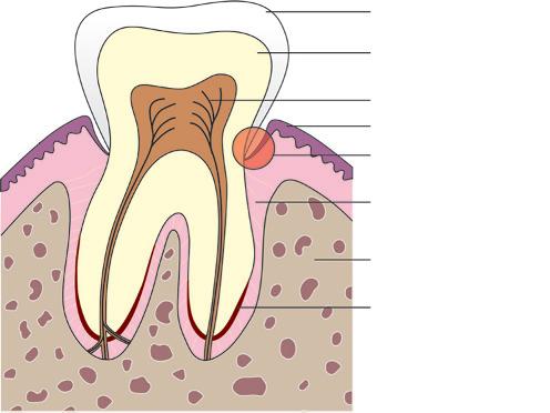
Dentin is the first hard tissue of the tooth to form. Enamel starts its formation after the first layer of dentin has formed. The enamel formation is from its junction with dentin outwards, first in the cuspal/incisal and later in the cervical regions. Dentin formation is similar, but from the dentinoenamel junction, the formation is pulp ward. Cementum formation occurs after the root form, size, shape and number of roots is outlined by the epithelial root sheath and dentin is laid down in these regions. Formation of enamel, dentin and cementum takes place as a daily event in phases or in increments, and hence they show incremental lines. In dentin and cementum formation, a layer of uncalcified matrix forms first, followed by its mineralization. While in enamel formation enamel matrix is calcified, but its maturation or complete mineralization occurs as a secondary event. Mineralization occurs as a result of supersaturation of calcium and phosphorus in the tissue fluid. The formative cells concentrate the minerals from calcium phosphate (apatite) and secrete them into the organic matrix, in relation to specific substances like collagen, which act as attractants or nucleators for mineralization. The mechanism of mineralization is quite similar in all the hard tissues of tooth and in bone (Fig. 1.1).
Enamel
Dentin
Pulp
Gingiva
Attachment of gingiva with tooth
Periodontal ligament
Alveolar bone
Cementum
ENAMEL
The enamel is the hardest tissue in the human body. It is the only ectodermal derivative of the tooth. Inorganic constituents account for 96% by weight and they are mainly calcium phosphate in the form of hydroxyapatite crystals. These apatite crystals are arranged in the form of rods. All other hard tissues of the body, dentin, cementum and bone also have hydroxyapatite as the principal inorganic constituent. Hydroxyapatite crystals differ in size and shape; those of the enamel are hexagonal and longest. Enamel is the only hard tissue, which does not have collagen in its organic matrix. The enamel present in the fully formed crown has no viable cells, as the cells forming it—the ameloblast degenerates, once enamel formation is over. Therefore, all the enamel is formed before eruption. This is of clinical importance as enamel lost, after tooth has erupted, due to wear and tear or due to dental caries, cannot be formed again. Enamel lacks not only formative cells but also vessels and nerves. This makes the tooth painless and no blood oozes out when enamel is drilled while making a cavity for filling.
DENTIN
The dentin forms the bulk of the tooth. It consists of dentinal tubules, which contains the cytoplasmic process of the odontoblasts. The tubules are laid in the calcified matrix—the walls of the tubules are more calcified than the region between the tubules. The apatite crystals in the matrix are plate like and shorter, when compared to enamel. The numbers of tubules near the pulp are broader and closer and they usually have a sinusoidal course, with branches, all along and at their terminus at the dentinoenamel or cementodentinal junction. The junction between enamel and dentin is scalloped to give mechanical retention to the enamel. Dentin is avascular. Nerves are present in the inner dentin only. Therefore, when dentin is exposed, by loss of enamel and stimulated, a pain-like sensation called sensitivity is experienced. The dentin forms throughout life without any stimulation or as a reaction to an irritant. The cells that form the dentin—the odontoblast lies in the pulp, near its border with dentin. Thus, dentin protects the pulp and the pulp nourishes the dentin. Though dentin and pulp are different tissues they function as one unit.
PULP
The pulp, the only soft tissue of the tooth, is a loose connective tissue enclosed by the dentin. The pulp responds to any stimuli by pain. Pulp contains the odontoblast. Odontoblasts are terminally differentiated cells, and in the event of their injury and death, they are replaced from the pool of undifferentiated ectomesenchymal cells in the pulp. The pulp is continuous with the periodontal ligament through the apical foramen or through the lateral canals in the root. Pulp also contains defense cells. The average volume of the pulp is about 0.02 cm3.
CEMENTUM
The cementum is comparable to bone in its proportion of inorganic to organic constituents and to similarities in its structure. The cementum is thinnest at its junction with the enamel and thickest at the apex. The cementum gives attachment to the periodontal ligament fibers. Cementum forms throughout life, so as to keep the tooth in functional position. Cementum also forms as a repair tissue and in excessive amounts due to low grade irritants. The cells that form the cementum; the cementoblast lines the cemental surface. Uncalcified cementum is usually seen, as the most superficial layer of cementum. The cells within the cementum, the cementocytes are enclosed in a lacuna and its process in the canaliculi, similar to that seen in bone, but in a far less complex network. Cementocytes presence is limited to certain regions. The regions of cementum containing cells are called cellular cementum and the regions without it, are known as the acellular cementum. The acellular cementum is concerned with the function of anchorage to the teeth and the cellular cementum is concerned with adaptation, i.e. to keep the tooth in the functional position. Like dentin, cementum forms throughout life, and is also avascular and noninnervated.
PERIODONTAL LIGAMENT
The periodontal ligament is a fibrous connective tissue, which anchors the tooth to the alveolar bone. The collagen fibers of the periodontal ligament penetrate the alveolar bone and cementum. They have a wavy course. The periodontal ligament has the formative cells of bone and cementum, i.e. osteoblast and cementoblast in addition to fibroblast and resorptive cells—the osteoclast. Cementoclasts are very rarely seen as cemental resorption is not seen in health. Fibroblast, also functions as a resorptive cell. Thus, with the presence of both formative and resorptive cells of bone, cementum and connective tissue, and along with the wavy nature of the fibers, the periodontal ligament is able to adjust itself to the constant change in the position of teeth, and also maintains its width. The periodontal fibers connect all the teeth in the arch to keep them together and also attach the gingiva to the tooth. The periodontal ligament nourishes the cementum. The presence of proprioceptive nerve endings provides the tactile sensation to the tooth and
Figure 1.1 Diagrammatic representation of tooth in situ.
excessive pressure on the tooth is prevented by pain originating from the pain receptors in the periodontal ligament.
ALVEOLAR BONE
Alveolar bone is the alveolar process of the jaws that forms and supports the sockets for the teeth. They develop during the eruption of the teeth and disappear after the tooth is extracted or lost. The basic structure of the alveolar bone is very similar to the bone found elsewhere, except for the presence of immature bundle bone amidst the compact bone lining the sockets for the teeth. The buccal and lingual plates of compact bone enclose the cancellous bone. The arrangement and the density of the cancellous bone varies in the upper and lower jaws and is related to the masticatory load, the tooth receives. The ability of bone, but not cementum, to form under tension and resorb under pressure makes orthodontic treatment possible.
TEMPOROMANDIBULAR JOINT
This only movable bilateral joint of the skull has a movable fibrous articular disk separating the joint cavity. The fibrous layer that lines the articular surface is continuous with the periosteum of the bones. The fibrous capsule, which covers the joint, is lined by the synovial membrane. The joint movement is intimately related to the presence or absence of teeth and to their function.
MAXILLARY SINUS
The maxillary posterior teeth are related to the maxillary sinus in that, they have a common nerve supply and that their roots are often separated by a thin plate of bone. Injuries to the lining and extension of infection from the apex of roots are often encountered in clinical practice. Developing maxillary canine teeth are found close to the sinus. Pseudostratified ciliated columnar epithelium lines the maxillary sinus.
ERUPTION AND SHEDDING OF TEETH
The eruption of teeth is a highly programed event. The teeth developing within the bony crypt initially undergo bodily and eccentric movements and finally by axial movement make its appearance in the oral cavity. At that time, the roots are about half to two thirds complete. Just before the tooth makes its appearance in the oral cavity the epithelium covering it, fuses with the oral epithelium.
The tooth then cuts through the degenerated fused epithelium, so that eruption of teeth is a bloodless event. Root growth, fluid pressure at the apex of the erupting teeth and dental follicle cells contractile force are all shown to be involved in the eruption mechanism. The bony crypt forms and resorbs suitably to adjust to the growing tooth germ and later to its eruptive movements.
The deciduous teeth are replaced by permanent successor teeth as an adaptation to the growth of jaws and due to the increased masticatory force of the masticatory muscles, in the process of shedding. The permanent successor teeth during the eruptive movement cause pressure on the roots of deciduous teeth and induce resorption of the roots. The odontoclast, which has a similar morphology to osteoclast and participates in this event, has the capacity to resorb, all dental hard tissues.
ORAL MUCOSA
The mucosa lining the mouth is continuous anteriorly with the skin of the lip at the vermilion zone and with the pharyngeal mucosa posteriorly. Thus, the oral mucosa and GI tract mucosa are continuous. The integrity of the mucosa is interrupted by the teeth to which it is attached. The oral mucosa is attached to the underlying bone or muscle by a loose connective tissue, called submucosa. The mucosa is firmly attached to the periosteum of hard palate and to the alveolar process (gingiva). The mucosa in these regions is a functional adaptation to mastication, hence, they are referred to as masticatory mucosa. Elsewhere, except in the dorsum of tongue, the mucosa is loosely attached as an adaptation to allow the mucosa to stretch. The mucosa in these regions is referred to as lining mucosa. The stratified squamous epithelium varies in thickness and is either keratinized as in masticatory mucosa or non-keratinized as in lining mucosa. The submucosa is prominent in the lining and is nearly absent in the masticatory mucosa. The cells that have the ability to produce keratin, called keratinocytes, undergo maturational changes and finally desquamate. The non-keratinocytes, do not undergo these changes, and they are concerned either with immune function (Langerhans cells) or melanin production (melanocytes). The mucosa that attaches to the tooth is unique, thin and permeable. The fluid that oozes through this lining into the crevice around the tooth is called gingival fluid. It aids in defense against entry of bacteria, through this epithelium. The mucus of the dorsum of tongue, is called specialized mucosa because it has the taste buds in the papillae.
SALIVARY GLANDS
The major salivary glands (parotid, submandibular and sublingual) and the minor salivary glands present in the submucosa, everywhere in the oral cavity except in gingivae and anterior part of the hard palate; secrete serous, mucous or mixed salivary secretion, into the oral cavity by a system of ducts. The acini, which are production centers of salivary secretion, are of two types—the serous and the mucous acini. They vary in size and shape and also in the mode of secretion. The composition and physical properties of saliva differ between mucous and serous secretions. The ducts, act not merely as passageways for saliva, but also modify the salivary secretion with regard to quantity and electrolytes. The ducts, which vary in their structure from having a simple epithelial lining to a stratified squamous epithelial lining, show functional modifications.