No part of this publication may be reproduced or transmitted in any form or by any means, electronic or mechanical, including photocopying, recording, or any information storage and retrieval system, without permission in writing from the publisher. Details on how to seek permission, further information about the Publisher's permissions policies and our arrangements with organizations such as the Copyright Clearance Center and the Copyright Licensing Agency, can be found at our website: www.elsevier.com/permissions.
This book and the individual contributions contained in it are protected under copyright by the Publisher (other than as may be noted herein).
Notices
Knowledge and best practice in this field are constantly changing. As new research and experience broaden our understanding, changes in research methods, professional practices, or medical
treatment may become necessary.
Practitioners and researchers must always rely on their own experience and knowledge in evaluating and using any information, methods, compounds, or experiments described herein. In using such information or methods they should be mindful of their own safety and the safety of others, including parties for whom they have a professional responsibility. With respect to any drug or pharmaceutical products identified, readers are advised to check the most current information provided (i) on procedures featured or (ii) by the manufacturer of each product to be administered, to verify the recommended dose or formula, the method and duration of administration, and contraindications. It is the responsibility of practitioners, relying on their own experience and knowledge of their patients, to make diagnoses, to determine dosages and the best treatment for each individual patient, and to take all appropriate safety precautions. To the fullest extent of the law, neither the Publisher nor the authors, contributors, or editors, assume any liability for any injury and/or damage to persons or property as a matter of products liability, negligence or otherwise, or from any use or operation of any methods, products, instructions, or ideas contained in the material herein.
International Standard Book Number: 978-0-323-40062-6
Senior Content Strategist: Kristin Wilhelm
Content Development Manager: Ellen Wurm-Cutter
Senior Content Development Specialist: Rebecca Leenhouts
Publishing Services Manager: Jeff Patterson
Senior Project Manager: Jodi M. Willard
Design Direction: Bridget Hoette
Personal Dedication
To Our husbands
Lawrence
and Jerry
For their years of support and encouragement of all our professional endeavors, especially each and every edition of Oral Pathology for the Dental Hygienist: With General Pathology Introductions. They have assisted us in innumerable ways, and we are most grateful to them. We appreciate their dedication to us and, ultimately, to the profession.
With our love,
Olga and Joan
Professional Dedication
We dedicate this edition of Oral Pathology for the Dental Hygienist: With General Pathology Introductions to the faculty who have chosen this text over the past 25 years and to past, present, and future students who have used or will use this text. Our goal in developing this text has been to enhance patient care by providing dental hygienists with a comprehensive knowledge of oral pathology. We encourage you to join us in using this knowledge to provide optimal care to the patients we serve.
Olga
A.C. Ibsen RDH, MS
Joan Andersen Phelan MS, DDS
Contributors
Olga A.C. Ibsen RDH, MS
Adjunct Professor
Department of Oral and Maxillofacial Pathology, Radiology and Medicine
New York University
College of Dentistry
New York, New York;
Adjunct Professor
University of Bridgeport Bridgeport, Connecticut
Joan Andersen Phelan MS, DDS
Professor and Chair
Department of Oral and Maxillofacial Pathology, Radiology and Medicine
New York University
College of Dentistry
New York, New York;
Diplomate, American Board of Oral and Maxillofacial Pathology
Margaret J. Fehrenbach RDH, MS
Dental Hygiene Educational Consultant
Dental Science Technical Writer
Seattle, Washington
Kenneth E. Fleisher DDS
Clinical Associate Professor
Oral and Maxillofacial Surgery
extraoral and intraoral examinations that will ensure identification of abnormal conditions as early as possible.
The student's ability to recognize the slightest deviation from normal is the first step in the evaluation process. The diagnostic principles described in this text should be applied to every lesion or condition identified. The application of these principles should begin at the student's very first clinical experience. As clinical experience increases, the student will become more proficient in the data collection process. In addition to recognizing abnormal findings while doing a clinical examination, students must develop the skills necessary to obtain more information from the patient. This is an important component of the diagnostic process. Besides providing a resource to identify abnormalities clinically, the information in this text will enhance the student's ability to ask appropriate questions.
Clinical faculty will also find this text helpful when describing the clinical characteristics of oral lesions and conditions encountered in the clinic. It is a very helpful resource, and the earlier students become familiar with the content, the more proficient they will become when describing a lesion using professional terminology.
About This Edition
The text is divided into 10 chapters. Chapter 10 has been revised and expanded to include additional conditions. Several chapters begin with a general pathology introduction that will assist the student in understanding the pathologic processes discussed within the chapter. The seventh edition includes a variety of learning features to help students achieve the best possible outcomes in both the classroom and clinical setting.
• Each chapter begins with a list of objectives that provides an overview of the topics covered in the chapter. These objectives clearly define what the student is expected to learn and guide the faculty in test question preparation.
• A detailed vocabulary list with definitions and pronunciations is
For the Instructor
Evolve Instructor Resources offer the following:
• Test Bank in ExamView. A test bank containing more than 750 questions divided by chapter is included along with rationales for all answer choices. The questions can be sorted by chapter or randomly, making the creation of quizzes and exams much easier for the instructor.
• Image Collection. This image collection includes every illustration from the book, making it easy for the instructor to incorporate a photo or drawing into a lecture or quiz.
• TEACH Instructor's Resource with the following assets:
• TEACH Lesson Plans organize chapter content into 50-minute class times and map it to CODA educational standards and to chapter learning objectives.
• TEACH PowerPoints provide lecture presentations with talking points for discussion, all mapped to chapter learning objectives.
• TEACH Student Handouts are PDFs of the lecture presentations for easy posting and sharing with students.
From the Authors
Oral Pathology for the Dental Hygienist: With General Pathology Introductions has been written specifically for dental hygiene students and dental hygiene practitioners. Our goal for this new edition has been to continue to provide the highest standard oral pathology textbook. We strongly encourage instructors to continue to use the text in other courses, such as radiology, oral histology, and clinical courses. Dental hygienists have earned a reputation for
Acknowledgments
Through the years, many individuals have contributed to the success of Oral Pathology for the Dental Hygienist: With General Pathology Introductions.
Our former editors at Elsevier have included Shirley Kuhn, who was with us through the first four editions; Content Strategy Director Penny Rudolph, who also guided us through the fourth edition; John Dolan, who directed us through the fifth edition; and Kristin Wilhelm, Senior Content Strategist, who assisted us with the sixth edition. We sincerely appreciated their encouragement, support, and direction.
This seventh edition was directed and supported by Kristin Wilhelm, Senior Content Strategist; Becky Leenhouts, Senior Content Development Specialist; and Jodi Willard, Senior Project Manager. They guided us through all the stages of production, and we thank them for their encouragement and attention to this outstanding text, especially their insight into the needs of the Evolve site.
We also appreciate all of the contributions from Dr. Joan M. Iannucci at Ohio State University. Dr. Iannucci contributed to the first five editions of this text.
We will always be grateful to our oral pathology teachers: Drs. Melvin Blake, Ernest Baden, Leon Eisenbud, Paul Freedman, Stanley Kerpel, Harry Lumerman, Michael Marder, James Sciubba, Philip Silverstein, Marshal Solomon, David Zegarelli, and Edward V. Zegarelli. Olga A.C. Ibsen is especially grateful to two individuals who influenced her personal and professional life: her
cemento-osseous dysplasia (cementoma).
• Define leukoplakia and erythroplakia.
• For the following lesions, state all of the diagnostic categories that can contribute to the diagnosis: tori, squamous cell carcinoma, linea alba, erythema migrans, leukoplakia, nutritional deficiencies, angular cheilitis, and necrotizing ulcerative gingivitis (NUG).
3. Do the following related to variants of normal:
• Define “variant of normal” and give three examples of these lesions involving the tongue.
• Describe the clinical appearance of Fordyce granules (spots), torus palatinus, mandibular tori, melanin pigmentation, retrocuspid papilla, lingual varicosities, linea alba, and leukoedema and identify them in the clinical setting or on a clinical illustration.
• Describe the clinical and histologic differences between leukoedema and linea alba.
4. Do the following related to other benign conditions with unique clinical features.
• Define lingual thyroid and list three symptoms associated with it.
• List and describe the clinical characteristics and identify a clinical picture of median rhomboid glossitis (central papillary atrophy), erythema migrans (geographic tongue), fissured tongue, and hairy tongue.
VOCABULARY
Clinical Appearance of Soft Tissue Lesions
Bulla (adjective, bullous; plural, bullae) A circumscribed, elevated lesion that is more than 5 mm in diameter, usually contains serous fluid, and looks like a blister.
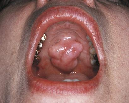
Lobule (adjective, lobulated) A segment or lobe that is a part of the whole; these lobes sometimes appear fused together (Fig. 1.1).
Macule An area that is usually distinguished by a color different from that of the surrounding tissue; it is flat and does not protrude above the surface of the normal tissue. A freckle is an example of a macule.
Papule A small, circumscribed lesion usually less than 1 cm in diameter that is elevated or protrudes above the surface of normal surrounding tissue.
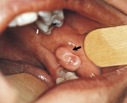
Pedunculated Attached by a stemlike or stalklike base similar to that of a mushroom (Fig. 1.2).
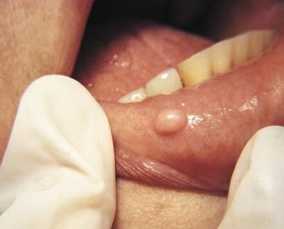
Sessile Describing the base of a lesion that is flat or broad instead of stemlike (Fig. 1.3).
FIGURE 1.2 Fibroma with a pedunculated base. Arrow points to the stemlike base.
FIGURE 1.3 Fibroma with a sessile base
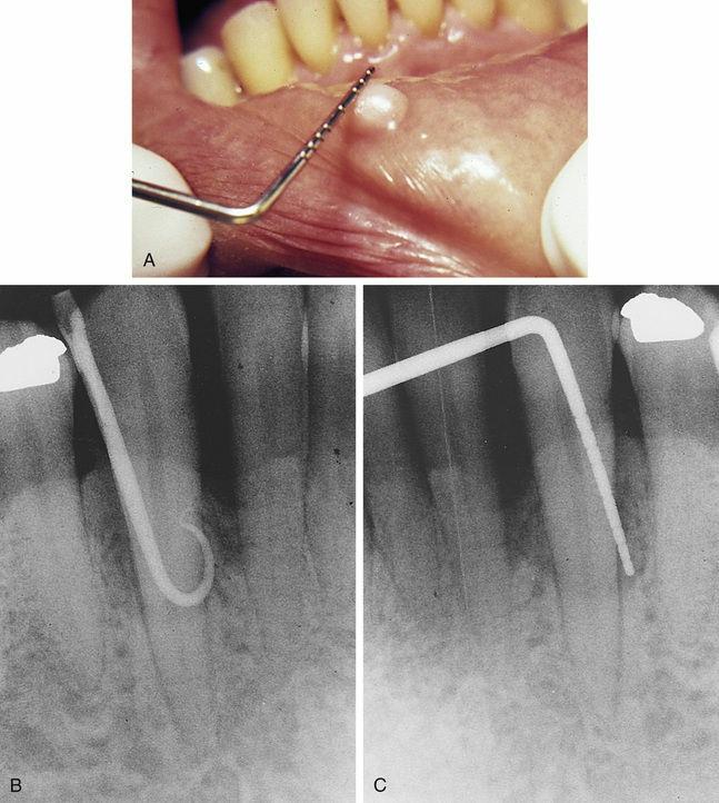
FIGURE 1.5 A, Probe measuring the diameter of a fibroma with a sessile base B, Gutta-percha point used to explore a radiographic defect. C, Periodontal probe placed before a radiograph.
Surface Texture
Corrugated Wrinkled.
Fissure A cleft or groove, normal or otherwise, showing prominent depth.
Papillary Resembling small, nipple-shaped projections or