Details on how to seek permission, further information about the Publisher’s permissions policies and our arrangements with organizations such as the Copyright Clearance Center and the Copyright Licensing Agency, can be found at our website: www.elsevier.com/permissions
This book and the individual contributions contained in it are protected under copyright by the Publisher (other than as may be noted herein).
Notices
Knowledge and best practice in this field are constantly changing. As new research and experience broaden our understanding, changes in research methods, professional practices, or medical treatment may become necessary.
Practitioners and researchers must always rely on their own experience and knowledge in evaluating and using any information, methods, compounds, or experiments described herein. In using such information or methods they should be mindful of their own safety and the safety of others, including parties for whom they have a professional responsibility.
With respect to any drug or pharmaceutical products identified, readers are advised to check the most current information provided (i) on procedures featured or (ii) by the manufacturer of each product to be administered, to verify the recommended dose or formula, the method and duration of administration, and contraindications. It is the responsibility of practitioners, relying on their own experience and knowledge of their patients, to make diagnoses, to determine dosages and the best treatment for each individual patient, and to take all appropriate safety precautions.
To the fullest extent of the law, neither the Publisher nor the authors, contributors, or editors, assume any liability for any injury and/or damage to persons or property as a matter of products liability, negligence or otherwise, or from any use or operation of any methods, products, instructions, or ideas contained in the material herein.
Previous edition copyrighted 2012.
Library of Congress Cataloging-in-Publication Data
Names: Woo, Sook-bin, author.
Title: Oral pathology : a comprehensive atlas and text / Sook-Bin Woo.
Description: Second edition. | Philadelphia, PA : Elsevier, [2017] | Includes bibliographical references and index.
Identifiers: LCCN 2016023952 | ISBN 9780323390545 (hardcover)
Classification: LCC RC815 | NLM WU 17 | DDC 616.3/1—dc23 LC record available at https://lccn.loc.gov/2016023952
Executive Content Strategist: William Schmitt
Senior Content Development Specialist: Anne Snyder
Publishing Services Manager: Patricia Tannian
Senior Project Manager: Cindy Thoms
Book Designer: Ryan Cook
Printed in China
Last digit is the print number: 9 8 7 6 5 4 3 2 1
This book is dedicated to my parents, who believe that education will lift us all, and to John, Sook-Yee, Chi-kan, I-Hwei, and Sarah.
PREFACE
This atlas was written to serve pathologists in training, dermatopathologists, general pathologists, as well as dental and medical specialists interested in diseases of the oral mucosa, minor salivary gland, and jawbones. Specific and detailed diagnostic criteria are provided for the diagnosis of pathology in the oral cavity. For the clinician, medications used in the treatment of inflammatory oral disease and viral and fungal infections are provided in Appendices A and B, which have been expanded in this edition. The chapter on infectious diseases has also been expanded to include most fungal diseases in the mouth and parasitic diseases that have become more common as a result of population migration. A new chapter on oral lymphomas, the second most common malignancy in the oral cavity, has been added.
Advances in molecular pathology in soft tissue and bone tumors, as well as salivary gland tumors, have occurred in leaps and bounds and brought new understanding to diagnosis, tumor biology, etiopathogenesis, and treatment. The chapter on salivary gland tumors has been expanded and now includes molecular findings in salivary gland tumors as are known at this time. Similar molecular findings are now included for soft tissue tumors often encountered in the oral cavity, primary bone tumors, and some odontogenic tumors.
However, it is inflammatory diseases of the mucosa, salivary glands, and jawbones that constitute the bulk of oral and maxillofacial pathology. Diagnosis of these conditions requires a deep understanding of the “lives of lesions,” as they evolve, resolve, and recur. Knowing the clinical presentation of oral mucosal disease is often essential for accurate diagnosis, especially for leukoplakia, the most common precancerous condition in the mouth. In this era of the ubiquitous smartphone, obtaining a photograph of a clinical lesion is simple and takes
only seconds, and all clinicians should be encouraged to send in photographs of such lesions to the pathologist with whom they collaborate. Similarly, clinicians should be encouraged to send oral radiographs, including computerized tomograms for accurate diagnosis of intra-bony lesions.
Practicing and teaching pathology teaches me humility on a regular basis. I continue to enjoy being challenged by my trainees and residents, revisiting old concepts and hopefully, debunking myths, or at least trying to view lesions in light of new concepts of etiopathogenesis and new diagnostic technologies. Pathology is a specialty that has much to offer the life-long learner and the curious mind. I hope that you will find this atlas to be informative and useful in your daily clinical or pathology practice.
Acknowledgments
I am indebted to my colleagues and friends who contributed slides and images for this atlas and to clinician colleagues who contribute cases to our biopsy service and have steadfastly supported the oral and maxillofacial pathology training program at Harvard School of Dental Medicine. Some cases were retrieved from the files of the American Board of Oral and Maxillofacial Pathology and the now defunct Armed Forces Institute of Pathology. Grateful thanks are due to my colleagues at the Ohio State University, Columbus, Ohio; Oral Pathology Laboratories, Queens, New York; University of Florida, Gainesville, Florida; and in Guatemala for their study sets. This endeavor would be less than what it is without the patients who have shared their stories and insights about their diseases with me, and whose images appear within these pages; it is an honor to care for them.
Unlike the skin , mucosal lesions in the oral cavity ma nosed as koilocytes. Muscle is present fairly superficially manifestonlyinalimitednumberofways-erythematous /'-- on the tongue and slightly deeper on the buccal and lip erosive (from epithelial atrophy, vascular ectasia, and ucosa. Minor salivary glands are predominantly mucous , inflammation), white (from keratosis or underlxing; fibro although serous acini and demilunes are frequently seen sis), yellow/ ulcerative (from fibrinous ex date), vesicu- ( Fig . 1.10 ); they are present everywhere in the mouth lobullous (often erosive), pigmented, papillary, diffuse or except on the keratinized gingiva (also known as nodular swelling, and mass. It is important for the pathol- "attached" gingiva because of its "attachment" to the periogist to be familiar with clinical presentations of mucosal osteum and bone). Serous salivary glands with a smaller disease for accurate diagnosis. Clinical images ( even those mucous component are frequently encountered on the taken with a smartphone) or radiographic images are anterior ventral tongue (glands of Blandin-Nuhn) and often indispensable for the diagnosi \Sf mucosal lesions posterior lateral and dorsal tongue (glands of van Ebner), and osseous pathology, respectively. sometimes invested in muscle.
ANATOMY
The oral mucosa varies clinically and histologically from site to site, and is divided into keratinized and nonkeratinized mucosa (Fig. 1. 1 ). Specific oral conditions correlate with oral anatomy: for example, recurrent aphthous ulcers occur primarily on the nonkeratinized mucosa , whereas recrudescent herpes simplex virus infections occur almost exclusively on the keratinized mucosa (such as the hard palatal mucosa and keratinized gingiva) in immune-competent patients. The tongue dorsum (with the thickest epithelium in the oral cavity) but not ventrum (with thin epithelium) is specialized for gustatory, masticatory, and deglutition functions. Filiform papillae cover the entire surface of the dorsum. Taste buds are present within fungiform ( on dorsum), circumvallate (8 to 14 on the posterior dorsum), and foliate (posterior lateral tongue) papillae but not within filiform papillae ( Fig.
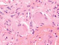
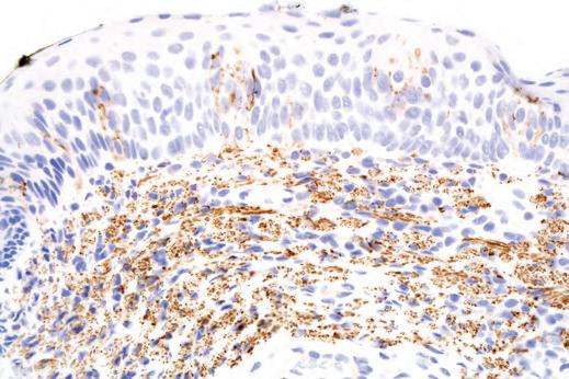
The subgemmal (gemma = Greek for bud) neurogenous plaque (also known as "subepithelial nerve plexus") is a normal nerve plexus located in the superficial lamina propria beneath the epithelium rich in taste buds and represent afferent fibers, and should not be mistaken for neurofibroma; ganglion cells are sometimes present (Fig. l. llA-B ). This neural plaque is positive for SIOO protein, neurofilament protein, and PGP9.5 ( Fig. l.llC-D . It is often noted in biopsies from the posterior lateral tongue in the area of the foliate papillae but is not exclusive to that site. Infrequently, ganglion cells and neuroepithelial islands similar to the organ of Chievitz may be seen within this neurogenous plaque.
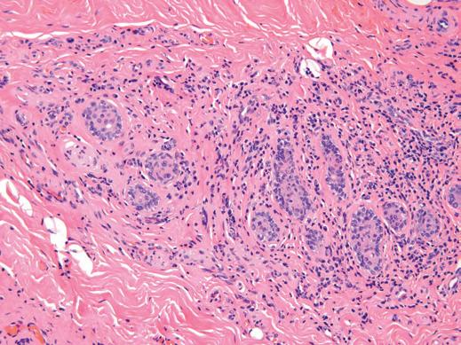
The juxtacortical organ of Chievitz is likely a remnant from embryogenesis and consists of small islands of benign epithelium with peripheral basal cells closely apposed to, or within nerve fibers (likely the buccal nerve) ( Fig. 1.12 ). This organ may be located in the area of the pterygomandibular raphe , medial to the mandible,
B
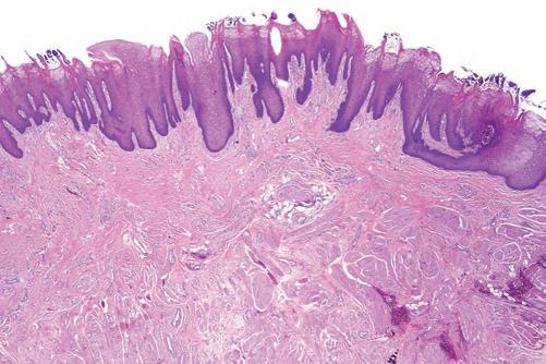
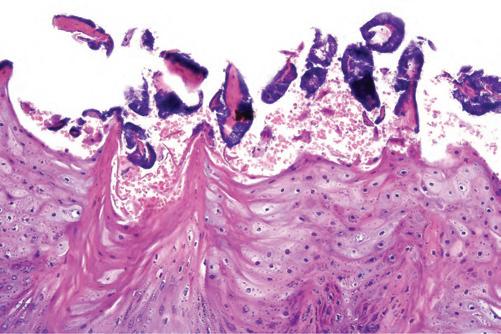
FIG 1.8 (A) tongue dor sum: thick epithelium with abundant muscle. (B) tongue dor sum: filiform papillae composed of par akeratin spires with bacter ial colonies.
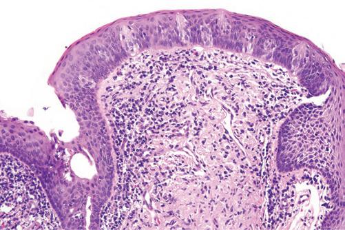
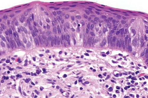
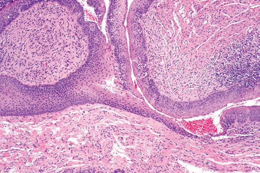
FIG 1.9 (A) Fungiform papilla with taste buds in the epithelium and many ner ve fiber s in the lamina propria. (B) taste buds: flask-shaped cluster s of sustentacular and gustator y sensor y cells.
A B
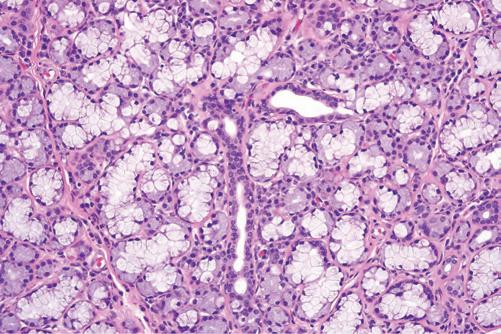
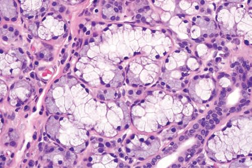
FIG 1.10 (A) mucous glands of the lower lip with some serous acini. (B) Serous acinar cells in caplike demilune ar rangement over mucous acini.
B
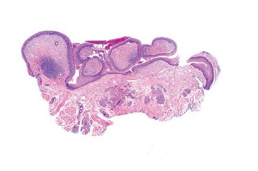
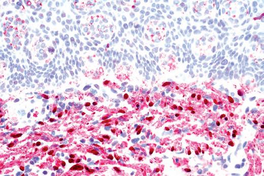
FIG 1.11 Subgemmal neurogenous plaque. (A) Foliate papillae of tongue with serous glands. (B) numerous taste buds in the epithelium and subepithelial neural plexus with ganglion cells (inset) (c) Spindle cells and taste buds are positive for S-100 protein. (d) Spindle cells and taste buds are positive for neurofilament protein.
and may extend toward the opening of the parotid duct. These structures must not be misinterpreted as perineural invasion by carcinomas in the vicinity.
TEETH
Teeth are made up mostly of tubular dentin that is capped by a thin shell of very hard enamel overlying the crown of the tooth. The root of the tooth is surrounded by a thin layer of cementum (Fig. 1.13). The periodontal membrane attaches to the cementum on one side and the alveolar bone on the other, allowing for slight movement of teeth within the bone. This membrane contains Sharpey fibers rich in type III (argyrophilic reticulin fibers) and type VI collagen. Although Sharpey fibers has been used to refer almost exclusively to these fibers of the periodontal membrane, they are also present in muscle-bone and periosteum-bone anchorage elsewhere in the body.
FIG 1.12 Juxtacortical organ of chievitz composed of nests of benign epithelium with closely associated nerve fibers.
histopathologic features is presented for inflammatory lesions in this atlas.
Dysplastic conditions are probably the most challenging for the pathologist, because the clinical appearance of the lesion plays an important role in the accurate diagnosis of dysplasia. To this end, clinicians are encouraged to send a photograph (even a smartphone image) of the lesion with the biopsy. Many dysplastic oral lesions show architectural rather than cytologic evidence of dysplasia. These concepts are covered in depth in Chapter 11
The teeth and salivary glands are adnexa of the oral cavity in the same way that hair and sweat glands are skin adnexa. As such, there are histopathologic similarities among odontogenic, salivary gland, skin adnexal, and breast neoplasms. Teeth and salivary glands are derived from the same primordium and it is therefore not surprising that primary salivary gland tumors may occur as primary tumors within the jawbones. The diagnosis of odontogenic cysts and tumors and primary intraosseous lesions can be accurately rendered only if radiographic images are provided.
Finally, a few words on terminology. The oral mucosa has no submucosa per se as mentioned previously, because it lacks a muscularis mucosae. There is only lamina propria, superficial and deep, and even then, the demarcation between the two is somewhat arbitrary The term hard palatal mucosa is preferable to hard palate, because the hard palate is the bony plate to which mucosa is attached.
Similarly, mandibular and maxillary mucosa are preferable to mandible and maxilla (implying bone) if one is referring to the mucosa overlying the bone. The term parakeratosis is used without the modifying “hyper” because all parakeratin is abnormal in the oral cavity, except on the tongue dorsum, which contains parakeratotic filiform papillae, so that hyperparakeratosis would be appropriate only for tongue dorsum lesions. The term hyperkeratosis is preferred over hyperorthokeratosis in keeping with usage by general pathologists and dermatopathologists.
REFERENCES
Aaron JE. Periosteal Sharpey’s fibers: a novel bone matrix regulatory system? Front Endocrinol (Lausanne) 2012;3:1-10.
Gueiros LA, Leon JE, Leao JC, et al. Subgemmal neurogenous plaque: clinical and microscopic evaluation of 7 cases. Oral Surg Oral Med Oral Pathol Oral Radiol Endod 2009;108:920-924.
Nanci A. Ten Cate’s Oral Histology: Development, Structure and Function 7th ed. St. Louis: Mosby; 2008.
Palazzolo MJ, Fowler CB, Magliocca KR, Gnepp DR. Neuroepithelial structures associated with the subepithelial nerve plexus of taste buds: a fortuitous finding resembling the juxtaoral organ of Chievitz. Oral Surg Oral Med Oral Pathol Oral Radiol 2014;117:497-501.
Pantanowitz L, Balogh K. Significance of the juxtaoral organ (of Chievitz). Head Neck 2003;25:400-405, discussion.
Triantafyllou A, Coulter P. Structural organization of subgemmal neurogenous plaques in foliate papillae of tongue. Hum Pathol 2004;35:991-999.