https://ebookmass.com/product/ophthalmology-5th-edition/
Instant digital products (PDF, ePub, MOBI) ready for you
Download now and discover formats that fit your needs...
Ophthalmology 5th Edition Myron Yanoff (Lead Editor)
https://ebookmass.com/product/ophthalmology-5th-edition-myron-yanofflead-editor/
ebookmass.com
Ophthalmology 5th Edition Jay S. Duker Md
https://ebookmass.com/product/ophthalmology-5th-edition-jay-s-dukermd/
ebookmass.com
Taylor and Hoyt’s Pediatric Ophthalmology and Strabismus 5th Edition Scott R. Lambert
https://ebookmass.com/product/taylor-and-hoyts-pediatricophthalmology-and-strabismus-5th-edition-scott-r-lambert/
ebookmass.com
Automated Market Makers: A Practical Guide to Decentralized Exchanges and Cryptocurrency Trading Miguel Ottina
https://ebookmass.com/product/automated-market-makers-a-practicalguide-to-decentralized-exchanges-and-cryptocurrency-trading-miguelottina/ ebookmass.com
eTextbook 978-0393265156 Psychology in Your Life 2nd Edition by
Sarah Grison
https://ebookmass.com/product/etextbook-978-0393265156-psychology-inyour-life-2nd-edition-by-sarah-grison/
ebookmass.com
Dental Anatomy Coloring Book 4th Edition Margaret J. Fehrenbach
https://ebookmass.com/product/dental-anatomy-coloring-book-4thedition-margaret-j-fehrenbach/
ebookmass.com
Management of Human Service Programs (SW 393T 16 Social Work Leadership in Human Services Organizations) 5th Edition, (Ebook PDF)
https://ebookmass.com/product/management-of-human-service-programssw-393t-16-social-work-leadership-in-human-services-organizations-5thedition-ebook-pdf/ ebookmass.com
(eTextbook PDF) for Making Sense of Change Management: A Complete Guide to the Models, Tools and Techniques of Organizational Change 5th Edition
https://ebookmass.com/product/etextbook-pdf-for-making-sense-ofchange-management-a-complete-guide-to-the-models-tools-and-techniquesof-organizational-change-5th-edition/ ebookmass.com
Medieval Ethiopian Kingship, Craft, and Diplomacy with Latin Europe Verena Krebs
https://ebookmass.com/product/medieval-ethiopian-kingship-craft-anddiplomacy-with-latin-europe-verena-krebs/
ebookmass.com
The Life of Henry James: A Critical Biography Peter Collister
https://ebookmass.com/product/the-life-of-henry-james-a-criticalbiography-peter-collister/
ebookmass.com
Nirali Bhatt, MD
Assistant Professor
Department of Ophthalmology
University of Pennsylvania Perelman School of Medicine Philadelphia, PA, USA
Orry C. Birdsong, MD
Clinical Fellow
Ophthalmology
Hoopes Vision Draper, UT, USA
Jyotirmay Biswas, MS, FMRF, FNAMS, FIC, Path, FAICO
Director
Uveitis and Ocular Pathology
Department
Sankara Nethralaya Chennai, Tamil Nadu, India
Bahram Bodaghi, MD, PhD, FEBOphth Professor of Ophthalmology
DHU ViewRestore
APHP, UPMC, Sorbonne University Paris, France
Swaraj Bose, MD
Associate Professor of Ophthalmology UCI and Attending Physician Cedars Sinai Medical Center
Los Angeles, CA, USA
Charles S. Bouchard, MD, MA Professor and Chairman of Ophthalmology
Loyola University Health System Maywood, IL, USA
Michael E. Boulton, PhD
Susan and Dowd Ritter/RPB Endowed Chair of Ophthalmology
University of Alabama Birmingham Birmingham, AL, USA
James D. Brandt, MD
Professor Department of Ophthalmology & Vision Science
Vice-Chair for International Programs and New Techology
Director - Glaucoma Service
University of California Davis Sacramento, CA, USA
Scott E. Brodie, MD, PhD Professor of Ophthalmology
NYU School of Medicine
New York, NY, USA
Michael C. Brodsky, MD Professor of Ophthalmology and Neurology
Knights Templar Research Professor of Ophthalmology
Mayo Clinic Rochester, MN, USA
Cassandra C. Brooks, MD
Resident in Ophthalmology
Duke Eye Center
Duke University School of Medicine Durham, NC, USA
Matthew V. Brumm, MD
Ophthalmologist
Cataract and Refractive Surgery
Brumm Eye Center
Omaha, NE, USA
Donald L. Budenz, MD, MPH
Kittner Family Distinguished Professor and Chairman
Department of Ophthalmology
University of North Carolina at Chapel Hill Chapel Hill, NC, USA
Igor I. Bussel, MS, MHA
Doris Duke Clinical Research Fellow
Department of Ophthalmology
University of Pittsburgh School of Medicine Pittsburgh, PA, USA
Louis B. Cantor, MD
Jay C. and Lucile L. Kahn Professor and Chair
Department of Ophthalmology
Indiana University School of Medicine Indianapolis, IN, USA
Hilda Capó, MD
Professor of Clinical Ophthalmology
Bascom Palmer Eye Institute
Division Chief Pediatric Ophthalmology and Adult Strabismus
Miller School of Medicine
John T. Flynn Professor of Ophthalmology Chair
University of Miami Miami, FL, USA
Antonio Capone, Jr., MD
Professor
Department of Ophthalmology
Oakland University
William Beaumont Hospital School of Medicine
Auburn HIlls, MI, USA
Alastair Carruthers, MA, BM, BCh, FRCP(Lon), FRCPC
Clinical Professor
Department of Dermatology and Skin Science
University of British Columbia Vancouver, BC, Canada
Jean Carruthers, MD, FRCSC, FRC(OPHTH)
Clinical Professor Department of Ophthalmology
University of British Columbia Fellow
American Society for Ophthalmic Plastic and Reconstructive Surgery Vancouver, BC, Canada
Keith D. Carter, MD, FACS
Lillian C. O’Brien and Dr. C.S. O’Brien Chair in Ophthalmology
Professor and Chair
Department of Ophthalmology & Visual Sciences
Carver College of Medicine
University of Iowa Iowa City, IA, USA
Rafael C. Caruso, MD
Staff Clinician
National Eye Institute
National Institutes of Health Bethesda, MD, USA
Harinderpal S. Chahal, MD
Oculofacial Plastic and Reconstructive Surgery
Eye Medical Center Fresno, CA, USA
Wallace Chamon, MD
Adjunct Professor
Department of Ophthalmology and Visual Sciences
University of Illinois at Chicago Chicago, IL, USA
Adjunct Professor
Department of Ophthalmology and Visual Sciences
Escola Paulista de Medicina (EPM)
Universidade Federal de São Paulo (UNIFESP)
São Paulo, SP, Brazil
Chi-Chao Chan, MD
Scientist Emeritus
Laboratory of Immunology
National Eye Institute
National Institutes of Health
Bethesda, MD, USA
Visiting Professor
Zhongshan Ophthalmic Center
Sun Yat-Sen University China
Melinda Y. Chang, MD
Assistant Professor of Ophthalmology
USC Roski Eye Institute and Children’s
Hospital Los Angeles
Keck School of Medicine of the University of Southern California
Los Angeles, CA, USA
Stanley Chang, MD
KK Tse and KT Ying Professor of Ophthalmology
Department of Ophthalmology
Columbia University
New York, NY, USA
Victoria S. Chang, MD
Assistant Professor of Clinical Ophthalmology
Ophthalmology, Cornea and External Disease
Bascom Palmer Eye Institute University of Miami Naples, FL, USA
David G. Charteris, MD, FRCS(Ed), FRCOphth
Professor
Vitreoretinal Unit
Moorfields Eye Hospital London, UK
Soon-Phaik Chee, MD
Professor
Cataract Service, Ocular Inflammation & Immunology Service
Singapore National Eye Centre Singapore
John J. Chen, MD, PhD
Assistant Professor
Department of Ophthalmology and Neurology
Mayo Clinic Rochester, MN, USA
Xuejing Chen, MD, MS
Clinical Fellow
Retina
Ophthalmic Consultants of Boston
New England Eye Center at Tufts Medical Center Boston, MA, USA
Paul T.K. Chew, MMed, FRCOphth Director Glaucoma Division Ophthalmology/Glaucoma
National University Hospital Singapore Singapore
Bing Chiu, MD
Ophthalmology Resident New York University New York, NY, USA
Clement C. Chow, MD
Partner Physician
Retinal Diagnostic Center Campbell, CA, USA
Mortimer M. Civan, MD
Professor of Physiology and Professor of Medicine
Department of Physiology University of Pennsylvania Perelman School of Medicine Philadelphia, PA, USA
Abbot (Abe) Clark, PhD, FARVO
Regents Professor of Pharmacology and Neuroscience
Executive Director, North Texas Eye Research Institute
University of North Texas Health Science Center Fort Worth, TX, USA
Jonathan C.K. Clarke, MD, FRCOphth Consultant Ophthalmologist
NIHR Moorfields Biomedical Research Centre
Moorfields Eye Hospital
UCL Institute of Ophthalmology London, UK
François Codère, MD
Associate Professor Ophthalmology/Oculoplastic and Orbital Surgery Section
Université de Montréal Montréal, QC, Canada
Ian P. Conner, MD, PhD
Assistant Professor Ophthalmology
UPMC Eye Center Pittsburgh, PA, USA
Peter Coombs, MD
Vitreoretinal Physician and Surgeon Utah Eye Centers Salt Lake City, UT, USA
Zélia M. Corrêa, MD, PhD
Tom Clancy Endowed Professor of Ophthalmology
Head of Ocular Oncology and Echography
Retina Service, Wilmer Eye Institute
Johns Hopkins University School of Medicine Baltimore, MD, USA
Steven M. Couch, MD, FACS
Assistant Professor Department of Ophthalmology & Visual Sciences
Washington University in St Louis St Louis, MO, USA
Stuart G. Coupland, PhD
Associate Professor Department of Ophthalmology
University of Ottawa Ottawa, ON, Canada
Claude L. Cowan, Jr., MD, MPH
Clinical Professor of Ophthalmology
Georgetown University Medical Center
Washington, DC, USA
Staff Physician
Surgical Service
Veterans Affairs Medical Center
Washington, DC, USA
E. Randy Craven, MD
Associate Professor, Glaucoma
Johns Hopkins University Baltimore, MD, USA
Catherine A. Cukras, MD, PhD
Director, Medical Retina Fellowship Program
National Eye Institute
National Institutes of Health
Bethesda, MD, USA
Linda R. Dagi, MD
Director of Adult Strabismus
Boston Children’s Hospital
Associate Professor of Ophthalmology
Director of Quality Assurance
Department of Ophthalmology
Children’s Hospital Ophthalmology
Foundation Chair
Harvard Medical School
Boston, MA, USA
Elie Dahan, MD, MMed, (Ophth)† Formerly Senior Consultant Pediatric Ophthalmology and Glaucoma
Department of Ophthalmology
Ein Tal Eye Hospital Tel Aviv, Israel
Iben Bach Damgaard, MD PhD Fellow Department of Ophthalmology
Aarhus University Hospital Aarhus, Denmark
Karim F. Damji, MD, FRCSC, MBA
Professor Department of Ophthalmology & Visual Sciences University of Alberta Edmonton, AL, Canada
Dipankar Das, MD
Senior Consultant & Ocular Pathologist Uveitis, Ocular Pathology and Neuroophthalmology Services Sri Sankaradeva Nethralaya Guwahati, Assam, India
Adam DeBusk, DO, MS Instructor Department of Ophthalmology Wills Eye Hospital
Sidney Kimmel Medical College Thomas Jefferson University Philadelphia, PA, USA
Jose de la Cruz, MD, MSc
Assistant Professor Ophthalmology, Cornea Refractive Surgery Service
University of Illinois Eye and Ear Infirmary Chicago, IL, USA
Joseph L. Demer, MD, PhD
Arthur L. Rosenbaum Chair in Pediatric Ophthalmology Professor of Neurology Chief, Pediatric Ophthalmology and Strabismus Division
Director, Ocular Motility Laboratories Chair, EyeSTAR Residency/PhD and Post-doctoral Fellowship Program in Ophthalmology and Visual Science Member, Neuroscience Interdepartmental Program Member, Bioengineering Interdepartmental Program University of California Los Angeles Los Angeles, CA, USA
Shilpa J. Desai, MD Assistant Professor Department of Ophthalmology Tufts University School of Medicine Boston, MA, USA
Deepinder K. Dhaliwal, MD, L.Ac Professor of Ophthalmology, University of Pittsburgh School of Medicine Director, Cornea and Refractive Surgery Services
Director and Founder, Center for Integrative Eye Care
Co-Director, Cornea and Refractive Surgery Fellowship
Associate Medical Director, Charles T. Campbell Ocular Microbiology Laboratory
Medical Director, UPMC Laser Vision Center
University of Pittsburgh Medical Center Pittsburgh, PA, USA
Gary R. Diamond, MD†
Formerly Professor of Ophthalmology and Pediatrics
Drexel University School of Medicine
Philadelphia, PA, USA
Daniel Diniz, MD
Surgical Optics Fellow
Department of Ophthalmology & Visual Sciences
Federal University of São Paulo (UNIFESP)
São Paulo, SP, Brazil
Diana V. Do, MD
Professor of Ophthalmology Byers Eye Institute
Stanford University School of Medicine Palo Alto, CA, USA
Peter J. Dolman, MD, FRCSC
Clinical Professor
Division Head of Oculoplastics and Orbital Surgery
Fellowship Director
Department of Ophthalmology & Visual Sciences
Division of Oculoplastics and Orbit
University of British Columbia Vancouver General Hospital Vancouver, BC, Canada
Sean P. Donahue, MD, PhD
Professor
Department of Ophthalmology & Visual Sciences
Vanderbilt University Nashville, TN, USA
Richard K. Dortzbach, MD
Professor Emeritus Department of Ophthalmology and Visual Sciences
University of Wisconsin School of Medicine and Public Health Madison, WI, USA
Kimberly A. Drenser, MD, PhD
Associated Retinal Consultants, PC Department of Ophthalmology
Oakland University
William Beaumont Hospital School of Medicine
Royal Oak, MI, USA
Jacob S. Duker, MD Resident Physician
Department of Ophthalmology Bascom Palmer Eye Institute University of Miami Miami, FL, USA
Jay S. Duker, MD
Director
New England Eye Center Professor and Chairman Department of Ophthalmology Tufts Medical Center
Tufts University School of Medicine Boston, MA, USA
Vikram D. Durairaj, MD, FACS
ASOPRS Fellowship Director and Managing Partner
Oculoplastic and Orbital Surgery TOC Eye and Face Austin, TX, USA
Jonathan J. Dutton, MD, PhD
Professor Emeritus Department of Ophthalmology University of North Carolina Chapel Hill, NC, USA
Bryan Edgington, MD
Associate Professor, Cornea Division
Casey Eye Institute
Oregon Health Sciences University
Staff Ophthalmologist
Veterans Health Administration
Portland Health Care System
Portland, OR, USA
Howard M. Eggers, MD
Professor of Clinical Ophthalmology
Harkness Eye Institute New York, NY, USA
Dean Eliott, MD
Stelios Evangelos Gragoudas Associate Professor of Ophthalmology
Harvard Medical School
Associate Director, Retina Service
Massachusetts Eye & Ear Boston, MA, USA
George S. Ellis, Jr., MD, FAAP, FAAO, FACS
Director Ophthalmology
Children’s Hospital New Orleans
Associate Clinical Professor of Ophthalmology and Pediatrics
Tulane University
Associate Clinical Professor of Ophthalmology and Pediatrics
Louisiana State Universities Schools of Medicine
New Orleans, LA, USA
Michael Engelbert, MD, PhD
Research Assistant Professor Department of Ophthalmology
NYU/VRMNY New York, NY, USA
Miriam Englander, MD
Attending Surgeon Vitreo-Retinal Surgery
Ophthalmic Consultants of Boston Boston, MA, USA
Bita Esmaeli, MD, FACS Professor of Ophthalmology
Director, Ophthalmic Plastic & Reconstructive Surgery Fellowship Program, Department of Plastic Surgery Chair, Graduate Medical Education Committee
University of Texas MD Anderson Cancer Center Houston, TX, USA
Joshua W. Evans, MD
Assistant Professor of Ophthalmology Division of Glaucoma University of Kentucky Lexington, KY, USA
Monica Evans, MD
Ophthalmology
San Jose, Costa Rica
Daoud S. Fahd, MD
Clinical Assistant Professor Department of Ophthalmology
Ophthalmic Consultants of Beirut
Jal el Dib, Metn, Lebanon
Lisa J. Faia, MD
Partner, Associated Retinal Consultants
Associate Professor
Oakland University
William Beaumont School of Medicine
Ophthalmology - Retina Royal Oak, MI, USA
Katherine A. Fallano, MD Department of Ophthalmology University of Pittsburgh School of Medicine Pittsburgh, PA, USA
Ayad A. Farjo, MD
President & Director
Brighton Vision Center Brighton, MI, USA
Eric Feinstein, MD
Surgical Retina Fellow
Department of Ophthalmology
Rocky Mountain Lions Eye Institute
University of Colorado School of Medicine
Denver, CO, USA
Karen B. Fernandez, MD Consultant
Department of Ophthalmology
The Medical City Pasig City, Metro Manila, Philippines
Yale L. Fisher, MD
Voluntary Clinical Professor
Department of Ophthalmology
Bascom Palmer Eye Institute
Miami, FL, USA
Voluntary Clinical Professor Department of Ophthalmology
Weill Cornell Medical Center New York, NY, USA
Gerald A. Fishman, MD Director
The Pangere Center for Inherited Retinal Diseases
The Chicago Lighthouse Professor Emeritus of Ophthalmology
Department of Ophthalmology & Visual Sciences
University of Illinois at Chicago College of Medicine Chicago, IL, USA
Jorge A. Fortun, MD
Associate Professor of Ophthalmology
Vitreoretinal Diseases and Surgery
Medical Director of Bascom Palmer
Eye Institute
Palm Beach Gardens Bascom Palmer Eye Institute
University of Miami Miller School of Medicine Miami, FL, USA
Veronica Vargas Fragoso, MD Refractive Surgery Fellow Vissum Corporation Alicante, Spain
Nicola Freeman, MBChB, FCOphth, MMed
Senior Specialist
Department of Pediatric Ophthalmology
Red Cross Children’s Hospital
Cape Town, Western Province, South Africa
David S. Friedman, MD, MPH, PhD
Director, Dana Center for Preventive Ophthalmology
Professor of Ophthalmology, Wilmer/ Glaucoma
Johns Hopkins University Baltimore, MD, USA
Deborah I. Friedman, MD, MPH Professor
Department of Neurology & Neurotherapeutics and Ophthalmology
University of Texas
Southwestern Medical Center Dallas, TX, USA
Neil J. Friedman, MD
Adjunct Clinical Associate Professor Department of Ophthalmology
Stanford University School of Medicine
Stanford, CA, USA
Paul P. Lee, MD, JD
F. Bruce Fralick Professor and Chair
Director W.K. Kellogg Eye Center
Department of Ophthalmology & Visual Sciences
University of Michigan Ann Arbor, MI, USA
Richard M.H. Lee, MSc, FRCOphth
Clinical Fellow
Department of Glaucoma Moorfields Eye Hospital London, UK
Dawn K.A. Lim, MBBS, MRCP, MMed(Int, Med), MMed(Ophth), FAMS Consultant, Ophthalmology/Glaucoma National University Hospital Singapore
Jennifer I. Lim, MD, FARVO
Marion H. Schenk Esq. Chair in Ophthalmology for Research of the Aging Eye
Professor of Ophthalmology
Director of the Retina Service University of Illinois at Chicago Illinois Eye and Ear Infirmary Chicago, IL, USA
Ridia Lim, MBBS, MPH, FRANZCO
Ophthalmic Surgeon Glaucoma Service
Sydney Eye Hospital Sydney, NSW, Australia
Tony K.Y. Lin, MD, FRCSC
Assistant Professor
Department of Ophthalmology
Schulich School of Medicine and Dentistry
Western University London, ON, Canada
John T. Lind, MD, MS
Associate Professor Department of Ophthalmology & Visual Sciences
Washington University in St Louis St Louis, MO, USA
Yao Liu, MD
Assistant Professor Department of Ophthalmology & Visual Sciences
University of Wisconsin-Madison Madison, WI, USA
Sidath E. Liyanage, MBBS, FRCOphth, PhD
Consultant Ophthalmologist Bristol Eye Hospital Bristol, UK
Alastair J. Lockwood, BM, BCh, FRCOphth, PhD
Consultant, Ophthalmology
Queen Alexandra Hospital Portsmouth, Hampshire, UK
Nils A. Loewen, MD, PhD
Associate Professor of Ophthalmology Vice Chair of Electronic Health Records in Ophthalmology University of Pittsburgh Pittsburgh, PA, USA
Reid A. Longmuir, MD
Assistant Professor
Department of Ophthalmology & Visual Sciences
Vanderbilt University Nashville, TN, USA
Pedro F. Lopez, MD
Professor and Founding Chair Department of Ophthalmology
Herbert Wertheim College of Medicine
Florida International University Director of Vitreoretina and Macular Division
Center for Excellence in Eye Care Miami, FL, USA
Mats Lundström, MD, PhD
Adjunct Professor Emeritus Department of Clinical Sciences, Ophthalmology
Faculty of Medicine
Lund University
Lund, Region Skåne, Sweden
Robi N. Maamari, MD
Ophthalmology Resident Department of Ophthalmology & Visual Sciences
Washington University School of Medicine in St Louis
St Louis, MO, USA
Assumpta Madu, MD, MBA, PharmD Vice Chair, Operations
Associate Clinical Professor of Ophthalmology
NYU School of Medicine
NYU Langone Medical Center New York, NY, USA
Maya H. Maloney, MD
Consultant, Medical Retina
Mayo Clinic Rochester, MN, USA
Naresh Mandava, MD
Professor and Chair Department of Ophthalmology
University of Colorado School of Medicine Denver, CO, USA
Michael F. Marmor, MD Professor Department of Ophthamology
Byers Eye Institute
Stanford University School of Medicine Palo Alto, CA, USA
Jeevan R. Mathura, Jr., MD
Private Practitioner and Owner Diabetic Eye and Macular Disease Specialists, LLC Washington, DC, USA
Cynthia Mattox, MD
Associate Professor, Ophthalmology
Tufts University School of Medicine Boston, MA, USA
Scott K. McClatchey, MD
Associate Professor, Ophthalmology
Naval Medical Center San Diego, CA, USA
Stephen D. McLeod, MD
Theresa M. and Wayne M. Caygill
Distinguished Professor and Chair, Ophthalmology
University of California San Francisco San Francisco, CA, USA
Brian D. McMillan, MD
Assistant Professor of Ophthalmology
WVU Eye Institute
West Virginia University School of Medicine
Morgantown, WV, USA
Alan A. McNab, DMedSc, FRANZCO, FRCOphth
Associate Professor and Director
Orbital Plastic and Lacrimal Clinic
Royal Victorian Eye and Ear Hospital Melbourne, VIC, Australia
Jodhbir S. Mehta, BSc, MD, MBBS, FRCS(Ed), FRCOphth, FAMS
Associate Professor, Cornea and External Disease
Singapore National Eye Centre
Singapore
Luis J. Mejico, MD
Professor and Chair of Neurology
Professor of Ophthalmology
SUNY Upstate Medical University Syracuse, NY, USA
Carolina L. Mercado, MD
Clinical Research Fellow, Ophthalmology
Bascom Palmer Eye Institute Miami, FL, USA
Shahzad I. Mian, MD
Associate Chair, Terry J. Bergstrom Professor
Associate Professor, Ophthalmology & Visual Sciences
University of Michigan Ann Arbor, MI, USA
William F. Mieler, MD, FACS Cless Family Professor of Ophthalmology
Vice-Chairman of Education
Illinois Eye and Ear Infirmary
University of Illinois at Chicago College of Medicine Chicago, IL, USA
David Miller, MD
Associate Clinical Professor of Ophthalmology
Harvard Medical School Boston, MA, USA
Kyle E. Miller, MD
Assistant Professor, Ophthalmology
Naval Medical Center Portsmouth Portsmouth, VA, USA
Tatsuya Mimura, MD, PhD
Tokyo Womens Medical University
Medical Center East Tokyo, Japan
Rukhsana G. Mirza, MD
Associate Professor Department of Ophthalmology
Northwestern University Feinberg School of Medicine Chicago, IL, USA
Mihai Mititelu, MD, MPH
Assistant Professor Department of Ophthalmology & Visual Sciences
University of Wisconsin-Madison School of Medicine and Public Health Madison, WI, USA
Ramana S. Moorthy, MD
Clinical Associate Professor, Ophthalmology
Indiana University School of Medicine
Founding Partner and CEO
Associated Vitreoretinal and Uveitis Consultants
Indianapolis, IN, USA
Andrew A. Moshfeghi, MD, MBA
Director, Vitreoretinal Fellowship
Associate Professor of Clinical Ophthalmology
University of Southern California
Roski Eye Institute
Keck School of Medicine Los Angeles, CA, USA
Majid Moshirfar, MD, FACS
Professor of Ophthalmology
Hoopes Vision and John A. Moran Eye
Center
Draper, UT, USA
Heather E. Moss, MD, PhD
Assistant Professor
Departments of Ophthalmology and Neurology & Neurological Sciences
Stanford University Palo Alto, CA, USA
Mark L. Moster, MD
Director, Neuro-Ophthalmology Fellowship
Professor, Neurology and Ophthalmology
Wills Eye Hospital
Sidney Kimmel Medical College of Thomas Jefferson University Philadelphia, PA, USA
Kelly W. Muir, MD, MHSc
Associate Professor of Ophthalmology, Glaucoma Division
Duke University School of Medicine Durham, NC, USA
Ann G. Neff, MD
Dermatology Associates Sarasota, FL, USA
Jeffrey A. Nerad, MD
Oculoplastic & Reconstructive Surgery
Cincinnati Eye Institute
Volunteer Professor, Ophthalmology
University of Cincinnati Cincinnati, OH, USA
Neda Nikpoor, MD
Clinical Instructor, Ophthalmology
Byers Eye Institute
Stanford University
Palo Alto, CA, USA
Robert J. Noecker, MD, MBA Director of Glaucoma
Ophthalmic Consultants of Connecticut Fairfield, CT, USA
Ricardo Nosé, MD
Clinical Research Fellow
New England Eye Center
Tufts Medical Center Boston, MA, USA
Annabelle A. Okada, MD, DMSc
Professor of Ophthalmology
Kyorin University School of Medicine Tokyo, Japan
Michael O’Keefe, FRCS
Professor, Ophthalmology
Mater Private Hospital Dublin, Ireland
Jeffrey L. Olson, MD
Associate Professor
Department of Ophthalmology
University of Colorado School of Medicine
Denver, CO, USA
Jane M. Olver, MB, BS, BSc, FRCS, FRCOphth
Consultant Ophthalmologist
Eye Department
Clinica London
London, UK
Yvonne A.V. Opalinski, BSc, MD, BFA, MFA
Clinical Associate Cardiovascular Surgery
Department of Cardiovascular Surgery
Trillium Health Partners
Toronto, ON, Canada
William Tasman, MD†
Formerly Professor and Emeritus
Chairman
Department of Ophthalmology
Wills Eye Hospital and Jefferson Medical College Philadelphia, PA, USA
David G. Telander, MD, PhD
Clinical Professor Department of Ophthalmology
University of California Davis Davis, CA, USA
Associate Professor California Northstate School of Medicine Sacramento, CA, USA
Edmond H. Thall, MD, MS Consultant in Aerospace Ophthalmology
Aeromedical Consultation Service
Ophthalmology Branch
United States Air Force School of Aerospace Medicine Wright–Patterson Air Force Base Dayton, OH, USA
Aristomenis Thanos, MD Retina Department
Devers Eye Institute Portland, OR, USA
Christos N. Theophanous, MD Resident Physician
Department of Ophthalmology and Visual Science
University of Chicago Medicine Chicago, IL, USA
Benjamin J. Thomas, MD Physician, Vitreoretinal Surgery Florida Retina Institute Jacksonville, FL, USA
Praneetha Thulasi, MD
Assistant Professor of Ophthalmology Cornea, External Diseases, and Refractive Surgery Emory University Atlanta, GA, USA
Michael D. Tibbetts, MD Director of Retina Services Tyson Eye Center Cape Coral, FL, USA
David P. Tingey, BA, MD, FRCSC
Associate Professor, Ophthalmology Western University London, ON, Canada
Faisal M. Tobaigy, MD
Associate Professor of Ophthalmology Jazan University
Jazan, Saudi Arabia
Bozho Todorich, MD, PhD Staff Physician
Pennsylvania Retina Specialists, PC Camp Hill, PA, USA
Stuart W. Tompson, PhD
Associate Scientist Department of Ophthalmology & Visual Sciences University of Wisconsin-Madison Madison, WI, USA
James C. Tsai, MD, MBA
President, New York Eye & Ear Infirmary of Mount Sinai, DelafieldRodgers Professor and System Chair Department of Ophthalmology Icahn School of Medicine at Mount Sinai New York, NY, USA
Julie H. Tsai, MD
Assistant Professor of Clinical Ophthalmology Albany, NY, USA
Nancy Tucker, MD, FRCSC Chief of Oculoplastics, Ophthalmology University of Toronto Toronto, ON, Canada
Sonal S. Tuli, MD, MEd Professor and Chair, Ophthalmology University of Florida Gainesville, FL, USA
Caroline W. Vargason, MD, PhD
Oculoplastic & Reconstructive Surgery Fellow
Cincinnati Eye Institute Cincinnati, OH, USA
Roshni A. Vasaiwala, MD
Assistant Professor of Ophthalmology Director of Cornea Service
Loyola University Medical Center Maywood, IL, USA
Daniel Vitor Vasconcelos-Santos, MD, PhD
Adjunct Professor of Ophthalmology Director of Uveitis
Universidade Federal de Minas Gerais Belo Horizonte, Minas Gerais, Brazil
Gregory J. Vaughn, MD Consultant, Global Healthcare Practice
Spencer Stuart Atlanta, GA, USA
Arthi Venkat, MD, MS, BA
Staff Physician in Medical Retina and Uveitis
Cleveland Clinic Cole Eye Institute Cleveland, OH, USA
Guadalupe Villarreal, Jr., MD
Attending Department of Ophthalmology Mid-Atlantic Permanente Medical Group Falls Church, VA, USA
Kateki Vinod, MD
Assistant Professor of Ophthalmology New York Eye and Ear Infirmary of Mount Sinai Icahn School of Medicine at Mount Sinai New York, NY, USA
Jesse M. Vislisel, MD Staff Physician, Cornea & External Disease
Associated Eye Care Stillwater, MN, USA
Ivan Vrcek, MD
Partner, Texas Ophthalmic Plastic, Reconstructive, and Orbit Surgery
President, Ivan Vrcek, M.D. PA
Associate Adjunct Professor of Ophthalmology and Oculoplastic Surgery, Texas A&M Medical School, Dallas Campus
Clinical Assistant Professor of Ophthalmology and Oculoplastic Surgery, UT Southwestern Medical Center Dallas, TX, USA
Hormuz P. Wadia, MD
Assistant Clinical Professor Department of Ophthalmology
James A. Haley VAMC Morsani School of Medicine University of South Florida Eye Institute Tampa, FL, USA
Brian D. Walker, BS
Medical Student
McGovern Medical School
Houston, TX, USA
David S. Walton, MD
President, Children’s Glaucoma Foundation
Clinical Professor of Ophthalmology
Harvard Medical School
Surgeon in Ophthalmology
Massachusetts Eye and Ear Infirmary Boston, MA, USA
Li Wang, MD, PhD
Associate Professor, Ophthalmology
Baylor College of Medicine Houston, TX, USA
Michelle Y. Wang, MD
Associate Physician Department of Ophthalmology/ Neuro-Ophthalmology
Southern California Permanente Medical Group Los Angeles, CA, USA
Robert C. Wang, MD
Partner
Texas Retina Assoc
Clinical Associate Professor of Ophthalmology
UT Southwestern Dallas, TX, USA
Martin Wax, MD
Chief Medical Officer and Executive Vice-President R&D
PanOptica, Inc. Bernardsville, NJ, USA
Joel M. Weinstein, MD
Professor of Ophthalmology and Pediatrics
Penn State University M.S. Hershey Medical Center
Hershey, PA, USA
John J. Weiter, MD, PhD
Associate Professor of Ophthalmology
Harvard Medical School Boston, MA, USA
Liliana Werner, MD, PhD
Professor of Ophthalmology & Visual Sciences
Co-Director Intermountain Ocular Research Center
University of Utah
John A. Moran Eye Center Salt Lake City, UT, USA
Mark Wevill, MBChB, FRCSE, FCS(SA) Consultant Ophthalmologist
Optegra Birmingham Eye Hospital Birmingham, West Midlands, UK
Janey L. Wiggs, MD, PhD
Paul Austin Chandler Professor of Ophthalmology
Harvard Medical School Boston, MA, USA
Andrew M. Williams, MD
Resident Department of Ophthalmology
University of Pittsburgh School of Medicine Pittsburgh, PA, USA
George A. Williams, MD Chair, Department of Ophthalmology
Oakland University
William Beaumont School of Medicine Royal Oak, MI, USA
Matthew T. Witmer, MD
Partner Physician, Vitreoretinal Surgery
Retina Associates of Western New York
Rochester, NY, USA
Clinical Instructor
University of Rochester Medical Center Rochester, NY, USA
Gadi Wollstein, MD
Professor of Ophthalmology
Vice Chairman for Clinical Research
Director of Ophthalmic Imaging Research Laboratory
Director of Research Education
NYU School of Medicine
New York, NY, USA
Maria A. Woodward, MD, MS
Assistant Professor of Ophthalmology & Visual Sciences
University of Michigan
Ann Arbor, MI, USA
Nicholas K. Wride, MB, ChB, FRCOphth Consultant Ophthalmologist
Sunderland Eye Infirmary City Hospitals Sunderland Sunderland, Tyne & Wear, UK
Albert Wu, MD, PhD
Associate Professor of Ophthalmology
Icahn School of Medicine at Mount Sinai
New York, NY, USA
David Xu, MD
Resident Physician
Stein Eye Institute
University of California Los Angeles Los Angeles, CA, USA
Joshua A. Young, MD
Clinical Professor
Department of Ophthalmology
New York University
School of Medicine
Chief Ophthalmologist Correspondent EyeWorld Magazine
Producer and Manager of Podcasting
American Society of Cataract and Refractive Surgery
New York, NY, USA
Edward S. Yung, MD
Clinical Instructor, Glaucoma Wills Eye Hospital Philadelphia, PA, USA
Cynthia Yu-Wai-Man, PhD, FRCOphth Postdoctoral Research Fellow
Rescue, Repair and Regeneration
UCL Institute of Ophthalmology London, UK
Wadih M. Zein, MD
Staff Clinician
Ophthalmic Genetics and Visual Function Branch
National Eye Institute, NIH Bethesda, MD, USA
Ivy Zhu, MD
Resident Physician
Department of Ophthalmology & Visual Sciences
Illinois Eye and Ear Infirmary
University of Illinois at Chicago College of Medicine Chicago, IL, USA
We are grateful to the editors and authors who have contributed to Ophthalmology and to the superb, dedicated Ophthalmology team at Elsevier. We especially would like to thank Sharon Nash and Russell Gabbedy for their tireless efforts in keeping us on track and making our job much easier. We would also like to thank Josh Mearns, Content Coordinator; Joanna Souch, Project Manager; Brian Salisbury, Designer; Karen Giacomucci, Illustration Manager; Richard Tibbitts, Illustrator; Vinod Kothaparamtath, Multimedia Producer; and Claire McKenzie, Marketing Manager.
Dedication
We would like to dedicate this book to our wives, Karin Yanoff and Julie Starr-Duker, and to our children—Steven, David, and Alexis Leyva-Yanoff; Joanne Grune-Yanoff; and Jake, Claire, Bear, Becca, Sam, Colette, and Elly Duker—all of whom play such an important part in our lives and without whose help and understanding we would have never come this far.
CENTRAL DOGMA OF MOLECULAR GENETICS
Fig. 1.1.2 the Central Dogma of Molecular Genetics. transcription of DnA into RnA occurs in the nucleus of the cell, catalyzed by the enzyme RnA polymerase. Mature mRnA is transported to the cytoplasm, where translation of the code produces amino acids linked to form a polypeptide chain, and ultimately a mature protein is produced.
ethical, legal, and social issues that may arise from the project. One of the most important goals, the complete sequence of the human genome, was completed in draft form in 2001.3 Catalogs of variation in the human genome sequence have also been completed, with the microsatellite repeat map in 1994,4 the release of the HapMap from the International HapMap Consortium in 2004,5 and more recently a catalog of variants from the 1000 genomes project.6 dbSNP (https://www.ncbi.nlm.nih.gov/projects/SNP/) is a database listing single nucleotide polymorphisms (SNPs) that are single-letter variations in a DNA base sequence. SNPs are bound together to form haplotypes, which are blocks of SNPs that are commonly inherited together. This binding occurs through the phenomenon of linkage disequilibrium. Within a haplotype block, which may extend for 10,000–100,000 bases of DNA, the analysis of only a subset of all SNPs may “tag” the entire haplotype. The International HapMap project has performed an initial characterization of the linkage disequilibrium patterns between SNPs in multiple different populations. The SNP haplotype blocks identified can be examined for association with human disease, especially common disorders with complex inheritance. Knowledge about the effects of DNA variations among individuals can lead to new ways to diagnose, treat, and prevent human disease. This approach has been used successfully to identify the risk loci for age-related macular degeneration,7–9 myopia,10,11 primary open-angle glaucoma,12–14 and Fuchs’ endothelial dystrophy.15
Mitosis and Meiosis
In order for cells to divide, the entire DNA sequence must be copied so that each daughter cell can receive a complete complement of DNA. The growth phase of the cell cycle terminates with the separation of the two sister chromatids of each chromosome, and the cell divides during mitosis. Before cell division, the complete DNA sequence is copied by the enzyme DNA polymerase in a process called DNA replication. DNA polymerase is an enzyme capable of the synthesis of new strands of DNA using the exact sequence of the original DNA as a template. Once the DNA is copied, the old and new copies of the chromosomes form their respective pairs, and the cell divides such that one copy of each chromosome pair belongs to each cell (Fig. 1.1.4). Mitotic cell division produces a daughter cell that is an exact replica of the dividing cell.
Meiotic cell division is a special type of cell division that results in a reduction of the genetic material in the daughter cells, which become the reproductive cells—eggs (women) and sperm (men). Meiosis begins with DNA replication, followed by a pairing of the maternal and paternal chromosomes (homologous pairing) and an exchange of genetic material
PACKAGING OF DNA INTO CHROMOSOMES
DNA
Chromosome
chromatin loop contains approximately 100, 000 bp of DNA chromatin strand
Fig. 1.1.3 the Packaging of DNA Into Chromosomes. strands of DnA are wound tightly around proteins called histones. the DnA–histone complex becomes further coiled to form a nucleosome, which in turn coils to form a solenoid. solenoids then form complexes with additional proteins to become the chromatin that ultimately forms the chromosome.
between chromosomes by recombination (Fig. 1.1.5). The homologous chromosome pairs line up on the microtubule spindle and divide such that the maternal and paternal copies of the doubled chromosomes are distributed to separate daughter cells. A second cell division occurs, and the doubled chromosomes divide, which results in daughter cells that have half the genetic material of somatic (tissue) cells.
BASIC MENDELIAN P r INCIPLES
Two important rules central to human genetics emerged from the work of Gregor Mendel, a nineteenth century Austrian monk. The first is the principle of segregation, which states that genes exist in pairs and that only one member of each pair is transmitted to the offspring of a mating couple. The principle of segregation describes the behavior of chromosomes in meiosis. Mendel’s second rule is the law of independent assortment, which states that genes at different loci are transmitted independently. This work also demonstrated the concepts of dominant and recessive traits. Mendel found that certain traits were dominant and could mask the presence of a recessive gene.
At the same time that Mendel observed that most traits segregate independently, according to the law of independent assortment, he unexpectedly found that some traits frequently segregate together. The physical arrangement of genes in a linear array along a chromosome is the
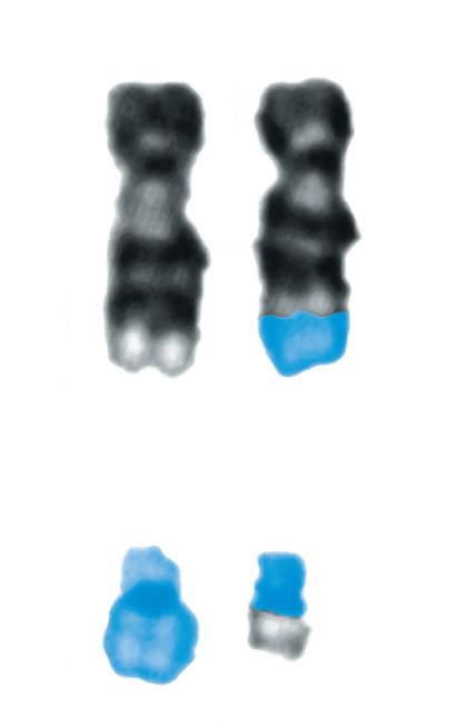
normal 9 der (9)
normal 22 der (22)
Fig. 1.1.7 reciprocal translocation Between two Chromosomes. the Philadelphia chromosome (responsible for chronic myelogenous leukemia) is shown as an example of a reciprocal chromosomal translocation that results in an abnormal gene product responsible for a clinical disorder. in this case an exchange occurs between the long arm of chromosome 9 and the long arm of chromosome 22.
GENES AND PHENOt YPES
The relationship between genes and phenotypes is complex. More than one genetic defect can lead to the same clinical phenotype (genetic heterogeneity), and different phenotypes can result from the same genetic defect (variable expressivity). Retinitis pigmentosa is an excellent example of genetic heterogeneity, as it may be inherited as an X-linked, autosomal dominant, autosomal recessive, or digenic trait, and more than 200 causative genes have been identified.23 Other ocular disorders that are genetically heterogeneous include congenital cataract, glaucoma, and age-related macular degeneration. Different genes may contribute to a common phenotype because they affect different steps in a common pathway. Understanding the role of each gene in the disease process can help define the cellular mechanisms that are responsible for the disease.
For many genes, a single mutation that alters a critical site in the protein results in an abnormal phenotype. For some diseases, the resulting phenotypes are remarkably similar regardless of the nature of the mutation. For example, a wide variety of mutations in RB1 cause retinoblastoma. Other diseases, however, exhibit variable expressivity, in which an individual’s mutation may be responsible for severe disease, mild disease, or disease that is not clinically detectable (incomplete penetrance). There are many examples of ocular disease demonstrating variable expressivity, including Kjer’s autosomal dominant optic atrophy,24 Axenfeld–Rieger syndrome,25 and aniridia.26
acid sequence of the polypeptide chain. The severity of the missense mutation is dependent on the chemical properties of the switched amino acids and on the importance of a particular amino acid in the function of the mature protein. Point mutations also may decrease the level of polypeptide production because they interrupt the promoter sequence, splice site sequences, or create a premature stop codon.
Gene expression can be affected by the insertion or deletion of large blocks of DNA sequence. These types of mutations are less common than point mutations but may result in a more severe change in the activity of the protein product. A specific category of insertion mutations is the expansion of trinucleotide repeats found in patients affected by certain neurodegenerative disorders. An interesting clinical phenomenon, “anticipation,” was understood on a molecular level with the discovery of trinucleotide repeats as the cause of myotonic dystrophy.16 Frequently, offspring with myotonic dystrophy were affected more severely and at an earlier age than their affected parents and grandparents. Examination of the disease-causing trinucleotide repeat in affected pedigrees demonstrated that the severity of the disease correlated with the number of repeats found in the myotonic dystrophy gene in affected individuals. This phenomenon has been observed in a number of other diseases, including Huntington’s disease.17
Chromosomal rearrangements may result in breaks in specific genes that cause an interruption in the DNA sequence. Usually, the break in DNA sequence results in a truncated, unstable, dysfunctional protein product. Occasionally, the broken gene fuses with another gene to cause a “fusion polypeptide product,” which may have a novel activity in the cell. Often, such a novel activity results in an abnormality in the function of the cell. An example of such a fusion protein is the product of the chromosome 9;22 translocation that is associated with many cases of leukemia (Fig. 1.1.7).18,19
A set consisting of one of each autosome as well as an X or a Y chromosome is called a haploid set of chromosomes. The normal complement of two copies of each gene (or two copies of each chromosome) is called diploidy. Rarely, as a result of abnormal chromosome separation during cell division, a cell or organism may have three copies of each chromosome, which is called triploidy. A triploid human is not viable, but some patients have an extra chromosome or an extra segment of a chromosome. In such a situation, the abnormality is called trisomy for the chromosome involved. For example, patients with Down syndrome have three copies of chromosome 21, also referred to as trisomy 21.20 If one copy of a pair of chromosomes is absent, the defect is called haploidy. Deletions of the X chromosome are frequently the cause of Duchenne’s muscular dystrophy.21
Polymorphisms are changes in DNA sequence that don’t have a significant biological effect. These DNA sequence variants may modify disease processes, but alone are not sufficient to cause disease. Human DNA sequence is highly variable and includes single nucleotide polymorphisms (SNPs), microsatellite repeat polymorphisms (20–50 bp repeats of CA or GT sequence), variable number of tandem repeat polymorphisms (VNTR, repeats of 50–100 bp of DNA), or larger insertion deletions.22 RECIPROCAL
Different mutations in the same gene can also result in different phenotypes (allelic heterogeneity). Allelic heterogeneity accounts for the different phenotypes of dominant corneal stromal dystrophies caused by mutations in the TGFB1/BIGH3.27 The phenotypic expression of a mutation may depend on its location within a gene. Such variable expressivity based on the location of the mutation is exemplified by mutations in the rds gene, which may cause typical autosomal dominant retinitis pigmentosa or macular dystrophy depending on the position of the genetic defect.28
PAt t E r NS OF HUMAN INHE r I tANCE
The most common patterns of human inheritance are autosomal dominant, autosomal recessive, X-linked recessive, and mitochondrial. Fig. 1.1.8 shows examples of these four inheritance patterns. Other inheritance patterns less commonly encountered in human disease include X-linked dominant, digenic inheritance (polygenic), pseudodominance, and imprinting. Fig. 1.1.9 defines the notation and symbols used in pedigree construction.
Autosomal Dominant
A disease-causing mutation that is present in only one of the two gene copies at an autosomal locus (heterozygous) is a dominant mutation. For example, a patient with dominant retinitis pigmentosa will have a defect in one copy of one retinitis pigmentosa gene inherited from one parent who, in most cases, is also affected by retinitis pigmentosa. The other copy of that gene, the one inherited from the unaffected parent, is normal (wild type). Affected individuals have a 50% chance of having affected siblings and a 50% chance of passing the abnormal gene to their offspring; 50% of children of an affected individual will be affected. For a dominant disease, males and females transmit the disease equally and are affected equally.
True dominant alleles produce the same phenotype in the heterozygous and homozygous states. In humans, most individuals affected by a disease caused by a dominant allele are heterozygous, but occasionally homozygous mutations have been described. In cases where the homozygous individual is more severely affected than the heterozygous individual, the disease is more appropriately noted to be inherited as a semidominant trait. For example, alleles in the PAX3 gene, causing Waardenburg’s syndrome, are semidominant, because a homozygote with more severe disease compared with their heterozygote relatives has been described.29
In some pedigrees with an autosomal dominant disease, some individuals who carry the defective gene do not have the affected phenotype. However, these individuals can still transmit the disease gene to offspring and have affected children. This phenomenon is called reduced penetrance. The gene responsible for retinoblastoma (RB1) is only 90% penetrant, which means that 10% of the individuals who inherit a mutant copy of the gene do not develop the tumor.30
Autosomal Recessive
Diseases that require both copies of a gene to be abnormal for development are inherited as recessive traits. Heterozygous carriers of mutant genes are
with an autosomal dominant trait
usually clinically normal. The same recessive defect might affect both gene copies, in which case the patient is said to be a homozygote. Different recessive defects might affect the two gene copies, in which case the patient is a compound heterozygote. In a family with recessive disease, both parents are unaffected carriers, each having one wild-type gene (allele) and one mutant gene (allele). Each parent has a 50% chance of transmitting the defective allele to a child. Because a child must receive a defective allele from both parents to be affected, each child has a 25% chance of being affected (50% × 50% = 25%), and 50% of the offspring will be carriers of the disease. If the parents are related, they may be carriers of the same rare mutations, and there is a greater chance that a recessive disease can be transmitted to offspring. Males and females have an equal chance of transmitting and inheriting the disease alleles.
X-Linked Recessive
Fig. 1.1.8 Patterns of Inheritance. For pedigrees with an autosomal dominant trait, panel 1 shows inheritance that originates from a previous generation, panel 2 shows segregation that originates in the second generation of this pedigree, and panel 3 shows an apparent “sporadic” case, which is actually a new mutation that arises in the most recent generation. this mutation has a 50% chance of being passed to offspring of the affected individual. For pedigrees with an autosomal recessive trait, panel 1 shows an isolated affected individual in the most recent generation (whose parents are obligatory carriers of the mutant gene responsible for the condition), panel 2 shows a pair of affected siblings whose father is also affected (for the siblings to be affected, the mother must be an obligate carrier of the mutant gene), and panel 3 shows an isolated affected individual in the most recent generation who is a product of a consanguineous marriage between two obligate carriers of the mutant gene. For pedigrees with an X-chromosomal trait, panel 1 shows an isolated affected individual whose disease is caused by a new mutation in the gene responsible for this condition, panel 2 shows an isolated individual who inherited a mutant copy of the gene from the mother (who is an obligate carrier), and panel 3 shows segregation of an X-linked trait through a multigeneration pedigree (50% of the male offspring are affected, and their mothers are obligate carriers of the disease). For pedigrees with a mitochondrial trait, the panel shows a large, multigeneration pedigree—men and women are affected, but only women have affected offspring.
gene, he will be affected. If a daughter inherits the defective gene, she will be a carrier. An important characteristic of X-linked recessive disorders is that males never transmit the disease to sons directly (male-to-male transmission).
Usually female carriers of an X-linked disease gene do not have any clinical evidence of the disease. However, for some X-linked diseases, mild clinical features can be found in female carriers. For example, in X-linked retinoschisis, affected males are severely affected, whereas carrier females have a visually insignificant but clinically detectable retinal abnormality.31 Mild phenotypic expression of the disease gene can be caused by the process of lyonization. In order for males (with one X chromosome) and females (with two X chromosomes) to have equal levels of expression of X-linked genes, female cells express genes from only one of their two X chromosomes. The decision as to which X chromosome is expressed is made early in embryogenesis, and the line of descending cells faithfully adheres to the early choice. As a result, females are mosaics, with some cells in each tissue expressing the maternally derived X chromosome and the remainder expressing the paternally derived X chromosome. When one of the X chromosomes carries an abnormal gene, the proportion of cells that express the mutant versus the normal gene in each tissue can vary.
If a son inherits the defective
Mutations of the X chromosome produce distinctive inheritance patterns, because males have only one copy of the X chromosome and females have two. Most X-linked gene defects are inherited as X-linked recessive traits. Carrier females are typically unaffected because they have both a normal copy and a defective copy of the disease-associated gene. Carrier males are affected because they only have one defective X chromosome and they do not have a normal gene copy to compensate for the defective copy. All of the daughters of an affected male will be carriers of the disease gene because they will inherit the defective X chromosome. None of the sons of an affected male will be affected or be carriers because they will inherit the Y chromosome. Each child of a carrier female has a 50% chance of inheriting the
Females can also be affected by an X-linked recessive disease if the father is affected and the mother coincidentally is a carrier of a mutation in the disease gene. In this case, 50% of daughters would be affected, because 50% would inherit the X chromosome from the mother carrying the disease gene, and all the daughters would inherit the X chromosome from the father carrying the disease gene. Because most X-linked disorders are rare, the carrier frequency of disease genes in the general population is
Pedigrees
Pedigrees with an autosomal recessive trait
Pedigrees with an X-chromosomal inheritance
Pedigrees with a mitochondrial trait
normal female fraternal twins (not identical)
normal male
single bar indicates mating
normal parents and normal offspring, two girls and a boy, in birth order indicated by the numbers; I and II indicate generations
single parent as presented means partner is normal or of no significance to the analysis
double bar indicates a consanguineous union (mating between close relatives)
identical twins number of children for each sex
darkened square or circle means affected individual; arrow (when present) indicates the affected individual is propositus, the beginning of the analysis
autosomal heterozygous recessive
X-linked carrier dead
aborted or stillborn
X-Linked Dominant Inheritance
This inheritance pattern is similar to X-linked recessive inheritance, except that all females who are carriers of an abnormal gene on the X chromosome are affected rather than unaffected. All of the male offspring are also affected. Incontinentia pigmenti is probably inherited as an X-linked dominant trait. Affected females have irregularly pigmented atrophic scars on the trunk and the extremities and congenital avascularity in the peripheral retina with secondary retinal neovascularization.34 This and other X-linked dominant disorders occur almost always in females, and it is likely that the X chromosome gene defects causing these diseases are embryonic lethals when present in males.
Digenic Inheritance and Polygenic Inheritance
Digenic inheritance occurs when a patient has heterozygous defects in two different genes, and the combination of the two gene defects causes disease. Individuals who have a mutation in only one of the genes are normal. Digenic inheritance is different from recessive inheritance, because the two mutations involve different disease genes. In some retinitis pigmentosa families, mutation analysis of the peripherin gene and the ROM1 gene showed that the affected individuals harbor specific mutations in both genes. Individuals with a mutation in only one copy of either gene were unaffected by the disease.35 Triallelic inheritance has been described in some families affected by Bardet–Biedl syndrome (BBS). In these pedigrees, affected individuals carry three mutations in one or two BBS genes (12 BBS genes have been identified),36 and unaffected individuals have only two abnormal alleles. In some families, it has been proposed that BBS may not be a single-gene recessive disease but a complex trait requiring at least three mutant alleles to manifest the phenotype. This would be an example of triallelic inheritance.37
low, and the chance that a carrier female would mate with a male affected by the same disease is quite low.
Mitochondrial Inheritance
Mitochondria are small organelles located in the cytoplasm of cells. They function to generate ATP for the cell and are most abundant in cells that have high energy requirements, such as muscle and nerve cells. Mitochondria have their own small chromosome—16,569 bp of DNA encoding for 13 mitochondrial proteins, 2 ribosomal RNAs, and 22 tRNAs. Mutations occurring in genes located on the mitochondrial chromosome cause a number of diseases, including Leber’s hereditary optic atrophy32 and Kearns–Sayre syndrome.33 Mutations occurring on the mitochondrial chromosome are inherited only from the mother because virtually all human mitochondria are derived from the maternal egg. Fathers do not transmit mitochondria to their offspring.
Cells vary in the number of mitochondria they contain, and when cells divide, the mitochondria are divided randomly. As a result, different cells can have varying numbers of mitochondria, and if a fraction of the mitochondria contain a mutated gene, different cells will have a varying proportion of healthy versus mutant mitochondria. The distribution of mutant mitochondria is called heteroplasmy, and the proportion of mutant mitochondria can vary from cell to cell and can also change with age. Differences in the relative proportions of mutant mitochondria can partly explain the observed variable severity of mitochondrial diseases and also the variable age of onset of mitochondrial diseases.
Pseudodominance
This term describes an apparent dominant inheritance pattern due to recessive defects in a disease gene. This situation arises when a parent affected by a recessive disease (two abnormal copies of the disease gene) has a spouse who is a carrier of one abnormal copy of the disease gene. Children from this couple will always inherit a defective gene copy from the affected parent and will have a 50% chance of inheriting the defective gene copy from the unaffected carrier parent. On average, half of the children will inherit two defective gene copies and will be affected. The pedigree would mimic a dominant pedigree because of apparent direct transmission of the disease from the affected parent to affected children and because approximately 50% of the children will be affected. Pseudodominant transmission is uncommon, because few people are asymptomatic carriers for any particular recessive gene.
If the expression of a heritable trait or predisposition is influenced by the combination of alleles at three or more loci, it is polygenic. Because of the complex inheritance, conditions caused by multiple alleles do not demonstrate a simple inheritance pattern. These complex traits may also be influenced by environmental conditions. Examples of phenotypes in ophthalmology that exhibit complex inheritance because of contributions of multiple genes and environmental factors are myopia,38 age-related macular degeneration,39 and adult-onset open-angle glaucoma.40
Imprinting
Some mutations give rise to autosomal dominant traits that are transmitted by parents of either sex, but they are expressed only when inherited from a parent of one particular sex. In families affected with these disorders, they would appear to be transmitted in an autosomal dominant pattern from one parent (either the mother or the father) and would not be transmitted from the other parent. Occasionally, the same mutation gives rise to a different disorder depending on the sex of the parent transmitting the trait. These parental sex effects are evidence of a phenomenon called imprinting. Although the molecular mechanisms responsible for imprinting are not completely understood, it appears to be associated with DNA methylation patterns that can mark certain genes with their parental origin.41
MOLECULA r MECHANISMS OF DISEASE
Autosomal Dominant
Disorders inherited as autosomal dominant traits result from mutations that occur in only one copy of a gene (i.e., in heterozygous individuals). Usually, the parental origin of the mutation does not matter. However, if the gene is subject to imprinting, then mutations in the maternal or paternal copy of the gene may give rise to different phenotypes.
Haploinsufficiency
Under normal circumstances, each copy of a gene produces a protein product. If a mutation occurs such that one copy of a gene no longer produces a protein product, then the amount of that protein in the cell has been reduced by half. Mutations that cause a reduction in the amount of protein or lead to inactivation of the protein are called loss-of-function mutations. For many cellular processes, this reduction in protein quantity does not have consequences, i.e., the heterozygous state is normal, and these mutations may be inherited as recessive traits (see later section). However, for some cellular processes there is an absolute requirement for the full dosage of protein product, which can only be furnished if both copies of
Fig. 1.1.9 Basic Pedigree Notation. typical symbols used in pedigree construction are defined.
a particular gene are active. Diseases that are caused by inheritance of a single mutation reducing the protein level by half are inherited as dominant traits.
Gain-of-Function Dominant Negative Effect
Autosomal dominant disorders can be caused by mutant proteins that have a detrimental effect on the normal tissue. Mutations in one copy of a gene may produce a mutant protein that can accumulate as a toxic product or in some other way interfere with the normal function of the cell. The mutant protein may also interfere with the function of the normal protein expressed by the remaining normal copy of the gene, thus eliminating any normal protein activity. It is possible to have gain-of-function mutations that can also be dominant negative because the new function of the protein also interferes with the function of the remaining normal copy of the gene.
Autosomal and X-Linked Recessive
Recessive disorders result from mutations present on both the maternal and paternal copies of a gene. Mutations responsible for recessive disease typically cause a loss of biological activity, either because they create a defective protein product that has little or no biological activity or because they interfere with the normal expression of the gene (regulatory mutations). Most individuals heterozygous for recessive disorders, both autosomal and X-linked, are clinically normal.
GENE t HE r APY
Mutations in the DNA sequence of a particular gene can result in a protein product that is not produced, works poorly, or has acquired a novel function that is detrimental to the cell. Gene-based therapies can involve delivery of a normal gene to disease tissue, replacing or augmenting protein activity with other proteins or small molecules, decreasing abnormal gene expression, or genome-editing techniques to repair the mutation. Therapeutic genes can be delivered to specific tissues using modified viruses as vectors42 (Fig. 1.1.10). A successful example of this approach is the restoration of vision in a canine model of Leber’s congenital amaurosis using a recombinant adeno-associated virus carrying the normal gene (RPE65).43 Human trials using a similar approach also successfully restored vision in patients with RPE65 mutations.44
Diseases caused by mutations that create a gene product that is destructive to the cell (dominant negative or gain of function mutations) need to be treated using a different approach. In these cases, genes or oligonucleotides—in particular antisense molecules—that can reduce expression of the mutated gene are introduced into the cell.45 Gene editing using CRISPR/Cas9 (Fig. 1.1.11) is another potentially useful approach for gain of function or loss of function mutations.46 Recent advances have produced highly potent in vivo gene therapy vectors for targeting retina.47 In addition, new methods are emerging to introduce therapeutic genes into damaged tissue using nonviral mechanisms based on nanotechnology.48
GENE THERAPY USING A RETROVIRUS VECTOR
Therapeutic gene engineered into retrovirus DNA
retrovirus
unpackagable helper provirus
Recombinant virus replicates in a packaging cell
therapeutic human gene
replace retroviral genes with therapeutic human gene
packaging cell
nucleus
RNA
human target cell therapeutic gene product reverse transcription DNA
virions
Fig. 1.1.10 Gene therapy Using a retrovirus Vector. A therapeutic gene is engineered genetically into the retrovirus DnA and replaces most of the viral DnA sequences. the “recombinant virus” that carries the therapeutic gene is allowed to replicate in a special “packaging cell“ that also contains normal virus that carries the genes required for viral replication. the replicated recombinant virus is allowed to infect the human diseased tissue, or “target cell.“ the recombinant virus may invade the diseased tissue but cannot replicate or destroy the cell. the recombinant virus inserts copies of the normal therapeutic gene into the host genome and produces the normal protein product.
Replicated recombinant virus infects the target cell and inserts copies of the therapeutic gene
1. Watson JD, Crick FHC. Molecular structure of nucleic acids: a structure for deoxyribose nucleic acid. Nature 1953;171:737–8.
2. Esteller M. Non-coding RNAs in human disease. Nat Rev Genet 2011;12(12):861–74.
3. Wolfsberg TG, McEntyre J, Schuler GD. Guide to the draft human genome. Nature 2001;409:824–6.
4. Murray JC, Buetow KH, Weber JL, et al. A comprehensive human linkage map with centimorgan density. Cooperative Human Linkage Center (CHLC). Science 1994;265:2049–54.
5. The International HapMap Consortium. The International HapMap Project. Nature 2003;426:789–96.
6. 1000 Genomes Project Consortium. A map of human genome variation from population-scale sequencing. Nature 2010;28:1061–73.
7. Haines JL, Hauser MA, Schmidt S, et al. Complement factor H variant increases the risk of age-related macular degeneration. Science 2005;308:419–21.
8. Seddon JM, Yu Y, Miller EC, et al. Rare variants in CFI, C3 and C9 are associated with high risk of advanced age-related macular degeneration. Nat Genet 2013;45(11):1366–70.
9. Fritsche LG, Igl W, Bailey JN, et al. A large genome-wide association study of age-related macular degeneration highlights contributions of rare and common variants. Nat Genet 2016;48(2):134–43.
10. Hysi PG, Young TL, Mackey DA, et al. A genome-wide association study for myopia and refractive error identifies a susceptibility locus at 15q25. Nat Genet 2010;42:902–5.
11. Verhoeven VJ, Hysi PG, Wojciechowski R, et al. Genome-wide meta-analyses of multiancestry cohorts identify multiple new susceptibility loci for refractive error and myopia. Nat Genet 2013;45(3):314–18.
12. Thorleifsson G, Walters GB, Hewitt AW, et al. Common variants near CAV1 and CAV2 are associated with primary open-angle glaucoma. Nat Genet 2010;42:906–9.
13. Wiggs JL, Yaspan BL, Hauser MA, et al. Common variants at 9p21 and 8q22 are associated with increased susceptibility to optic nerve degeneration in glaucoma. PLoS Genet 2012;8(4):e1002654.
14. Bailey JN, Loomis SJ, Kang JH, et al. Genome-wide association analysis identifies TXNRD2, ATXN2 and FOXC1 as susceptibility loci for primary open-angle glaucoma. Nat Genet 2016;48(2):189–94.
15. Baratz KH, Tosakulwong N, Ryu E, et al. E2-2 protein and Fuchs’s corneal dystrophy. N Engl J Med 2010;363:1016–24.
16. Lindblad K, Schalling M. Expanded repeat sequences and disease. Semin Neurol 1999;19:289–99.
17. Lee JM, Ramos EM, Lee JH, et al. CAG repeat expansion in Huntington disease determines age at onset in a fully dominant fashion. Neurology 2012;78:690–5.
18. Kato T, Kurahashi H, Emanuel BS. Chromosomal translocations and palindromic AT-rich repeats. Curr Opin Genet Dev 2012;22(3):221–8.
19. Vladareanu AM, Müller-Tidow C, Bumbea H, et al. Molecular markers guide diagnosis and treatment in Philadelphia chromosome-negative myeloproliferative disorders (Review). Oncol Rep 2010;23:595–604.
20. Roubertoux PL, Kerdelhue B. Trisomy 21: from chromosomes to mental retardation. Behav Genet 2006;36:346–54.
21. Soltanzadeh P, Friez MJ, Dunn D, et al. Clinical and genetic characterization of manifesting carriers of DMD mutations. Neuromuscul Disord 2010;20:499–504.
22. Little PF. Structure and function of the human genome. Genome Res 2005;15:1759–66.
23. Daiger SP, Sullivan LS, Bowne SJ. Genes and mutations causing retinitis pigmentosa. Clin Genet 2013;84(2):132–41.
24. Han J, Thompson-Lowrey AJ, Reiss A, et al. OPA1 mutations and mitochondrial DNA haplotypes in autosomal dominant optic atrophy. Genet Med 2006;8:217–25.
25. Hjalt TA, Semina EV. Current molecular understanding of Axenfeld–Rieger syndrome. Expert Rev Mol Med 2005;7:1–17.
26. Vincent MC, Gallai R, Olivier D, et al. Variable phenotype related to a novel PAX 6 mutation (IVS4+5G>C) in a family presenting congenital nystagmus and foveal hypoplasia. Am J Ophthalmol 2004;138:1016–21.
27. Schmedt T, Silva MM, Ziaei A, et al. Molecular bases of corneal endothelial dystrophies. Exp Eye Res 2012;95:24–34.
28. Stuck MW, Conley SM, Naash MI. RDS functional domains and dysfunction in disease. Adv Exp Med Biol 2016;854:217–22.
29. Wollnik B, Tukel T, Uyguner O, et al. Homozygous and heterozygous inheritance of PAX3 mutations causes different types of Waardenburg syndrome. Am J Med Genet A 2003;122:42–5.
30. Harbour JW. Molecular basis of low-penetrance retinoblastoma. Arch Ophthalmol 2001;119:1699–704.
31. Sikkink SK, Biswas S, Parry NR, et al. X-linked retinoschisis: an update. J Med Genet 2007;44:225–32.
32. Newman NJ. Hereditary optic neuropathies: from the mitochondria to the optic nerve. Am J Ophthalmol 2005;140:517–23.
33. Schmiedel J, Jackson S, Schafer J, et al. Mitochondrial cytopathies. J Neurol 2003;250: 267–77.
34. Shields CL, Eagle RC Jr, Shah RM, et al. Multifocal hypopigmented retinal pigment epithelial lesions in incontinentia pigmenti. Retina 2006;26:328–33.
35. Kajiwara K, Berson EL, Dryja TP. Digenic retinitis pigmentosa due to mutations at the unlinked peripherin/RDS and ROM1 loci. Science 1994;264:1604–8.
36. Sheffield VC. The blind leading the obese: the molecular pathophysiology of a human obesity syndrome. Trans Am Clin Climatol Assoc 2010;121:172–81.
37. Eichers ER, Lewis RA, Katsanis N, et al. Triallelic inheritance: a bridge between Mendelian and multifactorial traits. Ann Med 2004;36:262–72.
38. Hornbeak DM, Young TL. Myopia genetics: a review of current research and emerging trends. Curr Opin Ophthalmol 2009;20:356–62.
39. Deangelis MM, Silveira AC, Carr EA, et al. Genetics of age-related macular degeneration: current concepts, future directions. Semin Ophthalmol 2011;26:77–93.
40. Wiggs JL, Pasquale LR. Genetics of glaucoma. Hum Mol Genet 2017;26(R1):R21–7.
41. Lewis A, Reik W. How imprinting centres work. Cytogenet Genome Res 2006;113:81–9.
42. Bennett J, Chung DC, Maguire A. Gene delivery to the retina: from mouse to man. Methods Enzymol 2012;507:255–74.
43. Acland GM, Aguirre GD, Ray J, et al. Gene therapy restores vision in a canine model of childhood blindness. Nat Genet 2001;28:92–5.
44. Maguire AM, Simonelli F, Pierce EA, et al. Safety and efficacy of gene transfer for Leber’s congenital amaurosis. N Engl J Med 2008;358:2240–8.
45. Pelletier R, Caron SO, Puymirat J. RNA based gene therapy for dominantly inherited diseases. Curr Gene Ther 2006;6:131–46.
46. Hung SS, McCaughey T, Swann O, et al. Genome engineering in ophthalmology: application of CRISPR/Cas to the treatment of eye disease. Prog Retin Eye Res 2016;53: 1–20.
47. Zinn E, Pacouret S, Khaychuk V, et al. In silico reconstruction of the viral evolutionary lineage yields a potent gene therapy vector. Cell Rep 2015;12(6):1056–68.
48. Vasir JK, Labhasetwar V. Polymeric nanoparticles for gene delivery. Expert Opin Drug Deliv 2006;3:325–44.