Acromioclavicular Joint Injuries: Open Reduction and Internal Fixation
Michael D. McKee and Alireza Naderipour
INDICATIONS
• Acute injury
• Grades IV, V, and VI in most patients unless surgery is contraindicated owing to medical or psychological factors
• Grade III in selected patients, including heavy laborers (lifting, carrying) and overhead athletes/workers
• Chronic injury
• Grade II in patients with symptomatic anterior-posterior instability
• Grades III, IV, and V in patients with symptomatic instability
PHYSICAL EXAMINATION
• Evaluate shoulder posture.
• Determine the position of the distal clavicle relative to the acromion.
• The deformity is more visible in standing or sitting position without support for the injured arm.
• In grade IV dislocations, the clavicle is posterior to the acromion and stuck in the trapezius.
• The distal end of the clavicle is level or superior to the acromion in other grades.
• The distal clavicle is sitting subcutaneously, through the trapezius, in grade V injuries.
• In contrast to higher grades, the acromioclavicular (AC) joint is reducible in grade III by applying an upward force on the ipsilateral elbow.
• Assess horizontal stability by grasping and moving the clavicle.
• Examine sternoclavicular (SC) joint for possible bipolar dislocation (synchronous AC and SC dislocation).
• Assess active and passive shoulder motions.
• AC joint pain is accentuated by abduction and cross-body adduction.
• Manage glenohumeral stiffness prior to reconstruction of chronic separation.
• Isolated AC injury does not typically produce decreased shoulder range of motion.
• Evaluate deltoid and rotator cuff strength.
• Consider the rare occurrence of concomitant rotator cuff pathology.
• Perform neurovascular examination.
IMAGING
STUDIES
• Plain radiographs
• True anteroposterior view of the shoulder
• Evaluate the glenohumeral joint.
• Look for bony signs of rotator cuff pathology.
• Axillary view will demonstrate posterior displacement of the clavicle in grade IV injuries.
• Outlet/scapular Y view
• Evaluate acromial anatomy.
• The presence of a spur may warrant acromioplasty.
• Bilateral anteroposterior acromioclavicular views (Zanca view)
• Evaluate the acromioclavicular joint position.
• Look for possible arthritic changes.
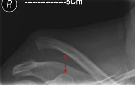
• Compare coracoclavicular distance on both sides.
• Normal coracoclavicular distance is 11 to 13 mm.
PITFALLS
• Acute injury
• Skin abrasion: wait until healed
• Noncompliant patient
• Patient with substance abuse
• Chronic injury
• Noncompliant patient
CONTROVERSIES
There is no consensus on
• Optimum timing of surgery
• Anatomic vs. nonanatomic reconstruction
• Best type of graft
• Acute repair of grade III injuries
• Operative treatment of acute injuries is the only treatment that will restore normal anatomy, but it is associated with greater risk of complications.
• Although often recommended, insufficient evidence exists to recommend surgery for heavy laborers or overhead athletes.
• Successful nonsurgical treatment of type III injuries in professional athletes has been reported.
• Inclusion of distal clavicle excision in management of chronic cases
• Preserving distal clavicle may add to the stability of reduction.
• Reduction of an already arthritic distal clavicle may produce or aggravate pain.
• Resection of distal 1 cm of clavicle results in a 32% increase in posterior translation.
• Resection of as little as 2.3 mm in women and 2.6 mm in men could release the clavicular insertion of the acromioclavicular (AC) ligaments in some patients.
• Some studies suggest improved outcomes with preservation of the distal clavicle during AC reconstruction.
TREATMENT OPTIONS
• Nonoperative treatment
• Indicated for grade I and II and most grade III injuries
• Good short-term results
• 10% to 20% of patients will have residual symptoms and may need subsequent surgery.
• Nonoperative treatment of high-grade injuries (IV, V) may be acceptable, but has a higher rate of poor outcome.
• A short course (1–3 weeks) of sling support or immobilization may be used for comfort,
but strict or prolonged immobilization should be avoided.
• Physical therapy
• Early passive and active assisted range of motion (ROM) exercises
• When painless ROM is achieved, proceed to isometric periscapular and rotator cuff strengthening, followed by isotonic exercises.
• Avoid contact sports and heavy lifting for 3 months.
• Operative treatment
• Components of optimal surgical technique
• Anatomic reduction of acromioclavicular joint
• Coracoclavicular ligament repair/ reconstruction
• Acromioclavicular ligament repair/ reconstruction
• Protection/augmentation of repair/ reconstruction
• Deltoid/trapezoid fascia repair
• Distal clavicle resection, if arthritic
• Acute injury
• Coracoclavicular ligament repair and augmentation
• Multiple techniques have been described to stabilize the AC joint with autograft/ allograft tendon or ligament augmentation devices around the coracoid.
• Transarticular acromioclavicular pin fixation
• Needs limited dissection
• Risk of pin migration/breakage significant, largely abandoned
• Acromioclavicular hook plate
• Mechanically very effective
• May result in acromial wear or fracture
• Newer hook designs that match acromial anatomy preferred
• Avoid over-reduction
• Most, but not all, patients require eventual hook plate removal.
• Weaver-Dunn acromioclavicular ligament transfer
• 40% failure rate, not used in isolation
• Provides 25% of intact coracoclavicular ligament strength
• Strength can be drastically increased by adding synthetic loop augmentation
• Coracoclavicular screw fixation
• Has a high failure rate, not used in isolation
• Acromioclavicluar ligament repair
• Imbrication of the torn AC ligaments
• Chronic injury
• Coracoclavicular ligament reconstruction with
• Tendon graft
• Synthetic loops
• Weaver-Dunn procedure
• Conjoined tendon transfer
• Acromioclavicluar ligament reconstruction with
• Suturing of the remaining coracocalvicular (CC) graft around the AC joint
• Intramedullary free tendon graft
• Reverse coracoacromial ligament
• Stress views
• Originally described to differentiate between type II and type III injuries
• Stress views are costly and uncomfortable for the patient and rarely provide new information to help diagnose an unstable injury.
• Advanced imaging should be considered only if evaluation suggests rotator cuff or intraarticular glenohumeral pathology.
• Magnetic resonance imaging may be indicated to evaluate the rotator cuff in chronic injury.
SURGICAL ANATOMY
• Clavicle
• The distal clavicle forms the medial articulation of the acromioclavicular joint.
• Acromion
• The acromion forms the lateral aspect of the acromioclavicular joint and typically slopes posteriorly and laterally. Newer designs of hook plates recognize this.
• The anterior acromion is also the site of coracoacromial ligament insertion, which is used in the Weaver-Dunn procedure.
• Acromioclavicular joint
• The orientation of the joint varies from vertical to 50 degrees oblique from inferomedial to superolateral.
• The intraarticular meniscus
• Made of fibrocartilage
• True function unknown
• Undergoes significant degeneration over time
• Acromioclavicular ligaments
• The posterior acromioclavicular ligament is an important restraint to posterior translation of the acromioclavicular joint.
• The superior acromioclavicular ligament contributes to a lesser extent to restraint of posterior translation of the acromioclavicular joint.
• The inferior acromioclavicular ligament contributes to restraint of anterior translation of the acromioclavicular joint.
• Isolated disruption of the acromioclavicular ligament occurs in grade II injuries.
• Coracoclavicular ligaments
• The conoid ligament is a more medial structure that attaches on the conoid tubercle on the underside of the distal clavicle. The conoid tubercle is located at the juncture of the lateral and medial thirds of the clavicle.
• The trapezoid ligament is more lateral and attaches on the trapezoid line of the inferior clavicle.
• Disruption of the acromioclavicular and coracoclavicular ligaments occurs in grades III, IV, V, and VI injuries.
• Muscular anatomy
• Trapezius, pectoralis major, and anterior deltoid muscles attach to the distal clavicle and acromion.
• Their combined action provides dynamic stability to the acromioclavicular joint.
• Neurologic anatomy
• Brachial plexus, suprascapular, and musculocutaneous nerves are in the vicinity and could be injured in reconstruction surgeries.
• AC joint is innervated by lateral pectoral, axillary, and suprascapular nerves.
• Vascular anatomy
• Branches of the thoracoacromial artery run in the vicinity of the distal clavicle and can bleed during the dissection and exposure of the base of the coracoid.
POSITIONING
• The patient is placed in the beach chair position, with the surgical field draped out, bony landmarks outlined, and the skin incision marked.
• Neck alignment should be in a neutral position with the head on an adjustable articulating headrest or gel pad “donut.”
• If desired, an articulating arm holder is used to support and position the arm during the procedure. Alternatively, the arm may be secured at the patient’s side.
• A side pad is placed against the lateral chest to keep the patient from falling off the side of the table.
PORTALS/EXPOSURES
• A superior surgical approach is used.
• An incision is made along Langer’s lines over the distal end of the clavicle.
• Begin just posterior to the clavicle and extend toward the coracoid process.
PROCEDURE: HOOK PLATE FIXATION
Step 1: Skin Incision and Surgical Dissection
• Surgical incision is made along Langer’s lines.
• Continue dissection through the subcutaneous tissue.
• The skin and subcutaneous tissue are elevated to extend exposure medially and laterally to expose the distal 3 to 4 cm of the clavicle and the acromion.
Step 2: Acromioclavicular Joint Exposure and Mobilization
• The deltotrapezial fascia is split over the distal clavicle and acromion.
• Typically the acromioclavicular joint capsule and ligaments are disrupted by the injury. Be alert for this disruption and work through any defects created by the injury.
• The meniscus is debrided.
• Look for arthritic changes. Distal clavicle resection should be considered in chronic cases with frank arthritic changes.
• Mobilize the distal clavicle and ensure that it can be reduced.
Step 3: Hook Plate Insertion
• Anterior deltoid is elevated off the distal clavicle, subperiosteally and retracted anteriorly.
• Cauterize vessels imbedded in subdeltoid fatty tissue.
• Open the subacromial space with a Cobb or periosteal elevator and insert the hook portion of the hook plate. This typically will be posterior in the subacromial space.
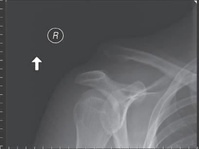
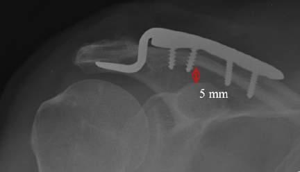
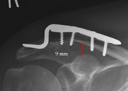
• Use the hook plate trials to determine the correct height of the hook plate to be inserted; be careful not to over-reduce the joint. The clavicle should not require excessive force to reduce (Fig. 1.1).
• Insert the chosen hook plate and then place the screws in the plate, which will bring the plate down to the clavicle.
• Be careful that insertion of the screws in the shaft portion of the clavicle does not “lever” the clavicle down further.
• If there is any question as to reduction, use radiographic imaging to ascertain this. Considerable variation exists in AC joint pathology: a preoperative radiograph of the opposite side can be useful to gauge proper reduction.
Step 4: Optional Coraco-Acromial (CA) Ligament Transfer
• If desired, especially in the chronic situation where an acute healing response will not occur, a CA ligament transfer can be performed in addition.
• This Weaver-Dunn transfer can be performed by releasing the CA ligament from the acromion and inserting it through drill holes in the distal clavicle.
• Alternatively, a small fragment of acromion can be resected with the CA ligament and then secured with a lag screw to a corresponding slot cut into the distal anterior acromion. This provides biologic healing and ligamentous stability following eventual hook plate removal.
Step 5: Optional Coracoclavicular Augmentation
• Acute repair
• The coracoclavicular sutures (nonabsorbable no. 5 suture or 5-mm suture tape) are passed under the coracoid.
• The clavicle is held reduced to the acromion with direct downward push on the distal clavicle and upward pressure on the arm through the elbow.
• Tie the sutures over the plate.
EQUIPMENT
• Articulating sterile arm holder
• Gel headrest
• Side pad
PITFALLS
• Keep the neck aligned in neutral rotation and flexion/extension position to protect the cervical spine and prevent brachial plexus injury.
PEARLS
• Drape high on the neck and inferior enough on the chest to have an adequate surgical field.
• If a difficult reduction is anticipated, drape the operative arm free.
• Position the shoulder in a way that imaging can be used if needed.
PEARLS
• An incision parallel to Langer’s lines will heal with a very cosmetic scar.
PITFALLS
• An incision that is too lateral limits exposure of the clavicle.
• An incision that is too medial limits access to the acromion.
• A longitudinal incision in line with the clavicle, across Langer’s lines, may heal with a thick, noncosmetic scar.
INSTRUMENTATION/IMPLANTATION
• Place a self-retaining retractor to hold the skin and subcutaneous tissue apart.
PEARLS
• Release enough capsule and soft tissue to facilitate anatomic reduction of the distal clavicle.
• Have a preoperative radiograph of the opposite side.
PITFALLS
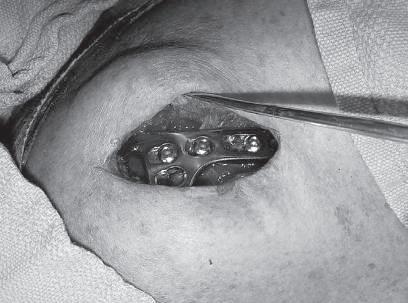
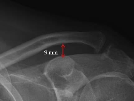
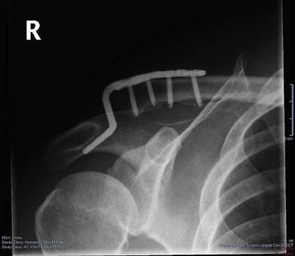
• Avoid over-reduction of the AC joint: this leads to a painful, stiff shoulder with a high rate of subsequent mechanical failure (plate pull-off, acromial fracture) (Fig. 1.2)
• Excessive distal clavicle resection potentially destabilizes the acromioclavicular joint by releasing the acromioclavicular ligaments.
INSTRUMENTATION/IMPLANTATION
• Hook plate implants, including trials and definitive implants
• Newer hook plate designs provide a better fit to the undersurface of the acromion and may minimize complication and removal rates (Fig. 1.3).
• Power saw, osteotome or chisel for distal clavicle resection
1.1 Proper alignment and positioning of the hook plate results in rapid healing in an anatomic position.
1.2 Over reduction of the clavicle is to be avoided as it increases pain and can lead to acromial erosion of the hook.
FIG.
FIG.
• Chronic reconstruction
• Tendon ends are prepared with passing sutures.
• Tendon ends are passed under the coracoid.
• The tendon ends are pulled up through clavicle drill holes or over the clavicle itself and tied into place. Avoid making the superior aspect of the graft too bulky: it will interfere with the hook plate placement.
• Stability is then enhanced by the addition of the hook plate over top of the tendon graft. Once graft healing has occurred, typically 6 to 8 months postoperatively, the hook plate may be removed.
Step 6: Deltotrapezial and Acromioclavicular Repair
• The acromioclavicular ligaments and capsule are repaired over the acromioclavicular joint, incorporating the lateral extension of the tendon graft for a chronic reconstruction.
• The deltotrapezial fascia is sutured over the clavicle with nonabsorbable suture.
POSTOPERATIVE CARE AND EXPECTED OUTCOMES
• A sling is used to support the arm for 6 weeks.
• Physiotherapy protocol
• 0–2 weeks: No shoulder motion is permitted.
• 2–6 weeks: The sling is discontinued and supine passive and active assisted external rotation and scapular plane elevation is begun.
• 6–12 weeks: Passive and active-assisted range of motion in all planes. Isometric deltoid and rotator cuff exercises below chest level are started.
• >12 weeks: Progressive resisted exercises are begun.
• 16 weeks: Return to sports is allowed if range of motion is full and strength is adequate.
• Most patients attain a shoulder rating of 90+ after hook plate fixation of acute AC joint disruptions. The major complication rate is low, as long as over-reduction is avoided.
• Most, but not all, patients require hook plate removal: it is recommended that the plate be left in place for at least 6 months prior to removal to allow adequate healing to occur to prevent re-displacement of the joint.
CONTROVERSIES
• Distal clavicle resection is controversial.
• Distal clavicle resection
• May facilitate reduction
• May prevent late acromioclavicular arthritis
• At least partial resection is required for Weaver-Dunn procedure for ligament reattachment.
• Preserving the distal clavicle
• May facilitate acromioclavicular ligament repair
• May improve acromioclavicular joint stability
• Isolated coracoclavicular ligament reconstruction does not require distal clavicle resection.
INSTRUMENTATION/IMPLANTATION
• Power drill or burr to make holes in the clavicle for suture and tendon passing
CONTROVERSIES
• Coracoclavicular fixation can be achieved with heavy sutures, acromioclavicular hook plate, coracoclavicular screw, transarticular acromioclavicular screw, or pins.
• When patient compliance is a concern, early motion is desired, or in a revision setting, the tendon graft is best supplemented with a hook plate.
• Supplementing the graft with hook plate has been shown to result in less displacement in biomechanical testing.
PEARLS
• Early motion is advantageous.
FIG. 1.3 The angle of the hook should match the usually sloped angle of the acromion.
PITFALLS
• Overly aggressive early rehabilitation can lead to attenuation or failure of the repair or reconstruction.
EVIDENCE
Li X, Ma R, Bedi A, Dines DM, Altchek DW, Dines JS. Management of acromioclavicular joint injuries. J Bone Joint Surg [Am]. 2014;96:73–84.
A comprehensive review of modern treatment methods for acromioclavicular joint injuries.
Galpin RD, Hawkins RJ, Grainger RW. A comparative analysis of operative versus nonoperative treatment of grade III acromioclavicular separations. Clin Orthop. 1985;193:150–155.
This older retrospective review revealed that there was little improvement with surgical treatment of acute acromioclavicular joint injuries and recommended nonoperative treatment in general.
Gstettner C, Tauber M, Hitzl W, Resch H. Rockwood type III acromioclavicular dislocation: surgical versus conservative treatment. J Shoulder Elbow Surg. 2008;17:220–225.
A retrospective study (mean follow-up 34 months) of 24 patients treated surgically with a hook plate and 17 patients treated conservatively. The mean Constant score was 80.7 in the conservative group and 90.4 in the hook plate group. The mean coracoclavicular distance was 15.9 mm in the conservatively treated group and 12.1 mm in the surgically treated group. In this study, better results were achieved by surgical treatment with the hook plate than by conservative treatment.
Salem KH, Schmelz A. Treatment of Tossy III acromioclavicular joint injuries using hook plates and ligament suture. J Orthop Trauma. 2009;23:565–569.
A study of 25 patients revealed the hook plate was a reliable fixation tool for complete AC joint dislocations, ensuring immediate stability and allowing early mobilization with good functional and cosmetic results (mean Constant score 97 points).
Bannister GC, Wallace WA, Stableforth PG, Hutson MA. The management of acute acromioclavicular dislocation. A randomized prospective controlled trial. J Bone Joint Surg. 1989;71B(5):848–850
This study of 60 patients failed to reveal any improvement with surgery, in general. The authors postulate that patients with severe displacement (>2 cm) may benefit from surgery.
von Heideken J, Windhamre HB, Une-larsson V, Ekelund A. Acute surgical treatment of acromioclavicular dislocation type V with a hook plate: superiority to late reconstruction. J Shoulder Elbow Surg 2013;22:9–17.
Patients treated with acute surgery (22) had a more satisfactory outcome than those with late surgery (15) after failed conservative treatment.
Pauly S, Kraus N, Greiner S, Scheibel M. Prevalence and pattern of glenohumeral injuries among acute high-grade acromioclavicular joint instabilities. J Shoulder Elbow Surg. 2013;22:760–766.
A review of 125 patients with high grade AC joint injuries who underwent shoulder arthroscopy revealed a high rate of intra-articular glenohumeral pathology (30%).
Canadian Orthopaedic Trauma Society. Multicenter randomized clinical trial of nonoperative versus operative treatment of acute acromio-clavicular joint dislocation. J Orthop Trauma. 2015;29(11):479–487.
A clinical trial of 83 patients randomized to hook plate fixation versus nonoperative treatment. Although hook plate fixation resulted in superior radiographic alignment, it was not clinically superior to nonoperative treatment of acute complete dislocations of the acromioclavicular joint. Both groups improved from a significant level of initial disability to a good or excellent result (mean DASH score, 5–6; mean Constant score, 91–95 in both groups) at 2 years.
Sacroiliac Joint Injuries: Iliosacral Screws
Milton Lee (Chip) Routt, Jr.
INDICATIONS PITFALLS
• Accurate assessment of SI joint instability is based on physical examination, plain pelvic radiographs, computed tomography (CT) scans, and dynamic imaging during stress examination.
• Complete and incomplete SI joint instability is commonly noted on pelvic imaging.
• SI joint instability may not be obvious if the pelvic imaging was performed after a circumferential pelvic wrap was applied; the pelvic wrap often produces an accurate SI joint reduction.
INDICATIONS CONTROVERSIES
• Controversy still exists in reliably diagnosing and safely treating incomplete posterior pelvic injuries.
• The role of posterior pelvic instability in chronic symptomatic symphysis pubis instability remains controversial.
TREATMENT OPTIONS
• Closed reduction and percutaneous fixation (CRPF) is used whenever possible.
• CRPF relies routinely on intraoperative fluoroscopy to both assess the reduction and direct the iliosacral screw insertion.
• Usually incomplete SI joint injuries will indirectly reduce when the anterior pelvic injury is reduced, or when the precisely oriented lag screw compresses the residual SI joint distraction.
• Open reduction internal fixation (ORIF) of the SI joint is selected when closed reduction techniques fail or are not possible.
• Open reduction of the SI joint is performed using either an anterior exposure with the patient positioned supine, or via posterior surgical exposure in the prone position.
PEARLS
• The folded blanket is adjusted in thickness to elevate the pelvis from the OR table sufficiently to allow iliosacral screw insertion.
• The surgeon must ensure that the eyes are free of pressure, the genitals are positioned appropriately, and that all bony prominences are well padded when the patient is positioned prone.
• Prior to draping, use the C-arm to ensure that the patient is well positioned so that all appropriate images can be easily obtained.
INDICATIONS
• Unstable sacroiliac (SI) joint traumatic disruptions
• Unstable SI fracture-dislocations
• Symptomatic sacroiliac joint arthritis
• Symptomatic chronic posterior pelvic instability
EXAMINATION/IMAGING
• The physical examination identifies open wounds, closed degloving injuries, ecchymoses, prior scars, urethral meatal blood, rectal blood, vaginal-labial injuries, and neurovascular injuries.
• Manual compression toward the midline applied over each iliac crest during the physical examination reveals instability.
• For the injured patient, anteroposterior (AP) pelvic radiograph prior to circumferential pelvic wrapping
• Same patient, AP pelvic radiograph after wrap application
• The pelvic CT reveals injury sites, displacements, deformities, body habitus, hematoma location and extent, and associated injuries.
SURGICAL ANATOMY
• The SI joint is an unusual articulation composed of iliac and sacral articular pads surrounded by strong ligaments.
• The fifth lumbar nerve root is located on the sacral ala just medial to the anterior SI joint.
• For reliable and safe iliosacral screw insertions, the upper sacral osteology (including sacral dysmorphism) must be identified and quantified on the preoperative imaging.
• Hip flexion during the anterior surgical exposure for ORIF relaxes the iliopsoas muscle, eases retraction, and improves exposure of the anterior joint surface.
• Aggressive medial retraction and/or clamp application along the lateral sacral ala during the anterior ORIF risks injury of the fifth lumbar nerve root.
• Wound complications are more common when the posterior exposure is selected for ORIF.
• Iliosacral screws can be safely inserted with the patient properly positioned either supine or prone.
POSITIONING
• When the supine position is selected, a folded operating room (OR) blanket is used to elevate the patient and pelvis from the OR table so the iliosacral screws can be inserted easily.
• Skeletal traction is used as a reduction aid when necessary.
• Positioning the patient supine allows surgical access to both the anterior pelvic ring and the anterior SI joint.
• Prone positioning is more difficult in patients with anterior external fixation devices.
• The prone position denies the anesthesiologist easy access to the airway, and the surgeon must ensure that there is no pressure on the eyes during the surgery.
• The upper extremities are positioned so they do not obstruct either pelvic imaging or iliosacral screw insertion.
PORTALS/EXPOSURES
• The anterior SI joint is accessed using the lateral surgical interval of the ilioinguinal exposure. Hip flexion relaxes the iliopsoas muscle for easier retraction and improved visualization.
• Because of the SI joint’s unusual osteology, the posterior surgical exposure only reveals the caudal articular facet, whereas the anterior articular reduction is assessed by palpation.
• The iliosacral screw’s starting point and directional aim are planned preoperatively using the pelvic CT scan and then determined intraoperatively using inlet, outlet, and true lateral sacral fluoroscopic imaging.
PORTALS/EXPOSURES PEARLS
• A comprehensive preoperative plan includes the details of patient positioning, reduction maneuvers, clamp application, and iliosacral screw insertion.
• The pelvic CT scan identifies and quantifies the parameters for the planned osseus fixation pathways.
• To optimize screw accuracy, the three-dimensional (3D) surface rendered pelvic CT models are correlated with the intraoperative fluoroscopy views.
PITFALLS
• SI joint malreduction decreases the area available for the iliosacral screw within the osseus fixation pathway.
• Reduction clamps or the screws used to attach them to the bone should be positioned so that they do not obstruct the iliosacral screw insertion.
PORTALS/EXPOSURES EQUIPMENT
• A poor quality C-arm unit will not produce sufficient images for safe screw insertion.
• A radiology technician who does not pay attention to the intraoperative imaging details will add unnecessary radiation exposure, time, and cost to the operation. For numerous reasons, an attentive and skilled radiology technician is a critical part of the procedure.
CONTROVERSIES
• When prone posterior ORIF is selected, the reduction clamp is applied to the anterior sacral ala through the greater sciatic notch based on digital palpation of the anterior SI joint alone. This “blind” clamp application remains quite controversial and is not advocated.
• The prone posterior surgical exposure remains controversial because it has been associated with higher wound complication rates.
PROCEDURE
Step 1
• In patients with an incomplete SI joint injury, accurate reduction of the anterior pelvic ring injury (symphysis pubis, pubic ramus, combination injury) often will indirectly reduce the SI joint. In these patients, iliosacral screws are inserted to stabilize the SI joint injury and support the overall fixation construct. Some evidence indicates that iliosacral screw fixation of incomplete SI joint injury decreases the rate of failure of anterior fixation. If compression is needed to complete the SI joint indirect reduction, an initial iliosacral lag screw is inserted.
• In patients with complete SI joint injuries, the anterior pelvic reduction may aid in the SI joint reduction. In these patients with residual SI joint uniform distraction after anterior pelvic reduction, an iliosacral lag screw is used to complete the reduction. Additional screws provide improved support for the SI joint. Multiple iliosacral screws inserted at multiple posterior pelvic levels have lower failure rates.
• Open reduction is selected for those injuries when closed reduction fails. The clamp is applied so that it does not injure the fifth lumbar nerve root and does not obstruct the iliosacral screw fixation.
PITFALLS
• If the folded blanket is too thick, the pelvis will be overly elevated from the OR table causing un unstable patient position.
• Once the patient is positioned and before draping, the necessary intraoperative fluoroscopy images should be obtained. Any positioning changes should be made prior to draping.
• The surgical draping should be inclusive of all necessary exposures and implants.
• Urethral meatal necrosis can result when the urinary catheter is poorly positioned. Similarly, the patient’s scrotum should not be crushed between his thighs during surgery.
• Femoral vein and/or artery catheters and suprapubic catheters should be prepared and draped into the sterile field when necessary rather than removed.
POSITIONING EQUIPMENT
• The C-arm is located on the opposite side from the surgeon.
• The C-arm unit tilts and positioning are adjusted after the patient is positioned and prior to draping. The x-ray technician should mark the floor and C-arm machine so the necessary intraoperative images remain consistent throughout the operation.
CONTROVERSIES
• Some surgeons prefer prone patient positioning for the ease of access to the posterior pelvic ring during iliosacral screw insertion.
• Supine positioning allows the surgeon to access the anterior pelvic ring without compromising surgical access to the SI joint.
• Insufficient imaging may result from poor patient positioning, morbid obesity, osteoporosis, residual bladder or bowel contrast agents, excessive flatus, among others.
PEARLS
• Accurate reduction of the anterior pelvic injury will often result in an excellent indirect reduction of the SI joint.
• In ORIF, the clamp must be properly located in order to provide uniform compression across the SI joint during the iliosacral screw fixation.
PITFALLS
• The reduction clamp should not obstruct the optimal iliosacral screw pathway.
• Poor positioning of the reduction clamp usually results in a poor reduction.
INSTRUMENTATION/IMPLANTATION
• The optimal location for the iliosacral screw is best planned preoperatively using the CT scan.
• For patients with a symmetric upper sacrum and a unilateral SI joint injury, the uninjured side is used for preoperative iliosacral screw planning.
CONTROVERSIES
• Controversy persists on the value of accurate anterior pelvic reduction prior to posterior.
Step 2
• The caudal anterior pathway of the sacral alar ellipsoid is selected because it is the most reliable initial iliosacral screw site.
• Using inlet and outlet posterior pelvic imaging, a narrow diameter smooth Kirschner wire (K-wire) is used to identify the optimal skin insertion site and ideal directional aim. The wire is then inserted approximately 1 cm through the lateral iliac cortical bone.
• The skin incision is then made and the cannulated drill is applied over the K-wire and oscillated into the lateral iliac bone.
• The caudal-anterior location allows the drill to be advanced safely until the drill tip is located 2 to 3 mL lateral to the visible S1 nerve root tunnel, best seen on the outlet image.
• The true lateral image is then obtained by superimposing the greater sciatic notches and iliac cortical densities.
• The true lateral image is used to confirm the accurate location of the drill tip within the safe osseous fixation pathway. The drill tip should be located caudal to the sacral ala-iliac cortical density, posterior to the anterior cortical limit of the vertebral body, cranial to the S1 tunnel, and well anterior to the spinal canal.
PEARLS
• Using the cannulated drill to prepare the pathway first instead of completely inserting the guide pin allows a more precise pathway preparation. Thinner diameter guide pins often become misdirected, resulting in a poorly located screw.
• The posterior iliac tangential image demonstrates the washer as it contacts the bone surface. The washer is used to decrease the chance of unwanted screw intrusion through the lateral iliac cortical bone surface.
PITFALLS
• If the cannulated drill exits the anterior vertebral body, the guide pin can inadvertently advance and injure the local neurovascular structures.
• If the washer intrudes through the lateral iliac cortical bone, the iliosacral screw stability is compromised.
INSTRUMENTATION/IMPLANTATION
• Oblique iliosacral screws are more perpendicular to the SI joint surfaces than trans-sacral screws.
• The oblique iliosacral lag screw compresses residual SI joint distraction.
• Oblique iliosacral screws usually spare the majority of the SI joint articular surfaces, whereas transsacral screws penetrate the articular surfaces.
CONTROVERSIES
• Trans-sacral screws are controversial because they penetrate the uninjured SI joint and are riskier than oblique screws because they traverse the alar areas on both sides.
• Trans-sacral screws result in better biomechanical construct strength, although it is unclear if this results in superior clinical outcomes.
PEARLS
• The intraoperative pelvic inlet image is optimized by superimposing the upper and second sacral vertebral bodies.
• The mid-sagittal image on the injury pelvic CT scan demonstrates the ideal inlet tilt for each patient.
• The intraoperative outlet tilt is best achieved when the cranial edge of the symphysis pubis is superimposed on the second sacral vertebral body. That tilt reveals the S1 nerve root tunnel anterior foramen.
• For morbidly obese patients, the injury CT scan lateral scout image alerts the surgeon to potential intraoperative lateral fluoroscopic imaging difficulties. If the sacrum is not distinct on the CT scout lateral image, then the intraoperative lateral will be similarly obstructed by the soft tissues.
PITFALLS
• Accepting a poorly located skin starting site will result in either an unacceptable lateral iliac bone insertion site or improper directional aim.
• In morbidly obese patients, standard cannulated screw system guide pins, measuring devices, and screw drivers may be of insufficient length. Special longer instrumentation is available and should be utilized.
CONTROVERSIES
• Controversy remains regarding the optimal iliosacral screw number, orientation, and length.
• Some surgeons use only the lateral sacral image for iliosacral screw insertion. This is controversial because it limits the surgeon to just one style of iliosacral screw use.
Step 3
• Depending on the planned pathway, the drill is either advanced into the vertebral body or across the contralateral ala and SI joint, exiting the lateral iliac cortical bone.
• If an oblique iliosacral screw is planned, the drill should not penetrate the anterior vertebral body cortical bone.
• The guide pin for the cannulated screw system is then inserted into the drilled pathway, and the depth is assessed using a measuring device or guide pin of the same length.
• The iliosacral screw and washer are inserted over the guide pin.
• The C-arm is used at frequent intervals during screw insertion to ensure that the guide pin is not being inadvertently advanced.
• At terminal tightening, the C-arm beam is oriented tangentially relative to the screw insertion site at the posterior lateral iliac cortical bone. The screw is tightened to approximate the washer against the lateral iliac cortical bone surface without intrusion.
Step 4
• Adding additional iliosacral screws improves stability and is performed whenever possible.
• If the initial oblique screw is inserted in the caudal-anterior portion of the upper sacral safe osseus fixation pathway, the subsequent screw should be located slightly posterior and cranial to the initial screw in order to be properly contained.
• If the initial screw has provided sufficient compression, the subsequent screw can be a fully threaded screw to maintain the reduction.
Step 5
• The overall fixation construct is strengthened when both the unstable SI joint and the anterior pelvic injured are stabilized and reduced.
• For more extensive injuries (e.g., “jumper’s fractures”), lumbopelvic fixation is added to augment the posterior pelvic stability.
• Posterior trans-iliac screw and plating fixation techniques also have been described to supplement the iliosacral screw fixation.
PEARLS
• The lumbopelvic supplemental fixation procedure is performed with the patient positioned prone after the SI joint injury has been reduced and stabilized.
• Iliosacral screws are inserted before the lumbopelvic iliac bolts are placed. The LPF iliac bolts can be positioned to accommodate the iliosacral screws.
PITFALLS
• Failure to recognize, reduce, and stabilize the associated unstable anterior pelvic ring traumatic injury can result in posterior fixation failure.
• Applying LPF or other implants prior to iliosacral insertion can obstruct the iliosacral screw’s optimal pathway.
INSTRUMENTATION/IMPLANTATION
• Malleable reconstruction plates and medullary ramus screws are used commonly to provide anterior pelvic fixation.
• Safe iliosacral screws have a limited bone pathway, especially when trans-sacral screws are used.
• LPF iliac bolts can be adjusted in position to avoid the iliosacral screws.
CONTROVERSIES
• Controversy remains concerning the number of iliosacral screws necessary to provide sufficient fixation
POSTOPERATIVE CARE AND EXPECTED OUTCOMES
• Rehabilitation is guided by a licensed physical therapist whenever possible.
• The patients use crutches or other assistive devices to unload the injured SI joint during gait. Protected weight bearing on the injured side is continued for 4 to 8 weeks after operation, depending on the injury and fixation details.
PEARLS
• Safe and reliable iliosacral screw insertion occurs when the screw pathway is well planned, the osteology and its intraoperative imaging are completely understood, and the intraoperative imaging is high quality and consistent.
PITFALLS
• Locating the initial screw in the middle area of the osseus fixation pathway improves the safety for that screw, but that location then adds risk to subsequent screw placement.
CONTROVERSIES
• Using multiple screws (and/or trans-sacral screws) at multiple levels to further stabilize the SI joint injury remains controversial. No study has identified how much fixation is required to predictably provide durable stability until complete healing.
PITFALLS
• The fixation construct should be enhanced (i.e., more screws, more levels, trans-sacral screws) at surgery if patient noncompliance is anticipated prior to surgery.
CONTROVERSIES
• Noncompliant patients who exhibit early unprotected weight bearing have an increased risk of fixation failure.
EVIDENCE
Lucas JF, Routt Jr ML, Eastman JG. A useful preoperative planning technique for transiliactranssacral screws. J Orthop Trauma. 2017;31(1):e25–e31.
This article is a well-illustrated technique guide describing “state-of-the-art” planning for the insertion of trans-iliac and trans-sacral screws.
Simonian PT, Routt Jr ML, Harrington RM, Mayo KA, Tencer AF. Biomechanical simulation of the anteroposterior compression injury of the pelvis. An understanding of instability and fixation. Clin Orthop Relat Res. 1994;309:245–256.
A biomechanical study using seven cadaveric pelvii showed that plate fixation of the symphysis pubis alone reduced symphysis pubis motion, but not sacroiliac motion. Use of sacroiliac fixation alone without a symphysis pubis plate did not affect symphysis pubis motion. Both single iliosacral screws and plates produced equivalent decreases in sacroiliac joint motion.
Keating JF, Werier J, Blachut P, Broekhuyse H, Meek RN, O’Brien PJ. Early fixation of the vertically unstable pelvis: the role of iliosacral screw fixation of the posterior lesion. J Orthop Trauma 1999;13(2):107–113.
This paper describes the early results of 38 patients treated with iliosacral screw fixation for injuries of the SI joint. Nearly 44% of patients had some loss of reduction on final follow-up radiographs (malunion). It was recommended that iliosacral screw fixation be protected by anterior ring fixation.
Carlson DA, Scheid DK, Maar DC, Baele JR, Kaehr DM. Safe placement of S1 and S2 iliosacral screws: the “vestibule” concept. J Orthop Trauma. 2000;14(4):264–269.
This study attempted to determine the optimal starting points for placement of S1 and S2 iliosacral screws using normal subject study evaluating helical CT scans of 30 normal pelvic rings. Finding was that the transversely placed (horizontal) iliosacral screw was the least safe of the screws tested. The safest lateral ilium starting point for our entire population was at the posterior sacral body sagittally and at the inferior S1 foramen coronally. S2 iliosacral screws had less cross-sectional area for placement than S1 screws. Placement of the S2 screw slightly to the S1 foraminal side of the S2 vertebral body increased the safety of placement.
Sagi HC, Ordway NR, DiPasquale T. Biomechanical analysis of fixation for vertically unstable sacroiliac dislocations with iliosacral screws and symphyseal plating. J Orthop Trauma. 2004;18(3): 138–143.
Anterior symphyseal plating for the vertically unstable hemipelvis significantly increases the stability of the fixation construct and restores the normal response of the hemipelvis to axial loading. A significant benefit to supplementary iliosacral screws, in addition to a properly placed S1 iliosacral screw, was not shown.
Marissa Bonyun, MD
Fellow Resident
Department of Orthopedic Surgery University of Toronto Toronto, Ontario, Canada
Steven Borland, MBChB, FRCS(Tr+Orth)
Consultant Trauma and Orthopaedic Surgeon Department of Orthopaedic Surgery
Royal Victoria Infirmary
Newcastle Upon Tyne, United Kingdom
Karine Bourduas, MD, FRCSC
Clinical Assistant Professor University of Montreal Montreal, Québec, Canada
Henry M. Broekhuyse, MD
Clinical Professor
Department of Orthopaedic Surgery University of British Columbia Vancouver, British Columbia, Canada
Richard E. Buckley, MD, FRCS
Professor of Orthopedic Trauma Department of Surgery Foothills Medical Center, University of Calgary Calgary, Alberta, Canada
Cory V. Carlston, MD
Surgeon
Department of Orthopaedics Adventist Medical Center Portland, Oregon, United States
Damian Clark
Oak Leigh House Bridge Road Leighwood, Bristol, United Kingdom
Joseph B. Cohen, MD
Assistant Professor of Orthopedic Trauma Department of Orthopedic Surgery and Rehabilitation Loyola University Medical Center Maywood, Illinois, United States
Peter A. Cole, MD
Chief
Orthopaedic Surgery Regions Hospital St. Paul, Minnesota, United States Professor
Orthopaedic Surgery University of Minnesota Minneapolis, Minnesota, United States
Chad P. Coles, MD, FRCSC
Associate Professor
Division of Orthopaedic Surgery
Dalhousie University
Halifax, Nova Scotia, Canada
David W. Cruickshank, BSc, MD, FRCSC
Assistant Professor
Department of Surgery
Queen’s University
Kingston, Ontario, Canada
Niloofar Dehghan, MD, MSc, FRCSC
Orthopaedic Surgeon
The CORE Institute
Banner University Medical Center Phoenix, Arizona, United States
Assistant Professor
Department of Orthopaedic Surgery
University of Arizona College of Medicine - Phoenix Phoenix, Arizona, United States
Johanna Charlotte Emilie Donders, MD
Department of Orthopedic Trauma Service
Hospital for Special Surgery
New York, New York, United States
Paul Duffy, BA (Hons), MD, FRCSC
Division Chief Orthopedic Trauma
Surgery
Foothills Medical Centre Calgary, Alberta, Canada
Uma E. Erard, DO, FAAOS
Orthopaedic Foot and Ankle Surgeon
San Antonio Military Medical Center
San Antonio, Texas, United States
Tym Frank, MD, MSc, FRCSC Fellow
Department of Surgery
Roth McFarlane Hand and Upper Limb Centre
St. Joseph’s Health Care
University of Western Ontario London, Ontario, Canada
Andrew Furey, MD, MSc, FRCSC, MSM
Associate Professor
Department of Surgery
Memorial University
St. Johns, Newfoundland and Labrador, Canada
Peter V. Giannoudis, MBBS, MD, FACS, FRCS
Professor of Trauma and Orthopaedic Surgery
School of Medicine
University of Leeds
Leeds, United Kingdom
Thomas J. Goetz, BSc(Eng), MD, FRCS(C)
Clinical Professor Orthopaedics
University of British Columbia Vancouver, British Columbia, Canada
Wade Gofton, BScH, MD, MEd, FRCSC Associate Professor Department of Surgery University of Ottawa Ottawa, Ontario, Canada
John T. Gorczyca, MD
C. McCollister Evarts Professor of Orthopaedics Chief, Division of Orthopaedic Trauma Department of Orthopaedic Surgery University of Rochester Medical Center Rochester, New York, United States
Ruby Grewal, MD, MSc, FRCSC Associate Professor University of Western Ontario
Roth | McFarlane Hand and Upper Limb Centre St Joseph’s Health Center London, Ontario, Canada
Pierre Guy, MD, MBA
Head Division of Orthopedic Trauma University of British Columbia Vancouver, British Columbia, Canada Director
Centre for Hip Health and Mobility University of British Columbia Vancouver, British Columbia, Canada
Jeremy A. Hall, MD, FRCSC, Med Assistant Professor Department of Surgery Division of Orthopaedics St. Michael’s Hospital University of Toronto Toronto, Ontario, Canada
Chris Hamilton, MD, MSc, FRCSC Upper Limb Fellow Orthopaedic Surgery Dalhousie University Halifax, Nova Scotia, Canada
Jonah Hébert-Davies, MD, FRCSC Assistant Professor
Harborview Medical Center
Seattle, Washington, United States
David L. Helfet, MD
Director
Orthopaedic Trauma Service
Hospital for Special Surgery/ New York Hospital
New York, New York, United States
Patrick Henry, MD
Assistant Professor Surgery
University of Toronto Toronto, Ontario, Canada
James L. Howard, MD, MSc, FRCSC
Program Director, Associate Professor Division of Orthopaedic Surgery
Western University London, Ontario, Canada
Adrian Huang, MB BCh BAO, FRCSC
Clinical Instructor
University of British Columbia Department of Orthopaedics Vancouver, British Columbia, Canada
Stephen Hunt, P. Eng, MD, FRCSC
Orthopedic Surgeon Department of Orthopedics
South Health Campus Hospital Calgary, Alberta, Canada
Clinical Lecturer
University of Calgary Calgary, Alberta, Canada
Ajmal Ikram, MMed(Orth), FC(Orth)SA
Division of Orthopaedic Surgery
Tygerberg Academic Hospital Division of Orthopaedic Surgery Department of Surgical Sciences
Stellenbosch University
Tygerberg, South Africa
The Polyclinic and Swedish Orthopaedic Institute Seattle, Washington, United States
Robert C. Jacobs, MD
Orthopaedic Trauma Fellow
Orthopaedic Surgery and Sports Medicine University of Washington Seattle, Washington, United States
Richard Jenkinson, MD, MSc, FRCSC Head of Orthopaedic Trauma Surgery
Sunnybrook Health Sciences Center
Toronto, Ontario, Canada
Assistant Professor Surgery
University of Toronto Toronto, Ontario, Canada
Assistant Professor
Institute of Health Policy, Management and Evaluation University of Toronto Toronto, Ontario, Canada
Aaron J. Johnson, MD Fellow
Orthopaedics
University of Maryland School of Medicine
Baltimore, Maryland, United States
Clifford B. Jones, MD, FACS
National Chief of Orthopaedic Trauma and Bone Health Center for Orthopedic Research and Education (CORE Institute®)
Phoenix, Arizona, United States
Professor
Orthopaedic Surgery
University of Arizona College of Medicine – Phoenix Center Chiefs for Orthopedic Trauma University Medical Center
Banner University Medicine Orthopedics & Spine Institute Phoenix, Arizona, United States
Graham King, MD, MSc, FRCSC
Professor
Department of Surgery
Roth | McFarlane Hand and Upper Limb Center London, Ontario, Canada
Paul R. King, MMed (Orth), FC(Orth)SA, Division of Orthopaedic Surgery
Tygerberg Academic Hospital
Division of Orthopaedic Surgery
Department of Surgical Sciences
Stellenbosch University
Tygerberg, South Africa
The Polyclinic and Swedish Orthopaedic Institute
Seattle, Washington, United States
Conor Kleweno, MD
Associate Professor
Harborview Medical Center
Seattle, Washington, United States
Hans J. Kreder, MD, MPH Professor
Orthopaedic Surgery and Health Policy Evaluation and Management
University of Toronto Toronto, Ontario, Canada
Division of Orthopedics
Sunnybrook Health Sciences Center Toronto, Ontario, Canada
Adrian Z. Kurz, MD, FRCSC
Resident
Orthopedic Surgery
McMaster University Hamilton, Ontario, Canada
Paul R.T. Kuzyk, MD, MASc, FRCSC
Assistant Professor
Department of Surgery University of Toronto Toronto, Ontario, Canada
G. Yves Laflamme, MD, FRCSC
Professor Department of Surgery University of Montréal Montréal, Québec, Canada
Sebastien Lalonde, MDCM, FRCS(C)
Assistant Professor, Hand and Microvascular Surgery Department of Orthopaedic Surgery University of Missouri Columbia, Missouri, United States
Robert P. Lamberts, MSc, PHD, FECSS
Division of Orthopaedic Surgery
Tygerberg Academic Hospital
Division of Orthopaedic Surgery
Department of Surgical Sciences
Stellenbosch University
Tygerberg, South Africa
The Polyclinic and Swedish Orthopaedic Institute
Seattle, Washington, United States
Jean Lamontagne, MD, FRCSC Division Head
Orthopaedic Surgery Québec, Québec, Canada
Abdel-Rahman Lawendy, MD, PhD, FRCSC
Associate Professor Department of Surgery
Orthopedic Trauma Fellowship Director
Chair Masters of Surgery Scientist
Lawson Health Research Institute London, Ontario, Canada
Vu Le, MD, FRCSC
Orthopaedic Trauma Fellow Department of Orthopaedics University of British Columbia Royal Columbian Hospital
New Westminster, British Columbia, Canada
Kelly A. Lefaivre, MD, MSc, FRCSC
Associate Professor
Orthopaedic Surgery
University of British Columbia Vancouver, British Columbia, Canada
Ross Leighton, MD, FRCSC, FACS
Orthopaedic Surgeon Surgery
Nova Scotia Health Authority – Halifax Infirmary Halifax, Nova Scotia, Canada Professor Surgery
Dalhousie University Halifax, Nova Scotia, Canada
Martin Lesieur, MD, B.Sc, FRCSC
Orthopaedic Surgeon Orthopaedics
Laval University
Québec, Québec, Canada
Allan S.L. Liew, MD, FRCSC
Associate Professor of Surgery University of Ottawa Director of Orthopaedic Trauma
The Ottawa Hospital Ottawa, Ontario, Canada
Tyler R.S. MacGregor, BSc, MD, FRCSC
Clinical Instructor
Department of Orthopaedic Surgery
Orthopaedic Surgeon Royal Inland Hospital (Kamloops B.C.) Kamloops, British Columbia, Canada
Mark D. Macleod, MD, FRCSC
Associate Professor Department of Surgery Western University London, Ontario, Canada
Theodore T. Manson, MD
Associate Professor
Department of Orthopaedic Surgery R Adams Cowley Shock Trauma Center, University of Maryland Baltimore, Maryland, United States
Jill M. Martin, MD
Assistant Professor
Department of Orthopaedic Surgery Medical College of Wisconsin Milwaukee, Wisconsin, United States
Christopher Ryan Martin MD, FRCSC Cumming School of Medicine University of Calgary Calgary, Alberta, Canada
Michael D. McKee, MD, FRCS(C) Professor and Chairman Department of Orthopaedic Surgery
University of Arizona College of Medicine – Phoenix
Physician Executive Director Orthopaedic and Spine Institute Banner University Medical Center Phoenix, Arizona, United States
Matthew Menon, MD, FRCSC, MHSc
Associate Professor Department of Surgery University of Alberta Edmonton, Alberta, Canada
Mark Miller, MD, FRCSC
Clinical Fellow
Department of Orthopaedics
Division of Orthopaedic Trauma University of British Columbia Vancouver, British Columbia, Canada
Saam Morshed, MD, PhD
Associate Professor in Residence
Orthopaedic Trauma Institute
University of California San Francisco Department of Orthopaedic Surgery
San Francisco, California, United States
Alireza Naderipour, MD, FRCSC Clinical Fellow
Orthopedic Surgery
St. Michael’s Hospital Toronto, Ontario, Canada
Aaron Nauth, MD, MSc
Assistant Professor
Division of Orthopaedic Surgery
St. Michael’s Hospital, University of Toronto Toronto, Ontario, Canada
Vasileios S. Nikolaou, MD, MSc, PhD
Assistant Professor of Orthopaedics
2nd Department of Orthopaedics
National and Kapodistrian University of Athens Athens, Greece
Markku T. Nousiainen, BA(Hons),MS, MEd, MD, FRCSC Program Director
Division of Orthopaedic Surgery
University of Toronto Toronto, Ontario, Canada
Tyler Omeis, BSc, MD Surgical Resident Plastic Surgery
University of British Columbia Vancouver, British Columbia, Canada
Peter J. O’Brien, MD, FRCSC
Associate Professor
Department of Orthopedics
The University of British Columbia Vancouver, British Columbia, Canada
Steven Papp, BSc, MSc, MDCM / FRCSC
Associate Professor
Orthopedic Surgery
University of Ottawa Ottawa, Ontario, Canada
Ryan A. Paul, BHSc, MD, FRCSC
Clinical Fellow
Roth | McFarlane Hand and Upper Limb Centre
St. Joseph’s Health Care
London, Ontario, Canada
Bertrand Perey, MD, FRCSC
Clinical Professor
Department of Orthopaedics Surgery
University of British Columbia Vancouver, British Columbia, Canada
Brad Petrisor, MD, FRCSC
Professor Surgery, Division of Orthopaedic Surgery
McMaster University Hamilton, Ontario, Canada
Orthopaedic Trauma and Foot and Ankle Reconstruction Surgeon Surgery
Hamilton Health Sciences Hamilton, Ontario, Canada
David Pichora, MD, FRCSC
Professor
Paul B. Helliwell Chair in Orthopedic research Professor of surgery and Mechanical and Materials
Engineering
Division of Orthopedic Surgery, Queen’s University Kingston, Ontario, Canada
James Nelson Powell, MD
Clinical Professor of Surgery
Orthopedic Surgery
Cumming School of Medicine University of Calgary Calgary, Alberta, Canada
Darryl N. Ramoutar, MA, MBBChir, FRCS (T&O)
Consultant Orthopaedic Trauma Surgeon University Hospitals Coventry and Warwickshire Warwick, United Kingdom
Lee M. Reichel, MD
Associate Professor Orthopedic Surgery
Department of Surgery and Perioperative Care
Dell Medical School Austin, Texas, United States
Rudolf Reindl, MD, FRCSC
Associate Professor Orthopedic Surgery
McGill University Health Centre Montréal, Québec, Canada
David Ring, MD, PhD
Associate Dean for Comprehensive Care Professor of Surgery and Psychiatry Department of Surgery and Perioperative Care
Dell Medical School – The University of Texas at Austin Austin, Texas, United States
Bill Ristevski, MD, MSc, FRCS(C)
Associate Professor Department of Surgery, Division of Orthopaedic Surgery
McMaster University Hamilton, Ontario, Canada
Aaron M. Roberts
Department of Orthopaedic Surgery University of Rochester Medical Center Rochester, New York, New York
Dominique M. Rouleau, MD, MSc, FRCSC
Associate Professor Université de Montréal Surgery Department Montréal, Québec, Canada
Marie-Ève Rouleau, MPS University of Québec at Montreal Montreal, Québec, Canada
Milton Lee (Chip) Routt, Jr., MD
Professor and the Andrew R. Burgess M.D. Endowed Chair Orthopaedic Surgery University of Texas Health, McGovern Medical School Houston, Texas, United States
H. Claude Sagi, MD, FACS Professor Director Division of Trauma Program Director Orthopedic Trauma Fellowship Department of Orthopedic Surgery and Sports Medicine Cincinnati, Ohio, United States
David W. Sanders, MD, FRCSC Professor Orthopedic Surgery Western University London, Ontario, Canada
Emilie Sandman, MD
Associate Clinical Professor Université de Montréal, Surgery Department Montréal, Québec, Canada
Bruce J. Sangeorzan, MD, FAAOS, FAOA
Professor Department of Orthopedics and Sports Medicine
University of Washington
Director CLiMB, VA Center for Limb Loss and Mobility
Deputy Editor Journal of Bone and Joint Surgery
Past President of American Orthopedic Foot and Ankle Society, AOFAS Rosemont, Illinois, United States
Emil H. Schemitsch, MD, FRCS(C)
Richard Ivey Professor and Chair/Chief Department of Surgery University of Western Ontario London, Ontario, Canada
Andrew H. Schmidt, MD
Chief Orthopaedic Surgery Hennepin County Medical Center Minneapolis, Minnesota, United States Professor Orthopedic Surgery University of Minnesota Minneapolis, Minnesota, United States
Prism S. Schneider, MD, PhD, FRCSC
Clinical Assistant Professor Department of Surgery
Division of Orthopaedic Trauma University of Calgary Calgary, Alberta, Canada
Karen N. Slater, MD
Chief Resident Department of Surgery Division of Plastic Surgery University of British Columbia Vancouver, British Columbia, Canada
Graham Sleat, MD
Locum Consultant in Orthopaedic Trauma Surgery Trauma Department, John Radcliffe Hospital Oxford University Hospitals NHS Foundation Trust Oxford, United Kingdom
Gerard Slobogean, MD
Associate Professor
Orthopaedics University of Maryland School of Medicine Baltimore, Maryland, United States
David Stephen, MD, FRCS(C)
Associate Professor
Division of Orthopaedics, University of Toronto Sunnybrook Health Sciences Centre Toronto, Ontario, Canada
Trevor Stone, MD, FRCSC
Clinical Associate Professor Department of Orthopedics University of British Columbia Vancouver, British Columbia, Canada
Max Talbot, MD, FRCSC
Assistant Professor
McGill University
Staff Surgeon
Montreal General Hospital
McGill University Health Centre
Major and Medical Director
Canadian Forces Trauma Centre (East)
National Defence Government of Canada Montreal, Québec, Canada
Michel A. Taylor, MD, MSc, FRCSC
Clinical Fellow
Department of Orthopedic Surgery
Victoria Hospital
London Health Sciences Centre London, Ontario, Canada
J. Andrew I. Trenholm, MD, MSc Associate Professor of Surgery Department of Surgery
Dalhousie University Halifax, Nova Scotia, Canada
Ted Tufescu, BSc, MD, FRCSC
Assistant Professor Department of Surgery University of Manitoba Program Director, Orthopaedic Surgery Residency Department of Surgery University of Manitoba Fellowship Director, Orthopaedic Trauma Department of Surgery University of Manitoba Winnipeg, Manitoba, Canada
Kayee Tung, RN, CCRP Surgeon Orthopaedics
Royal Children’s Hospital Melbourne, Victoria, Australia
Darius Viskontas, MD, FRCSC
Clinical Associate Professor Department of Orthopaedics
University of British Columbia
Royal Columbian Hospital
New Westminster, British Columbia, Canada
David Weatherby, MD
Fellow
Division of Orthopaedic Surgery
Dalhousie University
Halifax, Nova Scotia, Canada
Ian Whatley, MD
Resident Physician
Division of Orthopaedic Surgery
St. Michael’s Hospital, University of Toronto Toronto, Ontario, Canada
Daniel B. Whelan, MD, MSc, FRCSC
Associate Professor Department of Surgery
Division of Orthopaedics University of Toronto Toronto, Ontario, Canada
Jesse Wolfstadt, MD, MSc, FRCSC
Assistant Professor Granovsky-Gluskin Division of Orthopaedics Sinai Health System University of Toronto
Jeff Yach, MD, FRCS(C) Assistant Professor Surgery
Queen’s University Kingston, Ontario, Canada
Michelle L. Zec, MD, PhD Orthopaedic Surgeon Hand and Upper Extremity Surgery Department of Surgery Cumming School of Medicine University of Calgary