No part of this publication may be reproduced or transmitted in any form or by any means, electronic or mechanical, including photocopying, recording, or any information storage and retrieval system, without permission in writing from the publisher. Details on how to seek permission, further information about the Publisher’s permissions policies and our arrangements with organizations such as the Copyright Clearance Center and the Copyright Licensing Agency, can be found at our website: www.elsevier.com/permissions
This book and the individual contributions contained in it are protected under copyright by the Publisher (other than as may be noted herein).
Notices
Practitioners and researchers must always rely on their own experience and knowledge in evaluating and using any information, methods, compounds or experiments described herein. Because of rapid advances in the medical sciences, in particular, independent verification of diagnoses and drug dosages should be made. To the fullest extent of the law, no responsibility is assumed by Elsevier, authors, editors or contributors for any injury and/or damage to persons or property as a matter of products liability, negligence or otherwise, or from any use or operation of any methods, products, instructions, or ideas contained in the material herein.
ISBN: 978-0-323-54755-0
E-ISBN: 978-0-323-54756-7
Content Strategists: Russell Gabbedy/Kayla Wolfe
Content Development Specialist: Sharon Nash
Project Manager: Joanna Souch
Design: Brian Salisbury
Marketing Manager: Claire McKenzie
Printed in China
Last digit is the print number: 9 8 7 6 5 4 3 2 1
FOREWORD
Myron Yanoff did his residency at the Scheie Eye Institute of the University of Pennsylvania in Ophthalmology followed by a residency in the Department of Pathology. He then did a fellowship at the Armed Forces Institute of Pathology (AFIP) in Washington, DC, under the directorship of Lorenz Zimmerman. Yanoff’s colleague, Ben Fine, was also Zimmerman’s student. Ben Fine was an excellent electron microscopist, and he and Yanoff authored the book, Ocular Histology: A Text and Atlas Dr. Yanoff developed a series of lectures presented at the Annual Postgraduate Course in Ophthalmology at the Scheie Eye Institute and the Lancaster Course in Colby College, Maine, as well as the Biannual Course in Ophthalmic Pathology at the AFIP. These lectures led to the first edition of Ocular Pathology: A Text and Atlas by Drs. Yanoff and Fine, which was published in 1975. The text was presented in outline form, similar to the lecture series, with ample illustrations in black and white and a few color plates. This book became the standard ocular pathology text for residents in ophthalmology and, indeed, I used this textbook when I was an ophthalmology resident. Dr. Yanoff went on to be Chair of the Department of Ophthalmology at the University of Pennsylvania, then Chair at Hahnemann University and, subsequently, Drexel University, where he maintained a comprehensive ophthalmology practice. He and Dr. Fine updated their textbook every several years, with the second edition in 1982, third edition in 1989, fourth edition in 1996, and fifth edition in 2002.
By that time, Yanoff’s resident, Joe Sassani, was ready to replace Ben Fine as the coauthor of this textbook. Dr. Sassani completed his ophthalmology residency and fellowship in Ophthalmic Pathology at the University of Pennsylvania, and developed a practice focused on glaucoma. Dr. Sassani is currently on the faculty at Penn State University in Hershey, Pennsylvania. Drs. Yanoff and Sassani completed the sixth edition of Ocular Pathology in 2009 and the seventh edition in 2015. The textbook retained its outline format; however, virtually all of the illustrations are now in color, and the book is replete with references. The textbook has kept up with the times, as it has added new information including immunohistochemistry, molecular biology, and confocal microscopy over the years.
The story of ocular pathology is one of successive waves of confluences of technology, clinicopathologic correlations and, most importantly, people. An important confluence of technologies occurred in the mid-1800s when Hermann von Helmholtz in Heidelberg developed the ophthalmoscope and Rudolf Virchow in Berlin established cellular pathology as the basis of disease.
This enabled correlation of findings in the eye as seen with the ophthalmoscope with cellular pathology viewed under the microscope. This led to important clinicopathologic correlations in ocular pathology, including tumors such as retinoblastoma and melanoma. As time progressed, more and more disease entities were defined by clinicopathologic correlations. Zimmerman and his students, Yanoff being one of them, described the pathology of most ocular diseases during the so-called “golden age of eye pathology” from the late 1950s through the 1980s. Subsequently, many of Zimmerman’s students, and in turn, their students, Sassani being one of them, applied newer technologies to the descriptions of these ocular diseases. This enabled updates of their book, Ocular Pathology. Ocular pathology has advanced with the confluence of technologies now being molecular biology and digital technology, including imaging technology such as confocal microscopy. The most important element in advancing knowledge and teaching of ocular pathology are individuals, in this case Drs. Yanoff and Sassani. Remarkably, they have updated their textbook, now in its eighth edition, keeping up with new discoveries in ocular pathology and clinicopathologic correlations, including using modern methods of investigation, such as ocular coherence tomography. Some examples of the updates in the eighth edition include the ocular manifestations of Zika virus infection, descriptions of the pathology of intravitreal injections, ocular injuries associated with terrorism, stem cells in the conjunctiva, the latest genetic information regarding corneal dystrophies, the genetics of retinal dystrophies, the TNM classification in the latest edition of the AJCC Cancer Staging Manual, and others.
Indeed, Drs. Yanoff and Sassani have kept up with the times in a remarkable fashion. The definition of a classic textbook is that it endures the test of time and builds upon itself to a point where it becomes a standard text that remains current. In this case, the text began as an outgrowth from the long-standing and storied history of the venerable AFIP, including Yanoff, a disciple of Zimmerman, and Sassani, a student of Yanoff. Ocular Pathology has stood the test of time, remains current, and remains a standard textbook for the study of ocular pathology, the basis of ocular disease. I congratulate Drs. Yanoff and Sassani for their continued efforts in the production of this beautiful textbook, which is now the classic textbook for ophthalmology residents and fellows and pathology residents and fellows.
Hans E. Grossniklaus MD Professor of Ophthalmology and Pathology Emory University School of Medicine
FOREWORDS TO THE FIRST EDITION
During the year of the observance of the 100th anniversary (1874–1974) of the University of Pennsylvania’s Department of Ophthalmology, it is exciting to have the publication of a volume whose coauthors have contributed significantly to the strides in ocular pathology taken by the Department in the past several years.
Myron Yanoff, a highly regarded member of our staff, began a residency in ophthalmology in 1962, upon graduating from the University’s School of Medicine. The residency continued for the next five years, during the first two of which he also held a residency in the Department of Pathology. His keen interest and ability in ocular pathology were readily apparent, and I encouraged him to apply for a fellowship at the Armed Forces Institute of Pathology (AFIP), Washington, DC. From July 1964 through June 1965, he carried out exceptional research at the AFIP in both ophthalmology and pathology. He returned to our Department in July 1965, where the caliber both of his clinical and research work was of the highest. When he completed his residency in June 1967, I invited him to join the staff, and he has recently attained the rank of full professor. During the ensuing years, he has contributed substantially to the literature, particularly in the fields of ophthalmic and experimental pathology. He is Board certified in ophthalmology and in pathology.
Ben Fine, noted for his work in electron microscopy at AFIP and at George Washington University, has shared his expertise in the field through lectures presented as part of the curriculum of the annual 16-week Basic Science Course in the Department’s graduate program.
It can be said that 100 years ago ophthalmology was a specialty that had been gradually evolving during the preceding 100 years, dating from the time of the invention of bifocals by Benjamin Franklin in 1785. Few American physicians of that era, however, knew how to treat diseases of the eye, but as medical education became more specialized it was inevitable that ophthalmology would also become a specialty.
With the invention of the ophthalmoscope in 1851, great advances were made in the teaching and practice of ophthalmology. This contributed greatly, of course, to setting the scene for the establishment of the University’s Department of Ophthalmology. It was on February 3, 1874, that Dr. William F. Norris was elected First Clinical Professor of Diseases of the Eye. Similar chairs had been established earlier in only three other institutions. The chair at the University of Pennsylvania later became known as the William F. Norris and George E. de Schweinitz Chair of Ophthalmology.
Both Dr. Norris and Dr. de Schweinitz actively engaged in the study of ocular pathology. Dr. Norris stressed the importance of the examination of the eye by microscopy and of the correlation of findings from pathology specimens with the clinical signs. Dr. de Schweinitz was instrumental in having a member of his staff accepted as ophthalmic pathologist with the Department of Pathology.
In the years that followed under succeeding chairmen of the Department, other aspects of ophthalmology were stressed. Then, in 1947, during the chairmanship of Dr. Francis Heed Adler, Dr. Larry L. Calkins was appointed to a residency. Dr. Calkins, like Dr. Yanoff, displayed a keen interest in ocular pathology. Accordingly, he was instrumental in its study being revitalized during the three years of his residency. Another resident, Dr. William C. Frayer, who came to the Department in 1949, joined Dr. Calkins in his interest in ocular pathology. Dr. Frayer received additional training in the Department of Pathology and then became the ophthalmic pathologist of the Department.
The importance of ocular pathology was increasingly evident, but facilities for carrying out the work in the Department of Ophthalmology were unfortunately limited. Until 1964, the pathology laboratory had been confined to a small room in the outpatient area of the Department. Then we were able to acquire larger quarters in the Pathology Building of the Philadelphia General Hospital located next door to the Hospital of the University of Pennsylvania. Although the building was earmarked for eventual demolition, the space was fairly adequate for research and also for conducting weekly ophthalmic pathology teaching conferences. Despite the physical aspects, we saw to it that Dr. Yanoff and his team of workers had a well-equipped laboratory.
During the next several years as I saw that my dream for an eye institute with facilities for patient care, teaching, and research under one roof was to become a reality, I was delighted to be able to include prime space on the research floor for the ever enlarging scope of ocular pathology. In addition to all that Dr. Yanoff has had to build upon from the past tradition of our Department of Ophthalmology, I would like to think that the new facilities at the Institute have in some measure contributed to the contents of this excellent volume. With grateful appreciation, therefore, I look upon this book as the authors’ birthday present to the Department. From these same facilities, as Dr. Yanoff and Dr. Fine continue to collaborate, I can hope will come insights and answers for which all of us are ever searching in the battle against eye disease.
Harold G. Scheie, MD Chairman, Department of Ophthalmology University of Pennsylvania Director, Scheie Eye
Institute
From their earliest days in ophthalmology, Myron Yanoff and Ben Fine impressed me as exceptional students. As they have matured and progressed up the academic ladder, they have become equally dedicated and effective teachers. Their anatomical studies of normal and diseased tissues have always been oriented toward providing meaningful answers to practical as well as esoteric clinical questions. Their ability to draw upon their large personal experience in clinical ophthalmology, ocular pathology, and laboratory investigation for their lectures at the Armed Forces Institute of Pathology and at the University of Pennsylvania has contributed immeasurably to the success of those courses. Now
they have used the same time-tested approach in assembling their material for this book. Beginning with their basic lecture outlines, then expanding these with just enough text to substitute for what would have been said verbally in lecture, adding a remarkable amount of illustrative material for the amount of space consumed, and then providing pertinent references to get the more ambitious student started in the pursuit of a subject, Drs. Yanoff and Fine have provided us with a sorely needed teaching aid for both the student and the teacher of ocular pathology. It should prove to be especially popular among medical students and residents in both ophthalmology and ocular pathology. With it one gets good orientation from the well-conceived outlines and fine clinicopathologic correlations from the selection of appropriate illustrations.
It is with considerable pride and admiration that I’ve watched the evolution of the authors’ work and its fruition in the form
of this latest book. I am proud that both authors launched their respective careers with periods of intensive study at the Armed Forces Institute of Pathology and that ever since, they have remained loyal, dedicated, and highly ethical colleagues. I admire their youthful energy, their patient, careful attitude, their friendly cooperative nature, and their ability to get important things accomplished. I’m appreciative of this opportunity to express my gratitude for the work they have been doing. If it is true that “by his pupils, a teacher will be judged,” I could only wish to have had several dozen more like Drs. Yanoff and Fine.
Lorenz E. Zimmerman, MD Chief, Ophthalmic Pathology Division Armed Forces Institute of Pathology Washington, DC
This edition of Ocular Pathology has been revised extensively to reflect the many developments in the field since the publication of the 7th edition. While maintaining a focus on histopathologic and immunohistopathologic features upon which most diagnoses are made, we have expanded coverage of supplemental and correlative techniques such as clinical confocal microscopy and optical coherence tomography. Moreover, we have placed additional emphasis on the pathobiology underlying established and new diagnoses. This emphasis is reflected particularly in expanded coverage of genetics as it relates to disease entities. For a more in-depth analysis of the latest developments in genetics please see: Wiggs JL: Part 1 Genetics, in Yanoff M, Duker JS: Ophthalmology (5th Ed). London: Elsevier 2018. There are many online resources to catalog these conditions, including Online Mendelian Inheritance in Man (OMIM, http://www.ncbi.nlm.nih.gov/omim), RetNet (https://sph.uth.edu/Retnet/), and Retina International (http://www.retina-international.org/)
Virtually every chapter has seen extensive revision including the addition of salient new material. Chapter 1, on the Basic Principles of Pathology, incorporates an expanded discussion of the role of the complement system in ocular homeostasis and disease. The section on immunobiology includes the concept of the inflammasome as a component of innate immunity. A section on autoimmunity and autoinflammation has been added. The discussion of HIV infection has been expanded including newer developments relative to its complications. There are entirely new sections on epigenetics and on modern molecular pathology diagnostic techniques. All of these changes are found in only the first chapter!
Chapter 2, Congenital Anomalies, revises multiple topics including the phakomatoses, chromosomal anomalies, and syndromes such as Noonan syndrome and Walker–Warburg syndrome. Relevant genetic alterations are cited throughout the chapter.
Chapter 3, Nongranulomatous Inflammation: Uveitis, Endophthalmitis, Panophthalmitis, and Sequelae, includes new attention on the ocular manifestations of Zika virus infection.
Chapter 4, Granulomatous Inflammation, updates the discussion of sympathetic uveitis (ophthalmia, ophthalmitis) and nontraumatic infectious causes, such as tuberculosis.
Chapter 5, Surgical and Nonsurgical Trauma, includes an expanded discussion of ophthalmic operative and postoperative surgical complications. A section on intravitreal injections has been added. A discussion of ocular injuries associated with modern warfare and terrorism has been added to the section on nonsurgical trauma. Numerous sections have been expanded in scope including the information on radiation injuries.
Chapter 6, Skin and Lacrimal Drainage System, has an expanded discussion of congenital lesions and anomalies. The new classification system for ichthyosis has been added, as has information regarding its genetic correlates. The section on aging has been expanded significantly, as has the discussion of numerous individual entities, such as pseudoxanthoma elasticum,
epidermolysis bullosa, and erythema multiforme. There is an extensive revision of the sections on degenerative diseases, collagen diseases, and other inflammatory skin conditions, such as the vasculitides. Particular attention has been given to the section on adnexal tumors.
Chapter 7, Conjunctiva, contains an enhanced discussion of stem cells. The congenital anomalies section is expanded significantly, with new entities added. The degenerations section has been revised to include the new classification of amyloidosis. The information regarding multiple types of cystic and neoplastic lesions has been expanded significantly, with a particular emphasis on cancerous epithelial lesions.
Chapter 8, Cornea and Sclera, contains an extensive revision of the section on congenital lesions. The ever-changing classification of corneal dystrophies is reflected in further revisions to that section that also include the latest genetic information impacting our understanding of these disorders. The section on nonheredofamilial disorders also has been revised extensively. Significant new information is found in the section on sclera.
Chapter 9, Uvea, includes updates on aniridia, coloboma, and choroidal dystrophies such as those involving choriocapillaris atrophy.
Chapter 10, Lens, reflects particular attention on congenital cataracts and those associated with syndromes. The section on pseudoexfoliation has been revised, as have other sections including lens-related complications and ectopic lens.
Chapter 11, Neural (Sensory) Retina, reflects updates in congenital and hereditary retinal disorders, vascular diseases, and inflammatory disorders. Particular attention has been directed to retinal degenerations. Multiple modifications have been made to the section on retinal dystrophies with a particular emphasis on the genetics of these disorders.
Chapter 12, Vitreous, has seen a revision on the amyloid section and on familial exudative vitreoretinopathy and other familial disorders.
Chapter 13, Optic Nerve, reflects updates in the section on congenital and familial disorders including relevant syndromes. The section on ischemic optic neuropathies has been revised, as has been the section on optic nerve tumors.
Chapter 14, Orbit, has an extensively revised discussion of thyroid orbitopathy and muscular disorders. The section on the reticuloendothelial system and related disorders, including relative genetic anomalies, has been revised extensively, and the section on Lymphomas and related disorders has been significantly expanded.
Chapter 15, Diabetes Mellitus, includes a new section on ocular surface disease secondary to diabetes. Additional new information and pertinent diagnostic techniques are discussed relative to diabetic complications for each anatomic region of the eye. The information on the pathobiology of ocular diabetic complications is expanded greatly.
Chapter 16, Glaucoma, contains a comprehensively revised discussion of the anatomic basis for aqueous outflow and the
histopathologic correlates in glaucoma. The information on the genetics of glaucoma is revised extensively as is the discussion of pathobiology for each of the glaucomas where appropriate. Particular attention has been paid to the discussion of pseudoexfoliation. Much information has been added regarding the pathobiology of optic nerve damage in glaucoma.
Chapter 17, Ocular Melanocytic Lesions, reflects the TMN classification as found in the 8th edition of the AJCC Cancer Staging Manual. Additionally, particular emphasis has been placed on genetic and chromosomal correlates to prognosis in ocular melanoma. The pathobiology underlying the correlations also is discussed.
Chapter 18, Retinoblastoma and Simulating Lesions, is revised extensively, including the chapter title itself, which drops reference to “pseudoglioma.” The latest classifications for retinoblastoma are presented including the principles on which they are based. Genetic mutations and chromosomal abnormalities relative to retinoblastoma have been revised. Prognostic factors for
retinoblastoma and the latest information on overall survival are included. The section on simulating lesions includes new entities and the latest terminology. Particular attention has been paid to the latest developments in retinopathy of prematurity. Adjunctive diagnostic techniques are discussed.
The 8th edition of Ocular Pathology is replete with new information reflecting the rapidly evolving world of ophthalmic pathology. Nevertheless, as we state in the very first line of our textbook, “The most important tool that the pathologist has at his/her disposal is meaningful communication with the patient’s clinician regarding the suspected diagnosis so that the pathologist can choose the appropriate strategy for processing whatever tissue or other samples are received.” No matter how sophisticated our techniques become, accurate communication remains the bedrock for accurate pathologic diagnoses in support of the best care for our patients.
MY, JS
This book could not have been completed without the understanding and patience of our wives Karin L. Yanoff, PhD, and Gloria Sassani, MA. We also wish to acknowledge the help of our assistants, Kelly McAnally and Sherri Maslasics. Finally, the members of the Elsevier production and editorial team lead by Russell Gabbedy, Kayla Wolfe and Sharon Nash, and including project manager Joanna Souch, designer Brian Salisbury and illustration managers Paula Catalano and Teresa McBryan all have provided invaluable help and guidance in the production of this 8th edition of Ocular Pathology.
We dedicate this book to our wives, Karin and Gloria, and to our children.
The most important tool that the pathologist has at his/her disposal is meaningful communication with the patient’s clinician regarding the suspected diagnosis so that the pathologist can choose the appropriate strategy for processing whatever tissue or other samples are received. As will be seen in the discussion under Modern Molecular Pathology Diagnostic Techniques, there is a dizzying array of techniques at the pathologist’s disposal; however, it is only through communication with the clinician that the pathologist can determine which of these techniques to utilize to best serve the patient.
INFLAMMATION
Definition
I. Inflammation is the response of a tissue or tissues to a noxious stimulus.
A. The tissue may be predominantly cellular (e.g., retina), composed mainly of extracellular materials (e.g., cornea), or a mixture of both (e.g., uvea).
B. The response may be localized or generalized, and the noxious stimulus may be infectious or noninfectious.
II. In a general way, inflammation is a response to a foreign stimulus that may involve specific (immunologic) or nonspecific reactions. Immune reactions arise in response to specific antigens, but they may involve other components (e.g., antibodies, T cells) or nonspecific components (e.g., natural killer [NK] cells, lymphokines).
III. There is an interplay between components of the inflammatory process and blood clotting factors that shapes the inflammatory process.
Causes
I. Noninfectious causes
A. Exogenous causes: originate outside the eye and body, and include local ocular physical injury (e.g., perforating trauma), chemical injuries (e.g., alkali), or allergic reactions to external antigens (e.g., conjunctivitis secondary to pollen).
B Endogenous causes: sources originating in the eye and body, such as inflammation secondary to cellular immunity (phacoanaphylactic endophthalmitis [phacoantigenic uveitis]); spread from continuous structures (e.g., the sinuses); hematogenous spread (e.g., foreign particles); and conditions of unknown cause (e.g., sarcoidosis).
Basic Principles of Pathology 1
II. Infectious causes include viral, rickettsial, bacterial, fungal, and parasitic agents.
Phases of Inflammation
( Table 1.1 lists the actions of the principal mediators of inflammation.)
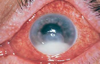
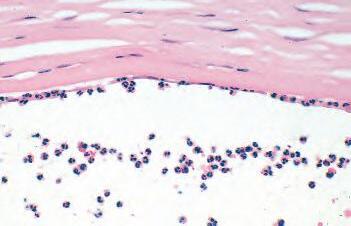
I. Acute (immediate or shock) phase (Fig. 1.1)
A. Five cardinal signs: (1) redness (rubor) and (2) heat (calor)—both caused by increased rate and volume of blood flow; (3) mass (tumor)—caused by exudation of fluid (edema) and cells; (4) pain (dolor) and (5) loss of function (functio laesa)—both caused by outpouring of fluid and irritating chemicals. Table 1.2 lists the roles of various mediators in the different inflammatory reactions.
B. The acute phase is related to histamine release from mast cells and factors released from plasma (kinin, complement, and clotting systems).
1. Histamine is found in the granules of mast cells, where it is bound to a heparin–protein complex. Serotonin (5-hydroxytryptamine), found in platelets and some neuroendocrine cells, has a similar effect to histamine.
2. The kinins are peptides formed by the enzymatic action of kallikrein on the α2-globulin kininogen. Kallikrein is activated by factor XIIa, which is the active form of the coagulation factor XII (Hageman factor). Factor XIIa converts plasma prekallikrein into kallikrein. Plasmin also can activate Hageman factor.
3. Plasmin, the proteolytic enzyme responsible for fibrinolysis, has the capacity to liberate kinins from their precursors and to activate kallikrein, which brings about the formation of plasmin from plasminogen. Plasmin cleaves C3 complement protein, resulting in the formation of C3 fragments. It also breaks down fibrin to form fibrin split products.
4. The complement system (see Table 1.3, which lists the complement molecules found in the normal eye, and Table 1.4, which lists the complement molecules found in diseased eyes) consists of almost 60 proteins present in blood plasma, on the cell surfaces, or within the cell. Its vital nature is evidenced by the fact that it has been preserved by evolution for more than a billion years.
TABLE 1.1 The Actions of the Principal Mediators of Inflammation
Fig. 1.1 Acute inflammation. A, Corneal ulcer with hypopyon (purulent exudate). Conjunctiva hyperemic. B, Polymorphonuclear leukocytes (PMNs) adhere to corneal endothelium and are present in the anterior chamber as a hypopyon (purulent exudate). C, Leukocytes adhere to limbal, dilated, blood-vessel wall (margination) and have emigrated through endothelial cell junctions into edematous surrounding tissue. D, PMNs in corneal stroma do not show characteristic morphology but are recognized by “bits and pieces” of nuclei lining up in a row. (C and D are thin sections from rabbit corneas six hours post-corneal abrasion.)
TABLE 1.2 Role of Mediators in Different Reactions of Inflammation
Role in Inflammation
Vasodilation
Increased vascular permeability
Mediators
Prostaglandins
Nitric oxide
Histamine
Histamine and serotonin
C3a and C5a (by liberating vasoactive amines from mast cells, other cells)
a. Initially named because it was seen to “complement” antibody and cell-mediated immune defenses against microbes.
b. Classic functions: Fig. 1.2 highlights some of the myriad functions performed by complement.
1) Removal of immune (antigen–antibody) complexes.
2) Labeling (opsonization) of foreign antigens for enhanced removal by phagocytes.
3) Recruitment and activation of nearby leukocytes.
4) Direct cytolysis of invading microorganisms.
c. Performs multiple functions in addition to those “classically” ascribed to it.
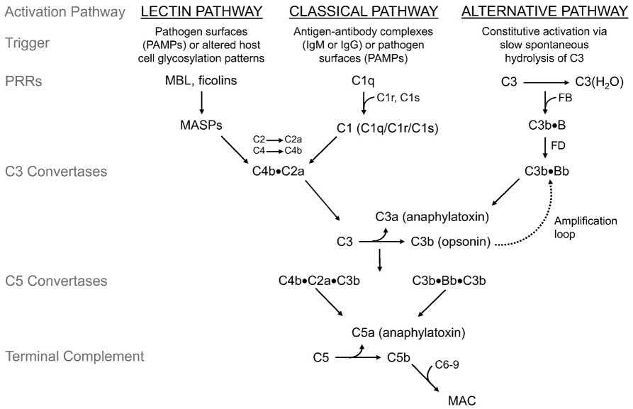
d. Complement achieves its effect through a cascade of the separate components working in coordination and in specific sequences leading through activation of C3. (Fig. 1.3 is a schematic representation of the three primary routes or pathways of complement cascade activation through C3.)
1) The three pathways leading to activation of C3 are:
a) Classical pathway.
b) Lectin pathway.
c) Alternative pathway.
TABLE 1.3 Complement Molecules Found in the Normal Eye
Complement Molecules
Expressed in the Healthy Eye Eye-Associated Remarks
(From Mohlin et al.: The link between morphology and complement in ocular disease. Mol Immunol 89:84–99, 2017. Table 1. Elsevier.)
2) Cleavage of C3 produces the active fragments C3a and C3b.
a) C3a is anaphylatoxin leading to chemotactic and proinflammatory responses.
b) C5a also is an anaphylatoxin.
c) C3b results in opsonization of foreign surfaces.
3) Thus, C3 has a major role in complement activation and generation of immune responses.
TABLE 1.4 Complement Molecules Found in the Human Diseased Eye, i.e., in Age-Related Macular Degeneration (AMD), Glaucoma, Neuromyolitis Optica (NMO) and in Uveitis
Complement Molecules
Expressed in the Diseased Eye Eye Disease–Associated Remarks
Complement Molecules Expressed in the Diseased Eye Eye Disease–Associated Remarks
Complement System Activators
Immunoglobulin Retina, optic nerve
Complement Proteins/Activation Products
C1q
Retina, ganglion cells (GCL) and nerve fiber layer (NFL)
C3, C3b Retina, GCL and NFL
C5b-9 (MAC)
Complement Regulators
Retina, GCL
Factor H GCL Uveitis
Complement System Activators
Immunoglobulin Ocular proteins
Complement Proteins/Activation Products
C3c, C3d Aqueous humor
C4a Aqueous humor
Factor B and Bb Aqueous humor
Complement Anaphylatoxins
C3a, C5a Aqueous humor
Neuromyelitis optica (NMO)
Complement System Activators
Immunoglobulin Optic nerve
aComplement-associated genes connected with AMD: (Adamus et al., 2017; Edwards et al., 2005; Hageman et al., 2005; Haines et al., 2005; Heckner et al., 2010; Klein et al., 2005; Gold et al., 2006; Maller et al., 2007; Park et al., 2009) and uveitis: (Thompson et al., 2013; Yang et al., 2011, 2013; Xu et al., 2015).
(From Mohlin et al., The link between morphology and complement in ocular disease. Mol Immunol 89:84–99, 2017. Table 2. Elsevier.)
e. C1 has been called the “defining component” of the classical complement pathway.
1) Functions as a molecular scaffold for binding of other complement components.
2) Activates and cleaves complement components to continue the complement cascade.
3) Helps to trigger Wnt receptor signaling.
4) Participates in the process of apoptosis.
5) Cleaves MHC class I molecule and other proteins.
6) Can adapt to multiple molecular and cellular processes besides the complement system.
f. Complement plays major roles in immune defense against microorganisms and in clearing damaged host components.
1) It responds to recognition of pathogenassociated molecular patterns (PAMPs) when they bind to host pattern-recognition receptors
(PRRs) and/or internally produced dangerassociated molecular patterns (DAMPs).
g Activation of complement pathways results in a proinflammatory response that includes the generation of membrane attack complexes (MACs), which mediate cell lysis, the release of chemokines to attract inflammatory cells to the site of damage, and the enhancement of capillary permeability. (See Fig. 1.3 for the steps leading to activation of MAC.)
1) Composed of five terminal complement proteins: C5b, C6, C7, C8, and C9. Multiple C9 molecules may be involved.
2) There are numerous levels regulating the activity of MAC and protecting heathy cells from attack. In fact, control of the system is the responsibility of almost half of its components.
a) Disorders resulting from impaired regulation of complement are termed complementopathies.
h. Complement proteins opsonize or lyse cells. Therefore, they may injure healthy tissue, particularly when there is a defect in complement regulation.
i. Complement is important in such diseases as macular degeneration, rheumatoid arthritis, multiple sclerosis, Alzheimer’s disease, schizophrenia, and angioedema.
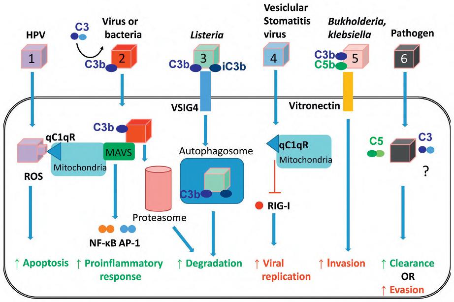
j. T cells and other cell types contain multiple complement components, which have been called the “complosome” in analogy to the inflammasome, which will be discussed later in this chapter. (Fig. 1.4 provides an overview of the multiple ways in which the cell complosome and other complement components may impact key cell processes when faced with various challenges.)
k. Other immune system cells that may produce or be involved in complement function are polymorphonuclear leukocytes, mast cells, monocytes, macrophages, dendritic cells, natural killer (NK) cells, and B cells.
l. Plays a role in adaptive immune response involving T and B cells, and functions as a bridge between innate and adaptive immunity.
m. Helps maintain tissue homeostasis and cellular integrity, and functions in tissue regeneration. Also functions in early sperm–egg interactions in fertilization, regulation of epiboly and organogenesis, and in refinement of cerebral synapses.
n. The complement system is implicated in multiple ocular diseases including age-related macular degeneration, glaucoma, and neuromyelitis optica (Table 1.4 lists elements of the complement system and how they may be involved in these disorders).
o. Complement system, components and their genetic deficiency.
1) Deficiency of early components of the classical pathway (C1q, C1r/s, C2, C4, and C3) is associated with autoimmune diseases resulting from failure of clearance of immune complexes and apoptotic materials and impairment of humoral response.
2) Deficiencies of mannan-binding lectin and the early components of the alternative (factor D and properdin) and terminal pathways (from C3 onward components C5, C6, C7, C8, and C9) increase susceptibility to infections and to their recurrence.
3) See also the discussion of monogenic autoinflammatory syndromes later in this chapter.
p Activation of complement in the tumor microenvironment enhances tumor growth and increases metastasis.
5. Prostaglandins (prostanoids), which have both inflammatory and anti-inflammatory effects, are 20-carbon, cyclical, unsaturated fatty acids with a 5-carbon ring and two aliphatic side chains.
a. They are produced by mast cells, macrophages, endothelial cells, and others.
b. With leukotrienes, they are designated eicosanoids. Leukotrienes are metabolized through the lipoxygenase pathway and prostaglandins through the cyclooxygenase pathway.
c Active in vascular and systemic reactions of inflammation, oxidative stress, and physiologic functions.
d. Cyclooxygenase helps catalyze the biosynthesis of prostaglandins from arachidonic acid.
e. Prostaglandins, cytokines, and leukotrienes function to dilate lymphatics at a site of injury.
f Prostaglandins play an important role in nociception and pain.
6. Major histocompatibility complex (MHC), called the human leukocyte antigen (HLA) complex in humans, is critical to the immune response.
a. HLAs are present on all nucleated cells of the body and platelets.
The HLA region is on autosomal chromosome 6. In practice, the blood lymphocytes are the cells tested for HLA.
Fig. 1.3 Schematic of the complement cascade. The three primary routes for activation of complement are: (1) the lectin pathway (LP), (2) the classical pathway (CP), and (3) the alternative pathway (AP). The LP and CP are activated when specific triggers are recognized by host pattern-recognition receptors (PRRs). The AP is constitutively active. Initial activation through the LP or CP generates a shared C3 convertase (C4b•C2a). In the AP, C3b pairs with factor B (FB) to form the AP proconvertase (C3b•B), which is processed by factor D (FD) to form the AP C3 convertase (C3b•Bb). Both types of C3 convertases cleave C3 to generate C3a and C3b. C3a is an anaphylatoxin, a substance that promotes an inflammatory response. C3b that lands on the surface of a healthy host cell is quickly inactivated; C3b that attaches to the surface of a pathogen or altered host cell triggers a rapid amplification loop to generate more C3b, resulting in opsonization. C3b also complexes with the C3 convertases to form the C5 convertases (C4b•C2a•C3b and C3b•Bb•C3b). In the terminal complement cascade, C5 convertases cleave C5 into C5a (an anaphylatoxin) and C5b. C5b combines with C6–9 to form the membrane attack complex (MAC), also referred to as the terminal complement complex (TCC). Regulatory factors act at various stages of the cascade to control complement activation via their decay accelerating activity and/or cofactor activity. Additional abbreviations: MASPs, mannose-binding lectin-associated serine proteases; MBL, mannose-binding lectin; PAMPs, pathogen-associated molecular patterns. (From Baines AC, Brodsky RA: Complementopathies. Blood Rev 31:213–223, 2017. Figure 1. Elsevier.)
b. The three genetic loci belonging to HLA class I are designated by the letters HLA-A, HLA-B, and HLA-C. Class II MHC molecules are encoded at the locus HLA-D with three subregions HLA-DP, HLA-DQ, and HLA-DR.
1) Class I MHC molecules display proteins derived from foreign antigens, which are recognized by CD8+ T lymphocytes.
2) Class II MHC molecules present antigens that are contained in intracellular vesicles and derived from foreign organisms and soluble proteins.
c. A tentatively identified specificity carries the additional letter “W” (workshop) and is inserted between the locus letter and the allele number— for example, HLA-BW 15.
d. The HLA system is the main human leukocyte isoantigen system and the major human histocompatibility system.
1) HLA-B 27 is positive in a high percentage of young women who have acute anterior uveitis and in young men who have ankylosing spondylitis or Reiter’s disease.
2) HLA-B 51 is strongly associated with Behçet’s disease.
7. Nonspecific soluble mediators of the immune system include cytokines, such as interleukins, which are mediators that act between leukocytes, interferons (IFNs), colony-stimulating factors (CSFs), tumor necrosis factor (TNF), transforming growth factor-β, and lymphokines (produced by lymphocytes).
Fig. 1.4 Suggestions on the potential impact of complosome-derived and/or pathogen-shunted intracellular complement on key cell processes during the host/pathogen interaction. Pathogens trigger an array of responses when interacting with complement during cell infection processes – some of which are beneficial for the microbe and some of which support host protection. For example, infection of human papillomavirus (HPV) triggers globular C1q receptor signaling (gC1qR), which leads to mitochondrial dysfunction and apoptosis (1). Opsonized bacteria trigger mitochondrial antiviral signaling, which increases the expression of AP-1- and NF-κB-controlled genes and proinflammatory cytokine responses. C3-opsonized viruses, on the other hand, are targeted for degradation via the proteosome (2). Opsonized Listeria is also targeted in an intracellular complement-dependent fashion for degradation after cell entry through v-set immunoglobulin domain containing 4 (VSIG4)-driven autophagosome formation (3). Supporting viral and bacterial propagation, gC1R signaling on mitochondria was also shown to block retinoic acid-inducible gene I (RIG-I) activation in a process that promoted the replication of vesicular stomatitis virus (4), while opsonized Klebsiella and other species use vitronectin to gain entry in nonphagocytic cells (5). Although in most of these processes, complement fragments were “dragged” into the cell by microbes, we propose that there will also be (subsequent) interactions of invading intracellular pathogens with components of the complosome, for example C3 and C5 activation fragments (6). In line with the “scheme” observed for the role of serum-derived complement, we further predict that in some cases the complosome will mediate clearance of the pathogen while in other cases, it will be utilized by the pathogen to promote its survival. (From Arbore G et al.: Intracellular complement – the complosome – in immune regulation. Mol Immunol 89:2–9, 2017. Figure 2. Elsevier.)
a. The TNF ligand family encompasses a large group of secreted and cell surface proteins (e.g., TNF and lymphotoxin-α and -β) that may affect the regulation of inflammatory and immune responses.
b. The actions of the TNF ligand family are somewhat of a mixed blessing in that they can protect against infection, but they can also induce shock and inflammatory disease.
C. Immediately after an injury, the arterioles briefly contract (for approximately five minutes) and then gradually relax and dilate because of the chemical mediators discussed previously and from antidromic axon reflexes.
After the transient arteriolar constriction terminates, blood flow increases above the normal rate for a variable time (up to a few hours) but then diminishes to below normal (or ceases) even though the vessels are still dilated. Part of the decrease in flow is caused by increased viscosity from fluid loss through the capillary and venular wall. The release of heparin by mast cells during this period probably helps to prevent widespread coagulation in the hyperviscous intravascular blood.
D. During the early period after injury, the leukocytes (predominantly the PMNs) stick to the vessel walls, at first
momentarily, but then for a more prolonged time; this is an active process called margination (see Fig. 1.1C).
1. Ameboid activity then moves the PMNs through the vessel wall (intercellular passage) and through the endothelial cell junctions (usually taking 2–12 minutes); this is an active process called emigration
2. PMNs, small lymphocytes, macrophages, and immature erythrocytes may also pass actively across endothelium through an intracellular passage in a process called emperipolesis.
3. Mature erythrocytes escape into the surrounding tissue, pushed out of the blood vessels through openings between the endothelial cells in a passive process called diapedesis.
E. Chemotaxis, a positive unidirectional response to a chemical gradient by inflammatory cells, may be initiated by lysosomal enzymes released by the complement system, thrombin, or the kinins.
F PMNs (neutrophils; Fig. 1.5) are the main inflammatory cells in the acute phase of inflammation.
All blood cells originate from a small, common pool of multipotential hematopoietic stem cells. Regulation of the hematopoiesis requires locally specialized bone marrow stromal cells and a coordinated activity of a group of regulatory molecules—growth factors consisting of four distinct regulators known collectively as CSFs.
1. PMNs are born in the bone marrow and are considered “the first line of cellular defense.”
2. CSFs (g lycoproteins that have a variable content of carbohydrate and a molecular mass of 18–90 kDa) control the production, maturation, and function of
PMNs, macrophages, and eosinophils mainly, but also of megakaryocytes and dendritic cells.
3. PMNs are the most numerous of the circulating leukocytes, making up 50–70% of the total.
4. PMNs function at an alkaline pH and are drawn to a particular area by chemotaxis (e.g., by neutrophilic chemotactic factor produced by human endothelial cells).
5. The PMNs remove noxious material and bacteria by phagocytosis and lysosomal digestion.
PMNs produce highly reactive metabolites, including hydrogen peroxide, which is metabolized to hypochlorous acid and then to chlorine, chloramines, and hydroxyl radicals—all important in killing microbes. Lysosomes are saclike cytoplasmic structures containing digestive enzymes and other polypeptides. Lysosomal dysfunction or lack of function has been associated with numerous heritable storage diseases: Pompe’s disease (glycogen storage disease type 2) has been traced to a lack of the enzymes α-1,4-glucosidase in liver lysosomes (see Chapter 11); Gaucher’s disease is caused by a deficiency of the lysosomal enzyme β-glucosidase (see Chapter 11). Metachromatic leukodystrophy is caused by a deficiency of the lysosomal enzyme arylsulfatase-A (see Chapter 11). Most of the common acid mucopolysaccharide, lipid, or polysaccharide storage diseases are caused by a deficiency of a lysosomal enzyme specific for the disease (see under appropriate diseases in Chapters 8 and 11). Chédiak–Higashi syndrome may be considered a general disorder of organelle formation (see section on congenital anomalies in Chapter 11) with abnormally large and fragile leukocyte lysosomes.
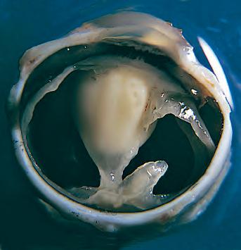
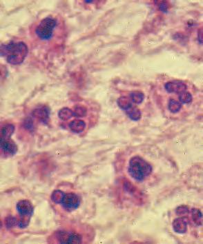
Fig. 1.5 Polymorphonuclear leukocyte (PMN). A, Macroscopic appearance of abscess—that is, a localized collection of pus (purulent exudate)—in vitreous body. B, PMNs are recognized in abscesses by their segmented (usually three parts or trilobed) nucleus. C, Electron micrograph shows segmented nucleus of typical PMN, and its cytoplasmic spherical and oval granules (storage granules or primary lysosomes).
A B
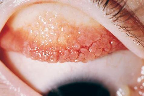
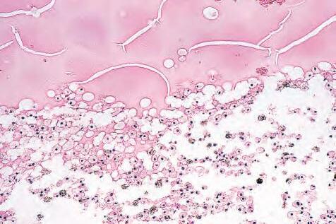
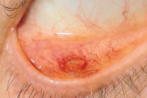
Fig. 1.6 A, Eosinophils are commonly seen in allergic conditions such as this case of vernal catarrh. B, Eosinophils are characterized by bilobed nucleus and granular, pink cytoplasm. C, Electron micrograph shows segmentation of nucleus and dense cytoplasmic crystalloids in many cytoplasmic storage granules. Some granules appear degraded.
6. PMNs are end cells; they die after a few days and liberate proteolytic enzymes, which produce tissue necrosis.
G Eosinophils and mast cells (basophils) may be involved in the acute phase of inflammation.
1. Eosinophils (Fig. 1.6) originate in bone marrow, constitute 1% or 2% of circulating leukocytes, increase in number in parasitic infestations and allergic reactions, and decrease in number after steroid administration or stress. They elaborate toxic lysosomal components (e.g., eosinophil peroxidase) and generate reactive oxygen metabolites.
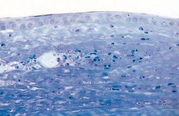
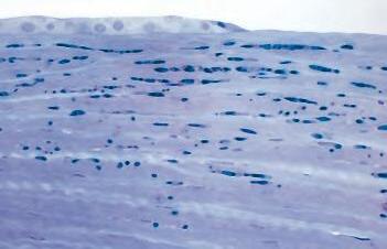
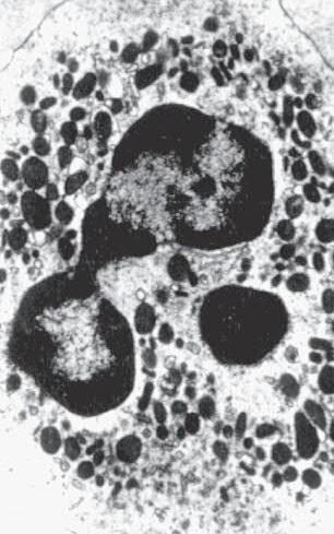
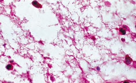
2. Mast cells (basophils; Fig. 1.7) elaborate heparin, serotonin, and histamine, and they are imperative for the initiation of the acute inflammatory reaction.
Except for location, mast cells appear identical to basophils; mast cells are fixed-tissue cells, whereas basophils constitute approximately 1% of circulating leukocytes. Basophils are usually recognized by the presence of a segmented nucleus, whereas the nucleus of a mast cells is large and nonsegmented.
H The acute phase is an exudative phase (i.e., an outpouring of cells and fluid from the circulation) in which the nature of the exudate often determines and characterizes an acute inflammatory reaction.
1. Serous exudate is primarily composed of protein (e.g., seen clinically in the aqueous “flare” in the anterior chamber or under the neural retina in a rhegmatogenous neural retinal detachment).
2. Fibrinous exudate (Fig. 1.8) has high fibrin content (e.g., as seen clinically in a “plastic” aqueous).
3. Purulent exudate (see Figs. 1.1 and 1.5) is composed primarily of PMNs and necrotic products (e.g., as seen in a hypopyon).
The term “pus” as commonly used is synonymous with a purulent exudate.
4. Sanguineous exudate is composed primarily of erythrocytes (e.g., as in a hyphema).
II. Subacute (intermediate or reactive countershock and adaptive) phase.
A. The subacute phase varies greatly and is concerned with healing and restoration of normal homeostasis
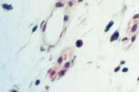
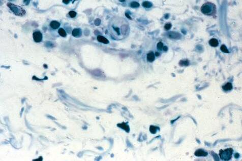
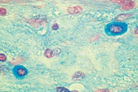
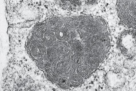
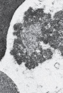
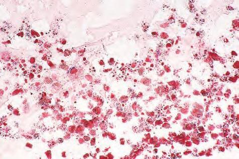
Fig. 1.7 A, Mast cell seen in center as round cell that contains slightly basophilic cytoplasm and round to oval nucleus. B, Mast cells show metachromasia (purple) with toluidine blue (upper right and left and lower right) and C, positive (blue) staining for acid mucopolysaccharides with Alcian blue. D, Electron microscopy of granules in cytoplasm of mast cell often shows typical scroll appearance.
(formation of granulation tissue and healing) or with the exhaustion of local defenses, resulting in necrosis, recurrence, or chronicity.
B. PMNs at the site of injury release lysosomal enzymes into the area.
1. The enzymes directly increase capillary permeability and cause tissue destruction.
2. Indirectly, they increase inflammation by stimulating mast cells to release histamine, by activating the kiningenerating system, and by inducing the chemotaxis of mononuclear (MN) phagocytes.
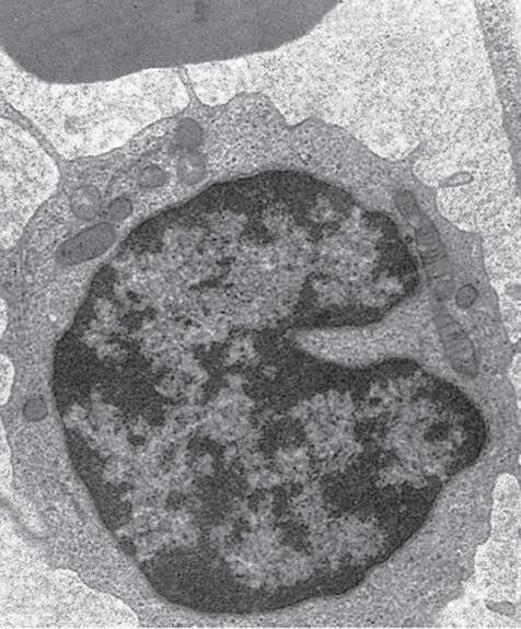
C. Mononuclear (MN) cells (Fig. 1.9) include lymphocytes and circulating monocytes.
1. Monocytes constitute 3%–7% of circulating leukocytes, are bone marrow-derived, and are the progenitor of a family of cells (monocyte–histiocyte–macrophage family) that have the same fundamental characteristics, including cell surface receptors for complement and the Fc portion of immunoglobulin, intracellular lysosomes, and specific enzymes; production of monokines; and phagocytic capacity.
2. Circulating monocytes may subsequently become tissue residents and change into tissue histiocytes, macrophages, epithelioid histiocytes, and inflammatory giant cells.
3. CSFs (g lycoproteins that have a variable content of carbohydrate and a molecular mass of 18–90 kDa) control the production, maturation, and function of MN cells.
4. These cells are the “second line of cellular defense,” arrive after the PMN, and depend on release of chemotactic factors by the PMN for their arrival.
a. Once present, MN cells can live for weeks, and in some cases even months.
b. MN cells cause much less tissue damage than do PMNs, and they are more efficient phagocytes
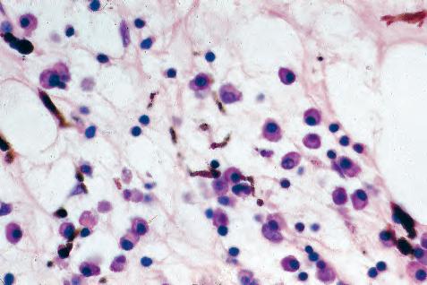
5. Monocytes have an enormous phagocytic capacity and are usually named for the phagocytosed material (e.g., blood-filled macrophages [erythrophagocytosis] and lipid-laden macrophages; Fig. 1.10).
6. Monocytes replace neutrophils as the predominate cell 24–48 hours after the onset of inflammation.
D. Lysosomal enzymes, including collagenase, are released by PMNs, MN cells, and other cells (e.g., epithelial cells and keratocytes in corneal ulcers) and result in considerable tissue destruction.
In chronic inflammation, the major degradation of collagen may be caused by collagenase produced by lymphokine-activated macrophages.
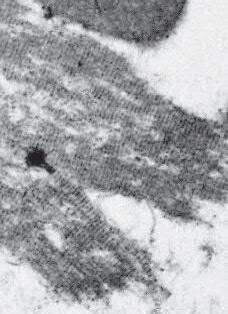
Fig. 1.8 A, Cobweb appearances of fibrinous exudate, stained with periodic acid–Schiff. Cells use fibrin as scaffold to move and to lay down reparative materials. B, Electron micrograph shows periodicity of fibrin cut in longitudinal section. C, Fibrin cut in cross-section.
E. If the area of injury is tiny, PMNs and MN cells alone can handle and “clean up” the area with resultant healing.
F In larger injuries, granulation tissue is produced.
1. Granulation tissue (Fig. 1.11) is composed of leukocytes, proliferating blood vessels, and fibroblasts.
2. MN cells arrive after PMNs, followed by an ingrowth of capillaries that proliferate from the endothelium of pre-existing blood vessels.
The new blood vessels tend to leak fluid and leukocytes, especially PMNs.
3. Fibroblasts (see Fig. 1.11), which arise from fibrocytes and possibly from other cells (monocytes), proliferate, lay down collagen ( Table 1.5), and elaborate ground substance.
4. With time, the blood vessels involute and disappear, the leukocytes disappear, and the fibroblasts return to their resting state (fibrocytes). This involutionary process results in shrinkage of the collagenous scar and a reorientation of the remaining cells into a parallel arrangement along the long axis of the scar.
5. If the noxious agent persists, the condition may not heal as described previously, but instead may become chronic.
6. If the noxious agent that caused the inflammation is immunogenic, a similar agent introduced at a future date can start the cycle anew (recurrence).
Histiocyte/macrophage
Activated macrophage
Activated macrophages
Multinucleated inflammatory giant cell
Fig. 1.9 A, Monocytes have lobulated, large, vesicular nuclei and moderate amounts of cytoplasm, and they are larger than the segmented polymorphonuclear leukocytes and the lymphocytes, which have round nuclei and scant cytoplasm. B, Possible origins of multinucleated inflammatory giant cells and of epithelioid cells.
Epithelioid cells
Langhans Touton
1.10 A, Foamy and clear lipid-laden macrophages in subneural retinal space. B, Cytoplasm of macrophages stains positively for fat with oil red-O technique.
III. Chronic phase
A The chronic phase results from a breakdown in the preceding two phases, or it may start initially as a chronic inflammation (e.g., when the resistance of the body and the inroads of an infecting agent, such as the organisms of tuberculosis or syphilis, nearly balance; or in conditions of unknown cause such as sarcoidosis).
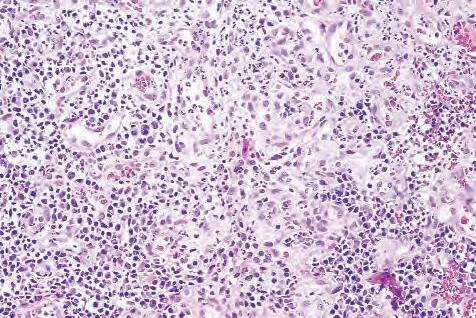
B. Chronic nongranulomatous inflammation is a proliferative inflammation characterized by a cellular infiltrate of lymphocytes and plasma cells (and sometimes PMNs or eosinophils).
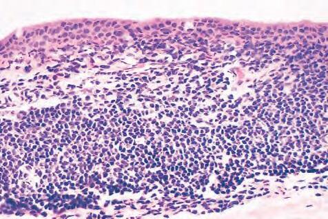
1. The lymphocyte (Fig. 1.12) constitutes 15%–30% of circulating leukocytes and represents the competent immunocyte.
a. All lymphocytes probably have a common stem cell origin (perhaps in the bone marrow) from which they populate the lymphoid organs: the thymus, spleen, and lymph nodes.
b. Two principal types of lymphocytes are recognized: (1) The bone marrow-dependent (or bursal equivalent) B-lymphocyte is active in humoral immunity, is the source of immunoglobulin production
(Fig. 1.13), and is identified by the presence of immunoglobulin on its surface; (2) the thymusdependent T lymphocyte participates in cellular immunity, produces a variety of lymphokines, and is identified by various surface antigens.
1) Helper-inducer T lymphocytes (CD4-positive) initiate the immune response in conjunction with macrophages and interact with (helper) B lymphocytes.
CD4+ T cells are activated after interaction with antigen–MHC complex and differentiate into Helper subsets. These functionally distinct T-helper subsets participate in host defense and immunoregulation. Classically, T-helper 1 (Th1) and T-helper 2 (Th2) cells secrete a distinctive suite of cytokines: Th1 express T-bet and produce interferon-γ and are involved predominantly in cellmediated immunity (e.g., cytotoxic T-cell response); Th2 express Gata3 and produce interleukins-4, -5, and -13. Regulatory T (Treg) cells also are CD4+-derived cells,
A B
Fig.
A
B
Fig. 1.11 Granulation tissue. A, Pyogenic granuloma, here in region of healing chalazion, is composed of granulation tissue. B, Three components of granulation tissue are capillaries, fibroblasts, and leukocytes.
TABLE 1.5 Heterogeneity of Collagens in the Cornea*
*At least 10 genetically distinct collagens have been described in the corneas of different animal species, ages, and pathologies. Types I, II, III, and V collagens are present as fibrils in tissues. Types IV, VI, VII, and VIII form filamentous structures. Types IX and XII are fibril-associated collagens. The sizes of the structures are not completely known. Type II collagen is found only in embryonic chick collagen associated with the primary stroma. Type III collagen is found in Descemet’s membrane and in scar tissue. Types I and V form the heterotypic fibrils of lamellar stroma. Type VII has been identified with the anchoring fibrils, and type VIII is present only in Descemet’s membrane. Type IX collagen, associated with type II fibrils in the primary stroma, and type XII collagen, associated with type I/V fibrils, are part of a family of fibril-associated collagens with interrupted triple helices. Both type IX and type XII are covalently associated with a chondroitin sulfate chain.
serve an immunosuppressive function, and express the master transcription factor FoxP3. There are thymic-derived natural, nTreg cells and peripherally induced iTreg cells that relate to autoimmunity. T-helper 17 (Th17) cells participate in protective tumor immunity; however, Th17-associated cytokines may be associated with tumor initiation and growth and also with autoimmune diseases. Finally, there are follicular T-helper (Tfh) cells that are in proximity to B cells in the germinal centers of lymphoid tissue. They promote class switching of B cells and express the master regulator Bc16 and the effector cytokine IL-21 as well as other surface molecules. Fig. 1.14 illustrates the complexity, flexibility and plasticity of the relationships between T-helper cells.
2) Suppressor-cytotoxic T lymphocytes (CD8positive) suppress the immune response and are capable of killing target cells (e.g., cancer cells) through cell-mediated cytotoxicity.
3) MHC molecules present antigenic peptides to CD8+ T cells, thereby providing the foundation for immune recognition.
2. The plasma cell (Fig. 1.15) is produced by the bone marrow–derived B lymphocyte, elaborates immunoglobulins (antibodies), and occurs in certain modified forms in tissue sections.
After germinal center B cells undergo somatic mutation and antigen selection, they become either memory B cells or plasma cells. CD40 ligand directs the differentiation of germinal center B cells toward memory B cells rather than toward plasma cells.
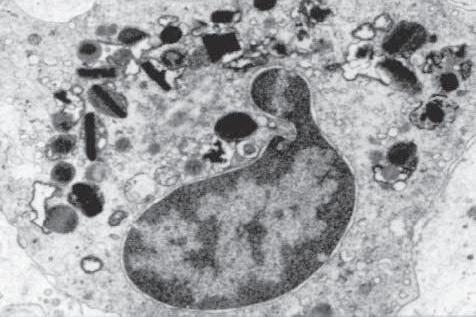
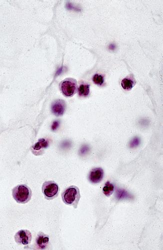
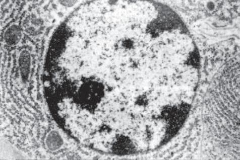
Fig. 1.12 Lymphocyte. A, Low magnification shows cluster of many lymphocytes appearing as a deep blue infiltrate. Cluster appears blue because cytoplasm is scant and mostly nuclei are seen. B, Electron micrograph shows lymphocyte nucleus surrounded by small cytoplasmic ring containing several mitochondria, diffusely arrayed ribonucleoprotein particles, and many surface protrusions or microvilli (rbc, red blood cell). C, Lymphocytes seen as small, dark nuclei with relatively little cytoplasm. Compare with polymorphonuclear leukocytes (segmented nuclei) and with larger plasma cells (eccentric nucleus surrounded by halo and basophilic cytoplasm).
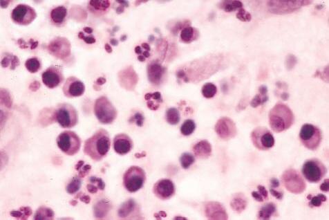
a. Plasmacytoid cell (Fig. 1.16A and B): This has a single eccentric nucleus and slightly eosinophilic granular cytoplasm (instead of the normal basophilic cytoplasm of the plasma cell).
b. Russell body (Fig. 1.16C and D): This is an inclusion in a plasma cell whose cytoplasm is filled and enlarged with eosinophilic grapelike clusters (morular form), with single eosinophilic globular structures, or with eosinophilic crystalline structures; usually the nucleus appears as an eccentric rim or has disappeared.
The eosinophilic material in plasmacytoid cells and in Russell bodies appears to be immunoglobulin that has become inspissated, as if the plasmacytoid cells can no longer release the material because of defective transport by the cells (“constipated” plasmacytoid cells).
C. Chronic granulomatous inflammation is a proliferative inflammation characterized by a cellular infiltrate of lymphocytes and plasma cells (and sometimes PMNs or eosinophils).
1. Epithelioid cells (Fig. 1.17) are bone marrow–derived cells in the monocyte–histiocyte–macrophage family (Fig. 1.18).
a. In particular, epithelioid cells are tissue monocytes that have abundant eosinophilic cytoplasm, somewhat resembling epithelial cells.
Fig. 1.14 Flexibility and plasticity of helper T cells. Recent studies continue to reveal surprising flexibility in expression of “master regulator” transcription factors. In addition, there are now many examples in which helper T cell phenotypes can change their pattern of expression of signature cytokines and gene expression. Striking examples exist in which apparently fully committed “lineages” readily switch their phenotype, and there are now many circumstances in which helper T cells have been shown to express more than one master regulator. This may be advantageous in terms of host defense, but it needs to be borne in mind in thinking about effective therapies for immune-mediated disease and vaccine development. (From Nakayamada S, Takahashi H, Kanno Y et al.: Helper T cell diversity and plasticity. Curr Opin Immunol 24:297, 2012.)
b. They are often found oriented around necrosis as large polygonal cells that contain pale nuclei and abundant eosinophilic cytoplasm whose borders blend imperceptibly with those of their neighbors in a pseudosyncytium (“palisading” histiocytes in a granuloma).
c. All cells of this family interact with T lymphocytes, are capable of phagocytosis, and are identified by the presence of surface receptors for complement and the Fc portion of immunoglobulin.
2. Inflammatory giant cells, probably formed by fusion of macrophages rather than by amitotic division, predominate in three forms:
a. Langhans’ giant cell (Fig. 1.19; see Fig. 1.17): This is typically found in tuberculosis, but it is also seen in many other granulomatous processes. When sectioned through its center, it shows a perfectly homogeneous, eosinophilic, central cytoplasm with a peripheral rim of nuclei.
If the central portion is not homogeneous, foreign material such as fungi may be present: the cell is then not a Langhans’ giant cell but a foreign-body giant cell. When a Langhans’ giant cell is sectioned through its periphery, it simulates a foreign-body giant cell.
b. Foreign-body giant cell (Fig. 1.20): This has its nuclei randomly distributed in its eosinophilic cytoplasm and contains foreign material.
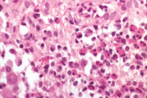
c. Touton giant cell (Fig. 1.21), frequently associated with lipid disorders such as juvenile xanthogranuloma, appears much like a Langhans’ giant cell with the addition of a rim of foamy (fat-positive) cytoplasm peripheral to the rim of nuclei.
3. Three patterns of inflammatory reaction may be found in granulomatous inflammations:
a. Diffuse type (Fig. 1.22A): This typically occurs in sympathetic uveitis, disseminated histoplasmosis
Fig. 1.15 Plasma cell. A, Plasma cells are identified by eccentrically located nucleus containing clumped chromatin and perinuclear halo in basophilic cytoplasm that attenuates opposite to nucleus. Plasma cells are larger than small lymphocytes, which contain deep blue nuclei and scant cytoplasm. B, Electron microscopy shows exceedingly prominent granular endoplasmic reticulum that accounts for cytoplasmic basophilia and surrounds nucleus. Mitochondria are also present in cytoplasm.