No part of this publication may be reproduced or transmitted in any form or by any means, electronic or mechanical, including photocopying, recording, or any information storage and retrieval system, without permission in writing from the publisher. Details on how to seek permission, further information about the Publisher’s permissions policies and our arrangements with organizations such as the Copyright Clearance Center and the Copyright Licensing Agency, can be found at our website: www.elsevier.com/permissions.
This book and the individual contributions contained in it are protected under copyright by the Publisher (other than as may be noted herein).
Notices
Knowledge and best practice in this field are constantly changing. As new research and experience broaden our understanding, changes in research methods, professional practices, or medical treatment may become necessary.
Practitioners and researchers must always rely on their own experience and knowledge in evaluating and using any information, methods, compounds, or experiments described herein. In using such information or methods they should be mindful of their own safety and the safety of others, including parties for whom they have a professional responsibility.
To the fullest extent of the law, neither the Publisher nor the authors, contributors, or editors, assume any liability for any injury and/or damage to persons or property as a matter of products liability, negligence or otherwise, or from any use or operation of any methods, products, instructions, or ideas contained in the material herein.
Library of Congress Cataloging-in-Publication Data
A catalog record for this book is available from the Library of Congress
British Library Cataloguing-in-Publication Data
A catalogue record for this book is available from the British Library
ISBN: 978-0-12-805298-3
For information on all Academic Press publications visit our website at https://www.elsevier.com/
Publisher: Mara Conner
Acquisition Editor: Melanie Tucker
Editorial Project Manager: Halima Williams
Production Project Manager: Edward Taylor
Designer: Matthew Limbert
Typeset by TNQ Books and Journals
7. Narrative and the
8.
II VITAMINS AND MINERALS
MULTIPLE SCLEROSIS
11.
9. Role of Vitamin D in Multiple Sclerosis
BEHAVIORAL MANAGEMENT OF ASSOCIATED CONDITIONS IN MULTIPLE SCLEROSIS
12. Developing and Applying the Theory of Psychological Adaptation Needs in Patients With Multiple Sclerosis
13. Assessment, Consequence, and Clinical Implication of Asymmetry
17. Looking Beyond Neurological Impairment in Patients With Multiple Sclerosis During Exercise Intervention: Evidence for Muscular, Cardiac, Pulmonary, and Metabolic Dysfunction Related to Exercise Intolerance and Prognosis
I. WENS AND D. HANSEN
IV
ENVIRONMENTAL FACTORS AND EXERCISE
14. Neuromuscular Taping and Multiple Sclerosis: Reality or Trend?
C. COSTANTINO AND O. LICARI
15. Constraint-Induced Movement Therapy: When Efficacious Motor Therapy Meets Progressive Disease
A. BARGHI, V.W. MARK AND E. TAUB
Multiple Sclerosis: A Progressive Disease That Is Responsive to Constraint-Induced Movement Therapy
Muscular, Pulmonary, Cardiac, and Metabolic Abnormalities
18. Exercise in the Treatment of Multiple Sclerosis: Pragmatic Exercise Intervention in People With Mild to Moderate Multiple Sclerosis—The ExIMS Project
A. CARTER, L. HUMPHREYS AND B. SHARRACK
19. Yoga and Pilates as Methods of Symptom Management in Multiple Sclerosis
R. FRANK, K. EDWARDS AND J. LARIMORE
16. Physical Activity Behavior in Multiple Sclerosis: Definition, Rates, Outcomes, and Safety
R.W. MOTL AND R.E. KLAREN
20. Exercise in Prevention and Treatment of Multiple Sclerosis
R. MARTÍN-VALERO, A.E. GARCÍA-RODRIGUEZ, M.J. CASUSO-HOLGADO AND J.A. ARMENTA-PEINADO
21. Physical Activity and Health Promotion for People With Multiple Sclerosis: Implementing Activities in the Community
M. MACDONALD, A. DIXON-IBARRA AND K. ROGERS
VI
FOODS IN MULTIPLE SCLEROSIS
25. The Role of Natural Products in the Prevention and Treatment of Multiple Sclerosis
A. SHAMSIZADEH, A. ROOHBAKHSH, F. AYOOBI AND A. MOGHADDAMAHMADI
22. Interdisciplinary Treatment of Patients With Multiple Sclerosis and Chronic Pain
A.B. SULLIVAN AND S. DOMINGO
V
DRUGS OF ABUSE, ALCOHOL AND TOBACCO, AND DISEASE OF MULTIPLE SCLEROSIS PATIENTS
23. Alcohol and Tobacco in Multiple Sclerosis
M. CARDOSO AND Y.D. FRAGOSO
24. Herbal Oil Supplement
26. Effects of B Vitamins in Patients With Multiple Sclerosis
S.P. KALARN AND RONALD ROSS WATSON
27. Eicosapentaenoic Acid in Myelinogenesis: Prospective in Multiple Sclerosis Treatment
A. DI BIASE, L. ATTORRI, R. DI BENEDETTO AND S. SALVATI
28. Biomarkers of Multiple Sclerosis and Their Modulation by Natural Products
Y.A. KULKARNI, M.S. GARUD, M.J. OZA AND A.B. GAIKWAD
List of Contributors
Y. Arima Hokkaido University, Sapporo, Japan
J.A. Armenta-Peinado University of Málaga, Málaga, Spain
L. Attorri Istituto Superiore di Sanità, Rome, Italy
F. Ayoobi Rafsanjan University of Medical Sciences, Rafsanjan, Iran
A. Barghi Harvard Medical School, Boston, MA, United States
G.S. Cantrell University of Oklahoma, Norman, OK, United States
M. Cardoso Universidade Metropolitana de Santos, Santos, SP, Brazil
A. Carter Sheffield Hallam University, United Kingdom
M.J. Casuso-Holgado University of Seville, Seville, Spain
İ. Coşkun Benlidayı Çukurova University, Adana, Turkey
C. Costantino University of Parma, Parma, Italy
R. Di Benedetto Istituto Superiore di Sanità, Rome, Italy
A. Di Biase Istituto Superiore di Sanità, Rome, Italy
A. Dixon-Ibarra Oregon State University, Corvallis, OR, United States
S. Domingo Mellen Center for Multiple Sclerosis, Cleveland, OH, United States
K. Edwards Precision Performance and Physical Therapy, Atlanta, Georgia
T. Elder Newcastle Community Fire Station, Newcastle Under Lyme, United Kingdom
J.W. Farrell University of Oklahoma, Norman, OK, United States
J. Fitzgerald Fort Lewis College, Durango, CO, United States
Y.D. Fragoso Universidade Metropolitana de Santos, Santos, SP, Brazil
R. Frank Georgia State University, Atlanta, Georgia
A.B. Gaikwad Birla Institute of Technology and Science, Pilani, Pilani, Rajasthan, India
A.E. García-Rodriguez University of Seville, Seville, Spain
M.S. Garud Shobhaben Pratapbhai Patel School of Pharmacy & Technology Management, SVKM’s NMIMS, Mumbai, India
D. Hansen Hasselt University, Diepenbeek, Belgium; Jessa Hospital, Hasselt, Belgium
C.E. Hayes University of Wisconsin–Madison, Madison, WI, United States
K. Higuchi Hokkaido University, Sapporo, Japan
L. Humphreys Sheffield Hallam University, United Kingdom
M. Iridoy Zulet Complejo Hospitalario de Navarra-IdiSNA (Navarra Institute for Health Research), Pamplona, Navarra, Spain
S.P. Kalarn University of Arizona, Tucson, AZ, United States
D. Kamimura Hokkaido University, Sapporo, Japan
J.M. Keppel Hesselink University of Witten/Herdecke, Witten, Germany
W.D.S. Killgore University of Arizona, Tucson, AZ, United States
R.E. Klaren University of Illinois at Urbana-Champaign, Urbana, IL, United States
A. Klimova University of Arizona, Tucson, AZ, United States
D.N. Krementsov University of Vermont, Burlington, VT, United States
Y.A. Kulkarni Shobhaben Pratapbhai Patel School of Pharmacy & Technology Management, SVKM’s NMIMS, Mumbai, India
D.J. Lantis University of Oklahoma, Norman, OK, United States
J. Larimore Agnes Scott College, Decatur, Georgia
R.D. Larson University of Oklahoma, Norman, OK, United States
O. Licari University of Parma, Parma, Italy
M. MacDonald Oregon State University, Corvallis, OR, United States
Sayed Mahdi Marashi Virology Department, School of Public Health, Tehran University of Medical Sciences (TUMS), Tehran, Iran
V.W. Mark University of Alabama at Birmingham, Birmingham, AL, United States
R. Martín-Valero University of Málaga, Málaga, Spain
M. Mendioroz Iriarte Complejo Hospitalario de Navarra-IdiSNA (Navarra Institute for Health Research), Pamplona, Navarra, Spain; Neuroepigenetics Laboratory, Navarrabiomed-IdiSNA (Navarra Institute for Health Research), Pamplona, Navarra, Spain
Y. Miyazaki Hokkaido Medical Center, Sapporo, Japan
A. Moghaddamahmadi Rafsanjan University of Medical Sciences, Rafsanjan, Iran
R.W. Motl School of Health Professions, Birmingham, AL, United States
M. Murakami Hokkaido University, Sapporo, Japan
M. Niino Hokkaido Medical Center, Sapporo, Japan
N. Nishikawa Hokkaido University, Sapporo, Japan
T. Ohki Hokkaido University, Sapporo, Japan
M. Orsini Centro Universitário Severino Sombra, Rio de Janeiro, Brazil; Centro Universitário Augusto Motta – UNISUAM
M.J. Oza Shobhaben Pratapbhai Patel School of Pharmacy & Technology Management, SVKM’s NMIMS, Mumbai, India; SVKM’s Dr. Bhanuben Nanavati College of Pharmacy, Mumbai, India
A. Palma da Cunha Matta Universidade Federal Fluminense, Rio de Janeiro, Brazil
B.A. Pribble University of Oklahoma, Norman, OK, United States
S. Rezapour-Firouzi Tabriz University of Medical Sciences, Tabriz, Iran; Urmia University of Medical Sciences, Urmia, Iran
K. Rogers Oregon State University, Corvallis, OR, United States
A. Roohbakhsh Mashhad University of Medical Sciences, Mashhad, Iran
S. Salvati Istituto Superiore di Sanità, Rome, Italy
A. Shamsizadeh Rafsanjan University of Medical Sciences, Rafsanjan, Iran
B. Sharrack Sheffield Teaching Hospital Foundation Trust, Sheffield, South Yorkshire, United Kingdom
Zabihollah Shoja Virology Department, Pasteur Institute of Iran (IPI), Tehran, Iran
P. Singh University of Arizona, Tucson, AZ, United States
A. Soundy University of Birmingham, Birmingham, United Kingdom
J.A. Spanier University of Minnesota, Minneapolis, MN, United States
A. Stofkova Hokkaido University, Sapporo, Japan
A.B. Sullivan Mellen Center for Multiple Sclerosis, Cleveland, OH, United States
E. Taub University of Alabama at Birmingham, Birmingham, AL, United States
Ronald Ross Watson University of Arizona, Tucson, AZ, United States
I. Wens Hasselt University, Diepenbeek, Belgium
Acknowledgments
The work of Dr. Watson’s editorial assistant, Bethany L. Stevens, in communicating with authors and working on the manuscripts was critical to the successful completion of the book. The support of Kristi L. Anderson is also very much appreciated. Support for Ms. Stevens’ and Dr. Watson’s work was graciously provided by Natural Health Research Institute www.naturalhealthresearch.org. It is an independent, nonprofit organization that supports
science-based research on natural health and wellness. It is committed to informing about scientific evidence on the usefulness and cost-effectiveness of diet, supplements, and a healthy lifestyle to improve health and wellness and reduce disease. Finally, the work of the librarian of the Arizona Health Science Library, Mari Stoddard, was vital and very helpful in identifying key researchers who participated in the book.
SECTION I MECHANISMS OF MS DISEASE
CAUSATION AND INTERVENTION
Epigenetic Changes in DNA Methylation and Environment in Multiple Sclerosis
M. Iridoy Zulet1, M. Mendioroz Iriarte1,2
1Complejo Hospitalario de Navarra-IdiSNA (Navarra Institute for Health Research), Pamplona, Navarra, Spain; 2Neuroepigenetics Laboratory, Navarrabiomed-IdiSNA (Navarra Institute for Health Research), Pamplona, Navarra, Spain
INTRODUCTION
Epigenetic Regulatory Mechanisms
The first time the term epigenetics appeared in literature dates back to mid-20th century (Conrad Waddington, 1905–1975).1 However, it has been only in the last decade that epigenetics has become one of the emerging research fields, as a promising source of knowledge, especially in medicine.
Epigenetics has been defined as the study of the mechanisms regulating gene expression without changing the sequence of deoxyribonucleic acid (DNA). This discipline has built a bridge between genetic and environmental influences on the development of a phenotype; that is, it provides the means by which genetic material can respond to the diverse environmental conditions not requiring structural changes. Epigenetic changes allow some genes to be expressed or not, depending on the external conditions,
and those changes are essential in cell and tissue differentiation that occurs during embryonic development as well as in adult organisms. Thus, mammalian cells undergo epigenetic changes throughout life. In fact, identical twins with the same genetic background build different epigenetic patterns depending on the environmental factors to which they are subjected, such as smoking, diet, or exercise. 2 In addition, these epigenetics patterns cause observable differences in the phenotype of both twins, either a different behavior or different risk of disease. 3
The main epigenetic mechanisms include DNA methylation, histone modifications, and action of noncoding RNAs. So far, DNA methylation is the best known of these mechanisms. Most studies have been focused on DNA methylation and how it is associated with the development of a disease. Therefore, our review has been focused on DNA methylation and its role in developing multiple sclerosis (MS).
DNA Methylation
DNA methylation is a biochemical process by which a methyl group is added to a cytosine residue in the DNA nucleotide chain. This binding occurs in cytosine–guanine dinucleotides (CpG), which are clustered in the genome, building the CpG islands. These are especially abundant in the promoter and other regulatory regions of genes. Methylation is performed by DNA methyltransferases (DNMTs) that catalyze the transfer of a methyl group from S-adenosyl-l-methionine (SAM) to carbon 5 of cytosine.4 This process may be carried out following two different models: the occurrence of a “de novo” methylation pattern catalyzed by the DNMT3a and DNMT3b5 enzymes, or by maintaining a methylation pattern in the following cycles of cell replication performed by DNMT1. The latter occurs during DNA replication. Therefore, when a CpG sequence acquires a certain methylation pattern, this modification becomes stable and is inherited as a clonal methylation pattern through subsequent cell divisions.6
Hypermethylation of CpG islands in the promoter region of the gene is typically a mechanism of gene repression as it inhibits transcription. This inhibition is basically performed through two processes: (1) by preventing the binding of transcription factors containing recognition sites for CpGs and (2) by means of adhering protein complexes known as methyl-binding domain (MBD) that are bound to the methylated CpG regions and block access to regulatory proteins or transcription factors.7
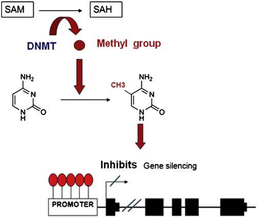
As mentioned earlier, the methyl group donor is the SAM molecule which, once it loses the methyl group, becomes S-adenosyl homocysteine (SAH). This molecule is hydrolyzed to homocysteine and then it is remethylated to methionine by 5-methyltetrahydrofolate cofactor (5mTHF). Finally, methionine is transformed back into a SAM molecule by the action of methionine adenosyltransferase (MAT). DNA methylation potential depends on the ratio between SAM level and SAH in blood. The higher the ratio, the more the methylation potential.8 Therefore, it can be inferred that for the process of DNA methylation, proper metabolism of homocysteine and methionine is critical, as well as the metabolism of the various enzymes involved in this metabolic route and of other substances, such as folic acid and vitamin B129 (Fig. 1.1).
Relevance of DNA Methylation in Clinical Practice
Disruption of epigenetic mechanisms involved in human disease has been, for the last few years, an area of emerging research, yielding positive results in various diseases, especially in oncology. The first tumor related to mechanisms of epigenetic regulation was colorectal cancer (CRC). Initially, a loss of overall methylation
FIGURE 1.1 The process of how DNA methylation within gene promoters regulates transcription. SAM, S-adenosyl-l-methionine. SAH, S-adenosyl homocysteine. DNMT, DNA methyltransferases.
was observed in cancer cells of CRC patients compared to healthy controls.10 Also, the promoters of tumor suppressor genes were shown to be hypermethylated, which caused lower expression of those genes.11 These findings supported that hypermethylation of tumor suppressor genes was associated with the occurrence of the disease. However, in other areas of medicine, such as neurological disorders, how disruption of DNA methylation is involved in the disease is not well known yet. In the case of MS, epigenetic changes that might be involved in the pathogenesis of the disease have been identified, which has led to an exciting and new route of research.
MS is considered the leading cause of severe neurological disease affecting young and middle-aged adults. It is a chronic disease causing inflammatory, demyelinating, and neurodegenerative damage in the central nervous system (CNS). Its etiology is still unknown, although an autoimmune and multifactorial origin has been presumed and several genetic and environmental factors of susceptibility have been described for MS. Given the complexity of the disease and the participation of diverse, both genetic and environmental, etiological mechanisms, it is conceivable that there may be an alteration in the epigenetic regulation involved in its progression.12,13
RISK FACTORS IN MS AND EPIGENETIC CHANGES
Epidemiological and family aggregation studies suggest that there is a genetic predisposition for MS. However, to date, the only locus consistently associated with MS is the major histocompatibility complex (MHC). This predisposition has been associated with DR2
haplotype (HLA-DRB1*1501-DQA1*0102-DQB1*0602), which determines a relative risk of four of having MS.14 Development of new technologies, such as polymorphism arrays, has helped to identify new candidate genes located outside the MHC region. Therefore, MS is considered as a polygenic disease in which each gene shows a different risk score (usually low or moderate).15 Moreover, environmental factors seem essential to the development of MS. For instance, the mismatch rate for disease occurring among monozygotic siblings is 70%, which supports the idea of other variables being involved.16 Indeed, there is a number of environmental factors described to be involved in the etiology of MS, such as vitamin D levels in serum, animal fats in the diet, injuries, and toxic substances17 (smoking, heavy metals, organic solvents, etc.). To date, the most consistent risk factors are smoking, vitamin D deficiency, and infection by Epstein–Barr virus (EBV).18,19
In the following sections, we summarize the three main environmental risk factors described for MS and the effects these factors may have on the various mechanisms of epigenetic regulation, both in MS and in the development of other diseases (Fig. 1.2).
Smoking and Epigenetic Mechanisms
Smoking is one of the environmental factors influencing the development of MS, as shown in different studies.
According to a study conducted by Rodriguez Regal et al., smoking involves an odds ratio (OR) of developing MS of 1.97.20 Cigarette smoke contains hundreds of potentially toxic elements, including nicotine, carbon monoxide, nitric oxide, cyanides, and polycyclic aromatic hydrocarbons, and some studies have suggested that these toxins might cause gene activation responsible for the MS autoimmune pathogenesis.21 In fact, smoking has been associated with an increased relapse frequency and with the number of active lesions in the brain MRI of patients with this disease.22
On the other hand, when the blood of adolescents whose mothers smoked during pregnancy was analyzed, it was observed that prenatal exposure to tobacco is associated with increased methylation of the promoter of the “brainderived neurotrophic factor” (BDNF), which promotes the differentiation and growth of new neurons.23 Likewise, another study conducted by Kjersti Aagaard et al. analyzed the DNA methylation pattern by PCR techniques, in two groups of smokers and nonsmokers. They found changes in DNA methylation in 25 genes in nonsmokers and 438 genes in smokers.24 Interestingly, epigenetic changes associated with smoking have also been found in oncology. In a study conducted in lung cancer patients, hypermethylation of CDKN2A, DAPK, and MGMT tumor suppressor genes was observed in smokers.25 Likewise, in a study conducted on cervical cancer among female smokers aged between 15 and 19, hypermethylation of
EPIGENETICS
GENETIC FACTORS
HLADRB1*1501
Candidate genes
FIGURE 1.2 The interplay between environmental and genetic factors. The interplay is where DNA methylation may play a role to develop multiple sclerosis.
the CDKN2A gene in cervical epithelial cells was also observed.26
Vitamin D and Epigenetic Mechanisms
Vitamin D deficiency is one of the leading risk factors in the development of MS.27 This vitamin can be synthesized by ultraviolet radiation or obtained by food, mainly fish oil. Many studies have been conducted studying the role of solar radiation as a protective factor in MS. The decrease in the amount of effective ultraviolet radiation in countries of higher latitude, involves an increased risk of MS.28,29 This is because vitamin D is a powerful regulator of the inflammatory and immunomodulatory response, working on both adaptive and innate immunity.30
Although the mechanism by which vitamin D causes these changes is still uncertain, a study conducted by Joshi et al. suggests that it might be due to epigenetic modifications. In this study, the effects of 1,25(OH)2D3 (active form of vitamin D produced in the skin after exposure to ultraviolet light) on human IL-17A production by T CD4+ cells were analyzed. They observed that 1,25(OH)2D3 directly inhibits the IL-17 locus, responsible for the transcription of proinflammatory cytokines, by means of a modification of the histone deacetylase 2 (HDAC2) in the promoter region of the IL17A gene.31 Other studies had previously shown that vitamin D is able to cause epigenetic changes, as in the case of colon cancer in which 1,25(OH)2D3 has been observed to be able to induce expression of the gene encoding the JMJD3 (lysine-specific demethylase).32
Epstein–Barr Virus and Epigenetic Mechanisms
To date, several infectious agents have been serologically and pathologically associated with MS. As an example, Sundstrom et al. analyzed the evidence to support whether a viral infection occurs in the preclinical stage of MS and only the EBV antigen showed a direct pathological correlation with the onset of MS.33 Moreover, it has been observed that a history of infectious mononucleosis (symptomatic form of EBV infection) doubles the risk of an individual to develop MS, while seronegativity for this virus is associated with a very low risk of developing MS (OR 0.06 compared with seropositive patients).34
Interestingly, EBV infection has been associated with epigenetic changes in the infected cells, and several tumor types have been related to EBV infection due to the hypermethylation of tumor suppressor gene promoters.35 This occurs in nasopharyngeal cancer and Hodgkin lymphoma induced by EBV, where the promoter hypermethylation has been observed to be triggered by an increase in DNMT1, DNMT3a, and DNMT3b enzymes,
and carried out by the LMP1 viral protein.36 MS epigenetic changes associated with EBV are also associated with the expression of microRNAs (miRNAs). The expression of miRNA-142-3p in MS patients has been linked to an increased immune tolerance, whereas miRNA-155 expression is associated with increased T cell differentiation and CNS inflammation.37
DNA METHYLATION IN MS
MS etiology remains not fully understood. However, the most accepted hypothesis postulates that MS is an immune disease mediated by autoreactive T cells, which are activated by exposure to one or more environmental factors in individuals with certain genetic predisposition. This disease is progressive with the occurrence of focal inflammatory (mainly white matter), demyelination (with relative preservation of axons in early stages) lesions, and neurodegeneration (in later stages). The exact pathophysiological mechanism mediating between environmental risk factors and genetic susceptibility to develop MS is unknown.38 It is precisely at this intersection where DNA methylation may provide new insights.
Inflammation and DNA Methylation
Several authors have linked the degree of methylation in specific genes with the occurrence of MS. In this regard, Kumagai et al. have found that the promoter of the sphingosine-1-phosphate enzyme (SPH-1) gene, which is involved in negative regulation of inflammatory signaling, is hypomethylated in MS patients compared to healthy controls. Promoter methylation of SPH-1 leads to a decreased expression of this enzyme and consequently to increased activity of inflammation mediated by lymphocytes.39
Furthermore, Janson et al. have analyzed the CD4 + T cells from a series of patients with relapsing–remitting multiple sclerosis (RRMS), and have found that such patients have a demethylation of the FOXP3 gene encoding for the scurfin protein, whose deficiency is associated with autoimmune disorders. FOXP3 gene demethylation can inhibit the differentiation to Th1 and Th2 cells and at the same time can promote regulatory T cells (Treg) and Th17 cells. The Th1/Th2 and Treg/Th17 balance influences the disease status so that changes therein may lead to the appearance of a new lesion or to its repair, and DNA methylation is one of the factors regulating this balance.40.41
Additionally, promoter hypomethylation of the gene encoding IL-17A, proinflammatory cytokine secreted by activated T lymphocytes, has also been observed. This finding has been linked to the development of autoimmune diseases and plays a core role in the MS pathogenesis.42
Demyelination and DNA Methylation
A study conducted by Mastronardi et al. shows that during the white matter demyelination in MS patients, the promoter of the peptidyl arginine deiminase 2 (PAD2) is demethylated and therefore PAD-2 is overexpressed in the brain. This enzyme makes the myelin basic protein (MBP) to be less stable as a result of enzymatic conversion of arginine into citrulline. Moreover, citrullination triggers the MBP to behave as an antigen to T lymphocytes. In this analysis, the methylation of PAD-2 promoter in the white matter of MS patients is reduced by 25% compared with healthy controls. Furthermore, this change only occurs in MS patients and not in patients with other neurological disorders, such as Alzheimer’s disease, Parkinson’s disease, or Huntington’s disease.43
Moreover, genome-wide methylation studies performed on CD4, CD8, and brain have revealed a number of genes to be differentially methylated in MS compared to controls.44,45 In this regard, Graves provides the first evidence for the association of DNA methylation at HLA-DRB1 with MS risk.46
Neurodegeneration and DNA Methylation
To date, no studies are available that have specifically analyzed the involvement of epigenetic mechanisms in the neurodegeneration of MS patients. However, changes in DNA methylation during neuronal death have been reported. For instance, Castaños et al. have analyzed cells from patients with amyotrophic lateral sclerosis (ALS) showing that overexpression of the DNMT3a enzyme triggers cell degeneration and death, whereas enzyme inhibition protects those cells, and DNA methylation is the mechanism that regulates DNMT3a expression.47 These results support that such a mechanism might be involved in the neurodegeneration occurring in MS patients, but this remains an open question for further research.
CONCLUSIONS
MS is a neurological disease with a major health, social, and family impact. Despite the significant advances in recent years, the exact etiopathogenic mechanism that causes MS remains unknown and a definitive therapy is not yet available.
Epigenetic changes, such as DNA methylation, are mechanisms by which environmental factors can influence individual gene expression. Epigenetic disruption is a research field of major interest in medicine, especially in the study of those diseases where combined genetic and environmental risk factors influence disease development, such as MS.
Although the number of studies conducted to date in patients with MS is low, the results are encouraging to further deepen this area. Currently available data show a relationship between regulation of DNA methylation in candidate genes that are key in the development of MS and autoimmune processes.48,49 While the results point in that direction, conducting cohort studies with more patients and controls is necessary.
Knowing epigenetic changes involved in the MS pathogenesis will help us to clarify the mechanisms causing the disease, and thus the way will be open to identify potential biomarkers and search for new therapeutic targets.
2. Feil R. Environmental and nutritional effects on the epigenetic regulation of genes. Mutat Res 2006;600:46–57.
3. Fraga MF, Ballestar E, Paz MF, Ropero S, Setien F, Ballestar ML, et al. Epigenetic differences arise during the lifetime of monozygotic twins. Proc Natl Acad Sci USA 2005;102:10604–9.
4. Rodríguez-Dorantes M, Téllez-Ascencio N, Cerbón MA, López M, Cervantes A, et al. Metilación del ADN: Un fenómeno epigenético de importancia médica. Rev Invest Clin 2004;56:56–71.
5. Turek-Plewa J, Jagodziński PP. The role of mammalian DNA methyltransferases in the regulation of gene expression. Cell Mol Biol Lett 2005;10:631–47.
6. Kar S, Deb M, Sengupta D, Shilpi A, Parbin S, Torrisani J, et al. An insight into the various regulatory mechanisms modulating human DNA methyltransferases stability and function. Epigenetics 2012;7:994–1007.
7. Urdinguio RG, Sanchez-Mut JV, Esteller M. Epigenetic mechanisms in neurological diseases: Genes, syndromes and therapies. Lancet Neurol 2009;8:1598–609.
8. Sugden C. One-carbon metabolism in psychiatric illness. Nutr Res Rev 2006;19:117–36.
9. Fryer AA, Emes RD, Ismail KM, Haworth KE, Mein C, Carroll WD, et al. Quantitative, high-resolution epigenetic profiling of CpG loci identifies associations with cord blood plasma homocysteine and birth weight in humans. Epigenetics 2011;6(1):86–94.
10. Feinberg AP, Vogelstein B. Hypomethylation distinguishes genes of some human cancers from their normal counterparts. Nature 1983;301:89–92.
11. Heyn H, Méndez-González J, Esteller M. Epigenetic profiling joins personalized cancer medicine. Expert Rev Mol Diagn 2013;13:473–9.
12. Renaudineau Y, Beauvillard D, Padelli M, Brooks WH, Youinou P1. Epigenetic alterations and autoimmune disease. J Dev Orig Health Dis October 2011;2(5):258–64.
13. Kamm CP, Uitdehaag BM, Polman CH. Multiple sclerosis: Current knowledge and Future Outlook. Eur Neurol July 30, 2014;72(3–4):132–41.
14. Yeo TW, De Jager PL, Gregory SG, Barcellos LF, Walton A, Goris A, et al. A second major histocompatibility complex susceptibility locus for multiple sclerosis. Ann Neurol 2007;61:228–36.
15. International Multiple Sclerosis Genetics Consortium. Risk alleles for multiple sclerosis identified by a genomewide study. N Engl J Med 2007;357:851–62.
17. MD1 N, Poole C, Satten GA, Ashley-Koch A, Ann Marrie R, Williamson DM. Heavy metals, organic solvents and multiple sclerosis: an exploratory look at gene-environment interactions. Arch Environ Occup Health 2016; 71(1):26–34
18. Mandia D, Ferraro OE, Nosari G, Montomoli C, Zardini E, Bergamaschi R. Environmental factors and multiple sclerosis severity: a descriptive study. Int J Environ Res Public Health June 19, 2014;11(6):6417–32.
19. Manouchehrinia A, Weston M, Tench CR, Britton J, Constantinescu CS. Tobacco smoking and excess mortality in multiple sclerosis: a cohort study. J Neurol Neurosurg Psychiatry October 2014; 85(10):1091–5
20. Rodríguez Regal A, del Campo Amigo M, Paz-Esquete J, Martínez Feijoo A, Cebrián E, Suárez Gil P, et al. Estudio de casos y controles sobre la influencia del hábito tabáquico en la esclerosis múltiple. Neurología 2009;24(3):177180.
21. Hernán MA, Jick SS, Logroscino G, Olek MJ, Ascherio A, Jick H Cigarette smoking and the progression of multiple sclerosis. Brain 2005;128:146165.
22. Healy BC, Ali EN, Guttmann CR, Chitnis T, Glanz BI, Buckle G, et al. Smoking and disease progression in multiple sclerosis. Arch Neurol 2009;66:8.
23. Toledo-Rodriguez M, Lotfipour S, Leonard G, Perron M, Richer L, Veillette S, et al. Maternal smoking during pregnancy is associated with epigenetic modifications of the brain-derived neurotrophic factor-6 exon in adolescent offspring. Am J Med Genet B Neuropsychiat Genet 2010;13B:1350–4.
24. Suter MA, Anders AM, Aagaard KM. Maternal smoking as a model for environmental epigenetic changes affecting birthweight and fetal programming. Mol Hum Reprod January 2013;19(1):1–6.
25. Koturbash I, Beland FA, Pogribny IP. Role of epigenetic events in chemical carcinogenesis—a justification for incorporating epigenetic evaluations in cancer risk assessment. Toxicol Mech Methods 2011;21:289–97.
26. Ma YT, Collins SI, Young LS, Murray PG, Woodman CB. Smoking initiation is followed by the early acquisition of epigenetic change in cervical epithelium: a longitudinal study. Br J Cancer 2011;104:1500–4.
27. Baarnhielm M. Multiple sclerosis is associated with low previous ultraviolet radiation exposure and low levels of current vitamin D: no interaction with HLA complex genes. Mult Scler 2010;16:S7–39.
28. Kimlin MG, Olds WJ, Moore MR. Location and vitamin D synthesis: is the hypothesis validated by geophysical data? J Photochem Photobiol B 2007;86:234–9.
29. Smolders J, Peelen E, Thewissen M, Cohen Tervaert JW, Menheere P, Hupperts R, et al. Safety and T cell modulating effects of high dose vitamin D3 supplementation in multiple sclerosis. PLoS One 2010;5:e15235.
30. Mahon BD, Gordon SA, Cruz J, Cosman F, Cantorna MT. Cytokine profile in patients with multiple sclerosis following vitamin D supplementation. J Neuroimmunol 2003;134:128–32.
31. Joshi S, Pantalena LC, Liu XK, Gaffen SL, Liu H, Rohowsky-Kochan C, et al. 1,25 dihydroxyvitamin D(3) ameliorates Th17 autoimmunity via transcriptional modulation of interleukin-17A. Moll Cell Biol 2011;31:3653–69.
32. Pereira F, Barbáchano A, Singh PK, Campbell MJ, Muñoz A, Larriba MJ. Vitamin D has wide regulatory effects on histone demethylase genes. Cell Cycle 2012;11:1081–9.
33. Sundstrom P. Evidence for virus infections in the presymptomatic stage of MS. Mult Scler 2010;16:S7–39.
34. Handel AE, Williamson AJ, Disanto G, Handunnetthi L, Giovannoni G, Ramagopalan SV. An updated meta-analysis of risk of multiple sclerosis following infectious mononucleosis. PLoS One September 1,2010;5(9)
35. Niller HH, Wolf H, Minarovits J. Epigenetic dysregulation of the host cell genome in Epstein-Barr virus associated neoplasia. Semin Cancer Biol 2009;19:158–64.
36. Tsai CL, Li HP, Lu YJ, Hsueh C, Liang Y, Chen CL, et al. Activation of DNA methyltransferase 1 by EBV LMP1 involves c-Jun NH2terminal kinase signaling. Cancer Res 2006;66:11668–76.
37. Junker A, Krumbholz M, Eisele S, Mohan H, Augstein F, Bittner R, et al. MicroRNA profiling of multiple sclerosis lesions identifies modulators of the regulatory protein CD47. Brain 2009;132:3342–52.
38. Bar-Or A. The immunology of multiple sclerosis. Semin Neurol 2008;28:29–45.
39. Kumagai C, Kalman B, Middleton FA, Vyshkina T, Massa PT. Increased promoter methylation of the immune regulatory gene SHP-1 in leukocytes of multiple sclerosis subjects. J Neuroimmunol 2012;246:51–7.
40. Janson PC, Linton LB, Bergman EA, Marits P, Eberhardson M, Piehl F. Profiling of CD4+ T cells with epigenetic immune lineage analysis. J Immunol 2011;186:92–102.
41. Liggett T, Melnikov A, Tilwalli S, Yi Q, Chen H, Replogle C, et al. Methylation patterns of cell-free plasma DNA in relapsing-remitting multiple sclerosis. J Neurol Sci Mar15, 2010;290(1–2):16–21.
42. Koch MW, Metz LM, Kovalchuk O. Epigenetic changes in patients with multiple sclerosis. Nat Rev Neurol January 2013;9(1):35–43
43. Mastronardi FG, Noor A, Wood DD, Paton T, Moscarello MA Peptidyl arginine deiminase 2 CpG island in multiple sclerosis white matter is hypomethylated. J Neurosci Res 2007;85:2006–16.
44. Graves M, Benton M, Lea R, Boyle M, Tajouri L, Macartney-Coxson D, et al. Methylation differences at the HLA-DRB1 locus in CD4+ T-Cells are associated with multiple sclerosis. Mult Scler December 12, 2013;20(8):1033–41.
45. Huynh JL, Garg P, Thin TH, Yoo S, Dutta R, Trapp BD, et al. Epigenome-wide differences in pathology-free regions of multiple sclerosis-affected brains. Nat Neurosci January 2014;17(1):121–30.
46. Maltby VE, Graves MC, Lea RA, Benton MC, Sanders KA, Tajouri L, et al. Genome-wide DNA methylation profiling of CD8+ T cells shows a distinct epigenetic signature to CD4+ cells in multiple sclerosis patients. Clin Epigenetics November 5, 2015;7:118.
47. Chestnut BA, Chang Q, Price A, Lesuisse C, Wong M, Martin LJ. Epigenetic regulation of motor neuron cell death through DNA methylation. J Neurosci 2011;31:16619–36.
48. Zhou Y, Simpson Jr S, Holloway AF, Charlesworth J, Van der Mei I, Taylor BV. Potential role of epigenetic modifications in the heritability of multiple sclerosis. Mult Scler February 2014;20(2):135–40.
49. Huynh JL, Casaccia P. Epigenetic mechanisms in multiple sclerosis: implications for pathogenesis and treatment. Lancet Neurol February 2013;12(2):195–206.
2
EBV Infection and Vitamin D in Multiple Sclerosis Patients
Sayed Mahdi Marashi1, Zabihollah Shoja2
1Virology Department, School of Public Health, Tehran University of Medical Sciences (TUMS), Tehran, Iran; 2Virology Department, Pasteur Institute of Iran (IPI), Tehran, Iran
MULTIPLE SCLEROSIS AND ENVIRONMENTAL FACTORS
Multiple sclerosis (MS) is a chronic, inflammatory, and debilitating autoimmune disease of the central nervous system (CNS) with unknown etiology. The inflammatory phenotype of MS is expressed as a relapsing–remitting course of neurological dysfunction, although the clinical symptoms vary between individuals.1 MS mostly occurs in young adults, although MS risk appears to decline largely after the age of 50.2,3
As documented for a variety of autoimmune diseases, MS is more prevalent in females.4,5 The female to male ratio ranges from 2:1 to 3:1 based on region,6 and it is tempting to speculate that the female bias in immune complex diseases such as MS might be a consequence of sex hormones. However, it is not clear whether sexrelated factors potentially exert deleterious effect or protective effect, although results from animal models remain conflicted, with some research showing no effect7 and other findings indicating a slight worsening of clinical experimental autoimmune encephalomyelitis (EAE) following ovariectomy.8 A number of MS-related genetic
variations have also been reported,9 highlighting the potential role of gene–environment interaction as well as the epigenetic phenomenon for female predominance.10 Indeed, the female to male ratio for MS susceptibility is shown to be higher in individuals with certain human leukocyte antigen (HLA) haplotypes. In addition, the female to male ratio is higher in MS patients with HLADRB1*15 haplotypes than those who are negative for DRB1*15. 9
Both infectious and noninfectious factors have been implicated in MS development.11,12. Rather than a sole trigger, the initiation of MS disease seems to be reliant on the elaborate interactions between infectious or noninfectious environmental risk factors with shared susceptibility genes.13
MS AND INFECTIONS
Infectious agents are considered an important environmental risk factor for autoimmune diseases. In this regard, viruses have long been considered as possible etiological agents of MS.14 As far back as 1946, the rabies
virus was the first to be considered as having an association with MS.15 Furthermore, aberrant immune reactivity against several viral infections has been reported.16
In an attempt to assess the temporal relationship between viral epidemics and MS relapses in the general population, the epidemics of influenza A and human herpesvirus-4 or Epstein–Barr virus (EBV) were shown to be temporally linked to the number of relapses in MS patients, supporting the notion that viral infections may influence MS development.17 Although infection with several viruses is considered as a potential candidates involved in MS development14,15 and no single virus has yet been proven solely responsible, infection with EBV and human herpesvirus 6 (HHV-6) has gained considerable attention.18,19 Like other members of herpesviridae family, these viruses establish lifelong latent infections and are neurotropic and lymphotropic.20
The correlative evidence for an MS association generally includes amplifying viral genome with polymerase chain reaction (PCR), assaying virus-specific antibodies in serum and cerebrospinal fluid (CSF), and spotting virus antigens within tissue sections obtained from brain and MS plaques.21,22 An etiological role for EBV infection in MS is supported by the most consistent findings including elevated EBV-specific antibody titers, dysfunctional EBV-specific CD8+ T cell responses, higher tendency to induce spontaneous transformation of peripheral blood B cells, and accumulation of EBV-infected B cells and plasma cells in the brain of MS patients.22
MS AND EBV INFECTION
EBV is a ubiquitous double-stranded DNA virus that establishes a lifelong persistent infection in over 90% of the adult population worldwide.20 Infection with EBV is usually asymptomatic in the first years of life; however, the risk of infection increases dramatically during adolescence or adulthood. Infectious mononucleosis (IM) is one of the most self-limiting clinical presentations of primary infection with this virus, which is characterized by fever, fatigue, pharyngitis, lymphadenopathy, and massive expansion of virus-specific T lymphocytes.23
EBV can infect both B cells and epithelial cells. At least three forms of EBV latency known as latency program I, II, and III have been described, and each program is reflected by a distinct set of proteins as well as the state of EBV-associated diseases or malignancies.20 In the blood, the virus establishes latency in memory B cells where there is usually no expression of viral proteins, except for EBV nuclear antigen 1 (EBNA-1) and sometime latent membrane protein 2a (LMP-2a).24
In general, EBV infection is vigilantly controlled by immune responses especially by EBV-specific CD8+ T cells.25 However, the virus can exploit the immune
system through encoding a number of proteins that have the ability to subvert the host’s immune surveillance.26 One of these proteins is the viral homolog of IL-10 (vIL-10), which is encoded by the EBV gene BCRF1 and has anti-inflammatory properties similar to human IL-10.27,28 The virus not only modulates B cell differentiation and function but also can elicit a vigorous and persistent cytotoxic T cell response. Infection with EBV can also interfere with the normal process of autoreactive B cell neutralization or control at several tolerance checkpoints.26,29,30
The role of EBV infection in patients with MS was first highlighted by studies reporting an increased tendency of spontaneous transformation of EBV-induced B lymphocyte in vitro31 and higher serum levels of anti-EBV antibodies in MS patients compared with healthy controls.32 The association between EBV seropositivity and risk of MS has been thoroughly confirmed by compelling evidence collected since mid-1990s demonstrating that virtually all MS patients are EBV seropositive compared with 90–95% of matched healthy controls.11,32–37 Seroconversion from EBV negative to EBV positive is associated with a higher risk of developing MS38 particularly in pediatric cases where around 80% of MS cases were shown to be EBV seropositive compared with 50% of matched healthy controls.39
Although elevated levels of serum or plasma IgG antibodies against different EBV antigens have been described,19,34,40–42 EBNA-1 is the only antigen that elicits the most robust antibody responses as supported by compelling evidence showing a significant association between elevated anti-EBNA-1 antibody titers and increased MS risk.19,35 This association has also been reported by magnetic resonance imaging (MRI)43 as well as clinical and radiological features of disease activity,44 although results remain conflicted45,46 and longitudinal studies are warranted to assess the serological profiles of anti-EBNA-1 in connection with the progression of MS.47
Evaluating anti-EBNA antibodies in sera collected prior to MS onset provided further evidence in which the relative risk of developing MS was found to be 30–36 times higher among those individuals who were EBV positive and had anti-EBNA titers ≥320 compared with those with titers <20.48,49 The increase in anti-EBNA-1 antibody titers seems to be age dependent at least before clinical onset of MS. In addition, coinfection with other herpes viruses or a new EBV strain is assumed to alter the host immune control on the latent EBV infection,50 which may support the relevance of EBV-specific immunity, particularly immune responses against EBNA-1 in MS pathogenesis.47
The antibody response to EBNA-1 is directed at multiple different epitopes, and further analysis highlighted that antibodies directed against particular epitopes especially within its glycine–alanine repeat are
of importance.51–53 While IgG antibodies against all EBNA-1 epitopes were shown to be associated with MS risk, IgG antibody against EBNA-1 epitopes 385–420 showed strong association to MS when compared with total EBNA-1 IgG levels.54
Patients with MS also have elevated levels of antiEBV IgG antibodies in the CSF,42,55 which may reflect the intrathecal synthesis of EBV-specific antibody or might simply be an indication of transport from the blood to the CSF.22 The intrathecal synthesis of viral-specific antibodies found in the CSF of MS patients might be an explanation for the occurrence of polyspecific humoral immune responses directed against viruses.56–59 Further evidence supporting the association between EBV and increased risk of MS came from studies where the relative risk of MS has shown to be 2–3 times higher in individuals with a history of IM than those without EBV infection.60–66 While consensus results have emerged from a serological point of view, studies investigating the EBV load have provided conflicting results, with some research showing no difference,67–72 and others reporting a significant difference of EBV load in MS patients.37,73 Despite conflicting results, the association of EBV reactivation and disease activity in MS patients was reported.74,75
Defective immune control of EBV in MS patients was suggested from the observation of increased spontaneous EBV transformation of B cells in vitro.31 Indeed, spontaneous EBV-induced transformation of peripheral blood B cells and production of EBV particles have been observed in the culture of lymphoblastoid cells obtained from patients with MS,31,76 which could be due to an increased frequency of EBV-infected B cells or impaired T cell function.22 Furthermore, in 2013, an increased frequency of EBV-infected B cells was reported in CSF and peripheral blood of MS patients.77
POTENTIAL MECHANISMS UNDERLYING EBV INFECTION IN MS
While there is compelling evidence for the epidemiological associations of MS and EBV infection, the underlying mechanisms by which EBV infection could contribute to MS development are yet to be understood. Therapeutic approaches using B cell depletion not only support the clinical efficacy but also the functionality of B cells in the pathogenesis of MS.78
Impaired regulatory immune functions,79–81 transactivation of human endogenous retrovirus element (HERV),82,83 impaired CD8+ T cell control of EBV-infected B cells,84–87 breakdown of immunologic self-tolerance to CNS antigens,88 molecular mimicry or cross-recognition of EBV gene products particularly EBNA-1-specific T cells with CNS antigens such as myelin basic protein (MBP) peptide,89–93 bystander damage to the CNS,94 activation of
innate immunity,95 expression of αB-crystallin in B cells,96 accumulation of EBV-infected autoreactive B cells,29 and inhibition of the activation-induced T cell apoptosis97 are all plausible explanations through which EBV infection may contribute to the induction of autoimmunity and MS development either directly or indirectly.
The pattern of increase in anti-EBV IgG antibodies in MS patients may reflect an altered T cell response to EBV-transformed B cells in MS, because anti-EBNA-1 titers were found to be positively correlated with the precursor frequency of T cells recognizing autologous EBV-transformed B cells.98 Although CSF T cells from MS patients are shown to recognize autologous EBVtransformed B cells99 and cross-recognize EBV and MBP,91,92 it is not clear how EBV infection drives local CNS inflammation.16
A significant increase in the EBV DNA load could result from either a large increase in the frequency of latently infected B cells or an increased number of EBV genomes in B lymphocytes or it may reflect a global effect on B cell activation, which may lead to increased EBV production.22 Increased frequency of EBV-infected plasma cells and plasmablasts in the peripheral blood of MS patients was described in 2013.77 Moreover, it has been shown that EBNA-1 is the only EBV protein that can be expressed by proliferating EBV-infected memory B cells,100 suggesting that higher expression of EBNA-1 as a consequence of higher viral load could result in stimulation and expression of larger number of EBNA1-specific B cells. This may lead to higher serum levels of EBNA-1 IgG. It is interesting to note that predominant EBNA-1-specific clonal expansions of T cells recognizing myelin antigens have been documented in MS patients, again suggesting that the increased EBV antigen load in these patients can hyperstimulate humoral- and cellmediated immune responses specific to EBV.16,92 The inflammation observed in a majority of MS patients may also reflect the combination of EBV infection and a dysregulated inflammatory response, although this needs to be addressed in larger studies.37
Numerous genetic variations associated with MS risk are described,101,102 and analysis of genome-wide association data support a causal role for the interaction between EBV and MS-associated gene variants,103 although the overall contribution of genetic factors seems to be modest and cannot explain how EBV infection increases MS susceptibility.104,105 To support this notion, statistical interactions between HLA alleles and EBV infection were carried out, suggesting that the risk factors may share a common biological pathway.54 The association found between HLA-DRB1*15 haplotypes and anti-EBNA-1 IgG levels54 and IM history106 are shown to increase the risk of MS disease, suggesting that HLA genes may influence the immune control of EBV infection. Given the fact that epigenetic changes such as