No part of this publication may be reproduced or transmitted in any form or by any means, electronic or mechanical, including photocopying, recording, or any information storage and retrieval system, without permission in writing from the publisher. Details on how to seek permission, further information about the Publisher’s permissions policies and our arrangements with organizations such as the Copyright Clearance Center and the Copyright Licensing Agency, can be found at our website: www.elsevier.com/permissions
This book and the individual contributions contained in it are protected under copyright by the Publisher (other than as may be noted herein).
Notice
Practitioners and researchers must always rely on their own experience and knowledge in evaluating and using any information, methods, compounds or experiments described herein. Because of rapid advances in the medical sciences, in particular, independent verification of diagnoses and drug dosages should be made. To the fullest extent of the law, no responsibility is assumed by Elsevier, authors, editors or contributors for any injury and/or damage to persons or property as a matter of products liability, negligence or otherwise, or from any use or operation of any methods, products, instructions, or ideas contained in the material herein.
Whose enthusiasm excites me to be a better teacher
Whose inquisitiveness drives me to dig deeper for answers
Whose joy to learn thrusts my dedication towards the educational mission
Whose rich personalities demonstrate how the human brain has incredible capability and compassion
-John Nolte, PhD, Todd W. Vanderah, PhD, and Jay B. Angevine Jr., PhD
When it comes to learning the anatomy and basic functions of the human nervous system, John “Jack” Nolte has played a role as an author and/or a professor for hundreds of thousands of students, residents, and physicians. Jack viewed the human brain as an endlessly fascinating playground that is forever changing. His
lifetime goal was not only to educate students but to educate future teachers as well. He continually scoured the primary literature and behaved with childhood excitement when discovering new explanations for human brain function. His own love and excitement for the nervous system would naturally bleed over to his students and colleagues, encouraging them to explore and question the human nervous system.
In working with Jack for over 15 years, my career and take on life changed from teaching as a “job” to teaching as an enjoyable hobby with benefits. His ability to make teaching fun, telling jokes and giving examples, led to an enriched student environment that resulted in students wanting more. Our meetings most often included discussions of how we could better educate students. Jack was on the cutting edge of designing “case-based” instruction, showing videos of patients for teaching purposes, having patients present in the classroom, and pushing ideas of working and taking exams as groups, using innovative technology, including the virtual brain and interconnected vocabulary terms across multiple fields of science. Jack’s desire to create a textbook that was cutting edge yet to the point for learning purposes was his continual love. He shared with me chapters, images, and novel ideas while writing the next edition to continually produce a product that students would enjoy.
Our time together was not all work. Although most know him as a professor of the nervous system, Jack also enjoyed playing handball, woodworking, traveling, cooking, wonderful deep red wines, a martini with blue cheese olives, and the joy of eating oysters (things that often appeared in his textbook as examples of nervous system function). In closing, I dedicate this new edition to my teacher, colleague, and friend. I dearly miss Jack’s enthusiasm for teaching, his humor, his friendship, and of course his infamous Birkenstocks.
Learning about the functional anatomy of the human central nervous system (CNS) is usually a daunting task. Structures that interdigitate and overlap in three dimensions contribute to the difficulty, as does a long list of intimidating names, many with origins in descriptive terminology derived from Latin and Greek. Here we have attempted to make the task a little easier by presenting a systematic series of whole-brain sections in three different sets of planes (coronal, sagittal, and axial—similar to what is seen in medical imaging), by relating these sections to three-dimensional reconstructions, and by trying to restrain ourselves when indicating structures.
Unlabeled photographs are presented throughout the book, juxtaposed with faded-out versions of the same photographs with important structures outlined and labeled. This circumvents the common need to mentally superimpose a labeled drawing on a photograph or to inspect a photograph through a thicket of lead lines. Photos of the CNS are comprehensive sets in each plane; sections illustrating major structures or major transitions are shown in greater detail and at a higher magnification. Every labeled structure is discussed briefly in an illustrated glossary at the end of the book. For this edition, minor adjustments were made to sections and photographs throughout the book; the functional pathways in chapter 8 were redone in color; an important new imaging modality (diffusion tensor imaging) was added to Chapter 9; and a number of new illustrations were added to the glossary. Chapter 10 is brand new in this edition as an introduction to neuropathology. Common types of CNS derived tumors and
neuro-diseases/disorders are displayed as representative images of what might be detected upon diagnosis.
The methods used in this book inevitably involve compromises. We labeled only structures that we believe are important for the knowledge base of undergraduate and professional students, and we omitted others dear to our hearts but perhaps not critical for these students. Hence the fasciola cinerea so prominent in Figure 7.8 is not labeled, and the indusium griseum is mentioned only briefly in a footnote. In addition, explicitly outlining structures required some simplifications, and complex entities are sometimes indicated more simply as single structures. We think the resulting pedagogical utility for students justifies these anatomical liberties.
Current technological methods allowed us to approach the construction of this atlas differently than we could have when it was first discussed. All the photographs of brains and sections used in the book were retouched digitally. Mounting medium, staining artifacts, and small cracks, folds, and scratches were removed from the digitized versions of the sections. The profiles of many small blood vessels were removed as well. The color balance was changed as appropriate to make the sections as uniform as possible. These procedures improved the illustrations aesthetically while leaving their essential content unchanged. In addition, computer-based surface-reconstruction techniques made possible the beautiful three-dimensional images that appear in Chapter 4 and elsewhere in the book.
John Nolte and Todd W. Vanderah
This book could never have happened without the hard work and endless efforts of Jack Nolte. He loved to teach and mentor colleagues of which I am forever indebted. Many colleagues and friends have helped with previous versions of the book, and that work is presented in this edition, including the photographic expertise of Nathan Nitzky and Jeb Zirato in the UofA Biocommunications. Grant Dahmer and Dr. Norman Koelling of the UofA prepared the prosections shown in Chapter 1. The sections shown in Chapter 2 were cut by Shelley Rowley, and those in Chapter 3 by Pam Eller. John Sundsten produced the three-dimensional images shown in Chapter 4. Paul Yakovlev, as detailed shortly, was the central figure in the production of the sections shown in Chapters 5 through 7. Cody Thorstenson played a major role in retouching the images of these sections. Cheryl Cotman produced the threedimensional reconstructions of the limbic system shown in Chapter 8. Drs. Ray Carmody, Robert Handy, Elena Plante, and Joe Seeger provided the images shown in Chapters 9 and 10. Drs. Agamanolis and Carmody supplied many of the pathology images in chapter 10 and helped with the description of the neuropathology.
I thank the co-author on the first three editions of this atlas, Jay B. Angevine Jr., who passed away in October 2011. Dr. Angevine was responsible for producing the whole-brain sections in Chapters 5 through 7
Drs. Nolte and Angevine both had an infectious love for the central nervous system and its incredible capability. Their personalities and enthusiasm made learning about the nervous system fun
and exciting. I hope this enjoyment for the nervous system feeds forward to future students of the human nervous system.
Todd W. Vanderah
Jay B. Angevine Jr. June 29, 1928–October 18, 2011.
A NOTE ON THE WHOLE-BRAIN SERIAL SECTIONS AND THEIR ORIGIN
As crucial as computer technology is to our book, the whole-brain serial sections are its foundation. They were prepared during 1966–1967 in the Warren Anatomical Museum at Harvard Medical School. The work, in which I took part, was performed under the direction of Dr. Paul I. Yakovlev (1894–1983), who was curator of the museum from 1955 to 1961 and then Emeritus Clinical Professor of Neuropathology until 1969. Each brain, embedded whole in celloidin, was sectioned in coronal, horizontal, or sagittal planes on a giant microtome with a standing oblique 36-inch blade and a sliding brain holder. The sections, each 35 µm thick, were rolled and stored in test tubes in a console of 100 numbered receptacles. After processing pilot sections for suitability and quality, we stained every twentieth section with Weigert’s hematoxylin (Loyez method) for myelin and mounted it between sheets of window glass. Each preparation is thus about 4 mm thick, yet great depth and detail of cells and fibers are visible.
Such preparations illustrate the white matter and tracts of the brain by staining the myelin sheaths of axons black; gray matter and nuclei appear as more or less pale areas, depending on the number and caliber of myelinated fibers present. These sections, all from essentially normal brains, were added to an already huge collection representing more than 900 cerebra that Dr. Yakovlev had been building since 1930. Now a national resource known and available to neurological scholars worldwide, this priceless compilation known as the Yakovlev-Haleem Collection is graciously housed in the National Museum of Health and Medicine by the Armed Forces Institute of Pathology in Washington, DC. Today it comprises about 1600 specimens, normal and pathological, processed in a rigorously consistent manner from the start.
In mid-1967, with Dr. Yakovlev’s blessing, I took with me to The University of Arizona some 1000 of the 8741 sections cut from the three normal brains used in Chapters 5 through 7 of this book. I had left Boston to join the faculty of the University’s new College of Medicine in Tucson. Paul, my mentor from the time I came to Harvard in 1956, wanted to support me as I began teaching in a far-off land that he believed (perhaps correctly) to be a frontier: the “Wild West.” As with everything else he did, it was thoughtful, kind, and generous. How he would have loved to see students studying the sections illustrated on these pages!
Unlike the fairly simple task of sectioning the brainstem, cutting perfect gapless whole-brain serial sections is difficult. The procedure was never more carefully undertaken or widely employed than by Paul, who used it at or in association with Harvard Medical School for 40 years. A central theme for him was this holistic method (“every part of the brain is there, nothing is left out…”), but no aspect of neuroanatomy or neuropathology failed to intrigue him. Although such sections had been made since the late 19th century (they are found in small numbers at many medical schools and
in profusion at a few research institutes), Paul’s are unique—in uniformity of preparation at every step from fixation to mounting, and in unity of general neurological interest and comparability. Of this legacy (he called it “over 40 tons of glass”), Derek Denny-Brown, Emeritus Professor of Neurology at Harvard, wrote in 1972: “The perspective given by serial whole brain sections provides at once an arresting view of anatomical relationships in patterns of striking beauty. After working in the collection for years, one still finds every occasion to view it illuminating and rewarding.”
In 2000, artist-scientist Cheryl Cotman, computer programmer Kevin Head, and I, an anatomist, traced and digitized structures from the serial sagittal sections shown in this atlas. We made a computer reconstruction and large hologram (three by five feet) of the human limbic system. We are indebted to Cheryl for her help in selecting images from her large collection of color-coded overall and regional views of the system. We are enlightened by her discovery that several limbic structures are quite differently shaped than traditionally believed. Although Paul and I would find this hard to accept, we would accept her fantastic findings with glee and laughter.
-Jay B. Angevine Jr.
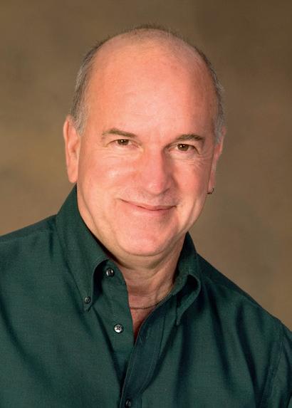
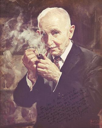
An autographed copy of an oil portrait of Paul
The original portrait was presented to the Warren Anatomical Museum at Harvard Medical School in 1978. (Courtesy the Warren Museum in the Francis A. Countway Library of Medicine, Boston, Massachusetts.)
Paul I. Yakovlev, MD 1894–1983.
Yakovlev by Bettina Steinke.
In Memoriam, v
Preface, vii
Acknowledgments, ix
A Note on the Whole-Brain Serial Sections and Their Origin, xi
1 External Anatomy of the Brain, 1
2 Transverse Sections of the Spinal Cord, 23
3 Transverse Sections of the Brainstem, 31
4 Building a Brain: Three-Dimensional Reconstructions, 49
5 Coronal Sections, 53
6 Axial Sections, 79
7 Sagittal Sections, 103
8 Functional Systems, 125
Long Tracts of the Spinal Cord and Brainstem, 125
Sensory Systems of the Brainstem and Cerebrum, 125
This atlas emphasizes views of the interior of the human central nervous system (CNS), sectioned in various planes. Here in the first chapter we lay some of the groundwork for understanding the arrangements of these interior structures by presenting the surface features with which they are continuous, and by giving a broad overview of the components of the CNS.
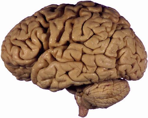
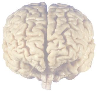
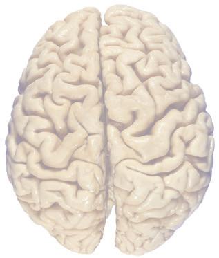
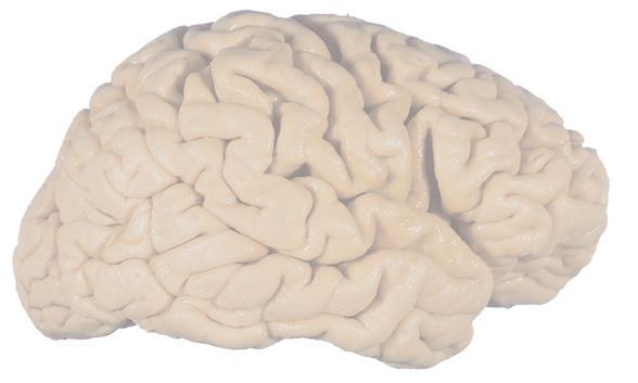
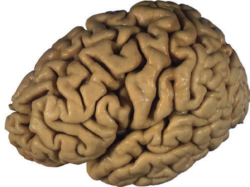
The CNS is composed of the spinal cord and the brain, the major components of which are indicated in Fig. 1.1. The human brain is dominated by two very large cerebral hemispheres, separated from each other by a deep longitudinal fissure. Each hemisphere is convoluted externally in a fairly consistent pattern into a series of gyri, separated from each other by a series of sulci (an adaptation that makes more area available for the cortex that covers each cerebral hemisphere). Several prominent sulci are used as major landmarks to divide each hemisphere into five lobesa frontal, parietal, occipital, temporal, and limbic—each of which contains a characteristic set of gyri (Figs. 1.3 to 1.8). The two hemispheres are interconnected by a massive bundle of nerve fibers, the corpus callosum, and two smaller bundles of fibers called the anterior and posterior commissures. Finally, certain areas of gray matter are embedded in the interior of each cerebral hemisphere. These include major components of the basal ganglia (or, more properly, basal nuclei) and limbic system (primarily the amygdala
aIn addition, the insula, an area of cerebral cortex buried deep in the lateral sulcus (see Fig. 5.7A), is usually considered as a separate lobe.
and hippocampus). They are apparent in the brain sections shown in Chapters 5 through 7
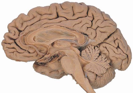
The cerebral hemispheres of humans are so massive that they overshadow or almost conceal the remaining major subdivisions of the brain—the diencephalon (made up of the thalamus, hypothalamus, epithalamus), brainstem, and cerebellum. Hemisecting a brain in the midsagittal plane, as in Fig. 1.1B, reveals these components.
The diencephalon (literally the “in-between brain”) is interposed between each cerebral hemisphere and the brainstem. The diencephalon contains the left and right thalamus, major waystations for information seeking access to the cerebral cortex; the hypothalamus, a major control center for visceral and drive-related functions; and the epithalamus, which includes the pineal gland and a set of nuclei called the habenula.
The brainstem, continuous caudally with the spinal cord, serves as a conduit for pathways traveling between the cerebellum or spinal cord and more rostral levels of the CNS. It also contains the neurons that receive or give rise to most of the cranial nerves
The cerebellum (literally the “little brain”) is even more intricately convoluted than the cerebral hemispheres, to make room for an extensive covering of its own cortex. It plays a major role in the planning and coordination of movement. A deep transverse fissure (normally occupied over most of its extent by the tentorium cerebelli) separates the cerebellum from the overlying occipital and parietal lobes and then continues deeper into the brain, partially separating the diencephalon from the cerebral hemispheres.
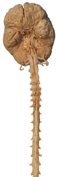
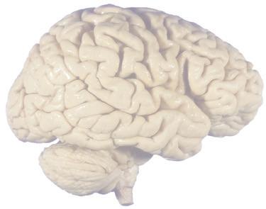
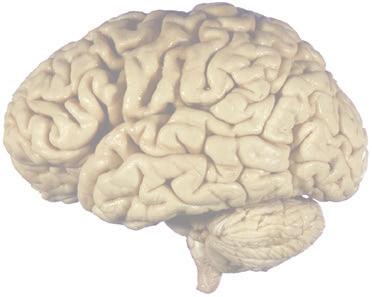
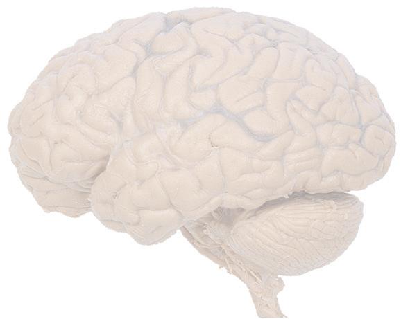
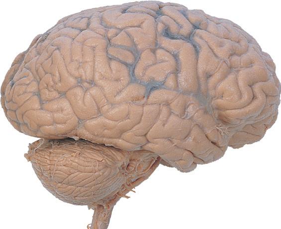
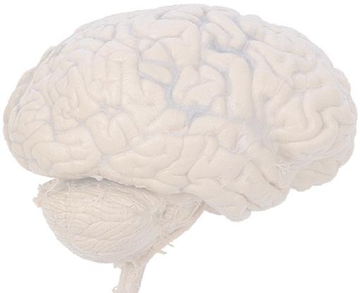
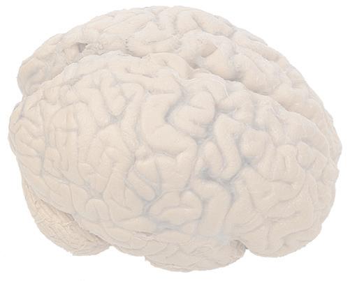
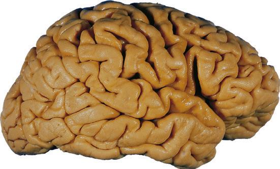
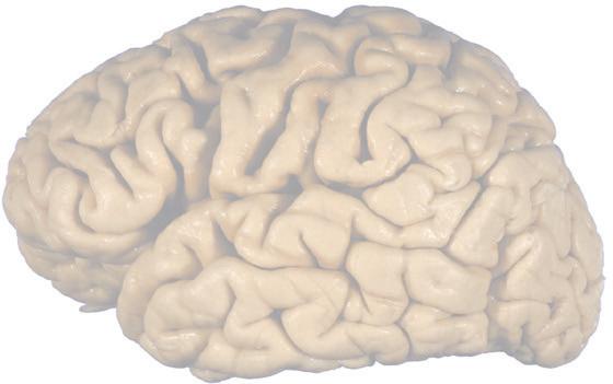
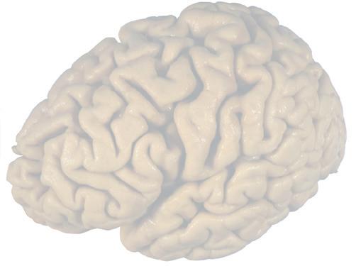
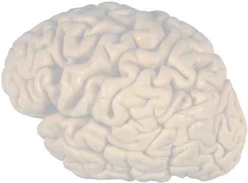
Figure 1.1 Lateral and medial surfaces of the brain. (A) The left lateral surface of the brain; anterior is to the left. (B) The medial surface of the right half of the sagittally hemisected brain; anterior is to the left. (Dissections by Grant Dahmer, Department of Cell Biology and Anatomy, The University of Arizona College of Medicine.)
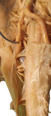
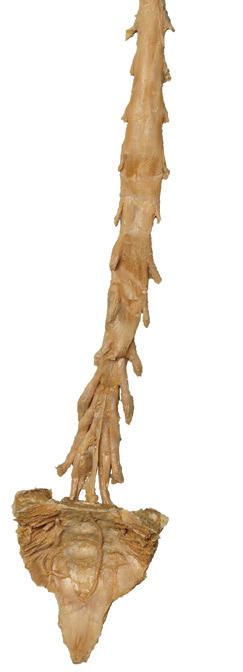
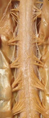
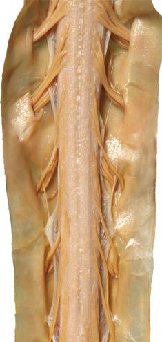
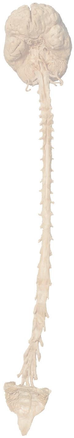
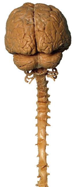
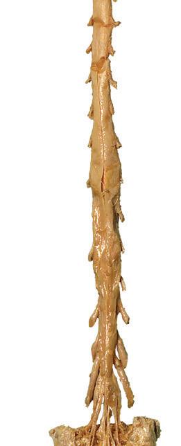
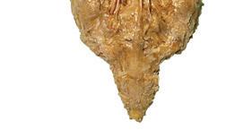
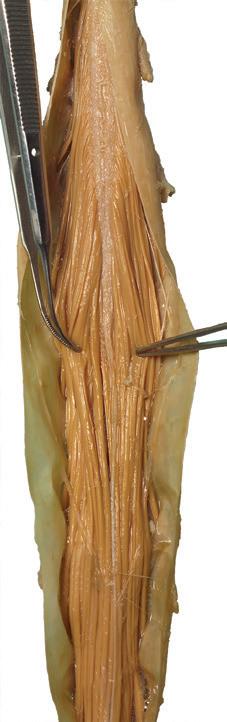
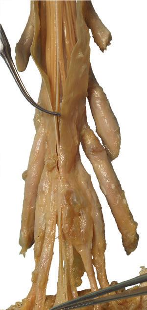
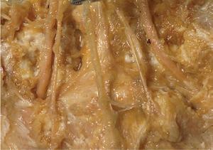
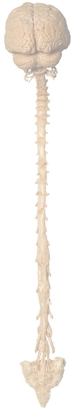
Figure 1.2 A masterful dissection of the entire CNS, with the spinal cord still encased in dura mater and arachnoid. (A) The anterior/inferior surface. Regions enlarged in the insets, after the dura mater and arachnoid were spread apart.
Longitudinal fissure
Parietal lobe
Occipital lobe
Temporal lobe
Cerebellum: vermis hemisphere
Posterior inferior cerebellar branches
Vertebral artery
Cut edge of dura & arachnoid
Cut edge of arachnoid
Cut edge of dura
Filum terminale (pial part)
Filum terminale (dural part)
Filum terminale (pial part)
Coccygeal
Sacrum
Filum terminale (dural part)
Conus medullaris
Cauda equina
Cauda equina T11
(B) The posterior surface of the entire CNS. The cauda equina and the caudal end of the spinal cord, enlarged in the insets after the dura mater and arachnoid were spread apart. (Dissection by Dr. Norman Koelling, Department of Cell Biology and Anatomy, The University of Arizona College of Medicine.)
Figure 1.2 (Continued)
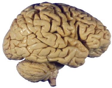
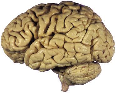
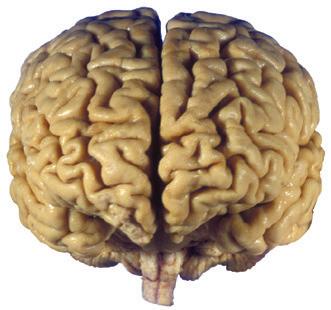
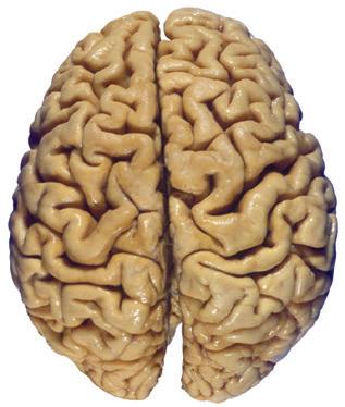
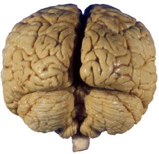
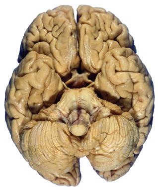
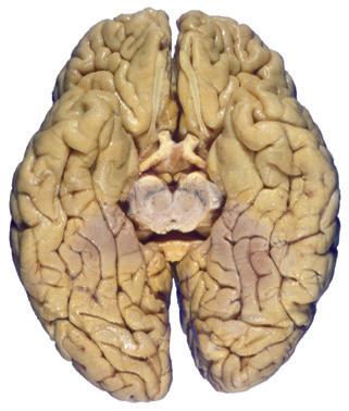
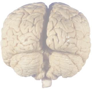
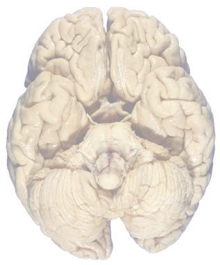
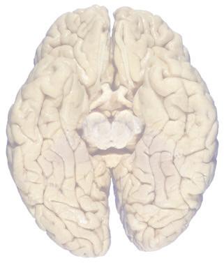
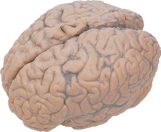
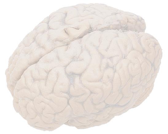
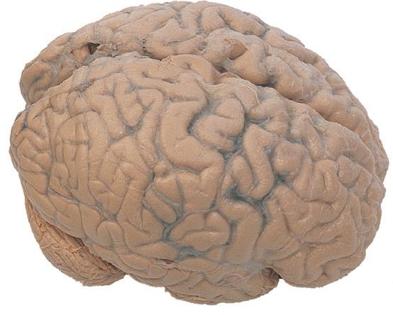
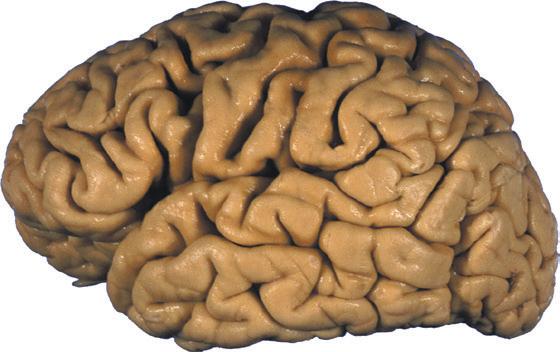
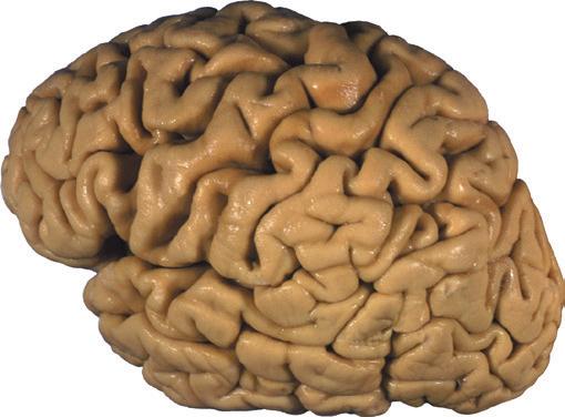
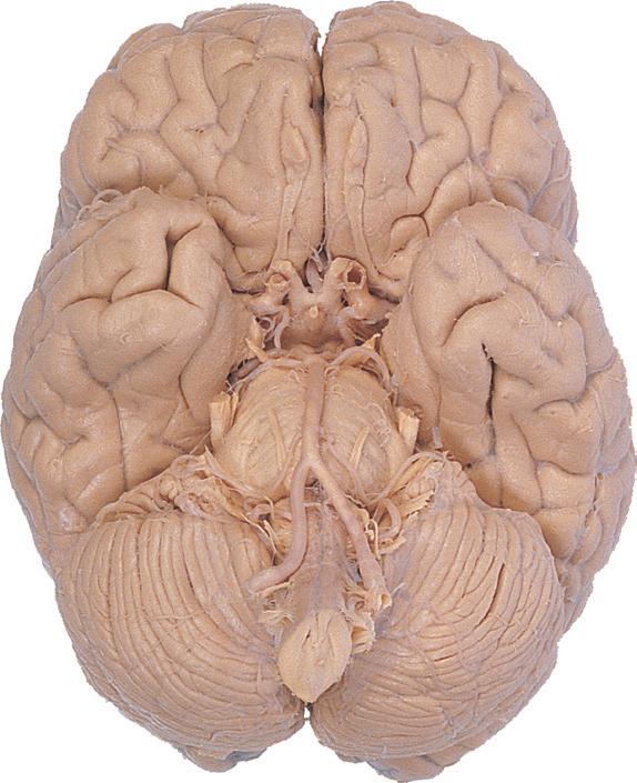
Figure 1.3 Multiple views of a brain. Only major structures are labeled here. (A) The right lateral surface (anterior toward the right). (B) The left lateral surface (anterior toward the left). (C) The anterior surface. (D) The superior surface (anterior toward the top of the page). (E) The posterior surface. (F) The inferior surface (anterior toward the top of the page). (G) The same inferior surface after removal of the cerebellum and most of the brainstem; the latter are shown in more detail in Fig. 1.9
Superior parietal lobule
Intraparietal sulcus
Central sulcus
Lateral sulcus
Cerebellum: hemisphere
Longitudinal fissure
Lateral sulcus
Orbital gyri
Cerebellum: hemisphere Brainstem
Orbital gyri
Olfactory bulb & tract
Cerebellum: flocculus
hemisphere vermis
Figure 1.3 (Continued)
Central sulcus
Superior parietal lobule
Intraparietal sulcus
Cerebellum: hemisphere Brainstem
Longitudinal fissure
Inferior
Inferior parietal lobule
frontal gyrus
Central sulcus
Intraparietal sulcus
Brainstem
Intraparietal sulcus
Longitudinal fissure
Optic chiasm
Infundibulum
Midbrain Thalamus
Medulla
Cerebellum: vermis hemisphere
Corpus callosum
Rhinal sulcus
Uncus
Parahippocampal gyrus
Collateral sulcus
Occipitotemporal gyrus
Longitudinal fissure
(The rhinal sulcus is drawn as a dashed line to indicate that it is separate from the collateral sulcus, even though in this particular brain the two are continuous.) IFG, Inferior frontal gyrus; IPL, inferior parietal lobule; MFG, middle frontal gyrus; Occ, occipital lobe; Po, postcentral gyrus; Pr, precentral gyrus; SFG, superior frontal gyrus; SPL, superior parietal lobule; Temp, temporal lobe. (Dissection by Grant Dahmer, Department of Cell Biology and Anatomy, The University of Arizona College of Medicine.)
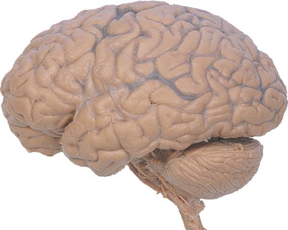
Figure 1.4 Lateral and superior surfaces of the cerebral hemispheres. The arachnoid was not removed from this specimen, but major gyri and sulci are still apparent. (A) Lateral view of the left hemisphere. Numerous branches of the left middle cerebral artery can be seen emerging from the lateral sulcus and spreading over the lateral surface of the hemisphere beneath the arachnoid. (B) The same hemisphere as in (A), seen from a more anterior and superior vantage point. Anterior and posterior cerebral branches can be seen emerging from the longitudinal fissure and extending for a short distance over the superior surface of the hemisphere beneath the arachnoid. (Middle cerebral branches, although visible in this view, are not indicated.)
Frontal gyri: superior middle inferior
Central sulcus 1 2 3
Precentral gyrus
Postcentral gyrus
Supramarginal gyrus
Orbital gyri
Lateral sulcus
Temporal gyri: superior middle inferior
Inferior frontal gyrus:
1. orbital part
2. triangular part
3. opercular part
Cerebellum (hemisphere)
Superior parietal lobule
Intraparietal sulcus
Angular gyrus
Occipital lobe
Transverse fissure
Preoccipital notch
Cerebellum (flocculus)
Postcentral gyrus
Central sulcus
Longitudinal fissure
Anterior cerebral branches
Precentral gyrus
Frontal gyri: superior middle inferior
Supramarginal gyrus
Superior parietal lobule
Arachnoid granulations
Intraparietal sulcus
Posterior cerebral branches
Angular gyrus
Temporal gyri: superior middle
Figure 1.4 (Continued)
(C) Lateral view of the right hemisphere of the same brain shown in (A) and (B). Numerous branches of the right middle cerebral artery can be seen emerging from the lateral sulcus and spreading over the lateral surface of the hemisphere beneath the arachnoid, and a few posterior cerebral branches emerge from the longitudinal fissure. Although the two cerebral hemispheres of human brains are approximately mirror images of each other, some slight asymmetries are common, particularly in certain language-related areas. Note in this specimen how much farther posteriorly the lateral sulcus extends in the left hemisphere (A), and how much larger the triangular part of the inferior frontal gyrus is on the left (see also Fig. 1.5). (D) The same hemisphere as in (C) seen from a more anterior and superior vantage point. Anterior and middle cerebral branches can be seen emerging from the longitudinal fissure and extending for a short distance over the superior surface of the hemisphere beneath the arachnoid. (Middle cerebral branches, although visible in this view, are not indicated.) (Dissection by Grant Dahmer, Department of Cell Biology and Anatomy, The University of Arizona College of Medicine.)
Frontal gyri: superior middle inferior Lateral sulcus
Temporal gyri: superior middle inferior Cerebellum (flocculus) Transverse fissure
Preoccipital notch Central sulcus Superior parietal lobule
Inferior frontal gyrus:
1. orbital part
2. triangular part
3. opercular part
Orbital gyri
Posterior cerebral branch
Postcentral gyrus Supramarginal gyrus
Intraparietal sulcus
Precentral gyrus
Longitudinal fissure Central sulcus Superior parietal lobule Anterior cerebral branch
Arachnoid granulations Angular gyrus
Temporal gyri: superior middle inferior
Frontal gyri: superior middle inferior
Figure 1.5 (A and B) The left and right cerebral hemispheres of the brain. Note in this specimen how much farther posteriorly the lateral sulcus extends in the left hemisphere (A), and how much larger the triangular and opercular parts of the inferior frontal gyrus are on the left. (A) Lateral view of the left hemisphere. (B) Lateral view of the right hemisphere.
Precentral gyrus
Frontal gyri: superior middle inferior
Orbital gyri
Olfactory bulb
sulcus
Postcentral gyrus Supramarginal gyrus
Lateral sulcus
Inferior frontal gyrus:
Temporal gyri: superior middle inferior
1. orbital part
2. triangular part
3. opercular part
Superior parietal lobule
Intraparietal sulcus Angular gyrus
Occipital gyri
Preoccipital notch
Superior parietal lobule
Intraparietal sulcus Angular gyrus
Occipital gyri
Supramarginal gyrus
Postcentral gyrus Precentral gyrus
sulcus
Frontal gyri: superior middle inferior Lateral sulcus
Preoccipital notch
Temporal gyri: superior middle inferior
Inferior frontal gyrus: 1. orbital part 2. triangular part 3. opercular part Orbital gyri
Figure 1.5 (Continued)
(C and D) Lateral and superior surfaces of the left cerebral hemisphere shown in (A). (C) The same hemisphere as in (A), seen from a more anterior and superior vantage point. (D) The same hemisphere as in (A), seen from a more posterior and superior vantage point. (Dissection by Grant Dahmer, Department of Cell Biology and Anatomy, The University of Arizona College of Medicine.)
Precentral gyrus
Frontal gyri: superior middle inferior
Inferior frontal gyrus:
1. orbital part
Postcentral gyrus
Supramarginal gyrus
sulcus
2. triangular part
3. opercular part
Superior parietal lobule
Intraparietal sulcus Angular gyrus
Occipital lobe
Preoccipital notch
Lateral sulcus
Temporal gyri: superior middle inferior
Frontal gyri: superior middle inferior
Inferior frontal gyrus:
1. orbital part
2. triangular part
3. opercular part
Lateral sulcus
Precentral gyrus
Central sulcus
Postcentral gyrus
Temporal gyri: superior middle inferior
Supramarginal gyrus
Superior parietal lobule
Intraparietal sulcus Angular gyrus
Occipital gyri
Preoccipital notch
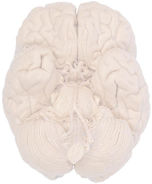
A Longitudinal fissure
Olfactory: sulcus bulb tract
Temporal gyri: superior middle inferior
Transverse fissure
Pons
Gyrus rectus
Orbital gyri
Uncus
Rhinal sulcus
Parahippocampal gyrus
Occipitotemporal (fusiform) gyrus
Medulla
Cerebellum (hemisphere)
Figure 1.6 (A) Inferior surface of the human brain.