Biodiversity of the Genus Aspergillus in Different Habitats
A.M. Abdel-Azeem1, F.M. Salem1, M.A. Abdel-Azeem2, N.A. Nafady3, M.T. Mohesien4 and E.A. Soliman1
1University of Suez Canal, Ismailia, Egypt, 2University of Sinai, North Sinai, Egypt, 3Assuit University, Assiut, Egypt, 4University of Damietta, New Damietta, Egypt
INTRODUCTION
Members of the genus Aspergillus are cosmopolitan and prevalent components of different ecosystems in a wide range of environmental and climatic zones (Klich, 2002a; Lević et al., 2013), because they can colonize a wide variety of substrates. Species belonging to the genus Aspergillus are widely distributed throughout the world biomes, for example, soil (Hill et al., 1983; Klich, 2002a; Abdel-Azeem and Ibrahim, 2004; Conley et al., 2006; Jaime-Garcia and Cotty, 2010), salterns (Butinar et al., 2011; Balbool et al., 2013), agroecosystems (Bayman et al., 2002; Horn, 2003; JaimeGarcia and Cotty, 2006; Abdel-Azeem et al., 2007; Marín et al., 2012; Muthomi et al., 2012), polar (Arenz et al., 2014), living plants, animals and lichens (Yu et al., 2012; Salem and Abdel-Azeem, 2014; Tripathi and Joshi, 2015), stones (Tang et al., 2012), water-related (Sivakumar et al., 2006; BonugliSantos et al., 2015), fossil records (Thomas and Poinar, 1988; Dörfelt and Schmidt, 2005), and human (Horré et al., 2010; Marguet et al., 2012; Findley et al., 2013).
The occurrence of Aspergillus species is controlled by several factors including microclimate, the availability of substrates, as well as water activity and complex ecological interactions (Mouchacca, 1995; Grishkan and Nevo, 2010; Pettersson and Leong, 2011). Survival in different environmental and geographical habitats can be related to metabolic diversity, high reproductive capacity, and competitive capabilities of Aspergillus strains in nature (de Vries and Visser, 2001; Horn and Dorner, 2002; Shehu and Bello, 2011; Mehl and Cotty, 2013).
The genus Aspergillus consists of about 339 species, including both pathogenic and beneficial species (Samson et al., 2014). Several species are pathogenic to plants, animals, and humans (eg, Aspergillus fumigatus, Aspergillus terreus) and/or produce different types of toxins, such as aflatoxins and ochratoxins (eg, Aspergillus flavus, Aspergillus ochraceous). On the other hand, several species are widely used in different industrial applications, for
example, production of foods, drinks, organic acids, and a large variety of enzymes (eg, Aspergillus niger, Aspergillus aculeatus, Aspergillus oryzae). The broad relevance and economic importance of the genus have pushed it to the forefront of fungal research, with one of the largest academic and industrial research communities dedicated to this genus. We searched major names of interest of Aspergillus species in both the web of Google Scholar and Research Gate on July 17, 2015. Results showed that A. niger came first by 307,000 and 79,900 recorded hits followed by A. fumigatus (199,000 and 55,500), A. oryzae (82,900 and 25,200) and A. flavus (79,000 and 43,100), respectively.
The aim of this chapter is to give an overview of the studies aimed at the investigation of Aspergillus biodiversity in a wide variety of different ecological habitats.
METHODOLOGY OF STUDYING ASPERGILLUS BIODIVERSITY
Phenotypic Studies
Microscopic features of Aspergillus and its teleomorphs are an important part of the species concept. However, many debatable taxonomic schemes in several sections of the genus have resulted due to the occurrence of much morphological variation. Phenotypic characters of aspergillum-like spore-bearing structure include conidial head shape (presence or absence of metulae, ie, uniseriate or biseriate), color, shape, texture and dimension of stipes, vesicles, conidia, and Hülle cells if present. Morphological characteristics, such as colony growth rates on identification media, texture, sporulation rate, production of sclerotia or cleistothecia, colors of mycelia, sporulation, diffusible pigments, exudates and reverses, and physiological characteristics (temperature, water activity) have been used with aforementioned criteria for charcterizing species. The preliminary identification of species can be performed with the aid of taxonomic keys and descriptions available in the
http://dx.doi.org/10.1016/B978-0-444-63505-1.00001-4
literature (Thom and Church, 1926; Thom and Raper, 1945; Raper and Fennell, 1965; Christensen and States, 1982; Christensen, 1981,1982; Gams et al., 1985; Samson and Gams, 1985; Pitt, 1985; Klich and Pitt, 1988; Kozakiewicz, 1989; Samson and Pitt, 2000; Klich, 2002b; McClenny, 2005; Varga and Samson, 2008; Pitt and Hocking, 2009; Samson et al., 2010; Hubka et al., 2013). Furthermore, all these phenotypic features have to be determined by trained mycologists under standardized laboratory conditions to obtain an accurate identification (Okuda et al., 2000). However, without professional expertise this may often lead to an incorrect description, therefore, the use of biochemical and molecular methods is recommended.
Secondary Metabolite Profiling and Chemotaxonomy
Aspergilli have a variety of biochemical characteristics that classify them as Eumycota. Their cell walls containing polysaccharide (chitin and glucan); ergosterol; fatty acid profile dominated by C16 and C18 chain lengths; and production of trehalose and polyols (Wessels, 2005).
Guarro et al. (1999) recommended other chemical markers or patterns of metabolites, secondary metabolite profiles, in conjunction with morphology and physiology approaches for further classification of Aspergillus. Raper and Fennell (1965) did not use any physiological, chemical, or biochemical characters, but in later physiological tests (Klich and Pitt, 1988) and secondary metabolites (Frisvad, 1989; Frisvad et al., 1998, 2004, 2007; Samson et al., 2004) have been introduced in the taxonomy of Aspergillus
Secondary metabolites have been the molecules most often used in species recognition due to their high species specificity (Frisvad, 1989; Larsen et al., 2005). All species produce a unique combination of different types of small organic compounds of mixed biosynthetic origin and even unique to a single species (Frisvad et al., 2007). Recently various studies have shown that major genomic differences between Aspergillus species are often related to the number and similarity of polyketide and nonribosomal peptide synthase genes (Galagan et al., 2005; Nierman et al., 2005; Pel et al., 2007). Hence, secondary metabolites are indeed excellent phenotypic characters for species recognition. Chemotaxonomy by using fatty acid profiles have been used extensively for bacteria and the characterization of microbial communities (Zelles, 1999; Kirk et al., 2004). In comparison with bacteria, fewer different fatty acids are produced by fungi (Lechevalier and Lechevalier, 1988), and by the end of the 20th century fatty acids analyses were increasingly used to distinguish different fungi (Welch, 1991; Stahl and Klug, 1996; Nemec et al., 1997; Silva et al., 1998; Guarro et al., 1999). Fatty acid methyl esters (FAME) prepared in most methods and analyzed by gas chromatography (GC) or gas chromatography–mass spectrometry
(GC-MS) and multivariate programs have been developed to apply fungal Fatty Acid data in routine taxonomy and identification work (Stahl and Klug, 1996). Few studies concerning the Fatty Acid methodology have been applied as a taxonomic tool for discriminating amongst Aspergillus (Fraga et al., 2008). Glassbrook (2008) studied the biochemical markers for the detection and classification of Aspergillus. In his study, reference strains of different Aspergillus species, Penicillium chrysogenum, Candida albicans, and Cryptococcus neoformans were characterized using liquid chromatography–mass spectrometry (LC-MS) and gas chromatography–mass spectrometry (GC-MS) biochemical profiling techniques in order to find specific small molecules, peptides, or biochemical profiles that can be used in addition to established methods to detect and classify Aspergilli to the species level.
Thus in various scenarios detection of a unique mixture or in some cases one or a few biomarkers can be used for species recognition based on the chemical nature of such small organic molecules which can be detected by different spectroscopic tools. These spectroscopic techniques (Infrared (IR), Ultra Violet (UV), Fluorescence Detection (FLD), Mass Spectroscopy (MS), and Nuclear Magnetic Resonance (NMR), UV, FLD, MS, and NMR) give complementary structural information, and are often used in a combined setup in connection with either gas or liquid chromatography (Nielsen et al., 2004).
In the last decade, other tools concerning chemoinformatics have been developed and applied in order to deal with large amounts of spectroscopic data that can be generated from analysis of numerous fungal taxa (Nielsen et al., 2004; Larsen et al., 2005). The use of electronic nose technologies, a similar but very different approach for species recognition combined with neural network analysis as a kind of “black box” approach for detection of fungal growth, is associated with certain kinds of feed or foodstuffs (Karlshøj et al., 2007).
Protein profiles, as a diagnostic tool, are not used extensively in the taxonomy of genus Aspergillus (Glassbrook, 2008). By using electrophoretic techniques different protein patterns will be observed and they directly related to the diversity of the coding genes and may indicate specific differences or similarities between examined species (Mitterdorfer et al., 2002). One-dimensional polyacrylamide gel electrophoresis (PAGE) of proteins has been used to compare different species of Aspergillus (Rath, 2001; Leila et al., 2010; Khosravi et al., 2012). Several investigators (Khosravi et al., 2012 Nealson and Garber, 1967; Nasuno, 1971, 1972a,b, 1974; Kurzeja and Gabber, 1973; Cruickshank and Pitt, 1990; Sugiyama and Yamatoya, 1990; Yamatoya et al., 1990) have studied enzyme profiles of a limited number of Aspergillus isolates. Slab polyacrylamide gel electrophoresis method was introduced by Saito et al. (1991) for the identification of the alkaline proteinases of A. flavus and Aspergillus parasiticus, but the result was not good enough.
Ubiquinone (coenzyme Q) is a lipid component of the mitochondrial electron transport chain and has been used as a taxonomic criterion for yeast and filamentous fungi (Yamada et al., 1989; Yaguchi et al. (1996)). The number of isoprene units attached to the benzoquinone varies, and such differences in ubiquinone structure are excellent indicators in the classification of genera and subgeneric taxa in bacteria and yeasts. In addition, the isoprene units of ubiquinone were highly correlated with morphological and physiological characters in the infrageneric taxa of Aspergillus (Kuriashi et al., 1990). Sugiyama et al. (1991) and Matsuda et al. (1992) reported that three major ubiquinone systems (Q-9, Q-10, and Q-10(H2)) occurred in Aspergillus and the ubiquinones were useful indicators for classification. Kuriashi et al. (1990) studied the ubiquinone systems in Aspergillus in relation to the taxonomy of Raper and Fennell (1965), who subdivided Aspergillus into uniseriate species, uniseriate or biseriate species, and biseriate species. Their study showed that nearly all species having Hülle cells possessed only the Q-10(H2) system while xerophilic species had Q-9 or Q-10. Yamatoya et al. (1990) determined the ubiquinone systems of 27 isolates assigned to Aspergillus sect. Flavi. Coenzyme Q systems for 190 (teleomorphic and anamorphic) isolates, and three samples of Dendrosphaera eberhardtii fruit bodies, which belonged to Eurotiales, Onygenales, and related taxa have been determined by Kuraishi et al. (2000).
Several biochemical and physiological techniques have been introduced to improve Aspergillus taxonomy, one of which is isoenzyme patterns (Cruickshank and Pitt, 1990; Yamatoya et al., 1990). It generally is most successful at distinguishing species and has been used to make recommendations on the separation or combination of species (Micales et al., 1992). Differences in isozyme banding patterns have been used to separate species of Aspergillus (Kurzeja and Gabber, 1973).
Cruickshank and Pitt (1990) used polyacrylamide gel electrophoresis to examine several kinds of exoenzymes (pectinases, ribonucleases, amylases, and proteases) from six isolates of Aspergillus. They found that four isolates (A. flavus, A. parasiticus, Aspergillus tamarii, and Aspergillus nomius) produced distinct patterns. On the other hand, A. oryzae produced very similar patterns to those of A. flavus, and patterns of Aspergillus sojae were very similar to those of A. parasiticus. In the above-mentioned studies taxonomic relationships could be elucidated, but until now isozyme profiles have not provided a practical system for identification because isoenzyme patterns could not be used to distinguish the domesticated species from their wild types.
Frisvad et al. (2007) discussed the particular interest of using mycotoxins, as secondary metabolites with bioactive properties, in taxonomy of Aspergillus species. As an important chemotaxonomic marker, aflatoxins have been used by several investigators (Frisvad et al., 1998; Klich
et al., 2000; Seifert and Levesque, 2004; Varga et al., 2004; Frisvad et al., 2007) in taxonomic studies of aflatoxinproducing taxa of Aspergillus
Evolution of the Approach: Polyphasic Taxonomy of Aspergillus
The polyphasic taxonomy takes into account all available phenotypic and genotypic data and integrates them in a consensus type of classification. Phylogenetic species recognition is increasingly being used with the internal transcribed spacers of the nrDNA (ITS) now accepted as the official DNA barcode for fungi (Schoch et al., 2012). Sequencing of genomic regions widely applied to the identification of a large number of Aspergillus species and the results of these techniques are generally well correlated with morphological and physiological characteristics (Rodrigues et al., 2011). Genomic regions that are sequenced for the identification of Aspergillus species include the ITS (internal transcribed spacer) region (White et al., 1990), β-tubulin (BenA) gene (Glass and Donaldson, 1995), and calmodulin (CaM) gene (Carbone and Kohn, 1999). The nuc rDNA internal transcribed spacer rDNA region (ITS1-5.8S-ITS2) is the official DNA barcode for fungi because it is the most frequently sequenced marker in fungi and has primers that work universally (Schoch et al., 2012). In contrast, BenA is easy to amplify, in comparison with the RNA polymerase II second largest subunit (RPB2), but has been reported to vary in the number of introns and amplification of paralogous genes sometimes resulting from PCR (Peterson, 2008; Hubka and Kolarik, 2012).
Isolates of Aspergillus species usually produce a diverse range of extrolite (secondary metabolites) that are characteristic of the different groups of sections of Aspergillus For example, production of kojic acid characterized species of Aspergillus section Flavi (Varga et al., 2011), while penicillic acid (small acidic molecules) produced by most species of Aspergillus section Circumdati (Frisvad et al., 2004). Production of a specific extrolite is considered an efficient identification tool for allocating a species of Aspergillus to section but some extrolites, for example, ochratoxin A, are produced by species in different sections, for example, Flavi, Circumdati, and Nigri (Frisvad et al., 2004, 2011; Varga et al., 2011; Samson et al., 2014). Various polyphasic studies have been carried on different sections of Aspergillus by several researches (Hong et al., 2005; Varga et al., 2007a; Houbraken et al., 2007; Silva et al., 2011; Samson et al., 2007, 2014). Samson et al. (2014) recommended an updated qualitative database on the verified production of secondary metabolites to identify isolates of Aspergillus up to species level.
Current knowledge pertaining to the diversity, detection, and distribution of Aspergillus taxa is still rudimentary. Obviously, improvements in traditional approaches
combined with other biochemical/serological methods and incorporation of various molecular techniques (DNAbased) have provided new data on these aspects but, for a clearer picture and a better understanding, a combination of all approaches (polyphasic) is essential. There is a need to unravel the taxonomic diversity of speciose groups (Jeewon and Hyde, 2007).
ASPERGILLUS DIVERSITY IN DIFFERENT HABITATS
Desert
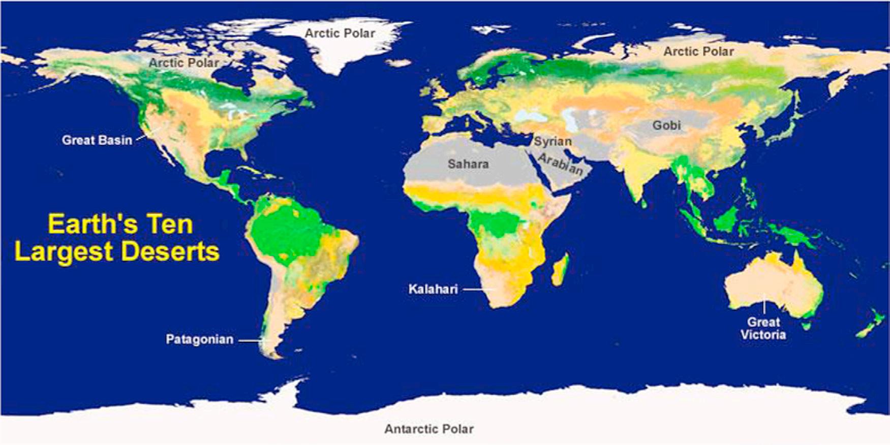
By definition a “desert” is a region that receives extremely low rains—less than 250 mm/year—far less than the amount required to support the growth of most plants. Approximately one-third of the earth’s land surface is desert, with an area more than 52,000 square kilometers (Fig. 1.1).
Deserts are extreme environments where intense solar radiation, limited nutrients, low organic matter content, and restricted water availability present formidable challenges for fungi inhabiting these areas. Desert soils generally are characterized by low propagule densities but high species diversity (Christensen, 1981; Mouchacca, 1995). Studies on mycobiota of soils may be dated back to 1886 when Adametz started his pioneer study by isolation and naming 4 species of yeasts and 11 species of filamentous fungi including Aspergillus (Watanabe, 2002). Species of Aspergillus are common and they may account for up to 20% of the total species isolated in the desert (Christensen and Tuthill, 1985).
The number of mycological studies on desert soil is rather limited in comparison with other ecological habitats.
Several authors assume the diversity of microbes including fungi is low compared to soil in moderate or tropical regions and they suggest these extreme ecosystems as suitable in situ models to study the relationship between phylogenetic biodiversity and function (Adams et al., 2006).
Desert mycobiota of Egypt have been the target of many studies, namely: Montasir et al. (1956a,b), Mahmoud et al. (1964), Besada and Yusef (1968), Moubasher and Moustafa (1970), Moubasher and El-Dohlob (1970), Salama et al. (1971), Mouchacca (1971, 1973a,b, 1977, 1982); Naguib and Mouchacca (1970-1971), Mouchacca and Nicot (1973), Mouchacca and Joly (1974, 1976), Samson and Mouchacca (1974, 1975), Moubasher et al. (1985, 1988, 1990), Nassar (1998), Abdel-Hafez et al. (1989a,b, 1990), Abdel-Sater (1990, 2000), Abdel-Hafez and El-Maghraby (1993), Abdel-Azeem and Ibrahim (2004), and Abdel-Azeem (1991, 2009).
Moubasher and Moustafa (1970) surveyed the Egyptian soil fungi with special reference to Aspergillus, Penicillium, and Penicillium-related genera in 32 soil samples collected from different localities in Egypt. They met 16 species of Aspergillus and the highest population and occurrence were recorded for A. niger, A. terreus, A. flavus, and Aspergillus sydowii, respectively.
Mouchacca and Joly (1976) studied the biodiversity of genus Aspergillus in arid soils of Egypt. They collected 31 soil samples from the western desert of Egypt. They collected 14 soils (set A) from regions receiving very weak to null winter rains and 17 (set B) samples from regions that benefit from an appreciable amount of wintry precipitation. In their study the taxonomic distribution is hardly affected by the dimensions of soil sand components, while regional localization exerts a certain influence. Twenty-seven species
FIGURE 1.1 Map shows the generalized location of Earth’s ten largest deserts on the basis of surface area (http://geology.com/records/largest-desert.shtml).
of Aspergillus were isolated, some are practically omnipresent (A. niger, A. flavus group), others develop preferentially in set A soil (Aspergillus nidulans, Aspergillus ustus, A. ochraceous, and possibly A. fumigatus groups) and/or have distribution positively affected (Aspergillus flavipes and A. terreus) or perhaps negatively (A. fumigatus group) due to soil reclamation.
In their extensive survey of Sinai terricolous fungi, Abdel-Azeem and Ibrahim (2004) and Abdel-Azeem (2009) recorded 17 species of Aspergillus. They recorded A. alutaceous, Aspergillus candidus, Aspergillus clavatus, A. flavus, A. fumigatus, Aspergillus japonicus, A. niger, A. ochraceous, A. sydowii, Aspergillus tamerii, A. terreus, A. ustus, Aspergillus versicolor, Aspergillus wentii, Emericella nidulans, Eurotium amstelodami, and Eurotium chevalieri
Six taxa are introduced to the genus Aspergillus as novel taxa based on type materials collected from Egyptian deserts namely: Aspergillus egyptiacus Moubasher and Moustafa (1972) (as Aspergillus aegyptiacus), Aspergillus floriformis Samson and Mouchacca (1975), Aspergillus pseudodeflectus Samson and Mouchacca (1975), Emericella desertorum Samson and Mouchacca (1974), Emericella purpurea Samson and Mouchacca (1975), and Eurotium xerophilum Samson and Mouchacca (1975).
Few investigations have been made on soil mycobiota in Libya. Naim (1967a,b) studied rhizosphere and soil fungi of Artemisia herba-alba and fungi under citrus trees in Tripoli, Libya. Youssef (1974) studied the fungal flora of Libyan soil. He examined 16 different localities in Libya for their fungal microflora. El-Said and Saleem (2008) studied soil fungi at the western region of Libya. Mansour (2010) studied the distribution and occurrence of various groups of fungi in different kinds of soils in the eastern region of Libya. Result showed that the most abundant species were Aspergillus flavus, A. fumigatus, A. niger, Aspergillus ochraceus, A. terreus, and A. ustus. For more details concerning the checklist of Libyan fungi check El-Buni and Rattan (1981)
Mycobiota of Algerian, Tunisian, and Moroccan deserts do not receive that much attention from mycologists and hence few studies have been published concerning the mycobiota of these deserts. Recently mycobiota of three chotts located in the northeast of Algerian Sahara have been studied by Dendouga et al. (2015). They isolated 327 colonies of fungi and Aspergillus was one of the most common genera isolated in this study.
Studies on micromycetes of desert soils of the Kingdom of Saudi Arabia showed that Aspergillus amstelodami, Aspergillus chevalieri, Aspergillus ruber, A. ochraceous, A. fumigatus, A. flavus, A. sydowii, A. terreus, and A. ustus are the most common species (Fathi et al., 1975; Ali, 1977; Ali et al., 1977; Abdel-Hafez, 1982a,b,c, 1994; Hashem, 1991, 1995; Arif and Hashem (1988); Barakat, 1999; Abdulmoniem and Saadabi (2006); Abou-Zeid and Abd
El-Fattah, 2007). Also, the teleomorph genera Emericella (E. nidulans) and Eurotium with E. amstelodami and E. chevalieri are common in Saudi Arabian desert soils.
Tolba et al. (1957), Al-Doory et al. (1959), Ismail and Abdullah (1977), and Abdullah et al. (1986) studied soil microfungi from different localities in Iraq. In these studies genus Aspergillus accounted for about 16% of the total species isolated. Aspergillus fumigatus was the most common species, being isolated from 70% of the sampling sites examined. Aspergillus candidus and A. niger were in the second and third positions in frequency, being isolated from 60% and 50% of the sampling sites examined, respectively. Imran and Al Rubaiy (2015) studied the molecular ecological typing of environmental isolates of A. terreus collected from the desert region in Iraq.
In Syria various species of Aspergillus were recorded by various investigators, such as: Sizova et al. (1967), Baghdadi (1968), Abdel-Hafez et al. (1983), and Abdel-Kader et al. (1983) Aspergillus niger, A. sydowii, A. flavus, A. wentii, and A. clavatus were the most prevalent species. Aspergillus kassunensis as a new species added to genus Aspergillus was introduced by Baghdadi (1968) from Syrian soil.
Al-Subai (1983) and Moubasher (1993) concluded that Aspergillus was consistently the most common genus in Qatari soils. Moubasher (1993) isolated fungi from 11 desert soil samples out of 42 samples representing different ecological habitats of Qatar. Aspergillus contributed by 23 species and 5 varieties, of which A. terreus, A. flavus, A. versicolor, and A. niger were the most frequent species.
Halwagy et al. (1982) found Aspergillus, Alternaria, and Drechslera constituted 16%, 5%, and 3% respectively of the total species isolated from desert soils in Kuwait. They recorded Aspergillus terreus, A. fumigatus, and A. niger with frequencies of occurrence of 70%. El-Said (1994) studied soil mycoflora of Bahreen (Bahrain) in which 39 species belonging to 20 genera were isolated from 50 soil samples on different isolation media. Aspergillus flavus, A. fumigatus, A. niger, A. sydowii and A. terreus, Eurotium amstelodami, and E. chevalieri were the most common species.
Mycobiota of the northern part of the Negev desert (Rayss and Borut, 1958; Borut, 1960; Guiraud et al., 1995; Steiman et al., 1995) represented by 159 species belonging to 58 genera in which 16 of them under genus Aspergillus Aspergillus fumigatus, Aspergillus sclerotiorum, and A. versicolor are the most common species in this region. Volz et al. (2001) concluded that the majority of Israel soil fungi (309 species—70%) belong to the division Ascomycota, but only 56 species of them were found to have a perfect stage in their life cycle. Concerning species diversity among genera, they showed that Aspergillus recorded only 48 species (15.53%) out of 309 species. Aspergillus niger, A. terreus, A. ustus, and A. versicolor are the most widely distributed species in Israel. Grishkan and Nevo (2010) isolated 185 species belonging to 76 genera from the soil of Makhtesh
Ramon hot desert in Israel. Ten species of Aspergillus, nine anamorphic and one teleomorphic, were isolated in which A fumigatus comprised a basic part of thermotolerant mycobiota obtained in this study.
Aspergillus as a xerotolerant and xerophilic genus can grow at or below a water activity (aw) of 0 (Pettersson and Leong, 2011). Several researchers have isolated genus Aspergillus from desert soils in Argentina, Chile, and Mexico (Giusiano et al., 2002, Piontelli et al., 2002, SamaniegoGaxiola and Chew-Madinaveitia, 2007). Conley et al. (2006) studied the fungal content of Atacama desert, the driest and oldest desert on Earth, without any record rainfall for decades. They reported 12 genera of fungi, with Aspergillus one of them. Aspergillus flavus and A. fumigatus reported from desert soils worldwide (Moubasher, 1993; Abdel-Hafez, 1981; Giusiano et al., 2002; Piontelli et al., 2002; El-Said and Saleem, 2008) and Aspergillus carneus recorded exclusively from desert soils in the Middle East (Abdullah et al., 1986; Ali-Shtayeh and Jamous, 2000, El-Said and Saleem, 2008) were missing in the Atacama soil.
Grishkan et al. (2015) examined the variations in microfungal communities inhabiting different biological crust types in the vicinity of the Shapotou Research Station in the Tengger Desert, China. The mycobiota isolated from the crusts sampled in 2011 and 2013 were composed of 123 and 67 identified species, respectively. Altogether 134 species were isolated: 6 of Mucoromycotina, 22 of teleomorphic (morphologically sexual) Ascomycota, and 106 of anamorphic (asexual) Ascomycota. These species belonged to 66 genera, with the most common being Aspergillus (12 species). Taxa of Aspergillus fumigatus, A. niger, A. nidulans, and Aspergillus rugulosus dominated.
Klich (2002a) published her biogeography of Aspergillus species in soil and litter and she concluded that there was no overall trend in distribution of the members of the entire genus by ecosystem, however, individual sections of the genus appeared to have distinct distribution patterns. Most members of sections Aspergillus, Nidulantes, Flavipedes, and Circumdati occurred at greater than expected frequencies in desert soils (Klich, 2002a). To conclude, in desert environments, the pan-global stable Aspergillus species are represented by A. niger, A. flavus, A. fumigatus, A. ochraceus, A. terreus, A. sydowii, A. tamerii, A. ustus, A. versicolor, A. wentii, Emericella nidulans, Eurotium amstelodami, and E. chevalieri.
Salterns
When evaporation of seawater accompanied with halite (NaCl) concentrations of greater than 10% (m/w), Thalassohaline hypersaline environments originated (Oren, 2002) and provide some of the most extreme habitats in the world. They are common all around the globe, and include, for example, marine ponds and salt marshes that
are subjected to evaporation, salt or soda lakes, and sea-salt and manmade salterns (Trüper and Galinski, 1986).
Life-limiting parameters in salterns are many, for example, variable water activities (aw), high concentrations of NaCl, low oxygen concentrations as well as high light intensity (Brock, 1979). Halotolerant and halophilic fungi were first reported as active inhabitants of solar salterns by Gunde-Cimerman et al. (2000). Later they were isolated by several investigators (Butinar et al., 2005a, b, c; Cantrell et al., 2006) from salterns around the world, for example, La Trinidad in the Ebro River Delta and Santa Pola on the Mediterranean coast of Spain, Camargue in France, and the salterns on the Atlantic coast in Portugal, and in Namibia, the Dominican Republic, and Puerto Rico.
After a decade of research into the fungal diversity in salterns, together with new taxa, a number of fungal genera with high diversities of halotolerant and halophilic species have been described. Different species of genus Aspergillus are among the filamentous fungi that appear with the highest frequencies in salterns (Butinar et al., 2011). The group of filamentous fungi that have been isolated from different salterns around the world is mainly represented by the order Eurotiales by the teleomorphic genera Eurotium and Emericella and the anamorphic Aspergillus and Penicillium (Tresner and Hayes, 1971; Cantrell et al., 2006; Butinar et al., 2011).
Global natural hypersaline waters are characterized by certain taxa mainly of Aspergillus niger and Aspergillus caesiellus, while hypersaline localities at higher environmental temperatures are characterized by primarily or exclusively taxa of A. ochraceus, A. flavus, Aspergillus roseoglobulosus, and Aspergillus tubingensis. Butinar et al. (2011) listed Aspergillus melleus, A. sclerotiorum, and Petromyces alliaceus (holomorphic species) within these taxonomic groups, although they have appeared only locally. Both Aspergillus versicolor and A. sydowii have also been identified as part of the fungal communities in the hypersaline environments, even they are common in marine environments and in dry foods. Aspergillus wentii, A. flavipes, A. terreus, and particularly A. candidus have been repeatedly isolated from Adriatic salterns, whereas A. penicillioides, A. proliferans, and A. restrictus have been found only sporadically at salinities below 10% NaCl. Aspergillus fumigatus is common in arid environments (deserts) at high temperatures, and has been found consistently in solar salterns, although it is also most abundant at salinities below 10% NaCl (Moustafa, 1975; El-Dohlob and Migahed, 1985; Moubasher et al., 1990; Abdel-Azeem, 2003; Abdullah et al., 2010; Butinar et al., 2011; Balbool et al., 2013).
Six different species of the known teleomorphic foodborne xerophilic genus Eurotium were repeatedly isolated in a mycodiversity study of hypersaline waters: Eurotium amstelodami, Eurotium herbariorum, and Eurotium repens as indigenous taxa in hypersaline water, while Eurotium
rubrum, E. chevalieri, and E. halotolerans are only impermanent inhabitants of brine at lower salinities (Butinar et al., 2005c).
The representatives of genus Emericella, which are recognizable by Hülle cells in the cleistothecial walls and ornamented ascospore, have frequently been isolated from dry substrata in hot and arid areas worldwide. These appear to be well adapted to dry and warm climates (Samson and Mouchacca, 1974) and low aw (Zalar et al., 2008). The new taxa of soil representative of Emericella was isolated also from desert saline soil as mentioned before (Samson and Mouchacca, 1974, 1975), while two newly described halotolerant species, Emericella filifera and Emericella stella-maris, were reported from hypersaline water of the Sečovlje salterns in Slovenia (Zalar et al., 2008). Emericella striata was described as a new taxon from Lake Enriquillo in Dominican Republic (Butinar et al., 2011).
To conclude, in hypersaline environments, the panglobal stable taxa of genus Aspergillus are represented by A. niger and E. amstelodami, and possibly also by A. sydowii, A. candidus, and E. herbariorum, which are also quite abundant, although more locally distributed (Butinar et al., 2011).
Aspergillus Xerophily in Different Habitats
The most xerophilic of the anamorphic Aspergilli are species in the section Restricti (Peterson, 2008), particularly Aspergillus restrictus and A. penicillioides. The later is regarded as an extreme xerophile (Andrews and Pitt, 1987), as it grows restrictedly or not at all at high aw, optimally at 0.91–0.93 aw and is capable of growth down to at least 0.73 aw in experimental systems.
Aspergillus candidus is an important xerophilic species, and has been reported from a wide range of commodities, but rarely as a primary cause of spoilage. The most tolerant of the Aspergilli to low oxygen tensions is A. candidus which can grow in 0.45% oxygen, which assists development to high populations in stored grain. It produces a range of secondary metabolites, but of these, only kojic acid is regarded as a significant toxin. Aspergillus flavus and A. parasiticus are perhaps the most widely reported food spoilage fungi, since the discovery in the early 1960s of their toxic carcinogenic metabolites, aflatoxins. Aspergillus parasiticus appears to be widely distributed in foodstuffs in the USA, Latin America, Africa, India, and Australia and rarely in Southeast Asia (Pettersson and Leong, 2011).
Aspergillus niger, Aspergillus carbonarius, A. japonicus, and A. aculeatus, as black Aspergilli, are widely distributed species. Aspergillus niger is widespread throughout the tropical and temperate zones and was regarded as a nontoxigenic species until it was demonstrated that certain strains produce ochratoxin A and fumonisin B2 (Frisvad et al., 2007). Aspergillus carbonarius is considered to be the
major producer of ochratoxin A. Aspergillus niger occurs in a range of foods (eg, peanuts, cereals, oilseeds, spices, dried fish, and meat products).
Aspergillus ochraceus and related species Aspergillus westerdijkiae and Aspergillus steynii produce the mycotoxin ochratoxin A. Like most Aspergilli, A. ochraceus is tolerant of a wide range of pH, growing well between pH 3 and 10, and weakly at pH 2.2. It is common in dried and stored products, has been reported in high numbers from green coffee beans, and may be a source of ochratoxin contamination in this commodity. It is less frequently reported from cereals and cereal products. Sterigmatocystin is produced by Aspergillus versicolor, which considered as an important species in the deterioration of stored grain and a major source of volatile compounds. It has also been implicated as one cause of the “Rio” off-flavor in coffee (pungent, medicinal, or iodine-like taste, musty cellar-like odor) due to formation of trichloroanisoles (Pettersson and Leong, 2011).
Eurotium species are perhaps the epitome of xerophilic fungi, being capable of rapid growth over wide temperature and aw ranges (minimum ∼ 0.70–0.72 aw), and having a cosmopolitan distribution. There are four common foodborne species of Eurotium: Eurotium amstelodami, E. chevalieri, E. repens, and E. rubrum. All are similar physiologically (halophilic xerophiles), and they appear to occupy similar ecological niches, namely causing deterioration of dried foods and also high-sugar products such as confectionery, dried fruit, jams, and conserves (Butinar et al., 2005c; Pettersson and Leong, 2011). Although most Eurotium species are capable of growth at high water activity, they compete poorly in natural substrates at water activity values above 0.92.
Agricultural
Globally the majority of the research which has involved the isolation and identification of Aspergillus strains from various agricultural and horticultural crop fields in different agro-climatic zones was undertaken in order to evaluate them for mycotoxin production (Klich, 2002b). Therefore, only a limited number of studies deal with biodiversity of the genus Aspergillus in specific crop fields or agroecosystems. Climatic factors, followed by edaphic and spatial patterning, are the best predictors of soil fungal richness and community composition at the global scale (Tedersoo et al., 2014). Biotic (plant species and their growth stage, microbial competition) and abiotic factors (soil physico-chemical characters, application of pesticides and/or fertilizers) as well as the geographical position affected populations and diversity of fungal communities in agroecosystems (Kredics et al., 2014).
In her biogeographic study of Aspergillus species in soil and litter, Klich (2002a) found that five species of
Aspergillus reported in over 100 studies were A. fumigatus, A. versicolor, A. terreus, A. flavus, and A. niger var. niger. With one exception, these five species occurred at the expected frequencies in all of the biomes; A. terreus occurred at greater than expected frequencies in cultivated soils and less than expected frequencies in forest soils. In many parts of Egypt several investigators studied soil fungi from cultivated soil, for example, Abdel-Hafez (1974), Moubasher and Abdel-Hafez (1978), and Abdel-Azeem (2003). They found taxa belonging to Aspergillus, Penicillium, Fusarium, Mucor, and some dematiaceous Hyphomycetes were the most common in various types of Egyptian soils. Mazen and Shaban (1983) studied the fluctuation of soil fungi in wheat fields and found that the most common fungi isolated were Aspergillus represented by five species Aspergillus niger, A. terreus, A. fumigatus, A. flavus, and A. versicolor Abdel-Hafez and coworkers (2000) isolated 118 species in addition to seven varieties belonging to 51 genera from cultivated and desert soils in Egypt. The results obtained from the three soil types were basically similar, and the most common Aspergillus species were A. flavus, A. flavus var. columnaris, A. fumigatus, A. niger, Aspergillus sydowii, and A. terreus.
Hafez (2012) made an ecological comparison on soil and rhizospheric fungi of maize and wheat plants in different areas in Minia Governorate in Egypt. She isolated 28 fungal species from wheat belonging to 18 genera and that 13 species were isolated from maize belonging to 9 genera. Aspergillus was the most dominant in both rhizospheric and nonrhizospheric soils and was represented by four species: A. niger, A. terreus, A. flavus, and A. ustus
Fusaria and other fungi associated with rhizosphere and rhizoplane of lentil and sesame at different growth stages from cultivated soil in Egypt have been studied by AbdelHafez et al. (2012). They isolated 16 Fusarium species and three Aspergillus species (A. flavus, A. niger, and A. ochraceous) were isolated.
Abdel-Azeem et al. (2007) studied the effects of longterm heavy metal contamination on diversity of terricolous fungi and nematodes in an agroecosystem in Egypt as a case study. They collected 100 soil samples in a randomized way to represent different stages of land reclamation during the period from September (2004) to February (2005). These profiles represented different land use periods of 0–20 years. Isolated species belonged to 21 genera. The prevailing genera were Aspergillus (12 species including anamorph stages of one Emericella and one Eurotium species; 52.63% of the total isolates). They found that the most abundant species were: Aspergillus niger var. niger, (21.15% of the total isolate number), Trichoderma pseudokoningii (12.65%), A. flavus (9.4%), and A. fumigatus (8.63%).
Aspergillus taxa distributed in different altitudes (24 m above sea level to 2000 m above sea level) of the eastern Himalayas were studied by Devi and Joshi (2012). They
recorded Aspergillus versicolor in samples collected from 1–500 m above sea level, Aspergillus nomius (500–1000 m), A. niger (1000–1500 m), A. fumigatus, A. flavus, A. terreus, and Aspergillus awamori (1500–2000 m).
Aspergillus species are able to produce a range of mycotoxins, including, for example, aflatoxins, ochratoxins, fumonisins, and patulin. Aflatoxins are mainly produced by members of Aspergillus section Flavi, and they contaminate various agricultural products in several parts of the world (Baranyi et al., 2013).
Taxonomically, based on Aspergillus species, mycotoxins in fruits can be divided into three major groups: (1) Aflatoxins produced by A. flavus, A. parasiticus, and A. nomius; (2) Ochratoxin A produced by A. ochraceus, A. carbonarius, A. niger aggregate, A. tubingensis, A. sclerotiorum, Aspergillus sulphureus, A. aculeatus, A. japonicus var. aculeatus, Aspergillus alliaceus, A. melleus, and other species; and (3) Other toxic metabolites produced by a variety of Aspergillus spp., the most important of these being sterigmatocystin, produced by A. flavus, A. flavipes, A. nidulans, and A. versicolor; cyclopiazonic acid, produced by A. flavus, A. tamarii, and A. versicolor; aflatrem, produced by A. flavus; citrinin, produced by A. flavipes, A. carneus, A. niveus, and A. terreus; and patulin, produced by A. terreus (Gill-Carey, 1949; Raper and Fennell, 1965; Semeniuk et al., 1971; Ciegler, 1972; Hesseltine et al., 1972; Buchanan et al., 1975; Durley et al., 1975; Lee et al., 1975; Mislivec et al., 1975; Sommer et al., 1976; Moss, 1977; Gallagher et al., 1978; Stack and Mislivec, 1978; GorstAllman and Steyn, 1979; Anke et al., 1980; Cole and Cox, 1981; Davis, 1981; Wicklow and Cole, 1982; Turner and Aldridge, 1983; Cole, 1984; Dorner et al., 1984; Scudamore et al., 1986; Kurtzman et al., 1987; Vesonder et al., 1988; Betina, 1989; Kim et al., 1993; Doster et al., 1996; Varga et al., 1996; Richard et al., 1999; Giridhar and Reddy, 2001; Sage et al., 2002, 2004; Battilani and Pietri, 2002; Bayman et al., 2002; Serra et al., 2003; Magnoli et al., 2004; Iamanaka et al., 2005; Medina et al., 2005; Perrone et al., 2006; Roussos et al., 2006; Barkai-Golan, 2008).
Fourteen species assigned to three sections of the genus Aspergillus are responsible for acute aflatoxicosis epidemics that occurred recently in several parts of Asia and Africa leading to the deaths of several hundred people. Taxa distributed among three sections: Flavi (A. flavus, Aspergillus pseudotamarii, A. parasiticus, A. nomius, Aspergillus bombycis, Aspergillus parvisclerotigenus, Aspergillus minisclerotigenes, Aspergillus arachidicola, Aspergillus togoensis), section Nidulantes (Emericella astellata, Emericella venezuelensis, Emericella olivicola), and section Ochraceorosei (Aspergillus ochraceoroseus, Aspergillus rambellii) (Varga et al., 2009; Rank et al., 2011). Potential aflatoxin-producing A. flavus isolates were also identified in other agricultural products including stored wheat, onions, grapes, and rice, and in cattle feed (Krnjaja et al., 2008). Aflatoxins
were also detected in sunflower flour samples (Masic et al., 2003) and in spices in Serbia (Saric and Skrinjar, 2008).
Several Aspergillus species are also able to produce patulin, including species assigned to Aspergillus sections Clavati (Varga et al., 2007b) and Terrei (Varga et al., 2005). These species frequently occur in cereals and cereal products (Lopez-Diaz and Flannigan, 1997; Abramson et al., 1987). The most well-known species A. clavatus can be isolated mainly from soil and dung, but it also occurs in stored products (mainly cereals) with high moisture content, for example, inadequately stored rice, corn, and millet (Flannigan and Pearce, 1994). Aspergillus clavatus isolates appeared to be particularly well adapted for growth during malting (Flannigan and Pearce, 1994).
Polar
Around 2.3% of the world’s fungal biota exists in the Arctic and fungi in this region have been isolated from various substrates and habitats (Ivarson, 1965; Reeve et al., 2002; Säwström et al., 2002; Callaghan et al., 2004; Ozerskaya et al., 2009; Pathan et al., 2009). More than 1000 species and over 400 genera of nonlichenized fungi have been reported from Antarctic regions (including the sub-Antarctic) (Bridge and Spooner, 2012; Arenz et al., 2014) including genus Aspergillus. The genus Aspergillus is also mesophilic to thermotolerant, yet some spores of Aspergillus and its associated teleomorphs are found in Arctic regions (GundeCimerman et al., 2005). The presence of “cosmopolitan” species such as Alternaria, Penicillium, Aspergillus, Cladosporium, and others may be referred to their wide dispersal potential and ubiquitous association with human activities and material (Ruisi et al., 2007).
However, fungal diversity in Arctic soils has been investigated only to a limited extent. Krishnan et al. (2011) isolated 28 isolates of fungi from bird-forming soil, pristine, and human-impacted soils collected from the Fildes Peninsula, King George Island, Antarctica, without any Aspergillus species. Singh et al. (2012a,b) studied filamentous soil fungi from Ny-Ålesund, Spitsbergen, and they isolated 19 species under 14 genera. Aspergillus were represented by three species, namely: A. aculeatus, A. flavus, and A. niger. Similarly, other genera seem to be absent in cold ecosystems, for example, Byssochlamys and its anamorphic state Paecilomyces Aspergillus species in general grow poorly below 12°C, and thus may have been recovered as spores in cold ecosystems (Gunde-Cimerman et al., 2003) because they are common as marine spores, transported by wind or birds, or are carried around due to human activity (Frisvad, 2008).
Water-Related
Shearer et al. (2007) estimated fungal biodiversity in freshwater, brackish, and marine habitats based on reports in the
literature. In their study they covered the ecological groups including fungi and taxa formerly treated as fungi, exclusive of yeasts, in freshwater, brackish, and marine habitats. They have reported approximately 3047 taxa from aquatic habitats thus far. The largest taxonomic group of fungi in aquatic habitats is comprised of teleomorphic and anamorphic Ascomycota, followed by the Chytridiomycota. Marine fungi are an ecological rather than a taxonomic group and comprise an estimated 1500 species, excluding those that form lichens (Hyde et al., 1998). Obligate marine fungi grow and sporulate exclusively in the marine or estuarine environment; facultative marine species may grow in marine as well as in freshwater (or terrestrial) habitats (Kohlmeyer and Kohlmeyer, 1979). A case in point is Aspergillus sydowii, isolated from diseased sea fans and causing the disease in laboratory experiments (Geiser et al., 1998).
Boutaiba (1997) studied the fungal flora of Lake El Golea in Algeria. He studied their taxonomy, ecology, and metabolite production. He isolated A. niger, A. terreus, A. sydowii, A. repens, A. ochraceous, A. fumigatus, A. flavus, A. candidus, and A. wentii
Singh et al. (2012a,b) investigated fungal diversity in two sediment cores w40 cmbsf (cm below seafloor) at a depth of w5000 m in the Central Indian Basin (CIB), by culture-dependent as well as culture-independent approaches. This resulted in recovering a total of 19 culturable fungi and 46 operational taxonomic units (OTUs), respectively. The majority of the fungi belonged to Ascomycota, within no single species dominating. It included members of filamentous fungi such as Aspergillus sp., Eurotium sp., Cladosporium sp., Pleospora sp., Chaetomium sp., Ascotricha sp., Penicillium sp., and Sagenomella sp.
Zhang et al. (2014) investigated the composition and abundance of fungal community in the deep-sea sediments of the Pacific Ocean. They identified 12 Ascomycetes belonged to 6 genera (Aspergillus, Aureobasidium, Candida, Exophiala, Fusarium, and Periconia). Aspergillus was represented only by two species A. sydowii and A. vitricola.
Abdel-Azeem et al. (2015) studied the occurrence and diversity of mycobiota in heavy metal-contaminated sediments of a Mediterranean coastal lagoon, El-Manzala, Egypt. They found that the prevailing genera were Aspergillus (11 species including anamorph stages of 2 Emericella species; 36.66% of the total isolates), Penicillium (4 species including anamorph of Talaromyces; 13.33%), and the remaining taxa were represented only by two to one species each. Aspergillus niger, A. flavus, and A. terreus showed the highest percentage of frequency of occurrence.
Mangrove
Mangroves are an assortment of tropical and subtropical trees and shrubs which have adapted to the inhospitable
zone between sea and land: the typical mangrove habitat is a muddy river estuary (Kathiresan and Bingham, 2001; Hogarth, 2007). Mangles are considered a dynamic ecotone and approximately 25% of the world’s coastline is dominated by mangroves distributed in 112 countries encompassing an area of 18,000,000 ha (Spalding et al., 1997). The biodiversity of biota associated with mangle ecosystem is well known for animals and plants, but poorly known for fungi (Khalil et al., 2013).
Species diversity of fungi, seasonal variation and frequency of occurrence in Muthupettai mangroves, on the east coast of Tamil Nadu, India, was studied at two different seasons by Sivakumar et al. (2006). A total number of 118 fungal species were isolated, of which maximum 94 species from sediment samples followed by water with 83 species in which genus Aspergillus came first as the common genus followed by Penicillium, Curvualria, and Alternaria.
Tariq et al. (2008) studied the rhizosphere fungi of four different species of mangrove plants collected from coastal areas in Pakistan. They found that A. flavus, A. fumigatus, and A. niger were common in the rhizosphere soil of the four species of mangrove plants sampled.
Behera et al. (2012) studied the diversity of soil fungi from mangroves of Mahanadi delta, Orissa, India. Twentytwo fungal species and A. oryzae, A. niger, A. flavus, and Aspergillus albus were recorded as occasionally frequent.
Madavasamy and Pannerselvam (2012) studied the phylloplane fungi of green, senescent, and brown leaves of Avicinnia marina. Recovered taxa included Aspergillus candidus, A. flavus, A. luchueusis, A. niger, A. sydowii, A. fumigatus, and A. sulphureus out of a total of 22 species.
The mycobiota composition of the mangrove soil located in costal area at the Red Sea in Egypt was investigated in 24 soil samples that were collected (Khalil et al., 2013). Aspergillus flavus, A. niger, A. versicolor, and A. fumigatus, were recorded at high species frequency in more than 15 cases out of 24.
Living Plants, Lichens, and Animals
Endophytes colonize symptomlessly the living, internal tissues of their host, even though the endophyte may, after an incubation or latency period, cause disease (Petrini, 1991). In literature the term “fungal endophytes” is normally used to describe fungal organisms, which in contrast to mycorrhizal fungi, reside entirely within the host tissues and emerge during host senescence (Rodriguez et al., 2001; Rodriguez and Redman, 2008).
Endophytic fungi have been classified into two groups based on differences in taxonomy, evolution, plant hosts, and ecological functions into clavicipitaceous, able to infect only some species of grasses, and nonclavicipitaceous, which are found in the asymptomatic tissues of bryophytes, ferns, gymnosperms, and angiosperms (Rodriguez et al., 2009).
There are 1.3 million species of endophytic fungi alone, the majority of which are likely found in tropical ecosystems (Verma et al., 2014). There has been great interest in endophytic fungi as potential producers of novel biologically active products (Schulz et al., 2002; Wildman, 2003; Strobel and Daisy, 2003; Tomita, 2003; Urairuj et al., 2003; Spiering et al., 2006; Manoharachary et al., 2013).
Unique species of endophytic fungi with a wide range of potential practical applications in plant protection as repellents, insecticides, antimicrobials, anthelmintics, and vermicides have been found (Strobel et al., 2008; Vega et al., 2008). In the last 5 years, there has been evidence of the use of endophytes for producing anticancer, antimicrobial, and antioxidant compounds, and also in a biotransformation process (Pimentel et al., 2011; Salem and Abdel-Azeem, 2014).
Species of Aspergillus as a member of nonclavicipitaceous endophytes attracted the attention of researchers as effective producers of bioactive metabolites. Such studies may result in the description of new Aspergillus species, for example, Zhao et al. (2009) described Aspergillus niger var. taxi as a new species variant of taxol-producing fungus isolated from Taxus cuspidata in China.
Endophytic fungi Aspergillus clavatus isolated from the Azadirachta indica plant have also been reported to synthesize silver nanoparticles, which have significant antibacterial and antifungal activity (Verma et al., 2010). Endophytic Aspergillus fumigatus, isolated from Juniperus communis as a novel source of the anticancer prodrug deoxypodophyllotoxin, has been isolated and chemically characterized by Kusari et al. (2009)
Mustafa et al. (2013) exploited some Egyptian endophytic taxa for extracellular biosynthesis of silver nanoparticles. They isolated endophytic fungi from medicinal plants in arid Sinai. Their results showed that Zygomycota represented by two species (9.5% of the total species number), teleomorphic Ascomycota (3 species, 14.2%), anamorphic Ascomycota (16 species, 76.19%). The prevailing genera were Aspergillus (3 species including anamorph stages of one Eurotium species; 14.28% of the total isolates), and Alternaria (2 species, 9.5%). The remaining taxa were represented only by one species each. The most abundant species were: Alternaria alternata (41.6%), Nigrospora oryzae (38.3%), and Chaetomium globosum (11.1%). A total 13 species belonging to 11 genera were screened for the production of AgNPs. They recorded that Aspergillus niger synthesized AgNPs in a moderate rate in comparison with other taxa.
Zhang et al. (2012a,b) isolated indolyl diketopiperazines (1–6) from the endophytic fungus Aspergillus tamarii of Ficus carica and examined its antiphytopathogenic potentiality in vitro for the first time. Thirty-nine fungal metabolites, including two new alkaloids, of endophytic fungus Aspergillus fumigatus isolated from the stem bark of
Melia azedarach and their antifungal, antifeedant, and toxic activities were tested by Li et al. (2012).
Palenicia (2012) studied endophytic associations of species in the Aspergillus section Nigri with Zea mays and Arachis hypogea and their mycotoxins. He developed a system to identify black Aspergilli from peanut and maize in the southeastern United States. His survey indicated that A. niger species complex is predominant in maize and peanut fields.
Raghunath et al. (2012) screened Aspergillus niger isolated from Taxus baccata for the production of lovastatin on a solid state fermentation. The presence of lovastatin was confirmed by different techniques, for example, spectroscopic method, nuclear magnetic resonance (NMR), thin layer chromatography (TLC), and high-performance liquid chromatography (HPLC) methods.
Silva et al. (2011) studied endophytic fungi from Laguncularia racemosa (Brazilian mangrove) and their antimicrobial potential. They recovered six isolates of A. niger out of 70 endophytic strains.
Guatam (2014) isolated endophytic fungi from leaf segments of five medicinal plants collected from Mandi district, Himachal Pradesh, India. Aspergillus niger, A. flavus, A. clavatus, and A. variecolor were isolated with 14 species belonging to 15 genera out of a total of 373 fungal strains.
Eight medicinal plants (Achillea fragrantissima, Artemisia herba-alba, Chiliadenus montanus, Origanum syriacum, Phlomis aurea, Tanacetum sinaicum, Teucrium polium, and Thymus decussates) were screened for their content of endophytic fungi on different altitudes by Salem and Abdel-Azeem (2014) in Saint Katherine Protectorate, South Sinai, Egypt. Salem and Abdel-Azeem isolated 32 genera belonging to 75 species in which nine species of Aspergillus, namely, A. alliaceus, A. bisporus, A. candidus, A. flavus, A. fumigatus, A. japonicus, A. niger, A. terreus, and A. versicolor were recovered.
Yu et al. (2012) studied the diversity of endozoic fungi in South China Sea sponges and their potential in synthesizing bioactive natural products suggested by PKS gene and cytotoxic activity analysis. They isolated 14 genera and Aspergillus was the predominant component in the culturable fungal community and was represented by Aspergillus insulicola, A. penicillioides, A. terreus, A. oryzae, and E. rubrum Genus Aspergillus was associated with more than 30 species of sponge all over the world (Abrell et al., 1996; Varoglu and Crews, 2000; Lin et al., 2003; Gao et al., 2008; Proksch et al., 2008; Ein-Gil et al., 2009; Li and Wang, 2009; Lee et al., 2010; Liu et al., 2010; Menezesa et al., 2010; Paz et al., 2010; Ding et al., 2011; Wiese et al., 2011; Zhou et al., 2011; Thirunavukkarasu et al., 2012; Yu et al., 2012). The most common species of Aspergillus recorded in those studies were A. aculeatus, A. insuetus, A. niger, A. ostianu, A. sclerotiorum, A. ustus, A. versicolor, and Eurotium cristatum (Suryanarayanan, 2012).
Bai et al. (2014) characterized two new aromatic butyrolactones, flavipesins A (1) and B (2), two new natural products (3 and 4), and a known phenyl dioxolanone (5) from marine-derived endophytic fungus Aspergillus flavipes Different species from the genus Aspergillus are cited as marine-derived producers of enzymes (Bonugli-Santos et al., 2015). Aspergillus terreus was most frequently isolated as an endosymbiont from green, brown, and red seaweeds namely: Caulerpa scalpelliformis, Helimeda macroloba, Ulva lactuca, Ulva fasciata, brown Lobophora variegata, Padina gymnospora, Stoechospermum marginatum, Sargassum ilicifolium, Portieria hornemanni, and Gracilaria edulis, respectively (Suryanarayanan et al., 2010). Marine-derived fungi such as Aspergillus spp., apart from dominating the endosymbiont assemblage of seaweeds (Suryanarayanan et al., 2010) also dominate the fungal consortium of marine invertebrates collected from different localities such as the great Barrier Reef, North Sea, the Mediterranean, and the Caribbean (Höller et al., 2000) attesting to their adaptation to occupy such a niche as the inner tissues of seaweeds or marine animals. Such a widespread occurrence of marine-derived fungi may be indicative of their passive migration from terrestrial habitats (Alva et al., 2002).
However, since these fungi are better adapted to marine environments than their terrestrial conspecifics (Zuccaro et al., 2004; König et al., 2006) and survive in seaweeds which produce antifungal metabolites (Kubanek et al., 2003; Lam et al., 2008), it is likely that they are not casual residents of the seas but have coevolved with the seaweeds (Zuccaro et al., 2004; Suryanarayanan et al., 2010). Common endosymbiont of algae and seaweeds are Aspergillus versicolor, A. terreus, A. niger, A. flavus, and A. oryzae (Suryanarayanan, 2012).
Kelecom (2002) predicted a relationship between the type of secondary metabolite and the source of fungus, rather than the fungi themselves. The latter was exemplified by the fungi in the genus Aspergillus that produce fumiquinazoline derivatives if they are obtained from fish, sesquiterpene nitrobenzoate derivatives if they originate from algae, and indole diketopiperazine derivatives if they are isolated from sponges.
To conclude, in association with seaweeds, the marinederived Aspergillus species are represented by Aspergillus versicolor, A. terreus, A. niger, A. flavus, and A. oryzae (Belofsky et al., 1998; Lee et al., 2003; Zhang et al., 2007a,b,c; Lin et al., 2008; Qiao et al., 2010).
Endolichenic fungi represent an important ecological group of species that form associations with lichens, and to extend the knowledge of their diversity within macrolichens, Tripathi and Joshi (2015) isolated and identified the endolichenic fungi from some healthy macrolichens of Kumaun Himalaya. The majority of endolichenic fungi belonged to anamorphic Ascomycota (Hyphomycetes), and the lowest
were obtained from Zygomycetes. Aspergillus flavus and A. niger were common as endolichenic species and were recorded during various studies (Suryanarayanan et al., 2005; Li et al., 2007; Tripathi et al., 2014a,b,c; Tripathi and Joshi, 2015).
Air and Settled Dust
Over 225 species of fungi have been reported from indoor environments, which represent a few of the proposed estimate of 1.5 million species of fungi (McGinnis, 2007). The most common allergenic fungal genera are Cladosporium, Alternaria, Aspergillus, and Fusarium, where more than 80 genera of fungi have been linked with symptoms of respiratory tract allergies (Horner et al., 1995). Exposure to the large concentration of conidia of the four genera is considered the main causative agent of aspergillosis (Anderson et al., 1996), asthma and pneumonitis (Cuijpers et al., 1995; Hu et al., 1997), allergic alveolitis, and toxicosis (Flannigan et al., 1991).
Fröhlich-Nowoisky et al. (2012) studied the biogeography and fungal diversity in the air. They found ascomycota species were represented by 67–85% of the total isolated taxa and taxonomically distributed in four taxonomic classes namely: Sordariomycetes, Dothideomycetes, Eurotiomycetes, and Leotiomycetes, respectively. They represent plant and animal pathogens, symbionts, saprophytes, endophytes and epiphytes, and allergenic taxa (eg, Cladosporium spp., Aspergillus spp.).
In the United States, Shelton et al. (2002) evaluated the presence of indoor airborne fungi in 1717 buildings from 1996 to 1998, including hospitals, homes, schools, and industries. They determined Aspergillus versicolor as the predominant taxon, followed by A. flavus, A. fumigatus, and A. niger. Studies of Samson et al. (2010) and Flannigan et al. (2011) listed 100 fungal species common in indoor environments. In these lists, A. fumigatus and A. sydowii were common in the collected house dust.
As part of a worldwide survey of the indoor mycobiota, dust was collected from nine countries (Australia, Indonesia, Mexico, Micronesia, New Zealand, South Africa, Thailand, United Kingdom, and Uruguay). Mycological analyses of samples included the culture-dependent dilution-to-extinction method and culture-independent 454-pyrosequencing. They found 2717 isolates out of the 7904 isolates were identified as belonging to Aspergillus, Penicillium, and Talaromyces, respectively (Visagie et al., 2014). Studies showed that A. versicolor was considered to be very common in indoor environments and recently it was shown to represent a species complex, with nine new species introduced (Jurjević et al., 2012).
The diversity of air mycobiota showed the highest diversity in countries that are also listed as biodiversity hotspots of the world (Myers et al., 2000). This might refer to the
origin of at least a considerable proportion of these species isolated from house dust as being from outdoors. However, the prevalence of specific species commonly isolated from indoor surveys suggests that the indoor environments do select for the growth of specific species. In addition, much of the metagenomics diversity may come from transient, dormant, or dead spores (Visagie et al., 2014).
Júnior et al. (2012) studied the biodiversity of Aspergillus spp. and Penicillium spp. residing in libraries in Brazil. The genus Aspergillus was highlighted as one of the principal airborne fungi present in indoor environments. Aspergillus spp. were identified in 1277 (89.6%) samples and Penicillium spp. in 148 (10.4%). The dry period exhibited a greater number of isolates of the two taxa. Frequency of species of 34 taxa of genus Aspergillus (anamorph and teleomorph) isolated from library units in the dry (2009) and wet season (2010) in the city of Cuiabá, MT, Brazil were studied. The taxa belonged to 13 sections. Aspergillus niger var. niger came first with a recorded 30.2% frequency of occurrence, followed by A. flavus (19.7%).
In Egypt, Abdel-Azeem and Rashad (2013) studied mycobiota of outdoor air that can cause asthma: a case study from Lake Manzala, Egypt. They isolated a total of 71,780 mold and 560 yeast colony-forming units from 600 exposures and the isolated taxa were assigned to 28 genera and 43 species. They found that the greater presence of fungal spores occurred in the summer. Aspergillus niger, Cladosporium cladosporioides, Epicoccum nigrum, Aureobasidium pullulans, Alternaria cheiranthi, P. chrysogenum, Aspergillus fumigatus, and Alternaria alternata were the predominant species. They found that Aspergillus, Cladosporium, Penicillium, and Alternaria that had the greatest frequencies in air of Lake Manzala are strongly associated with allergic respiratory disease, especially asthma, in Port Said and Ismailia governorates.
Decaying Wood and Mummies
Wood deterioration by fungi may occur from several sources. These include the following: surface molds that cause localized discoloration; stain fungi that penetrate deep into the sapwood causing blue, gray, green, red, or other dark coloration; and wood-destroying fungi that decompose cell-wall polymers (Blanchette, 1998). Many ascomycetous fungi, such as Aspergillus nidulans, A. fumigatus, and A. oryzae, Magnaporthe grisea, Neurospora crassa, and Fusarium gramineum have a higher number of cellulases, with 34–44 hemicellulase encoding genes, and even 1–5 of the most efficient cellobiohydrolases (Hatakka and Hammel, 2010). Research on microbial and enzymatic degradation of wood and wood components has provided a great deal of information that has been useful in helping to protect and conserve historic and archeological wood. Ascomycetes fungi (anamorphic and teleomorphic) usually cause soft-rot
decay of wood with soft brown appearance and cracked and checked when dry (Nilsson et al., 1989; Blanchette, 1995). Two forms of soft rots were described by Blanchette (1995), type I consisting of biconical or cylindrical cavities that are formed within secondary walls, while type II refers to an erosion form of degradation. The knowledge of lignocellulose degradation by Ascomycetes is rather limited in comparison with other basidiomycetous fungi, and very little is known about how they degrade lignin (Nilsson et al., 1989).
Zidan et al. (2006) studied the conservation of a wooden Graeco-Roman coffin box and they isolated Paecilomyces variotii, Penicillium aurantiogriseum, Aspergillus niger, Aspergillus flavus, Aspergillus terreus, Emericella nidulans, and Mucor racemosus. These fungi were found in various parts of the coffin box, and their growth rate varied from one part to the other.
In Latvia, during the period from 1996 to 2007, a total of 300 private and public buildings, as well as more than 20 cultural monuments had been inspected regarding the damage by wood decay basidiomycetes and discoloring microfungi (Irbe et al., 2009). Wood decay fungi in constructions occurred in 338 cases. Brown-rot damage occurred more frequently (78.1%) than the white-rot (21.9%). Wood discoloring fungi (molds and blue stain) on construction and decorative materials were recorded in 55 cases where frequent genera were Penicillium, Cladosporium, Aspergillus, and Trichoderma.
Aspergillus candidus, A. ustus, and A. terreus were isolated from two wooden masks dating back to the GreekRoman period in Egypt (Darwish et al., 2013). Abu Deraz (2014) studied the soft rot fungi deteriorating archeological wood in Al-Aqsa mosque, Jerusalem, Israel. He isolated Aspergillus flavus, A. fumigatus, A. glaucus, A. niger, A. ochraceopetaliformis, and Emericella nidulans. Both A. flavus and A. niger showed high frequency of occurrence in all examined samples.
Mummies have been widely investigated by phenotypic and molecular techniques, particularly the study of ancient bacteria and micromycetes. There are several well-known examples showing the colonization of preserved bodies by opportunistic fungi, such as the case of the restoration of the body of Ramses II, performed in Paris in 1976–77. The mummy showed a dense fungal population with species belonging to the genera Aspergillus and Penicillium (Mouchaca, 1985). In his study, Mouchacca isolated 21 species and one variety of Aspergillus from debris (D) and abdominal materials (A) of Ramses II mummy. The most common species of D and A were A. niger, A. flavus, A. versicolor, A. sydowii, A. amstelodami, and A. restrictus Aspergilli also dominated the microbial communities of the air and dust of the Egyptian mummy chamber at the Baroda Museum in India (Arya et al., 2001).
Additionally, saprophytic fungi belonging to the genera Monilia, Penicillium, Alternaria, Aspergillus, Rhizopus,
and Chrysosporium as well as saprophytic bacteria of the genus Bacillus were isolated from a mummy from the collection of the Archaeological Museum in Zagreb, Croatia (Čavka et al., 2010). Fungal genera more related to the mummy materials were: Botryotinia, Giberella, Didymella, Fusarium, Verticillium, Tritirachium, Coprinus, and Coniosporium (Piñar et al., 2013).
Microscopic fungi were isolated from different materials including muscles, bones, skin, and funeral clothes from the mummified human remains of three members of the Kuffner’s family and from the surrounding air environments in Slovakia by Šimonovičová et al. (2015). Their hydrolytic abilities such as cellulolytic, lipolytic, and proteolytic keratinolytic were also assessed. The most commonly isolated fungi, from human remains, belonged mainly to the species of Aspergillus (A. candidus, A. calidoustus, A. fumigatus, A. niger, A. sydowii, A. terreus, A. ustus, A. venenatus, A. versicolor, and A. westerdijkiae).
Stones
The tiny pores and cracks in rocks which buffer microbial communities from a number of physical stresses, such as desiccation, rapid temperature variations, and UV radiation is defined as endolithic environment. The diversity of microorganisms in these ecosystems gained considerable attention, but few culture-independent studies have been carried out on the diversity of fungi to date.
Raghukumar et al. (1992) studied the endolithic fungi from deep sea calcareous substrata from calcareous animal shells at 100–860 m depth in the Bay of Bengal. They found that conidia of an isolate of A. niger obtained from intertidal calcareous shells did not germinate above 1 atm. Up to 512 Mu calcium was leached out upon growth of A. restrictus on 1 g of calcareous shell substrata at 100 atm. in 25 days.
The diversity of endolithic fungal communities in dolomite and limestone rocks from Nanjiang Canyon in Guizhou karst area was China studied by Tang et al. (2012). The most common genus in the investigated carbonate rocks was Verrucaria Aspergillus and Penicillium were also identified from the rock samples.
The diversity of culturable fungi associated with six species of healthy South China Sea gorgonians was investigated using a culture-dependent method followed by analysis of fungal ITS sequences (Zhang et al., 2012a,b). A total of 121 fungal isolates belonged to 41 fungal species from 20 genera. Of these, 30 species and 12 genera are new records for gorgonians, and the genera Aspergillus and Penicillium were the most diverse and common. Fourteen Aspergillus were isolated, they were: Aspergillus carneus, A. flavus, A. fumigatus, A. gracilis, A. insulicola, A. niger, A. nomius, A. ochraceopetaliformis, A. penicillioides, A. sclertoiorum, A. sydowii, A. terreus, A. tubingensis, and A. versicolor.
Abu Deraz (2014) recovered seven species of endolithic fungi from archeological stones of Al-Aqsa Mosque, Jerusalem, Israel. Surface sterilized stones were incubated on modified Czapek’s medium supplemented with calcium carbonate, as sole carbon source, as described by Kurakov et al. (1999). Five species of genus Aspergillus were common they were: A. flavus, A. fumigatus, A. niger, A. terreus, and Emericella nidulans.
Human
The fungal biota in an environment (mycobiome) is an important component of the human microbiome (Cui et al., 2013). Every human has fungi as part of their microbiota, however, the impact of fungi on human health is significant, especially as a reservoir for pathogenic fungi when the host is compromised and as a potential cofactor in inflammatory diseases and metabolic disorders (Huffnagle and Noverr, 2013).
Findley et al. (2013) studied the skin mycobiota of 10 healthy Americans, six men and four women. Genera included the potentially medically significant Candida, Chrysosporium, Cryptococcus, and otherwise unnamed dermatophytes assigned to the Arthrodermataceae. Common saprobic genera such as Aspergillus, Cladosporium, Epicoccum, Leptosphaerulina, Penicillium, Phoma, and Rhodotorula were also frequently detected or isolated.
A survey of oral fungal genera has been carried out by Ghannoum et al. (2010). They found that Candida and Cladosporium were most common, present in 75% and 65% of participants, respectively. The fungi of the oral cavity were previously believed to be few and relatively nondiverse based on culture-dependent or genus/species-focused culture-independent methods of identification. In contrast with fungal genera associated with local oral and invasive diseases, they were Aspergillus, Cryptococcus, Fusarium, and Alternaria, indicating that these genera are present in the oral microbiome even during healthy state (Ghannoum et al., 2010). Different studies (Schuster, 1999, Salonen et al., 2000; Williams and Lewis (2006); Jabra-Rizk et al., 2001; Seed, 2015) reported different genera of yeasts and filamentous fungi, for example, Candida, Saccharomyces, Penicillium, Aspergillus, Geotrichum, and Scopulariopsis and the abundant presence of Candida, Aspergillus, and Fusarium was recorded among the HIV-infected.
A large number of newly emerging pathogens have been described, besides the most prevalent and well-known fungal pathogens such as Candida albicans and Aspergillus fumigatus (Horré et al., 2010; Marguet et al., 2012). The lung mycobiome of healthy people is comprised of various geni and species principally controlled by environment
agents including Aspergillus species (van Woerden et al., 2013; Underhill and Iliev, 2014).
Aspergilloses are commonly caused by the fumigatus, flavus, and niger groups of genus Aspergillus. Other groups rarely act as agents of pulmonary disease, but it is assumed that any species can cause hypersensitivity reactions (Londero and Guadalupe-Cortés, 1990). Aspergillus species responsible for pulmonary aspergillosis were A. amstelodami, A. candidus, A. carneus, A. fischeri, A. flavus, A. fumigatus, A. glaucus, A. niger, A niveus, A. phialiseptus, A. restrictus, A. sydowii, A. terreus, and A. versicolor (Londero and Guadalupe-Cortés, 1990; Júnior et al., 2012). Finally, common taxa of Aspergillus and human biome are represented by A. fumigatus, A. flavus, A. niger, and A. versicolor.
Fossils
Today there are reports of representatives of many different groups of fungi in amber because the translucent nature of the matrix makes it relatively easy to determine even very delicate features useful in systematics, as well as those useful in determining interactions with other organisms (Taylor et al., 2015). Some examples including genus Aspergillus have been recorded. Thomas and Poinar (1988) described Aspergillus from a piece of Eocene amber originating from the Dominican Republic as Aspergillus janus Aspergillus collembolorum, a novel species was introduced in 2005 by Dörfelt and Schmidt when studying a piece of Baltic amber (Tertiary, Eocene) which contains an inclusion of a springtail (Collembola).
CONCLUSIONS
The studies discussed above reflect that the genus Aspergillus can be characterized with high adaptability to various ecological environments (Fig. 1.2). However, it is important to mention that the results of any study aimed at the examination of Aspergillus biodiversity should always be evaluated in the context of the developmental stage of Aspergillus taxonomy and the species identification methods available at the time of the publication of the respective paper. Due to the constant development of the taxonomy of the genus and the description of new species, more recent examinations of a specific habitat may reveal higher biodiversity of the genus and refine the results of previous studies. By introducing new techniques and methods in biodiversity studies during the past two decades, the amount of information available about the distribution of Aspergillus taxa is constantly growing, therefore it can be expected that the biogeography of the genus will be understood more deeply in the near future.
Polar A aculeatus
A. flavus
A. niger
Desert
A. candidus
A. flavipes
A. flavus
A. fumigatus
A. niger
A. ochraceous
A. sydowii
A. terreus
A. ustus
A. versicolor
Emericella nidulans
Eurotium amstelodami
E. chevalieri
Water-related environment
A candidus
A flavus
A fumigatus
A. niger
A. ochraceous
A. repens
A. sydowii
A. terreus
A. vitricola
A. wentii
Endozoic
A. flavipes
A. flavus
A. niger
A. oryzae
A. penicillioides
A. sclerotiorum
A. terreus
A. ustus
A. versicolor
Eurotium cristatum
E. rubrum
Saltern
A. candidus
A. flavus,
A. fumigatus
A. niger
A. penicillioides
A. restrictus
A. sydowii
A. terreus
A. versicolor
A. wentii
Eurotium amstelodami
E. chevalieri
E. halotolerans
E. herbariorum
E. repens
E.rRubrum
Petromyces alliaceus
FIGURE 1.2 Distribution of Aspergillus species among the different biomes of the world by Abdel-Azeem & Salem.
Living plants, lichens and animals
A. alliaceus
A. bisporus
A. candidus
A. Clavatus
A. flavus, A. fumigatus
A. japonicus
A. niger
A. tamarii
A. terreus
A. variecolor
A. versicolor
Agricultural
A. carbonarius
A. flavipes
A. flavus
A. fumigatus
A. japonicus
A. nidulans
A. niger
A. ochraceous
A. parasiticus
A. sydowii
A. tamarii
A. terreus
A. versicolor
REFERENCES
Abdel-Azeem, A.M. (1991). Effect of overgrazing on vegetation, microbes and soil in Ismailia-desert habitat. Biological Diversity Symposium, Madrid-Spain, pp. 241–246.
Abdel-Azeem, A.M. 2003. Ecological and taxonomical studies on ascospore-producing fungi in Egypt. PhD Thesis, Faculty of Science, Suez Canal University, Egypt.
Abdel-Azeem, A.M., 2009. Operation Wallacea in Egypt. I- A preliminary study on diversity of fungi in the world heritage site of Saint Katherine, Egypt. Assiut Univ. J. Bot. 38 (1), 29–54.
Abdel-Azeem, A.M., Ibrahim, M.E., 2004. Diversity of terrophilous mycobiota of Sinai. Egyptian J. Biol. 6, 21–31.
Abdel-Azeem, A.M., Rashad, H.M., 2013. Mycobiota of outdoor air that can cause asthma: a case study from Lake Manzala, Egypt. Mycosphere 4 (4), 1092–1104.
Abdel-Azeem, A.M., Abdel-Moneim, T.S., Ibrahim, M.E., Hassan, M.A.A., Saleh, M.Y., 2007. Effect of long-term heavy metal contamination on diversity of terricolous fungi and nematodes in Egypt- a case study. Water Air Soil Pollut. 186 (1), 233–254.
Abdel-Azeem, A.M., El-Morsy, E.M., Nour El-Dein, M.M., Rashad, H.M., 2015. Occurrence and diversity of mycobiota in heavy metal contaminated sediments of Mediterranean coastal lagoon El-Manzala, Egypt. Mycosphere 6 (2), 228–240.
Abdel-Hafez, A.I.I., Mazen, M.B., Galal, A.A., 1989a. Keratinophilic and cycloheximide resistant fungi in soils of Sinai Governorate, Egypt. Cryptogam. Mycol. 10 (3), 265–275.
Abdel-Hafez, A.I.I., Mazen, M.B., Galal, A.A., 1989b. Some ecological studies of osmophilic and halophilic soil fungi of Sinai Peninsula, Egypt. J. Sohag Pure Appl. Sci. Bull. 5, 67–83.
Abdel-Hafez, A.I.I., Mazen, M.B., Galal, A.A., 1990. Glycophilic and cellulose-decomposing fungi from soils of Sinai Peninsula, Egypt. Arab Gulf J. Sci. Res. 8 (1), 153–168.
Abdel-Hafez, S., 1981. Halophilic fungi of desert soils in Saudi Arabia. Mycopathologia 75, 75e80.
Abdel-Hafez, S.I., 1982a. Survey of microflora of desert soils in Saudi Arabia. Mycopathologia 80, 3–8.
Abdel-Hafez, S.I., 1982b. Osmophilic fungi of desert soils in Saudi Arabia. Mycopathologia 80, 9–14.
Abdel-Hafez, S.I., 1982c. Thermophilic and thermotolerant fungi of desert soils in Saudi Arabia. Mycopathologia 80, 15–20.
Abdel-Hafez S.I.I. 1974. Ecological studies on Egyptian soil fungi, PhD Thesis. Department of Botany, Faculty of Science, Assiut University, Egypt.
Abdel-Hafez, S.I.I., 1994. Studies on soil mycoflora of desert soils in Saudi Arabia. Mycopathologia 80, 3–8.
Abdel-Hafez, S.I.I., El-Maghraby, O.M.O., 1993. Thermophilic and thermotolerant fungi of Wadi-Bir-El-Ain soils. Eastern desert, Egypt. Abhath Al-Yarmouk. Pure Sci. Eng. 2, 55–66.
Abdel-Hafez, S.I.I., Ismail, M.A., Hussein, N.A., Abdel-Hameed, N.A., 2012. Fusaria and other fungi taxa associated with rhizosphere and rhizoplane of lentil and sesame at different growth stages. Acta Mycol. 47 (1), 35–48.
Abdel-Hafez, S.I.I., Moharram, A.M., Abdel-Sater, M.A., 2000. Monthly variations in the mycobiota of wheat fields in El-Kharga Oasis, Western Desert, Egypt. Bull. Fac. Sci. Assiut Univ. 29 (2-D), 195–211.
Abdel-Hafez, S.I.I., Abdel-Kader, M.I.A., Abdel-Hafez, A.I.I., 1983. Composition of the fungal flora of Syrian soils. Mycopathologia 81 (3), 161–166.
Abdel-Kader, M.I.A., Abdel-Hafez, A.I.I., Abdel-Hafez, S.I.I., 1983. Composition of the fungal flora of Syrian soils. II Cellulosedecomposing fungi. Mycopathologia 81, 167–171.
Abdel-Sater M.A. 1990. Studies on the mycoflora of the New Valley area, Western Desert, Egypt. PhD Thesis, Faculty of Science, Assiut University.
Abdel-Sater, M.A., 2000. Soil fungi of the New Valley area, Western desert, Egypt. Bull. Fac. Sci. Assiut Univ. 29 (2-D), 255–271.
Abdullah, S.K., Al-Khesraji, T.O., Al-Edany, T.Y., 1986. Soil mycoflora of the southern desert of Iraq. Sydowia 39, 8e16.
Abdullah, S.K., Al-Dossari, M.N., Al-Imara, F.J., 2010. Mycobiota of surface sediments in marshes of southern Iraq. Marsh Bull. 5 (1), 14–26.
Abdulmoniem, M.A., Saadabi, A.M.A., 2006. On the fungal flora of Saudi Arabian soils. Res. J. Microbiol. 1, 280–284.
Abou-Zeid, A.M., El-Fattah, R.I.A., 2007. Ecological Studies on the Rhizosperic Fungi of some halophytic plants in Taif Governorate, Saudi Arabia. World J. Agric. Sci. 3, 273–279.
Abramson, D., Sinha, R.N., Mills, J.T., 1987. Mycotoxin formation in moist 2-row and 6-row barley during granary storage. Mycopathologia 97, 179–185.
Abrell, L., Borgeson, B., Crews, P., 1996. Chloro polyketides from the cultured fungus (Aspergillus) separated from a marine sponge. Tetrahedron Lett. 37, 2331–2334.
Abu Deraz, S.S. 2014. Isolation and characterization of microbiota inhabiting Al-Aqsa Mosque, Al-Quds, Palestine. Master thesis, Faculty of Science, University of Suez Canal.
Adametz, L. 1886. Untersuchungen über die niederen Pilze der Ackerkrume. Inaug. Diss., 1–78. Leipzig.
Adams, B.J., Bardgett, R.D., Ayres, E., Wall, D.H., Aislabie, J., Bamforth, S., et al., 2006. Diversity and distribution of Victoria Land biota. Soil Biol. Biochem. 38, 3003–3018.
Al-Doory, Y., Tolba, M.K., Al-Ani, A., 1959. On the fungal flora of Iraqi soil.II: Central Iraq. Mycologia 51, 429–439.
Ali, M.I., 1977. Studies on the fungal flora of Saudi Arabia. 1-Wadi Hanif. Bull. Fac. Sci. Riyadh Univ. 8, 7–20.
Ali, M.I., Abu-Zinada, A.H., El-Mashharawi, Z., 1977. On the fungal flora of Saudi Arabia. 11-Seasonal fluctuations of fungi in the rhizosphere of some plants. Bull. Fac. Sci., Riyadh Univ., 215–228.
Ali-Shtayeh, M.S., Jamous, R.M., 2000. Keratinophilic fungi and related dermatophytes in polluted soil and water habitats. Revista Iberoamericana de Micologia 17, 51–59.
Al-Subai, A.A.T. 1983. Soil fungi in state of Qatar. M.Sc. Thesis, Botany Department, Faculty of Science, Qatar University, Qatar.
Alva, P., Mckenzie, E.H.C., Pointing, S.B., et al., 2002. Do seagrasses harbour endophytes?. In: Hyde, K.D. (Ed.), Fungi in Marine Environments, Fungal Diversity Research Series Hong Kong University Press, Hong Kong.
Anderson, K., Morris, G., Kennedy, H., Croall, J., Michie, J., Richardson, M., et al., 1996. Aspergillosis in immunocompromised paediatric patients: associations with building hygiene, design, and indoor air. Thorax 51, 256–261.
Andrews, S., Pitt, J.I., 1987. Further studies on the water relations of xerophilic fungi, including some halophiles. J. Gen. Appl. Microbiol. 133, 233–238. http://dx.doi.org/10.1099/00221287-133-2-233
Anke, H., Kolthoum, I., Zähner, H., Laatsch, H., 1980. Metabolic products of microorganisms. The anthraquinones of Aspergillus glaucus group. I. Occurrence,isolation, identification and antimicrobial activity. Arch. Microbiol. 126, 223–230.
Arenz, B.E., Blanchette, R.A., Farrell, R.L., 2014. Fungal diversity in Antarctic soils. In: Cowan, D. (Ed.), Antarctic Terrestrial Microbiology: Physical and Biological Properties of Antarctic Soils Springer, Berlin, pp. 35–53.
Arif, I.A., Hashem, A.R., 1988. Soil analysis and mycoflora of Gizan City, Saudi Arabia. Phyton 62, 109–113.
Arya, A., Shah, A.R., Sadasivan, S., 2001. Indoor aeromycoflora of Baroda museum and deterioration of Egyptian mummy. Curr. Sci. 81, 793–799.
Baghdadi, V.Ch. (1968). De speciebus novis Penicilli Fr. et Aspergüli Fr. e terris Syriae isolatis notula. - Novosti Sistematiki Vysshikh i nizshikh Rastenii 1968, pp. 96–114.
Bai, Z.Q., Lin, X.P., Wang, Y.Z., Wang, J.F., Zhou, X.F., Yang, B., et al., 2014. New phenyl derivatives from endophytic fungus Aspergillus flavipes AIL8 derived of mangrove plant Acanthus ilicifolius Fitoterapia 95, 194–202.
Balbool, B.A., Abdel-Azeem, A.M., Khalil, W.F. El-Kazzaz, W.M. (2013). Bioprospecting as a conservation tool: the genus Aspergillus (Eurotium) in Egypt. Third International Congress on Fungal Conservation, Akyaka, Mugla, Turkey, 11–15 November 2013. Abstract book: 36.
Barakat, A., 1999. Incidence of halophilic and osmophoilic soil fungi and glycerol biosynthesis by Eurotium amstelodami Mangin from Riyadh, Saudi Arabia. Bull. Fac. Sci. Assiut Univ. 28 (2-D), 377–390.
Baranyi, N., Kocsubé, S., Vágvölgyi, C., Varga, J., 2013. Current trends in aflatoxin research. Acta Biologica Szegediensis 57 (2), 95–107.
Barkai-Golan, R., 2008. In: Barkai-Golan, R., Paster, N. (Eds.), Mycotoxins in fruits and vegetables Academic Press, San Diego, pp. 115–151.
Battilani, P., Pietri, A., 2002. Ochratoxin A in grapes and wine. Euro. J. Plant Pathol. 108, 639–643.
Bayman, P., Baker, J.L., Doster, M.A., Michailides, T.J., Mahoney, N.E., 2002. Ochratoxin production by the Aspergillus ochraceus group and Aspergillus alliaceus. Appl. Environ. Microbiol. 68, 2326–2329.
Behera, B.C., Mishra, R.R., Thatoi, H.N., 2012. Diversity of soil fungi from mangroves of Mahanadi delta, Orissa, India. J. Microbiol. Biotechnol. Res. 2, 375–378.
Belofsky, G.N., Jensen, P.R., Renner, M.K., et al., 1998. New cytotoxic sesquiterpenoid nitrobenzoyl esters from a marine isolate of the fungus Aspergillus versicolor. Tetrahedron 54, 1715–1724.
Besada, W.H., Yusef, H.M., 1968. On the mycoflora of UAR soil. Proc. Egyp. Acad. Sci. 21, 103–109.
Betina, V., 1989. Mycotoxins – Chemical, Biological and Environmental Aspects. Elsevier, Amsterdam.
Blanchette, R.A., 1995. Biodeterioration of archaeological wood. CAB Biodeterioration Abstracts 9, 113–127.
Blanchette, R.A., 1998. A guide to wood deterioration caused by microorganisms and insects. In: Dardes, K., Rotne, A. (Eds.), The Structural Conservation of Panel Paintings Getty Conversion Institute, Los Angeles, pp. 55–68.
Bonugli-Santos, R.C., Vasconcelos, M.R., Passarini, M.R.Z., Vieira, G.A.L., Lopes, V.C.P., Mainardi, P.H., et al., 2015. Marine-derived fungi: diversity of enzymes and biotechnological applications. Front. Microbiol. http://dx.doi.org/10.3389/fmicb.2015.00269
Borut, S., 1960. An ecological and physiological study on soil fungi of the Northern Negev (Israel). Bull. Res. Coun. E. Israel 8, 65–80.
Boutaiba, S. 1997. Contribution à l’étude de la flore fongique du lac d’El Goléa: taxonomie, écologie et production de metabolites. These de Magister en Biologie, Universite Abou-Bekr Belkaid, Tlemcen, Institut des Sciences de la Nature.
Bridge, P.D., Spooner, B.M., 2012. Non-lichenized Antarctic fungi: transient visitors or members of a cryptic ecosystem. Fungal Ecol 5, 381–394.
Brock, T.D., 1979. Ecology of saline lakes. In: Shilo, M. (Ed.), Strategies of Microbial Life in Extreme Environments Dahlem Konferenzen, Berlin, pp. 29–47.
Buchanan, J.R., Sommer, N.F., Fortlage, R.J., 1975. Aspergillus flavus infection and aflatoxin production in fig fruits. Appl. Microbiol. 30, 238–241.
Butinar, L., Santos, S., Spencer-Martins, I., Oren, A., Gunde-Cimerman, N., 2005a. Yeast diversity in hypersaline habitats. FEMS. Microbiol. Lett. 244 (2), 229–234.
Butinar, L., Sonjak, S., Zalar, P., Plemenitaš, A., Gunde-Cimerman, N., 2005b. Melanized halophilic fungi are eukaryotic members of microbial communities in hypersaline waters of solar salterns. Bot. Mar. 48 (1), 73–79.
Butinar, L., Zalar, P., Frisvad, J.C., Gunde-Cimerman, N., 2005c. The genus Eurotium—members of indigenous fungal community in hypersaline waters of salterns. FEMS Microbiol. Ecol. 51 (2), 155–166.
Butinar, L., Frisvad, J.C., Gunde-Cimerman, N., 2011. Hypersaline waters- a potential source of foodborne toxigenic aspergilli and penicillia. FEMS Microbiol. Ecol. 77, 186–199.
Callaghan, T.V., Björn, L.O., Chernov, Y., Chapin, T., Christensen, T.R., Huntley, B., et al., 2004. Biodiversity, distributions and adaptations of Arctic species in the context of environmental change. Ambio 33, 404–417.
Cantrell, S.A., Casillas-Martinez, L., Molina, M., 2006. Characterization of fungi from hypersaline environments of solar salterns using morphological and molecular techniques. Mycol. Res. 110, 962–970.
Carbone, I., Kohn, L.M., 1999. A method for designing primer sets for speciation studies in filamentous ascomycetes. Mycologia 91, 553–556.
Čavka, M., Glasnović, A., Janković, I., Šikanjić, P.R., Perić, B., Brkljačić, B., et al., 2010. Microbiological analysis of a mummy from the archeological museum in Zagreb. Coll. Antropol. 34, 803–805.
Christensen, M., 1981. A synoptic key and evaluation of species in the Aspergillus flavus group. Mycologia 73, 1056–1084.
Christensen, M., 1982. The Aspergillus ochraceous group: two new species from western soils and a synoptic key. Mycologia 74 (2), 210–225.
Christensen, M., States, J.S., 1982. Aspergillus nidulans group: Aspergillus navahoensis and a revised synoptic key. Mycologia 74, 226–235.
Christensen, M., Tuthill, D.E., 1985. Aspergillus: an overview. In: Samson, R.A., Pitt, J.I. (Eds.), Advances in Pencillium and Aspergillus systematics Plenum Press, New York, NY, pp. 195–209.
Ciegler, A., 1972. Bioproduction of ochratoxin A and penicillic acid by members of the Aspergillus ochraceus group. Canad. J. Microbiol. 18, 631–636.
Cole, R.J., 1984. Cyclopiazonic acid and related toxins. In: Betina, V. (Ed.), Mycotoxins: Production, Isolation, Separation and Purification Elsevier, Amsterdam, pp. 405–414.
Cole, R.J., Cox, R.H., 1981. Handbook of Toxic Fungal Metabolites. Academic Press, New York, NY.
Conley, C.A., Ishkhanova, G., McKay, C.P., Cullings, K., 2006. A preliminary survey of non-lichenized fungi cultured from the hyperarid Atacama Desert of Chile. Astrobiology 6, 521–526.
Cruickshank, R.H., Pitt, J.I., 1990. Isoenzyme patterns in Aspergillus fiavus and closely related taxa. In: Samson, R.A., Pitt, J.I. (Eds.), Modern Concepts in Penicillium and Aspergillus Classification Plenum Press, New York and London, pp. 259–264.
Cui, L., Morris, A., Ghedin, E., 2013. The human mycobiome in health and disease. Genome Med. 5, 63. <http://genomemedicine.com/ content/5/7/63>.
Cuijpers, C.E.J., Swaen, G.M.H., Wesseling, G., Sturmans, F., Wouters, E.F.M., 1995. Adverse effects of the indoor environment on respiratory health in primary school children. Environ. Res. 68, 11–23.
Darwish, S.S., El Hadidi, N., Mansour, M., 2013. The effect of fungal decay on Ficus sycomorus wood. Int. J. Conserv. Sci. 4 (3), 271–282.
Davis, N.D., 1981. Sterigmatocystin and other mycotoxins produced by Aspergillus species. J. Food. Prot. 44, 711–714.
Dendouga, W., Boureghda, H., Belhamra, M., 2015. Edaphic factors affecting distribution of soil fungi in three chotts located in Algeria desert. Courrier du Savoir 19, 147–152.
Devi, L.S., Joshi, S.R., 2012. Antimicrobial and synergistic effects of silver nanoparticles synthesized using soil fungi of high altitudes of eastern Himalaya. Mycobiology (40), 27–34.
de Vries, R.P., Visser, J., 2001. Aspergillus enzymes involved in degradation of plant cell wall polysaccharides. Microbiol. Mol. Biol. Rev. 65, 497–522.
Ding, B., Yin, Y., Zhang, F., Li, Z., 2011. Recovery and phylogenetic diversity of culturable fungi associated with marine sponges Clathrina luteoculcitella and Holoxea sp. in the South China Sea. Mar. Biotechnol. 13, 713–721.
Dörfelt, H., Schmidt, A.R., 2005. A fossil Aspergillus from Baltic amber. Mycol. Res. 109, 956–960.
Dorner, J.W., Cole, R.J., Diener, U.L., 1984. The relationship of Aspergillus flavus and Aspergillus parasiticus with reference to production of aflatoxins and cyclopiazonic acid. Mycopathologia 87, 13–15.
Doster, M.A., Michailides, T.J., Morgan, D.P., 1996. Aspergillus species and mycotoxins in figs from Californian orchards. Plant Dis. 80, 484–489.
Durley, R.C., MacMillan, J., Simpson, T.J., et al., 1975. Fungal products. XIII Xanthomegnin, viomellein, rubrosulphin and viopurpurin, pigments from Aspergillus sulphureus and Aspergillus melleus. J. Chem. Perkin Trans. 1, 163–169.
Ein-Gil, N., Ilan, M., Carmeli, S., Smith, G.W., Pawlik, J.R., Yarden, O., 2009. Presence of Aspergillus sydowii, a pathogen of gorgonian seafans in the marine sponge Spongia obscura. ISME J. 3, 752–755. http://dx.doi.org/10.1038/ismej.2009.18
El-Buni, A.M., Rattan, S.S., 1981. Check List of Libyan Fungi. Department of Botany, Al Faateh University, Tripoli, 169 pages.
El-Dohlob, S.M. F.F. Migahed (1985): Seed Borne and Rhizosphere Fungi of Four Varieties of Crop Plants. 2nd Agric. Conf. Bot. Sci. 21–23 Sept.
El-Said, A.H.M., 1994. Studies on soil mycoflora of Bahreen. Microbiol. Res. 149, 263–269.
El-Said, A.H.M., Saleem, A., 2008. Ecological and physiological studies on soil fungi at western region, Libya. Mycobiology 36, 109.
Fathi, S.M., El-Husseini, T.M., Abu-Zinada, A.H., 1975. Seasonal variations of soil microflora and their activities in Riyadh region, Saudi Arabia. Bull. Fac. Sci., Riyadh Univ. 7, 17–30.
Findley, K., Oh, J., Yang, J., Conian, S., Deming, C., et al., 2013. Topographic diversity of fungal and bacterial communities in human skin. Nature <http://dx.doi.org/10.1038/nature12171f> (online 22 May 2013).
Flannigan, B., Pearce, A.R., 1994. Aspergillus spoilage: spoilage of cereals and cereal products by the hazardous species Aspergillus clavatus. In: Powell, K.A., Renwick, A., Peberdy, J.F. (Eds.), The Genus Aspergillus from Taxonomy and Genetics to Industrial Application Plenum Press, New York, NY, pp. 55–62.
Flannigan, B., McCabe, E.M., McGarry, F., 1991. Allergenic and toxigenic micro-organisms in houses. J. Appl. Bacteriol. Sympos. 70, 61S–73S.
Microorganisms in home and indoor workFlannigan, B., Samson, R.A., Miller, J.D. (Eds.), 2011. Environments: Diversity, Health Impacts, Investigation and Control, second ed. CRC Press, Boca Raton.
Fraga, M.E., Santana, D.M.N., Gatti, M.J., Direito, G.M., Cavaglieri, L.R., Rosa, C.A.R., 2008. Characterization of Aspergillus species based on fatty acid profiles. Mem. Ist. Oswaldo Cruz 103 (6), 540–544.
Frisvad, J.C., 1989. The use of high-performance liquid chromatography and diode array detection in fungal chemotaxonomy based on profiles of secondary metabolites. Bot. J. Linnean Soc. 99, 81–95.
Frisvad, J.C., 2008. Fungi in cold ecosystems. In: Margesin, R., Schinner, F., Marx, J.-C., Gerday, C. (Eds.), Psychrophiles: from Biodiversity to Biotechnology Springer, Berlin, pp. 137–156.
Frisvad, J.C., Frank, J.M., Houbraken, J.A.M.P., Kuijpers, A.F.A., Samson, R.A., 2004. New ochratoxin A producing species of Aspergillus section Circumdati. Stud. Mycol. 50, 23–43.
Frisvad, J.C., Andersen, B., Thrane, U., 2007. The use of secondary metabolite profiling in chemotaxonomy of filamentous fungi. Mycol. Res. http://dx.doi.org/10.1016/j.mycres.2007.08.018.
Frisvad, J.C., Thrane, U., Filtenborg, O., 1998. Role and use of secondary metabolites in fungal taxonomy. In: Frisvad, J.C., Bridge, P.D., Arora, D.K. (Eds.), Chemical Fungal Taxonomy Marcel Dekker, New York, NY, pp. 289–319.
Frisvad, J.C., Larsen, T.O., Thrane, U., 2011. Fumonisin and ochratoxin production in industrial Aspergillus niger strains. PLoS ONE 6, e23496.
Fröhlich-Nowoisky, J., Burrows, S.M., Xie, Z., Engling, G., Solomon, P.A., Fraser, M.P., et al., 2012. Biogeography in the air: fungal diversity over land and oceans. Biogeosciences 9, 1125–1136. http://dx.doi. org/10.5194/bg-9-1125-2012
Galagan, J.E., Calvo, S.E., Cuomo, C., Ma, L.J., Wortman, J.R., Batzoglou, S., et al., 2005. Sequencing of Aspergillus nidulans and comparative analysis with A. fumigatus and A. oryzae. Nature 438, 1105–1115.
Gallagher, R.T., Richard, J.L., Stahr, H.M., Cole, R.J., 1978. Cyclopiazonic acid production by aflatoxigenic and non-aflatoxigenic strains of Aspergillus flavus. Mycopathologia 66, 31–36.
Gams, W., Christensen, M., Onions, A.H.S., Pitt, J.I., Samson, R.A., 1985. Infrageneric taxa of Aspergillus. In: Samson, R.A., Pitt, J.I. (Eds.), Advances in Penicillium and Aspergillus Systematics Plenum Press, New York, NY, pp. 55–64.
Gao, Z., Li, B., Zheng, C., Wang, G., 2008. Molecular detection of fungal communities in the Hawaiian marine sponges Suberites zeteki and Mycale armata. Appl. Environ. Microbiol. 74, 6091–6101.
Geiser, D.M., Taylor, J.W., Ritchie, K.B., Smith, G.W., 1998. Cause of sea fan death in the West Indies. Nature 394, 137–138.
Ghannoum, M.A., Jurevic, R.J., Mukherjee, P.K., Cui, F., Sikaroodi, M., Naqvi, A., et al., 2010. Characterization of the oral fungal microbiome (mycobiome) in healthy individuals. PLoS Pathog. 6, e1000713. Gill-Carey, D., 1949. The nature of some antibiotics from aspergilli. Brit. J. Exp. Path 30, 123.
Giridhar, P., Reddy, S.M., 2001. Incidence of mycotoxigenic fungi on raisins. Adv. Plant Sci. 14, 291–294.
Giusiano, G., Piontelli, E., Mangiaterra, M., Sosa, M.A., 2002. Distribución altitudinal de hongos queratinófilos, epífitos y endófitos en suelos semiáridos del noroeste argentino (Prov. De Jujuy, 23°l.S Y 66°l.W). Boletín Micológico 17, 51–62.
Glass, N.L., Donaldson, G.C., 1995. Development of primer sets designed for use with the PCR to amplify conserved genes from filamentous ascomycetes. Appl. Environ. Microbiol. 61, 1323–1330.
Glassbrook, N.J. 2008. Biochemical markers for the detection and classification of Aspergillus. PhD Thesis, Cardiff University. Google Scholar, <https://scholar.google.com.eg/> (Accessed 17. 07. 2015).
Gorst-Allman, C.P., Steyn, P.S., 1979. Screening methods for the detection of thirteen common mycotoxins. J. Chromat. 175, 325–331.
Grishkan, I., Nevo, E., 2010. Spatiotemporal distribution of soil microfungi in the Makhtesh Ramon area, central Negev desert, Israel. Fungal Ecol. 3, 326e337.
Grishkan, I., Rong-Liang, J., Kidron, G.J., Xin-Rong, L., 2015. Cultivable microfungal communities inhabiting biological soil crusts in the Tengger Desert, China. Pedosphere 25 (3), 351–363.
Guarro, J., Gene, J., Stchigel, A.M., 1999. Developments in fungal taxonomy. Clin. Microbiol. Rev. 12, 454–500.
Guatam, A.K., 2014. Diversity of fungal endophytes in some medicinal plants of Himachal Pradesh, India. Arch. Phytopathol. Plant Protect. 47 (5), 537–544.
Guiraud, P., Steiman, R., Seigle-Murandi, F., Sage, L., 1995. Mycoflora of soil around the Dead Sea II—Deuteromycetes (except Aspergillus and Penicillium). Syst. Appl. Microbiol. 18, 318–322.
Gunde-Cimerman, N., Zalar, P., de Hoog, S., Plemenitaš, A., 2000. Hypersaline waters in salterns –natural ecological niches for halophilic black yeasts. FEMS Microbiol. Ecol. 32 (3), 235–240.
Gunde-Cimerman, N., Sonjak, S., Zalar, P., Frisvad, J.C., Diderichsen, B., Plemenitaš, A., 2003. Extremophilic fungi in Arctic ice: a relationship between adaptation to low temperature and water activity. Phys. Chem. Earth Pt. B. 28, 1273–1278.
Gunde-Cimerman, N., Oren, A., Plemenitaš, A., Butinar, L., Sonjak, S., Turk, M., et al., 2005. Halotolerant and halophilic fungi from coastal environments in the Arctics. In: Seckbach, J. (Ed.), Adaptation to life at High Salt Concentrations in Archaea, Bacteria, and Eukarya, vol 9, Cellular Origin, Life in Extreme Habitats and Astrobiology Springer, Netherlands, pp. 397–423.
Hafez, W.A. 2012. Comparative ecological studies on soil and rhizospheric fungi of maize and wheat plants in different areas in Minia Governorate Egypt. M.S. Thesis, Faculty of Science, El-Minina University.
Halwagy, R., Moustafa, A.F., Kamel, S.M., 1982. Ecology of the soil mycoflora in the desert soil of Kuwait. J. Arid Environ. 5, 109–125.
Hashem, A.R., 1991. Studies on the fungal flora of Saudi Arabian soil. Crypt. Bot. 2/3, 179–182.
Hashem, A.R., 1995. Soil analysis and mycoflora of the Jubail industrial city in Saudi Arabia. J. Univ. Kuwait (Sci) 22, 231–237.
Hatakka, A. and Hammel, K.E. (2010). Fungal biodegradation of lignocelluloses, In: Esser, K. (series Ed.), The Mycota, A Comprehensive Treatise on Fungi as Experimental Systems for Basic and Applied Resarch. In: Hofrichter, M. (vol. Ed.), Industrial Applications, second ed., vol. 10. Springer Berlin Heidelberg, pp. 319–340.
Hesseltine, C.W., Vandegraft, E.E., Fennell, I., et al., 1972. Aspergilli as ochratoxin producers. Mycologia 64, 539–550.
Hill, R.A., Blankenship, P.D., Cole, R.J., Sanders, T.H., 1983. Effects of soil moisture and temperature on preharvest invasion of peanuts by the Aspergillus flavus group and subsequent aflatoxin development. Appl. Environ. Microbiol. 45, 628–633.
Hogarth, P.J., 2007. The Biology of Mangroves and Seagrasses. Oxford University Press, Oxford, 272 pp.
Höller, U., Wrigh, A.D., Matthee, G.F., et al., 2000. Fungi from marine sponges: diversity, biological activity and secondary metabolites. Mycol. Res. 104, 1354–1365.
Hong, S.B., Go, S.J., Shin, H.D., Frisvad, J.C., Samson, R.A., 2005. Polyphasic taxonomy of Aspergillus fumigatus and related species. Mycologia 97, 1316–1329.
Horn, B.W., 2003. Ecology and population biology of aflatoxigenic fungi in soil. J. Toxicol.—Toxin Rev. 22, 351–379.
Horn, B.W., Dorner, J.W., 2002. Effect of competition and adverse culture conditions on aflatoxin production by Aspergillus flavus through successive generations. Mycologia 94, 741–751.
Horner, W.E., Helbling, A., Salvaggio, J.E., Lehrer, S.B., 1995. Fungal allergens. Clin. Microbiol. Rev. 8, 161–179.
Horré, R., Symoens, F., Delhaes, L., Bouchara, J.-P., 2010. Fungal respiratory infections in cystic fibrosis: a growing problem. Med. Mycol. 48, S1–S3. http://dx.doi.org/10.3109/13693786.2010.529304
Houbraken, J., Due, M., Varga, J., et al., 2007. Polyphasic taxonomy of Aspergillus section Usti. Stud. Mycol. 59, 107–128.
Hu, F.B., Persky, V., Flay, B.R., Richardson, J., 1997. An epidemiological study of asthma prevalence and related factors among young adult. Br. Med. J. 34, 67–76.
Hubka, V., Kolarik, M., 2012. β-Tubulin paralogue tubC is frequently misidentified as the benA gene in Aspergillus section Nigri taxonomy: primer specificity testing and taxonomic consequences. Persoonia 29. http://dx.doi.org/10.3767/003158512X658123
Hubka, V., Kolarik, M., Alena Kubátova, A., Peterson, S.W., 2013. Taxonomic revision of Eurotium and transfer of species to Aspergillus Mycologia 105 (4), 912–937.
Huffnagle, G.B., Noverr, M.C., 2013. The emerging world of the fungal microbiome. Trends Microbiol. 21, 334–341. Available from: http:// dx.doi.org/10.1016/j.tim.2013.04.002
Hyde, K.D., Jones, E.B.G., Leaño, E., Pointing, S.B., Poonyth, A.D., Vrijmoed, L.L.P., 1998. Role of fungi in marine ecosystems. Biodivers. Conserv. 7, 1147–1161.
Iamanaka, B.T., Taniwaki, M.H., Menezes, H.C., et al., 2005. Incidence of toxigenic fungi and ochratoxin A in dried fruits sold in Brazil. Food Addit. Contam. 22, 1258–1263.
Imran, Z.K., Al Rubaiy, A.A., 2015. Molecular ecological typing of wild type Aspergillus terreus from arid soils and screening of lovastatin production. Afr. J. Microbiol. Res. 9 (8), 534–542.
Irbe, I., Andersone, I., Andersons, B., 2009. Diversity and distribution of wood decay fungi and wood discoloring fungi in buildings in Latvia. LLU Raksti 23 (318), 91–102.
Ismail, A.L.S., Abdullah, S.K., 1977. Studies on the soil fungi of Iraq. Proc. Indian Acad. Sci. 86, 151–154.
Ivarson, K.C., 1965. The microbiology of some permafrost soils in the McKenzie Valley, N.W.T. Arctic 18, 256–260.
Jabra-Rizk, M.A., Ferreira, S.M., Sabet, M., Falkler, W.A., Merz, W.G., Meiller, T.F., 2001. Recovery of Candida dubliniensis and other yeasts from human immunodeficiency virus associated periodontal lesions. J. Clin. Microbiol. 39, 4520–4522.
Jaime-Garcia, R., Cotty, P.J., 2006. Spatial relationships of soil texture and crop rotation to Aspergillus flavus community structure in South Texas. Phytopathology 96, 599–607.
Jaime-Garcia, R., Cotty, P.J., 2010. Crop rotation and soil temperature influence the community structure of Aspergillus flavus in soil. Soil Biol. Biochem. 42, 1842–1847.
Jeewon, R., Hyde, K.D., 2007. Diversity and detection of fungi from environmental samples: traditional versus molecular approaches.
In: Varma, A., Oelmuller, R. (Eds.), Advanced Techniques in Soil Microbiology. Soil Biology Series Springer-Verlag Press.
Junior, P.R.J., Yamamoto, A.C.A., Amadio, J.V.R., Martins, E.R., Leal, F.A., et al., 2012. Trichocomaceae: biodiversity of Aspergillus spp and Penicillium spp residing in libraries. J. Infect. Dev. Ctries 6 (10), 734–743.
Jurjević, Ž., Peterson, S.W., Horn, B.W., 2012. Aspergillus section Versicolores: nine new species and multilocus DNA sequence based phylogeny. IMA Fungus 3, 59–79.
Karlshøj, K., Nielsen, P.V., Larsen, T.O., 2007. Fungal volatiles: biomarkers of good and bad food quality. In: Dijksterhuis, J., Samson, R.A. (Eds.), Food Mycology. A multifaceted Approach to Fungi and Food CRC Press, Boca Raton, pp. 279–302.
Kathiresan, K., Bingham, B.L., 2001. Biology of mangroves and mangrove ecosystems. Adv. Mar. Biol. 40, 81–251.
Kelecom, A., 2002. Secondary metabolites from marine microorganisms. An. Acad. Bras. Cienc. 74, 151–170.
Khalil, A.M.A., El-sheikh, H.H., Sultan, M.H., 2013. Distribution of fungi in mangrove soil of coastal areas at Nabq and Ras Mohammed protectorates. Int. J. Curr. Microbiol. App. Sci. 2 (12), 264–274.
Khosravi, A.R., Arash, C.N., Hojjatollah, S., 2012. Protein profiles of Aspergillus species isolated from the tea gardens and factories air in northern Iran. Jundishapour J. Microbiol. 6 (1), 4–10.
Kim, K.W., Sugawara, F., Yoshida, S., et al., 1993. Structure of malformin A, a phytotoxic metabolite produced by Aspergillus niger. Biosci. Biotech. Biochem. 57, 240–243.
Kirk, J.L., Beaudette, L.A., Hart, M., Moutoglis, P., Klironomos, J.N., Lee, H., 2004. Methods of studying soil microbial diversity. J. Microbiol. Methods. 58, 169–188.
Klich, M.A., 2002a. Biogeography of Aspergillus species in soil and litter. Mycologia 94 (1), 21–27.
Klich, M.A., 2002b. Identification of Common Aspergillus Species. Centraalbureau voor Schimmelcultures, Utrecht.
Klich, M.A., Pitt, J.I., 1988. A Laboratory Guide to Common Aspergillus Species and Their Teleomorphs. CSIRO Division of Food Processing, North Ryde, NWS.
Klich, M.A., Mullaney, E.J., Daly, C.B., Cary, J.W., 2000. Molecular and physiological aspects of aflatoxin and Sterigmatocystin biosynthesis by Aspergillus tamarii and A. ochraceoroseus. Appl. Microbiol. Biotechnol. 53, 605–609.
Kohlmeyer, J., Kohlmeyer, E., 1979. Marine Mycology The Higher Fungi. Academic Press, New York, NY.
König, G.M., Kehraus, S., Seiber, S.F., Abdel-Lateff, A., Müller, D., 2006. Natural products from marine organisms and their associated microbes. Chem. Biochem. 7, 229–238.
Kozakiewicz, Z., 1989. Aspergillus species on stored products. Myc. Papers 161, 1–188.
Kredics, L., Hatvani, L., Naeimi, S., et al., 2014. Biodiversity of the genus Hypocrea/Trichoderma in different habitats. In: Gupta, V.G., Schmoll, M., Herrera-Estrella, A. (Eds.), Biotechnology and Biology of Trichoderma Elsevier, pp. 3–24.
Krishnan, A., Alias, S.A., Michael Wong, C.V.L., Pang, K.L., Convey, P., 2011. Extracellular hydrolase enzyme production by soil fungi from King George Island, Antarctica. Polar Biol. 4, 1535–1542.
Krnjaja, V., Stojanovic, L.J., Tomic, Z., Nesic, Z., 2008. The presence of potentially toxigenic fungi in dairy cattle feed with focus on species of genus Аspergillus. J. Mountain Agric. Balkans 11, 621–630.
Kubanek, J., Jensen, P.R., Keifer, P.A., Sullards, M.C., Collins, D.O., Fenical, W., 2003. Seaweed resistance to microbial attack: a targeted
chemical defense against marine fungi. Proc. Nat. Acad. Sci. USA 100, 6916–6921.
Kuraishi, H., Itoh, M., Katayama, Y., Ito, T., Hasegawa, A., Sugiyama, J., 2000. Ubiquinone systems in fungi. V. Distribution and taxonomic implications of ubiquinones in Eurotiales, Onygenales and the related plectomycete genera, except for Aspergillus, Paecilomyces, Penicillium, and their related teleomorphs. Antonie Van Leeuwenhoek. 77 (2), 179–186.
Kurakov, A.V., Somova, N.G., Ivanovskii, R.N., 1999. Micromycetes populating limestone and red brick surfaces of the Novodevichii Convent masonry. Microbiologia 68, 232–241.
Kuriashi, H., Itoh, M., Tsuzaki, N., Katayama, Y., Yokoyama, T., Sugiyama, J., 1990. The ubiquinone system as a taxonomic aid in Aspergillus and its teleomorphs. In: Samson, R.A., Pitt, J.I. (Eds.), Modern Concepts in Penicillium and Aspergillus Classification Plenum Press, New York, NY, pp. 407–421.
Kurtzman, C.P., Horn, H.B., Hesseltine, C.W., 1987. Aspergillus nomius: a new aflatoxin-producing species related to Aspergillus flavus and Aspergillus tamarii. J. Microbiol. 12, 85–87.
Kurzeja, K.C., Gabber, E.D., 1973. A genetic study of electrophoretically variant extracellular amylolytic enzymes of wild-type strains of Aspergillus nidulans. Can. J. Genet. Cytol. 15, 275–287.
Kusari, S., Lamshöft, M., Spiteller, M., 2009. Aspergillus fumigatus Fresenius, an endophytic fungus from Juniperus communis L. Horstmann as a novel source of the anticancer pro-drug deoxypodophyllotoxin. J. Appl. Microbiol. 107, 1019–1030.
Lam, C., Stang, A., Harder, T., 2008. Planktonic bacteria and fungi are selectively eliminated by exposure to marine macroalgae in close proximity. FEMS Microbiol. Ecol. 63, 283–291.
Larsen, T.O., Smedsgaard, J., Nielsen, K.F., Hansen, M.E., Frisvad, J.C., 2005. Phenotypic taxonomy and metabolite profiling in microbial drug discovery. Nat. Prod. Rep. 22, 672–693.
Lechevalier, H., Lechevalier, M.P., 1988. Chemotaxonomic use of lipids an overview In: Ratledge, C. Wilkison, S.G. (Eds.), Microbiol. Lipids, Vol. I Academic Press, London, pp. 869–902.
Lee, O.O., Wang, Y., Yang, J., Lafi, F.F., Al-Suwailem, A., Qian, P.Y., 2010. Pyrosequencing reveals highly diverse and species-specific microbial communities in sponges from the Red Sea. ISME J. 5, 650–664.
Lee, L.S., Bennett, J.W., Cucullu, A.F., Stanley, J.B., 1975. Synthesis of versicolorin A by a mutant of Aspergillus parasiticus deficient in aflatoxin production. J. Agric. Food Chem. 23, 1132–1134.
Lee, S.M., Li, X.F., Jiang, H., Cheng, J.G., Seong, S., Choi, H.D., et al., 2003. Terreusinone, a novel UV-A protecting dipyrroloquinone from marine algicolous fungus Aspergillus terreus. Tetrahedron Lett. 44, 7707–7710.
Leila, S., Azar, S., Alireza, K., Mansour, B., Amir, B., 2010. Determining protein patterns for three fungus species Aspergillus fumigatus, Asp. flavus and Asp. niger, obtained from outdoor air in Iran. Global Veterinaria 4 (2), 130–134.
Lević, J., Gošić-Dondo, S., Ivanović, D., Stanković, S., Krnjaja, V., Boćarov-Stancić, A., et al., 2013. An outbreak of Aspergillus species in response to environmental conditions in Serbia. Pestic. Phytomed. (Belgrade) 28 (3), 167–179.
Li, Q., Wang, G., 2009. Diversity of fungal isolates from three Hawaiian marine sponges. Microbiol. Res. 164, 233–241.
Li, W.C., Zhou, J., Guo, S.Y., Guo, L.D., 2007. Endophytic fungi associated with lichens in Baihua mountain of Beijing, China. Fungal Divers. 25, 69–80.
Li, X.J., Zhang, Q., Zhang, A.L., Gao, J.M., 2012. Metabolites from Aspergillus fumigatus, an endophytic fungus associated with Melia azedarach, and their antifungal, antifeedant, and toxic activities. J. Agr. Food Chem. 60, 3424–3431.
Lin, A., Lu, X., Fang, Y., et al., 2008. Two new 5-Hydroxy-2-pyrone derivatives isolated from a marine-derived fungus Aspergillus flavus J. Antibiot. 61, 245–249.
Lin, W., Brauers, G., Ebel, R., Wray, V., Berg, A., Sudarsono, P.P., 2003. Novel chromone derivatives from fungus Aspergillus versicolor isolated from the marine sponge Xestospongia exigua. J. Nat. Prod. 66, 57–61.
Liu, W., Li, C., Zhu, P., Yang, J., Cheng, K., 2010. Phylogenetic diversity of culturable fungi associated with two marine sponges: Haliclona simulans and Gelliodes carnosa, collected from the Hainan Island coastal waters of the South China Sea. Fungal Divers. 42, 1–15.
Londero, A.T., Guadalupe-Cortés, J.M., 1990. Aspergiloses Pulmonares. J. Pneumologia 16, 78–90.
Lopez-Diaz, T.M., Flannigan, B., 1997. Production of patulin and cytochalasin E by Aspergillus clavatus during malting of barley and wheat. Int. J. Food. Microbiol. 35, 129–136.
Madavasamy, S., Pannerselvam, A., 2012. Isolation, identification of fungi from Avecinnia marina Muthupet Mangroves Thiruvarur Dt. Asian J. Plant Sci. Res. 2 (4), 452–459.
Magnoli, C., Astoreca, A., Ponsone, L., et al., 2004. Survey of mycoflora and ochratoxin A in dried vine fruits from Argentina markets. Lett. Appl. Mycobiol 39, 326–331.
Mahmoud, S.A.Z., Abou El-Fadle, M., El-Mofty, M., 1964. Studied on the rhizosphere microflora of a desert plants. Folia. Microbiol. (Praha). 9, 1–8.
Manoharachary, C., Kunwar, I.K., Tilak, K.V., 2013. Diversity and characterization of fungi and its relevance. Indian Phytopath. 66 (1), 10–13.
Mansour, A.M.A., 2010. Contribution to knowledge of some soil fungi in Eastern region, in Libya. J Prod. Dev. (Agricultural Research) 15 (3), 10.
Marguet, C., Favennec, L., Matray, O., Bertout, S., Giraud, S., Couderc, L., et al., 2012. Clinical and microbiological efficacy of micafungin on Geosmithia argillacea infection in a cystic fibrosis patient. Med. Mycol. Case Rep 1, 79–81. Available from: http://dx.doi.org/10.1016/j. mmcr.2012.08.004
Marín, S., Ramos, A.J., Sanchis, V., 2012. Modeling Aspergillus flavus growth and aflatoxins production in pistachio nuts. Food Microbiol. 32, 378–388. http://dx.doi.org/10.1016/j.fm.2012.07.018.
Masic, Z., Bocarov-Stancic, A., Sinovec, Z., Dilas, S., Adamovic, M., (2003) Mycotoxin in feed for animals in the Republic of Serbia. 10th Symposium Food Technology for Animal Safety and Quality, Vrnjačka Banja, Serbia and Montenegro. Book of Prooceedings.
Matsuda, H., Kohno, S., Maesaki, S., Yamada, H., Koga, H., Tamura, M., et al., 1992. Application of ubiquinone systems and electrophoretic comparison of enzymes to identification of clinical isolates of Aspergillus fumigatus and several other species of Aspergillus. J. Clin. Microbiol. 30, 1999–2005.
Mazen, M.B., Shaban, G.M., 1983. Air-borne fungi of wheat fields in Egypt. Qatar Univ. Sci. Bull. 3, 131–139.
McClenny, N., 2005. Laboratory detection and identification of Aspergillus species by microscopic observation and culture: the traditional approach. J. Med. Vet. Mycol. 43 (Suppl. 1), S125–S128.
McGinnis, Mr, 2007. Indoor mould development and dispersal. Med. Mycol. 45 (1), 1–9.
Medina, A., Mateo, R., López-Ocaná, L., et al., 2005. Study of Spanish grape mycobiota and ochratoxin A production by isolates of
Aspergillus tubingensis and other members of Aspergillus section Nigri. Appl. Environ. Microbiol. 71, 4696–4702.
Mehl, H.L., Cotty, P.J., 2013. Influence of plant host species on intraspecific competition during infection by Aspergillus flavus. Plant Pathol. 62, 1310–1318.
Menezes, C.B.A., Bonugli-Santos, R.C., Miqueletto, P.B., Passarini, M.R.Z., Silva, C.H.D., Justo, M.R., et al., 2010. Microbial diversity associated with algae, ascidians and sponges from the north coast of Sao Paulo state, Brazil. Microbiol. Res. 165, 466–482. Available from: http://dx.doi.org/10.1016/j.micres.2009.09.005
Micales, J.A., Bonde, M.R., Peterson, G.L., 1992. Isozyme analysis in fungal taxonomy and molecular genetics. In: Arora, D.K., Elander, R.P., Mukerji, K.G. (Eds.), Handbook of Applied Mycology, vol. 4, Fungal Biotechnology Dekker, New York, NY, pp. 57–79.
Mislivec, P.B., Dieter, C.T., Bruce, V.R., 1975. Effect of temperature and relative humidity on spore germination of mycotoxic species of Aspergillus and Penicillium. Mycologia 67, 1187–1189.
Mitterdorfer, G., Mayer, H.K., Kneifel, W., Viernstein, H., 2002. Clustering of Saccharomyces boulardii strains within the species S. cerevisiae using molecular typing techniques. J. Appl. Microbiol. 93 (4), 521–530.
Montasir, A.H., Mostafa, M.A., Elwan, S.H., 1956a. Development of soil microflora under Zygophyllum album L. and Zygophyllum coccineum L. Ain Shams Sci. Bull. 1, 9–22.
Montasir, A.H., Mostafa, M.A., Elwan, S.H., 1956b. Development of soil microflora in relation to vegetation along a transect line at yellow hills, North Cairo. Ain Shams Sci. Bull. 1, 23–32.
Moss, M.O., 1977. Aspergillus mycotoxins. In: Smith, J.E., Patlman, J.A. (Eds.), Genetics and Physiology of Aspergillus Academic Press, New York and London, pp. 499–524.
Moubasher, A.H., 1993. Soil Fungi of Qatar and Other Arab Countries. The Scientific and Applied Research Centre, University of Qatar.
Moubasher, A.H., Abdel-Hafez, S.I.I., 1978. Studies on the mycoflora of Egyptian soils. Mycopathologia 63 (1), 3–10.
Moubasher, A.H., El-Dohlob, S.M., 1970. Seasonal fluctuation of Egyptian soil fungi. Trans. Brit. Mycol. Soc. 54, 45–51.
Moubasher, A.H., Moustafa, A.F., 1970. A survey of Egyptian soil fungi with special reference to Aspergillus, Penicillium and Penicillium related genera. Trans. Brit. Mycol. Soc. 54, 35–44.
Moubasher, A.H., Moustafa, A.F., 1972. Aspergillus aegyptiacus sp. nov. Egypt. J. Bot. 15, 153–154.
Moubasher, A.H., Abdel-Hafez, S.I.I., El-Maghraby, O.M.O., 1985. Studies on soil mycoflora of Wadi Bir- El- Ain, Eastern Desert, Egypt. Cryptogamie Mycologie 6, 129–143.
Moubasher, A.H., Abdel-Hafez, S.I.I., El-Maghraby, O.M.O., 1988. Seasonal fluctuations of soil and air borne fungi of Wadi Bir- El-Ain in Eastern Desert of Egypt. Nat. Monspel. Ser. Bot. 52, 57–70.
Moubasher, A.H., Abdel-Hafez, S.I.I., Bagy, M.M.K., Abdel-Sater, M.A., 1990. Halophilic and halotolerant fungi in cultivated, desert and salt marsh soils from Egypt. Acta Mycologica 27, 65–81.
Mouchacca, J., 1971. Pseudeurotium desertorum sp. nov. Revue de Mycologie 36, 123–127.
Mouchacca, J., 1973a. Deux Alternaria des sols arides d’Egypte: A. chlamydospora sp. nov. et A. phragmospora van Emden. Mycopathol. Mycol. Appl. 50, 217–225.
Mouchacca, J., 1973b. Les Thielavia des sols arides: espèces nouvelles et analyse générique. Bulletin de la Société Mycologique de France 89, 295–311.