No part of this publication may be reproduced or transmitted in any form or by any means, electronic or mechanical, including photocopying, recording, or any information storage and retrieval system, without permission in writing from the publisher. Details on how to seek permission, further information about the Publisher’s permissions policies and our arrangements with organizations such as the Copyright Clearance Center and the Copyright Licensing Agency, can be found at our website: www.elsevier.com/permissions
This book and the individual contributions contained in it are protected under copyright by the Publisher (other than as may be noted herein).
Notice
Practitioners and researchers must always rely on their own experience and knowledge in evaluating and using any information, methods, compounds or experiments described herein. Because of rapid advances in the medical sciences, in particular, independent verification of diagnoses and drug dosages should be made. To the fullest extent of the law, no responsibility is assumed by Elsevier, authors, editors or contributors for any injury and/or damage to persons or property as a matter of products liability, negligence or otherwise, or from any use or operation of any methods, products, instructions, or ideas contained in the material herein.
Library of Congress Control Number : 2020941988
Senior Content Strategist: Melanie Tucker
Content Development Specialist: Deborah Poulson
Publishing Services Manager: Deepthi Unni
Senior Project Manager: Manchu Mohan
Design Direction: Brian Salisbury
in the United States of America
This book is dedicated to our patients and their caregivers—to those thriving after 20 years and those battling at 3 months; to those who have returned to fantastic trips around the world and to those who are struggling after having lost work, friends, and a former life; to caregivers who are excited by the opportunity of a clinical trial and to those who are estranged by a disease that can at times seem to take away the very soul. You asked that we tell your story and excite the world to find treatments, make advances, and improve care. It is a privilege and honor to humbly present these cases, tell parts of your story, and teach others.
Prakash Ambady, MD Department of Neurology
Oregon Health and Science University Portland, Oregon
Stephen J. Bagley, MD, MSCE Assistant Professor Department of Medicine, Division of Hematology/Oncology
University of Pennsylvania Perelman School of Medicine
Philadelphia, Pennsylvania
Jaishri Blakeley, MD Department of Neurology
Johns Hopkins University School of Medicine Baltimore, Maryland
Taylor Brooks, MD
Wake Forest Baptist Medical Center Winston-Salem, North Carolina
Marc R. Bussière, MSc Department of Radiation Oncology
Massachusetts General Hospital Boston, Massachusetts
Jian L. Campian, MD, PhD Department of Medicine Division of Oncology
Washington University School of Medicine
St. Louis, Missouri
Michael D. Chan, MD Department of Radiation Oncology Co-Director, Gamma Knife Program
Wake Forest School of Medicine Winston-Salem, North Carolina
Contributors List
Ugonma N. Chukwueke, MD Center for Neuro-Oncology
Dana-Farber/Brigham and Women’s Cancer Center
Boston, Massachusetts and Harvard Medical School Boston, Massachusetts
Christina K. Cramer, MD
Department of Radiation Oncology
Wake Forest School of Medicine Winston-Salem, North Carolina
Daniel E. Couture, MD Department of Neurosurgery
Wake Forest School of Medicine Winston-Salem, North Carolina
Tiffany L. Cummings, PsyD Department of Neurology
Wake Forest School of Medicine Winston-Salem, North Carolina
Sonika Dahiya, MBBS, MD Division of Neuropathology Department of Pathology and Immunology
Washington University School of Medicine St. Louis, Missouri
Peter de Blank, MD, MSCE Associate Professor of Pediatrics Department of Pediatrics University of Cincinnati Medical Center and Cincinnati Children’s Hospital Medical Center Cincinnati, Ohio
Luisa A. Diaz-Arias, MD Postdoctoral Research Fellow Department of Neurology
Johns Hopkins University School of Medicine Baltimore, Maryland
Federica Franchino, MD Department of Neuro-Oncology University and City of Health and Science Hospital Turin, Italy
Jennifer L. Franke, BS Department of Neurosurgery
Johns Hopkins University School of Medicine Baltimore, Maryland
Carol Parks Geer, MD Department of Radiology
Wake Forest School of Medicine and Department of Radiology
Wake Forest Baptist Medical Center Winston-Salem, North Carolina
Elizabeth R. Gerstner, MD Division of Neuro-Oncology Department of Neurology Massachusetts General Hospital Boston, Massachusetts
Stuart Grossman, MD Sidney Kimmel Comprehensive Cancer Center
Johns Hopkins University Baltimore, Maryland
Jacob J. Henderson, MD Section of Pediatric Hematology/Oncology
Mary Bridge Children’s Hospital Tacoma, Washington
Lauren L. Henke, MD, MSCI Department of Radiation Oncology
Washington University School of Medicine
St. Louis, Missouri
Contributors List
Matthias Holdhoff, MD, PhD
The Sidney Kimmel Comprehensive Cancer Center at Johns Hopkins
The Johns Hopkins University School of Medicine Baltimore, Maryland
Wesley Hsu, MD Department of Neurosurgery
Wake Forest Baptist Health Winston-Salem, North Carolina
Jiayi Huang, MD, MSCI Department of Radiation Oncology
Washington University School of Medicine
St. Louis, Missouri
Christina Jackson, MD Department of Neurosurgery
Johns Hopkins University School of Medicine
Baltimore, Maryland
Justin T. Jordan, MD, MPH
Stephen E. and Catherine Pappas Center for Neuro-Oncology
Massachusetts General Hospital Boston, Massachusetts
David Olayinka Kamson, MD, PhD Department of Neurology
Johns Hopkins Hospital Baltimore, Maryland
Ahmad N. Kassem, MD
Resident Physician Department of Internal Medicine Metrohealth Medical Center Cleveland, Ohio
Albert E. Kim, MD
Division of Neuro-Oncology Department of Neurology
Massachusetts General Hospital Boston, Massachusetts
Teddy E. Kim, MD Department of Neurosurgery
Wake Forest Baptist Hospital Winston-Salem, North Carolina
Molly Knox, MD Department of Neurology
Mayo Clinic Arizona Scottsdale, Arizona
David E. Kram, MD
Section of Pediatric HematologyOncology
Wake Forest School of Medicine
Winston-Salem, North Carolina
Priya Kumthekar, MD
Assistant Professor Department of Neurology
Northwestern University Feinberg School of Medicine
Chicago, Illinois
Shannon Langmead, CRNP
Department of Neurology
Johns Hopkins University School of Medicine
Baltimore, Maryland
Adrian W. Laxton, MA, MD, FRCSC, FAANS
Department of Neurosurgery
Wake Forest School of Medicine
Winston-Salem, North Carolina
Emily S. Lebow, MD
Department of Radiation Oncology
Memorial Sloan Kettering Cancer Center
New York, New York
Michael Lim, MD
Department of Neurosurgery
Johns Hopkins University School of Medicine
Baltimore, Maryland
Mary Jane Lim-Fat, MD
Center for Neuro-Oncology
Dana-Farber/Brigham and Women’s Cancer Center
Boston, Massachusetts and Harvard Medical School
Boston, Massachusetts
K. Ina Ly, MD
Stephen E. and Catherine Pappas Center for Neuro-Oncology
Massachusetts General Hospital
Boston, Massachusetts
Sarah E. Mancone, MD
Department of Neurology
Yale School of Medicine
New Haven, Connecticut
Nimish Mohile, MD
Associate Professor of Neurology and Oncology
University of Rochester
Rochester, New York
Maciej M. Mrugala, MD, PhD, MPH
Department of Neurology and Medical Oncology
Mayo Clinic Cancer Center Phoenix, Arizona
Carl M. Nechtman, MD
Department of Neurosurgery
Wake Forest Baptist Hospital
Winston-Salem, North Carolina
Sapna Pathak, MD
Department of Neurology
Wake Forest Baptist Medical Center Winston-Salem, North Carolina
Joao Prola Netto, MD
Providence St Vincent’s Medical Center Portland, Oregon
David M. Peereboom, MD
Professor
Brain Tumor and Neuro-Oncology Center Cleveland Clinic Cleveland, Ohio
Alessia Pellerino, MD
Department of Neuro-Oncology
University and City of Health and Science Hospital Turin, Italy
John C. Probasco, MD
Associate Professor of Neurology Department of Neurology
Johns Hopkins University School of Medicine
Baltimore, Maryland
Amy Pruitt, MD
Department of Neurology
University of Pennsylvania Philadelphia, Pennsylvania
Shakti Ramkissoon, MD, PhD
Department of Pathology
Wake Forest School of Medicine
Winston-Salem, North Carolina and Foundation Medicine, Inc. Morrisville, North Carolina
David Wayne Robinson, MD
Department of Radiology
Wake Forest Baptist Medical Center
Winston-Salem, North Carolina
Carlos G. Romo, MD
Department of Neurology
Brain Cancer Program
The Johns Hopkins University School of Medicine Baltimore, Maryland
Roberta Rudà, MD
Department of Neuro-Oncology
City of Health and Science Hospital Turin, Italy
Colette Shen, MD, PhD
Department of Radiation Oncology
UNC School of Medicine Chapel Hill, North Carolina
Helen A. Shih, MD
Department of Radiation Oncology
Massachusetts General Hospital Boston, Massachusetts
Mary Silvia, MD
Tuberous Sclerosis Clinic
Wake Forest Baptist Medical Center Winston-Salem, North Carolina
Ananyaa Sivakumar
The Sidney Kimmel Comprehensive Cancer Center at Johns Hopkins
The Johns Hopkins University School of Medicine
Baltimore, Maryland
Riccardo Soffietti, MD
Department of Neuro-Oncology
University and City of Health and Science Hospital Turin, Italy
Michael H. Soike, MD
Hazelrig-Salter Radiation Oncology Center
University of Alabama at Birmingham Birmingham, Alabama
Roy E. Strowd, III, MD, MEd, MS
Associate Professor of Neurology and Oncology
Department of Neurology, Internal Medicine and Translational Sciences Institute
Wake Forest Baptist Comprehensive Cancer Center
Wake Forest School of Medicine Winston-Salem, North Carolina
Shivani Sud, MD
Department of Radiation Oncology
UNC School of Medicine Chapel Hill, North Carolina
Laszlo Szidonya, MD, PhD
Department of Diagnostic Radiology
Oregon Health and Science University Portland, Oregon
Stephen B. Tatter, MD, PhD
Department of Neurosurgery
Wake Forest School of Medicine Winston-Salem, North Carolina
Jigisha Thakkar, MD
Assistant Professor
Department of Neurology and Neurosurgery
Stritch School of Medicine
Loyola University Chicago Maywood, Illinois
Kutluay Uluc, MD
Department of Neurology
Oregon Health and Science University Portland, Oregon
Cristina Valencia-Sanchez, MD, PhD Department of Neurology
Mayo Clinic Arizona Scottsdale, Arizona
Courtney M. Vaughn, MPhil
Department of Radiation Oncology
UNC School of Medicine
Chapel Hill, North Carolina
Thuy M. Vu, MS, CGC
Department of Genetics
Wake Forest Baptist Medical Center
Winston-Salem, North Carolina
Andrea Wasilewski, MD
Assistant Professor of Neurology University of Rochester, Rochester, New York
Patrick Y. Wen, MD
Center for Neuro-Oncology
Dana-Farber/Brigham and Women’s Cancer Center
Boston, Massachusetts and Harvard Medical School Boston, Massachusetts
Michelle Marie Williams, MD
Department of Neurosurgery
Wake Forest School of Medicine
Winston-Salem, North Carolina
Foreword
Neuro-oncology is an exciting field. Providers from across medicine work collaboratively to provide multidisciplinary care to patients who suffer from some of the most complex, life-changing, and at times devastating diseases. Tumors of the brain, spinal cord, and leptomeningeal space are among the most difficult to diagnose, refractory to treatment, and complex to manage. Patients and their caregivers who are afflicted by these tumors are amongst the most engaged in their care of any population that I have encountered. As new advances are made, novel techniques discovered, and models of care refined, it is critical that providers in all areas of medicine understand the practice of neuro-oncology and help provide unparalleled care to this population.
Who is this book for?
This book is intended for clinicians—providers who see patients in outpatient and inpatient medicine. While the title speaks to neurologists, the audience of the book is much broader and includes:
1. Physicians: internists, medical oncologists, radiologists, emergency medicine physicians
2. Advanced practice providers: physicians assistants and nurse practitioners
3. Trainees: medical students, residents in neurology, neurosurgery, radiation oncology, radiology, pathology, neuropsychology, pediatrics, and other fields
Importantly, while the text is focused on many aspects of tumor management in adults, selected chapters heavily represent the clinical scenarios that are relevant for pediatric patients. These sections (Chapters 7, 11, 16, 17, 18, 25, and 27) include both common tumors in pediatric patients, as well as late adult sequelae of pediatric brain tumor treatment that are critical for adult providers who manage these patients into adulthood.
How should this book be used?
This is a case-based review of neuro-oncology. Each chapter is organized into real world cases that reflect the diseases, clinical questions, and consultations that are encountered in clinical practice. While the book is organized with the intention that you may read the text cover-to-cover, it is written more with the intention to provide a quick clinical reference that clinicians can use in the clinic just in time to manage new consultations, address patient care questions, and answer clinical conundrums. The text is formatted to allow providers to quickly reference case scenarios when they are referred a new patient with an abnormal MRI that might be tumor (Section 2); when they are seeing a brain tumor patient and need to integrate their recommendations into standard of care treatment algorithms (Section 3); when they are seeing a patient with a tumor syndrome that runs in the family (Section 4); or when they are sent a cancer patient who may have a suspected neurologic complication of cancer and its treatment (Section 5–8)
1. Section 1 is a neuro-oncology primer. This section reviews the foundational clinical principles for taking care of neuro-oncology patients. Read at your leisure or when you have the following questions:
a. How do I interpret a neuropathology report? (Chapter 1)
b. What are the surgical options for my patient with a new brain mass? (Chapter 2)
c. What should I monitor when my patient is receiving radiation or chemotherapy? (Chapters 3–4)
2. Section 2 is to be used when seeing a new imaging consult for a patient with an abnormal scan. This section reviews common abnormal images. A new dural-based mass; is it a meningioma?
A new spinal cord lesion; what is it? Each case provides a differential diagnosis and practical steps for managing the patient, including:
a. Dural-based lesions (Chapter 5)
b. Lesions in the brain parenchyma (Chapter 6)
c. Lesions in the posterior fossa (Chapter 7)
d. Lesions in the spinal cord (Chapter 8)
e. Lesions of the peripheral nerves (Chapter 9)
3. Section 3 is for managing patients with a known central nervous system tumor diagnosis. This section summarizes the clinical scenarios that arise for patients with common tumors of the nervous system and discusses how these patients are evaluated and treated, including:
a. Meningiomas (Chapter 10)
b. Low-grade gliomas (Chapter 11)
c. High-grade gliomas (Chapter 12)
d. Central nervous system lymphomas (Chapter 13)
e. Brain metastasis (Chapter 14)
f. Leptomeningeal disease (Chapter 15)
4. Section 4 focuses on familial tumor syndromes that affect the nervous system. These patients often present first to a neurologist or primary care physician and require familiarity with the myriad manifestations that are often characteristic of these conditions. These chapters review common clinical scenarios for patients with:
a. Neurofibromatosis type 1, type 2, and schwannomatosis (Chapter 16)
b. Tuberous sclerosis (Chapter 17)
c. Von Hippel Lindau disease (Chapter 18)
d. Cowden syndrome (Chapter 18)
e. Li-Fraumeni syndrome (Chapter 18)
f. Lynch syndrome (Chapter 18)
5. Sections 5–7 are for when providers are seeing consults of known cancer patients with neurologic complaints.
Neurologic symptoms are the second most common reason for a cancer patient to be admitted to the hospital (behind admission for scheduled chemotherapy!). Neurologic deficits can occur from the cancer itself (Section 5) or from treatment of the cancer (Sections 6 and 7).
a. Section 5 reviews neurologic complications of the cancer itself including direct effects of tumors that involve the nervous system and indirect effects of tumors that do not involve the nervous system. These latter disorders may contribute to neurologic deficits through paraneoplastic syndromes which are reviewed.
i. Neurologic complications in brain tumor patients including seizures, cerebral edema, stroke, hydrocephalus (Chapter 19)
ii. Non-neurologic complications to watch out for in brain tumor patients including endocrinopathy and hypercoagulability (Chapter 19)
iii. Neurologic complications of hematologic malignancies including leukemia and lymphoma (Chapter 23)
iv. Direct perineural cancer invasion causing cranial neuropathy in head and neck cancer patients (Chapter 21)
v. Brachial plexopathy from cancer spread to the brachial plexus (Chapter 22)
vi. Paraneoplastic neurologic syndromes (Chapter 20)
b. Sections 6 and 7 review common neurologic complications of cancer treatment including radiation and systemic therapies. Both the central nervous system (CNS) and peripheral nervous system (PNS) can be affected. These chapters review common CNS symptoms (neurocognition), PNS symptoms (neuropathy), and other relevant complications including:
i. Post-treatment radiation necrosis (Chapter 24)
ii. Treatment-induced neurocognitive dysfunction (Chapter 25)
iii. Chemotherapy induced peripheral neuropathy (Chapter 28)
iv. Complications of systemic therapies (Chapter 27)
v. Complications of immune-based therapies for cancer (Chapters 29–30)
vi. And rare complications of radiation therapy (Chapter 26)
What is included in this book?
In addition to the chapters highlighted above, several additional resources are included to provide a quick review and reference of teaching points in neuro-oncology. These are intended to help the practicing clinician as a quick guide and include:
1. Summary of clinical pearls: this is a summary of all of the clinical teaching points from each chapter of the book. This is intended to be a quick reference guide on important, clinically relevant, actionable items for managing patients. This is a quick and easy read to jog your memory of key points in neuro-oncology.
2. Reference list of clinical cases: this is a list of the clinical cases that are covered in this book. Do you want to read a case about how to manage recurrent glioblastoma? Do you need a refresher case about how to diagnose a patient with tuberous sclerosis complex or neurofibromatosis? Do you need to review a case of the neurologic complications of immune checkpoint inhibitors? The list of cases is organized alphabetically by tumor type to serve as a quick reference to teaching cases that are covered in this book. Not all brain and spinal cord tumors are intended to be covered in this text and this reference list highlights the key and clinically relevant tumors that are included in these cases.
Acknowledgements
It is only through the generous efforts and tireless work of the authors and coauthors that this text is available and will improve care for patients. The future of our field lies in the next generation of practitioners. This text draws from the seasoned expertise of senior practitioners who have decades of experience, as well as from the excitement of early career clinicians and investigators—it is through these opportunities that the future of our field grows.
Section 1: Basic principles of neuro-oncology
1. Fundamentals of neuropathology: introduction to neuropathology and molecular diagnostics ���������������������������������������� 1
Sonika Dahiya and Shakti Ramkissoon
2. Surgical considerations for brain and spine tumors
Michelle Marie Williams, Stephen B. Tatter, and Adrian W. Laxton
18
3. Introduction to radiation therapy 28
Emily S. Lebow, Marc R. Bussière, and Helen A. Shih
4. Evidence-based approaches to chemotherapy for gliomas ������������������������������ 38
Alessia Pellerino, Federica Franchino, Roberta Rudà, and Riccardo Soffietti
Section 2: Neuro-oncology imaging consults
5. Evaluation of a dural-based lesion 53
David Wayne Robinson and Carol Parks Geer
6. Evaluation of a supratentorial parenchymal lesion
Albert E. Kim and Elizabeth R. Gerstner
69
7. Evaluation of an infratentorial lesion 81
David E. Kram, Jacob J. Henderson, and Daniel E. Couture
8. Imaging of spinal lesions 93
Teddy E. Kim, Carl M. Nechtman, and Wesley Hsu
9. Evaluation of peripheral nerve lesions 104
K. Ina Ly and Justin T. Jordan
Section 3: Approach to patients with primary and secondary CNS tumors
10. Approach to the meningioma patient 118
Jennifer L. Franke, Christina Jackson, and Michael Lim
11. Approach to the low-grade glioma patient �� 136
Carlos G. Romo, Ananyaa Sivakumar, and Matthias Holdhoff
12. Approach to the high-grade glioma patient
David Olayinka Kamson and Stuart Grossman
13. Approach to the patient with CNS lymphoma 172
Ahmad N. Kassem and David M. Peereboom
14. Approach to a patient with brain metastasis
Michael H. Soike and Michael D. Chan
15. Approach to the patient with leptomeningeal metastases
Jigisha Thakkar and Priya Kumthekar
Section 4: Inherited tumor syndromes in neuro-oncology
16. Approach to patients with the neoplasms associated with neurofibromatosis type 1, neurofibromatosis type 2, and schwannomatosis 210
Jaishri Blakeley, Shannon Langmead, and Peter de Blank
17. Approach to the patient with tuberous sclerosis
Mary Silvia and Roy E. Strowd, III
18. Approach to von Hippel Lindau, Cowden disease, and other inherited conditions
Sapna Pathak, Thuy M. Vu, and Roy E. Strowd, III
229
242
Section 5: Neurologic complications of cancer
19. Neurologic complications of cancer ������������� 251
Section 7: Neurologic complications of systemic therapy
27. Approach to the patient with a delayed posttreatment CNS neurotoxicity ����������������� 341
Amy Pruitt
28. Approach to chemotherapy-induced peripheral neuropathy ����������������������������������� 356
Taylor Brooks and Roy E. Strowd, III
Section 8: Neurologic complications of immune-based cancer therapies
29. Neurologic complications of immune checkpoint inhibitors ������������������������������������ 371
Sarah E. Mancone and Roy E. Strowd, III
30. Neurologic complications associated with CAR T-cell therapy 381
Stephen J. Bagley
Appendix 1: Summary of clinical pearls 389
Appendix 2: Reference list of clinical cases covered in this book 406
Fundamentals of neuropathology: introduction to neuropathology and molecular diagnostics
Sonika Dahiya and Shakti Ramkissoon
Introduction
In typical cases, the clinical workflow for brain tumors begins with imaging studies; however, despite the increased sensitivity and capabilities of these methodologies, diagnosing brain tumors requires histopathologic evaluation of tissue. It is imperative for clinicians to understand how brain tumors are classified so that they can better counsel patients at the time of initial presentation, accurately describe prognosis, and prioritize management of medical or neurological comorbidities based on the anticipated behavior of the tumor.
Tumor classification has historically relied primarily upon morphologic features identified by light microscopy. In the past decade, integration of high-throughput genomic testing into routine clinical workflows has refined the approach to diagnosing brain tumors.1 In this chapter, we explore tumor classification from traditional microscopebased approaches to currently available methodologies. We review genomic profiling and introduce the concept of an integrated diagnosis.
The chapter begins with a discussion of the histologic assessment of brain tumors, which is often the first data to be reported after a tumor surgery. This is followed by a discussion of molecular profiling, which is used to further classify these tumors, guide prognosis, and predict response to therapy. Molecular data typically returns in the weeks following surgery. Finally, we review a series of common case scenarios and integrate the histologic and molecular data into a final integrated diagnosis that clinicians can use to help manage and counsel these patients.
Histologic classification of central nervous system tumors
The primary method of diagnostic neuropathology and brain tumor classification remains microscopic evaluation of hematoxylin and eosin (H&E)–stained tissue sections by light microscopy. This approach has experienced relatively few changes over the past several decades with generations of pathologists still undergoing specialized training focused on learning to read or interpret stained sections on glass slides.
The process of converting a portion of resected brain tissue to an H&E stained section is the first step to achieve a tissue-based diagnosis. Brain tissues removed at the time of surgery can range from small biopsies, such as needle core biopsies, to large resections that often occur during a tumor debulking surgery. Once the fresh tissue reaches the laboratory, the sample is subjected to fixation in formalin-based solutions that serve to preserve tissues and cells.
Formalin-fixed tissues then undergo a series of steps, commonly referred to as tissue processing, which prepares the formalin-fixed tissues for embedding into a block of paraffin wax. Once the tissue fragment is embedded in paraffin and mounted into a cassette, this “block” serves as the final medium where the tissue can be safely stored for years (even decades). Importantly, this processing method preserves the integrity of the tissues for later use. Formalin-fixed paraffin-embedded (FFPE) samples are the mainstay of both traditional microscopic analysis and serve as the starting medium for most tissue-based molecular and genomic assays currently available.
Once a tissue sample is converted to an FFPE block, sections (5 microns thickness) can be cut from the block, placed onto glass slides, and subjected to staining with the dyes hematoxylin and eosin. Hematoxylin stains nucleic acids a deep blue-to-purple color, which readily highlights the nuclei of cells. Eosin stains other cellular components, like proteins, with a bright pink color that contrasts with the blue hematoxylin-stained nuclei—together these two dyes serve as the basis for all H&E-based tumor classification.
Brain tumor classification based on H&E features relies on the fact that the histologic appearance of these tumors is consistent from one patient to another. In fact, the features are so reproducible that they have been codified into the World Health Organization (WHO) Classification of Tumors of the Central Nervous System, which outlines the microscopic features present in all brain tumors and serves as the central resource to ensure all patients around the world are diagnosed and graded using the same criteria.
Tumor Grading. Unlike other tumors, which are staged based on size, lymph node involvement, and presence or extent of metastases (e.g., TNM classification), staging is not used for gliomas, which rarely spread beyond the central
nervous system (CNS). Instead, gliomas are graded from grade I to IV and are broadly divided into low- and highgrade tumors. Grade I and II are considered “low grade” and grade III and IV as “high grade”; of these, grade I tumors are generally well circumscribed, whereas grades II–IV are diffusely infiltrating, albeit with some exceptions such as ependymoma and pleomorphic xanthoastrocytoma which tend to be well circumscribed despite conforming to grade II. The histologic features assessed to establish glioma grading include atypia, mitoses, endothelial or microvascular proliferation, and necrosis. Grade I tumors such as pilocytic astrocytomas are most commonly encountered in the pediatric setting. Although such tumors do occur in the adult population, the use of the term “lower-grade gliomas” in adults largely refers to infiltrating grade II tumors such as diffuse astrocytoma and oligodendroglioma. Grade III tumors include anaplastic astrocytoma (AA3) and anaplastic oligodendroglioma (AO3), whereas glioblastoma (GBM) is considered a grade IV astrocytoma. Progression of brain tumors from low to high grade does occur, most commonly in adults from grade II diffuse astrocytoma to higher-grade gliomas such as AA3 or GBM, which are termed “secondary” GBM. Not all GBMs present as progression from a lower-grade neoplasm. In fact, the vast majority of GBMs present as de novo tumors without any prior history of a lower-grade glioma and are referred to as “primary” GBMs. Ependymomas are also considered a subtype of glioma, albeit with combined glial and epithelial lineage, and are graded from I–III; these are generally well circumscribed, with the exception of WHO grade III tumors.
Tumor Lineage. Tumor lineage is another important concept that goes hand in hand with grading. Lineage refers to the presumed cell of origin to which a tumor morphologically resembles. This designation then leads to broad categories of tumors such as astrocytoma, oligodendroglioma, and ependymoma among others.
When reviewing H&Es for tumor classification, the first step is determining whether tumor cells are present. An initial feature to assess is the overall cellularity of the tissue compartment (white matter, cortex, thalamus, cerebellum) being analyzed. This cellularity should then be compared to what is normally expected for these tissues. When a tumor is moderate-to-densely cellular, this step can be trivial; however, in instances of low tumor content or scattered tumor cells infiltrating white matter (e.g., gliomatosis pattern), this step can be surprisingly difficult, as these tumors can be indistinguishable from reactive processes (gliosis) seen in the brain.
Once tumor cells are recognized, a grade must be assigned. Among diffuse gliomas, this is accomplished by cataloging the presence or absence of low- and high-grade histologic features, including nuclear atypia, presence/ absence of mitotic figures, microvascular proliferation, and necrosis. Grade II diffuse gliomas are typically moderately cellular tumors that can occur as both solid lesions with
diffuse infiltration of adjacent brain tissues or as largely diffusely infiltrating tumors. Grade II diffuse gliomas by definition lack mitoses, microvascular proliferation, and necrosis but show increased cellularity and nuclear atypia. The presence of mitoses (a marker of cell division) in a diffuse glioma warrants upgrading to an anaplastic glioma (grade III), and if necrosis and/or microvascular proliferation are present in astrocytic lineage tumors, a diagnosis of GBM (grade IV) would be warranted.
It should be noted that there are differences in grading based on tumor lineage. For example, oligodendrogliomas are restricted to grade II and III tumors, with no grade I or IV oligodendroglioma categories; in contrast, astrocytomas are graded on a scale of I to IV. Oligodendrogliomas with necrosis and microvascular proliferation are grade III, whereas astrocytomas with the same features are considered grade IV.
In addition to features important to grading, other histologic features readily apparent on H&E sections include tumor cell morphology and growth features, which are characteristic of certain lineages. Oligodendroglioma tumor cells are classically associated with round nuclei within cells that have clear cytoplasm, resulting in a “fried egg” appearance. These tumor cells are often distributed within a network of fine capillary-like vessels reminiscent of a “chicken wire” pattern. Astrocytomas often have irregularly shaped nuclei that may be eccentrically displaced (or pushed to the side of the cell); when associated with abundant amounts of pink cytoplasm, it is commonly referred to as gemistocytic morphology.
For many decades, the assessment of histologic features by H&E has been the foundation by which tumors were defined and served as the basis for much of our current understanding of natural history of diseases. Although this approach has proven invaluable, it is not without known limitations. Such challenges include tumors that demonstrate overlapping features between different lineages, thereby creating a challenge when attempting to assign a diagnosis and grade. Additionally, classification is entirely dependent on the tissue sampled, which introduces the concept of “under sampling.” In some cases wherein neuroimaging studies highlight tumors that likely represent high-grade gliomas, on H&E sections the resected tissue may only demonstrate low-grade glioma characteristics. The discrepancy frequently results from undersampling of the more overtly malignant portions of the tumor.
In practice, H&E analysis is paired with immunohistochemistry (IHC) to support tumor classification particularly for lineage assignment and, more recently, to identify specific mutations that can be detected at the protein level. IHC is a method of applying antibodies against specific protein antigens that are evaluated using light microscopy. Common markers that are assessed by IHC include GFAP (glial fibrillary acidic protein), a cytoplasmic marker of
astrocytes and glial cells, NeuN (neuronal nuclei), a marker of neuronal differentiation, and OLIG2 (oligodendrocyte transcription factor 2), a nuclear marker of glial lineage. In recent years, antibodies capable of detecting specific mutant proteins, such as H3F3A(K27M), BRAF(V600E), and IDH1(R132H), have become widely available for routine clinical use. Integrating IHC results with histologic features provides neuropathologists with critical information that is used to accurately assign a tumor lineage and grade. Additionally, as we will discuss below, determining mutational status can also provide therapeutic and prognostic information for the patient and clinical providers.
Molecular characterization of central nervous system tumors
Understanding the genomic drivers of each patient’s tumor is important to achieving the goals of precision medicine. Traditional methodologies for understanding oncogenic drivers have been limited to single gene sequencing methods that interrogate hotspot mutations in specific genes such as BRAF or IDH1/2. Moreover, fluorescence in situ hybridization (FISH) analyses allow for gene level or arm-level evaluation to identify copy number alterations commonly associated with CNS tumors such as EGFR amplification in GBMs or chromosome 1p/19q co-deletion in oligodendrogliomas. Although these methodologies provide powerful insights into the molecular mechanisms driving gliomagenesis, they require significant tumor input if multiple probes need to be tested, and need specialized personnel for interpretation, all of which can limit the availability of testing.
The ability to extract nucleic acids (DNA and RNA) from FFPE samples has revolutionized integration of genomic data into diagnostic algorithms. Extracted FFPE DNA can now be used in massively paralleled or next-generation sequencing (NGS) assays that have largely replaced the need for single gene assays or FISH testing in brain tumors. Common approaches to NGS testing of DNA range from targeted panels that interrogate 30–500 cancer-related genes to whole-exome (WES) or whole-genome sequencing (WGS). RNA testing options range from gene expression profiling to whole transcriptome RNA sequencing. In many cases, the amount of tissue (and DNA) needed to perform a single gene assay is equivalent to that needed for a large panel that includes over 300 genes. The benefit of panel-based sequencing or WES lies in the ability to identify single nucleotide variants, small insertion-deletions (indels), rearrangements, and even, in some cases, copy number alterations in hundreds of genes in parallel compared to single gene assays.
Application of these advanced technologies has identified key genomic signatures of specific tumor lineages.
Seminal studies from The Cancer Genome Atlas demonstrated that adult gliomas are driven more by copy number alterations than mutations contrary to what is observed in other malignant tumors such as lung, colon, or breast carcinomas. Indeed, adult GBMs are typically characterized by polysomy of chromosome 7, EGFR amplification, CDKN2A/B deletion, and monosomy of chromosome 10 (leading to single copy loss of PTEN).2 Conversely, oligodendroglial lineage tumors harbor IDH1/2 mutations in association with chromosome 1p/19q co-deletion and frequent alterations involving CIC and FUBP1. Young adult diffuse astrocytomas are characterized by a combination of IDH1/2, TP53, and ATRX mutations. In the pediatric setting, grade I pilocytic astrocytomas are frequently characterized by BRAF alterations, including point mutations (BRAF V600E) or gene fusions (KIAA1549-BRAF). Pediatric midline high-grade gliomas are now categorized based on their mutational profile. The presence of an H3 mutation (e.g., H3F3A K27M) in a midline diffuse glioma now warrants a grade IV designation independent of other histologic features. Focused analysis of rare glioma subtypes has also identified novel oncogenic drivers, as is the case with angiocentric gliomas, which are essentially defined by the presence of MYB alterations such as MYB-QKI fusions.
By recognizing that many brain tumors can be readily distinguished from each other based on genomic signatures, tumor classification has been refined to incorporate these data. The field is moving toward a model of an “integrated diagnosis” that combines histologic findings with those of genomic/molecular data. In this approach, tumors are classified and graded first based on H&E and IHC features which yields a histopathologic diagnosis. In parallel, a portion of tissue is subjected to NGS testing that yields genomic characteristics such as mutations, fusions, and copy number alterations identified in the tumor. The histologic and genomic findings are then collected together into a unified final and integrated diagnosis—often several weeks after a histopathologic diagnosis has been rendered.3
Given the time required to perform genomic testing, treatment is typically initiated based on the histopathologic diagnosis; however, the value of additional genomic and molecular data is to enhance diagnostic accuracy, provide prognostic biomarkers (e.g., IDH1/2 mutational status), and refine the treatment plan to better align with the patient’s genomic profile. In this approach, the integrated diagnosis serves to overcome limitations associated with traditional histopathology diagnoses and ensures optimal clinical management.
Clinical cases
In the following case presentations, we will explore how histopathologic findings can be combined with genomic
data to support diagnosis and prognosis, and guide clinical decision-making.
CASE 1.1 CHARACTERISTIC HISTOPATHOLOGICAL AND MOLECULAR FEATURES OF GLIOBLASTOMA
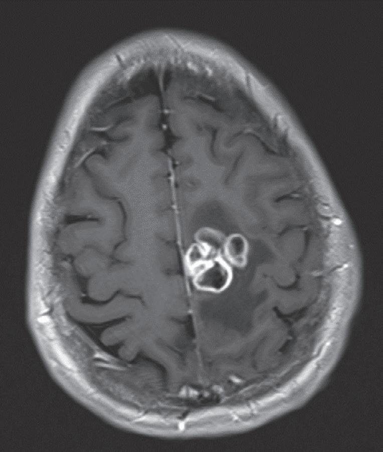
Case. A 69-year-old male presented to the emergency department with new onset seizures and rapidly progressive right sided weakness. Brain MRI with contrast revealed a centrally necrotic, multifocal ring-enhancing lesion in the left posterior frontal lobe (Fig. 1.1).
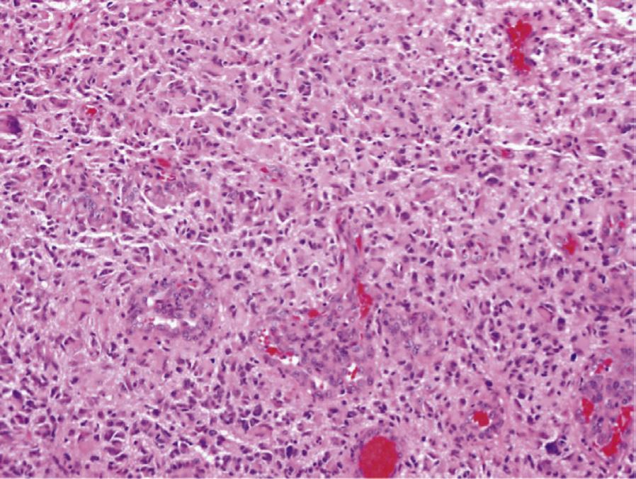
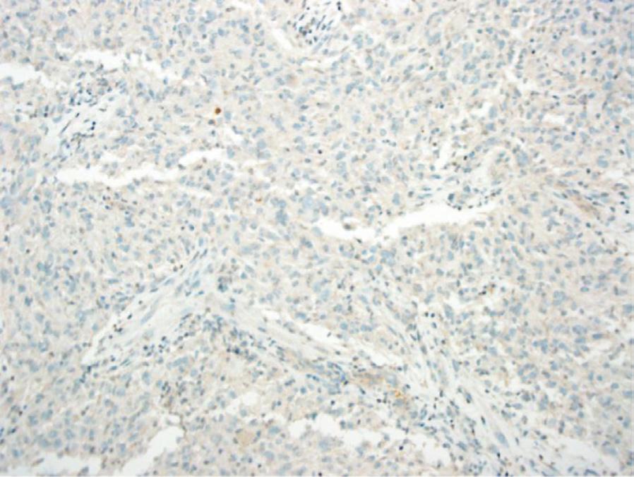
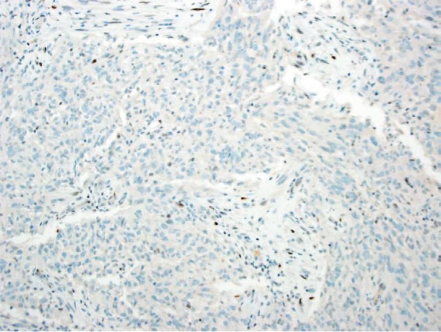
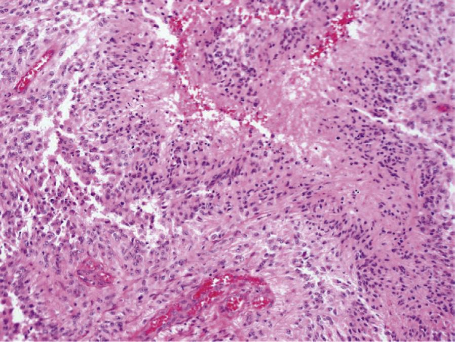
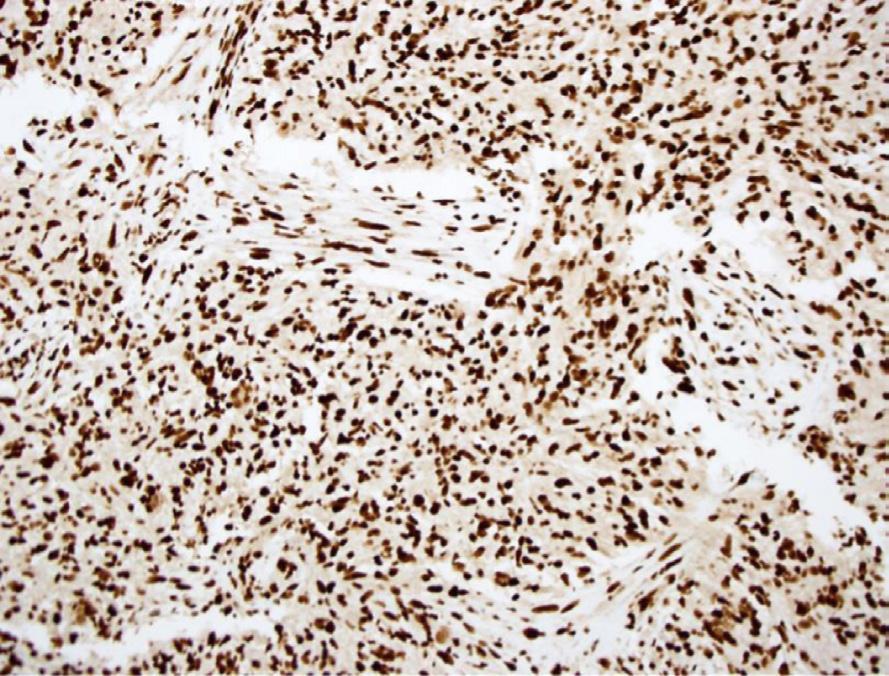
Histology. Microscopic examination of H&E sections (Fig. 1.2 A,B) demonstrates a densely cellular glial neoplasm composed of cells with irregular and hyperchromatic nuclei associated with moderate amounts of eosinophilic cytoplasm. Mitoses are readily identified. Multifocal microvascular proliferation is present, as well as areas of multifocal necrosis including palisading necrosis (Fig. 1.2B, black arrows). Immunohistochemistry reveals positive staining in tumor cells for GFAP (Fig. 1.2F), retained expression of ATRX (Fig. 1.2D), and negative staining for IDH1(R132H) (Fig. 1.2C).
Genomic and molecular analyses. Genomic profiling of tumor DNA by NGS reveals EGFRvIII amplification, TERT promoter mutation and homozygous deletion of CDKN2A/B. Alterations were not detected in the following genes: IDH1, IDH2, BRAF, ATRX, TP53, PDGFRA. Arm-level copy number analysis revealed polysomy of chromosome 7 and monosomy of chromosome 10. There was no evidence of 1p/19q co-deletion. Additionally, MGMT promoter methylation analysis demonstrated no evidence of methylation.
Integrated Diagnosis: GLIOBLASTOMA, WHO Grade IV, IDH1/2 wild-type, EGFRvIII amplified; MGMT promoter unmethylated
Teaching Points. This case illustrates the classic clinical, histologic, and genomic presentation of adult glioblastoma. Patients present with neurologic symptoms, typically nonspecific but which can include new-onset seizures in the fifth to seventh decades of life. Imaging studies reveal important characteristics such as location, size, enhancement, and involvement of adjacent brain tissues. The histologic analysis of the tumor in this case demonstrated characteristic features of GBM including increased cellularity, cells with irregular nuclei, and high-grade features (mitoses, microvascular proliferation, and necrosis). Immunohistochemical stains for GFAP, OLIG2, and SOX2 support a glial lineage. The genomic profile of this tumor also demonstrates the classic alterations associated with adult GBM, notably gains of chromosome 7, single copy loss of chromosome 10, and homozygous deletion of CDKN2A/B. Amplification of EGFR is detected in approximately 40% of adult GBMs and the EGFRvIII variant is seen in half of these cases. The EGFRvIII variant results from deletion of exons 2–7, which generates a constitutively activated, ligand-
A
B
Fig. 1.1 MRI Brain including (A) axial T2-weighted sequence showing a 2.9 × 2.4 cm mass with surrounding edema in the left posterior frontal lobe, and (B) axial T1-weighted gadolinium enhanced sequence showing multiple foci of ring-enhancement with central necrosis concerning for a diagnosis of high-grade glioma.
independent kinase that promotes cell proliferation through the PI3 kinase, RAS, and MAPK signaling pathways. Although no specific therapies have demonstrated utility against EGFRvIII, it is an active area of investigation and serves as a key player of tumor growth. Loss of one copy of chromosome 10 is the most frequently detected genomic alteration in GBM, occurring in 60–80% of cases.2 The tumor suppressor PTEN is located at 10q23.3 and considered a critical candidate gene in gliomagenesis as it is an important negative regulator of the PI3 kinase pathway. TERT promoter mutations are detected in 70–80% of GBMs and are associated with a worse prognosis in the absence of IDH1/2 mutations.
As standard of care, patients diagnosed with GBM are treated with combination chemo- and radiation (adjuvant) therapy (see Chapters 4 and 12 for further discussion of treatment regimens for GBM). Analysis of the MGMT promoter is typically performed to evaluate the tumor’s sensitivity to the alkylating agent temozolomide. O6-methylguanine methyltransferase (MGMT) is a DNA repair enzyme that is associated with resistance to alkylating agents when expressed. Methylation of the MGMT promoter leads to reduced protein expression in tumor cells allowing for accumulation of alkylatinginduced DNA damage that promotes cell death. In this tumor, the absence of MGMT promoter methylation (unmethylated) indicates decreased responsiveness to temozolomide.
In summary, this case highlights the classic pathologic features of adult GBM and illustrates how the genomics can inform the signaling pathways driving gliomagenesis and provide potential therapeutic targets and important prognostic information that can inform clinical decision-making.
Clinical Pearls
1. Microvascular proliferation and palisading necrosis are pathologic hallmarks of glioblastoma and, when present, establish the diagnosis of GBM.
2. IDH1/2 gene mutation is rare in glioblastoma; EGFR amplification and MGMT promoter methylation are observed in approximately 40% of GBMs.
CASE 1.2 USE OF MOLECULAR TESTING
TO DEFINE THE DIAGNOSIS OF WHO GRADE 2, OLIDOENDROGLIOMA
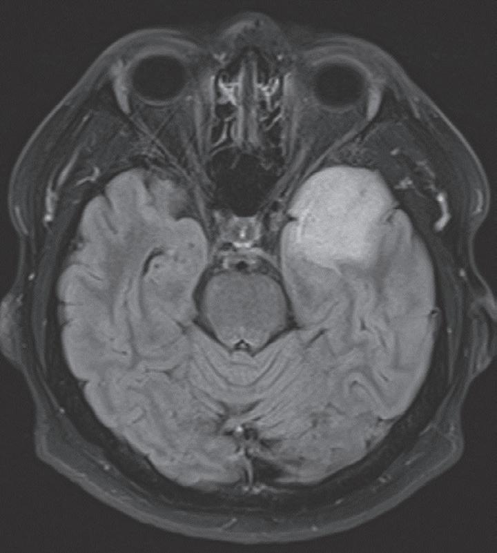
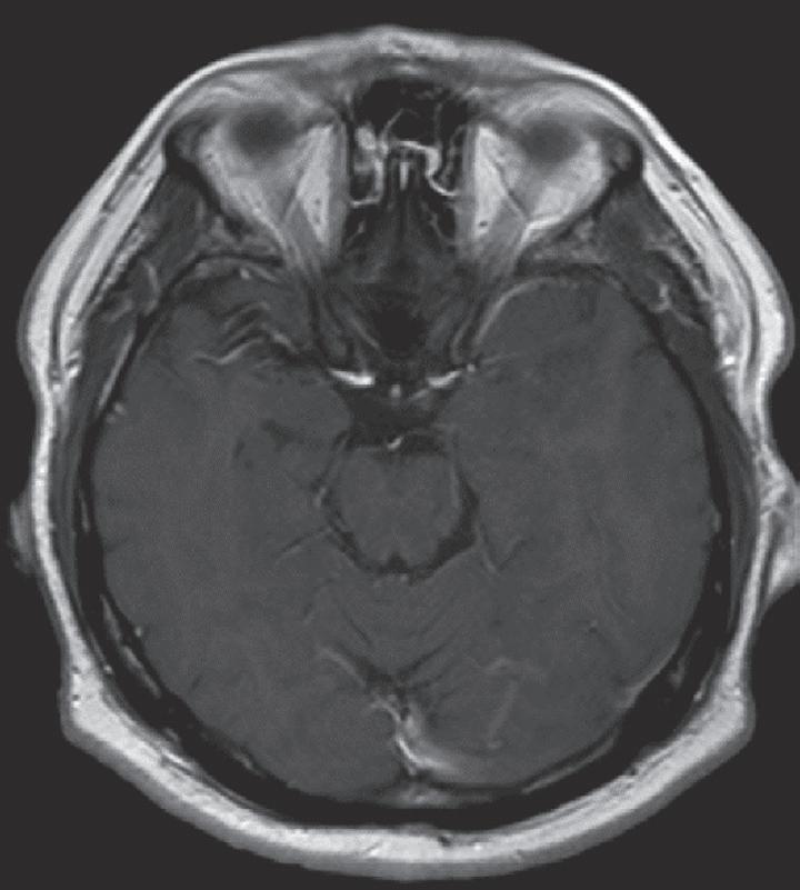
Case. A 39-year-old female presenting with new onset headaches. Brain MRI reveals a nonenhancing temporal lobe lesion (Fig. 1.3).
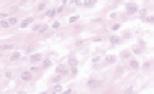
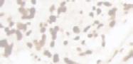
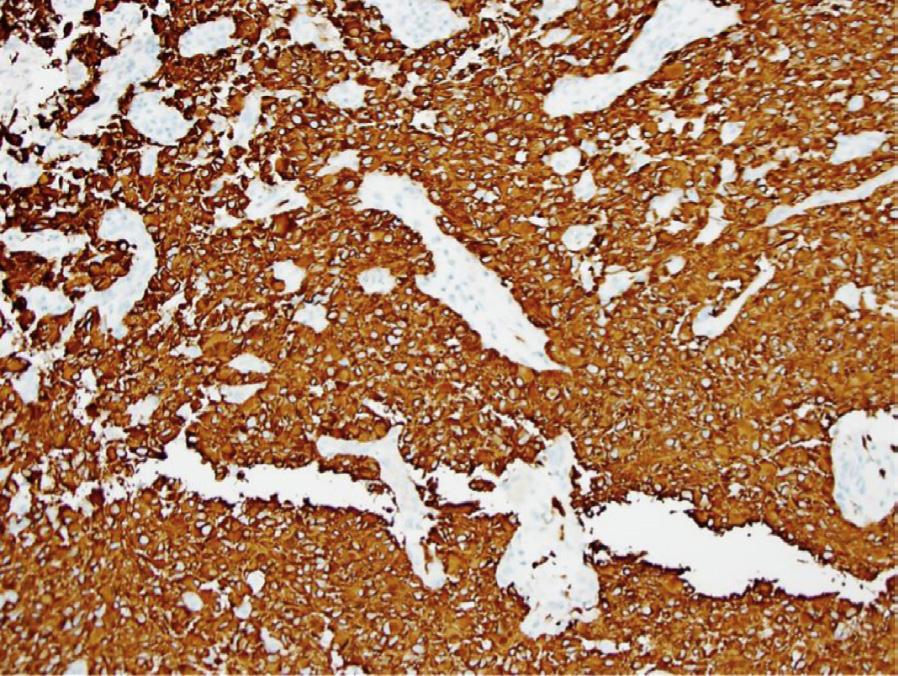
Histology. Microscopic examination of H&E sections (Fig. 1.4A) reveals a moderately cellular glial neoplasm composed of cells with centrally located round nuclei associated with modest amounts of clear cytoplasm. Mitoses are not readily identified and occur in less than 1 per 10 high-power fields (HPFs). Highgrade features including microvascular proliferation and necrosis are not detected. Immunohistochemistry reveals positive staining in tumor cells for IDH1 (R132H) (Fig. 1.4B) and OLIG-2 (Fig. 1.4D), and negative staining for TP53 (image not shown), with low Ki-67 proliferation index (Fig. 1.4E). An immunostain for ATRX shows positive staining in tumor nuclei (Fig. 1.4C).
Fig. 1.2 Microscopic examination showing histologic features consistent with glioblastoma including H&E stained sections (A,B) showing microvascular proliferation (A, black arrowheads), and palisading necrosis (B, black arrows). Immunohistochemistry shows negative staining for IDH1(R132H) (C), retained expression of ATRX (D), negative staining for p53 (E), and positive staining in tumor cells for GFAP (F).
P53
GFAP
IDH1
A
B
Fig. 1.3 MRI Brain including (A) axial fluid attenuation inversion recovery sequence showing a nonenhancing mass with minimal surrounding edema in the left anterior temporal pole, and (B) axial T1-weighted gadolinium enhanced sequence showing no evidence of contrast enhancement.
Genomics and molecular analysis. Genomic profiling of tumor cells reveals IDH1 p.R132H and CIC p.R215W mutations. Alterations were not detected in the following genes: IDH2, BRAF, ATRX, TP53, or EGFR. Arm-level copy number analysis revealed co-deletion of chromosomes 1p/19q without evidence of deletions involving CDKN2A/B MGMT promoter methylation was detected.
Integrated Diagnosis. OLIGODENDROGLIOMA, WHO Grade II, IDH1(R132H)-mutant, 1p/19q co-deleted; MGMT promoter methylated
Teaching Points. This case illustrates the classic histologic and genomic presentation of oligodendroglioma. In contrast to glioblastoma, these tumors typically present at younger ages, with a peak incidence in the fourth decade. Clinical symptoms can vary greatly but often include headaches, seizures, and other signs of increased cranial pressure. Combined CT and MRI studies frequently demonstrate wellcircumscribed lesions often associated with calcifications. Histologic analysis revealed the classic cellular appearance of oligodendroglial lineage tumor cells, with round nuclei surrounded by abundant clear cytoplasm. A low mitotic index and absence of microvascular proliferation and necrosis support classification as a lower-grade glioma. The genomic profile is diagnostic of an oligodendroglial neoplasm as evidenced by 1p/19q co-deletion and presence of an IDH1 (R132H) mutation. In addition to the diagnostic alterations, CIC mutations are typically observed in oligodendrogliomas and rarely present in astrocytic tumors. CIC functions as a transcriptional repressor to counteract activation of genes that are targets of
receptor tyrosine kinase pathways. The absence of an ATRX mutation, and therefore retained ATRX protein expression, also supports an oligodendroglial lineage tumor. In general, oligodendrogliomas are slow-growing tumors and have relatively favorable prognostic indications compared to diffuse astrocytic tumors; however, recurrence and progression to a more malignant neoplasm can occur. The detection of MGMT promoter methylation indicates a more favorable response to alkylating chemotherapy agents.
In summary, this case illustrates how the presence of specific genomic alterations now defines a tumor diagnosis. Classification of oligodendrogliomas now requires presence of 1p/19q co-deletion and an IDH1/2 mutation. Given the overlapping histologic features or limitations of sampling, incorporation of molecular data provides critical information for diagnosis and, importantly, prognosis.
Clinical Pearls
1. When molecular profiling of a brain tumor reveals the presence of 1p/19q co-deletion and IDH1/2 mutation, a diagnosis of oligodendroglioma is made.
2. Histologically, oligodendrogliomas are characterized by the presence of round nuclei and perinuclear halo that has a “fried egg” appearance.
3. Grading of oligodendrogliomas is restricted to grade II and III tumors; there is no grade I or IV oligodendroglioma; oligodendrogliomas with necrosis and microvascular proliferation are grade III.