Activate the eBook version of this title at no additional charge.
Unlock your eBook today.
Expert Consult eBooks give you the power to browse and find content, view enhanced images, share notes and highlights—both online and offline.
1 Visit expertconsult.inkling.com/redeem 2 Scratch off your code 3 Type code into “Enter Code” box 4 Click “Redeem” 5 Log in or Sign up 6 Go to “My Library” It’s that easy!
Scan this QR code to redeem your eBook through your mobile device: Place Peel Off Sticker Here
For technical assistance: email expertconsult.help@elsevier.com call 1-800-401-9962 (inside the US) call +1-314-447-8200 (outside the US)
Use of the current edition of the electronic version of this book (eBook) is subject to the terms of the nontransferable, limited license granted on expertconsult.inkling.com. Access to the eBook is limited to the first individual who redeems the PIN, located on the inside cover of this book, at expertconsult.inkling.com and may not be transferred to another party by resale, lending, or other means. 2015v1.0
NETTER’S 2nd Edition
Surgical Anatomy Review P.R.N.
Robert B. Trelease, PhD Professor
Division of Integrative Anatomy
Department of Pathology and Laboratory Medicine
David Geffen School of Medicine
University of California, Los Angeles
Los Angeles, California
Illustrations by Frank H. Netter, MD
Contributing Illustrators
Carlos A.G. Machado, MD
Kristen Wienandt Marzejon, MS, MFA
Tiffany DaVanzo, MA, CMI
John A. Craig, MD
1600 John F. Kennedy Blvd. Ste 1800 Philadelphia, PA 19103-2899
No part of this book may be produced or transmitted in any form or by any means, electronic or mechanical, including photocopying, recording or any information storage and retrieval system, without permission in writing from the publishers.
Permissions for Netter Art figures may be sought directly from Elsevier’s Health Science Licensing Department in Philadelphia, PA, USA: phone 1-800-523-1649, ext. 3276 or (215) 239-3276; or email H.Licensing@elsevier.com
Notice
Neither the Publisher nor the Author assumes any responsibility for any loss or injury and/or damage to persons or property arising out of or related to any use of the material contained in this book. It is the responsibility of the treating practitioner, relying on independent expertise and knowledge of the patient, to determine the best treatment and method of application for the patient.
Library of Congress Cataloging-in-Publication Data
Names: Trelease, Robert Bernard, author. | Netter, Frank H. (Frank Henry), 1906-1991, illustrator.
Title: Netter’s surgical anatomy review P.R.N. / Robert B. Trelease ; illustrations by Frank H. Netter ; contributing Illustrators, Carlos A.G. Machado, Kristen Wienandt Marzejon, Tiffany DaVanzo, John A. Craig.
Other titles: Netter’s surgical anatomy review pro re nata | Surgical anatomy review PRN
Description: Second edition. | Philadelphia, PA : Elsevier, [2017] | Includes index.
Identifiers: LCCN 2015047555 | ISBN 9780323447270 (pbk.)
Classification: LCC QM531 | NLM WO 517 | DDC 611/.9–dc23 LC record available at http://lccn.loc.gov/2015047555
Content Strategist: Elyse O’Grady
Content Development Specialist: Marybeth Thiel
Publishing Services Manager: Patricia Tannian
Project Manager: Ted Rodgers
Designer: Julia Dummitt
The Publisher Printed in China
Working together to grow libraries in developing countries www.elsevier.com | www.bookaid.org | www.sabre.org
This book is dedicated to My parents, Florence and Robert Trelease (Sr.), who always supported my pursuit of learning and science; My wife, Barbara, and our daughters, Cristin and Heather, who have motivated all my work; My students, who will put their anatomical knowledge to good use in caring for their patients.
This page intentionally left blank
About the Author
Robert B. Trelease, PhD, is Professor in the Division of Integrative Anatomy, Department of Pathology and Laboratory Medicine, in the David Geffen School of Medicine (DGSOM) at UCLA. In 1996, Dr. Trelease became a founding member of and Faculty Advisor to the Instructional Design and Technology Unit (IDTU), part of the DGSOM Dean’s Office established to develop online learning resources for medical education. IDTU currently provides and manages a broad range of web server– and mobile device–based educational resources for all 4 years of the medical school curriculum, as well as developing new multimedia teaching tools and course management applications. Dr. Trelease currently serves as Associate Director of IDTU, in addition to teaching medical gross anatomy, embryology, and neuroanatomy.
This page intentionally left blank
Preface
Netter’s Surgical Anatomy Review P.R.N. is a justin-time, point-of-contact review of anatomy for the most common of the surgically treated diseases and diagnoses encountered during medical student clerkships and general surgery residencies.
This second edition includes new chapters on Heart Diseases and Lungs and Respiratory Diseases, content requested by users of the first edition and its electronic versions. This extends the coverage of material from general surgery into thoracic surgery. There are also new updated Netter Figures contributed by Dr. Carlos Machado, Kristen Wienandt Marzejon, and Tiffany DaVanzo.
I thank the prior readers and institutional adopters for their confidence and support. In particular, special thanks go out to Dr. David Chen, Associate Professor of Clinical Surgery, and the medical students and residents of the David Geffen School of Medicine at UCLA (DGSOM) for their ongoing use of the Web-based version for surgical clerkships and in-service learning.
I am also grateful for the continuing support and good counsel of my Department Chair, Dr. Jonathan Braun, and feedback from former Senior Associate Dean of Medical Education, Dr. LuAnn Wilkerson, who originally suggested that I develop a PDA-based learning resource for surgical clerkships.
Great appreciation is due to my colleagues at DGSOM’s Instructional Design and Technology Unit, directed by Dr. Anju Relan and including master developers Zhen Gu, Katherine Wigan, Sam Payne, and Jason Rock. Their continuing multimedia learning projects and dedicated support of the online medical school curriculum have provided many practical lessons on the complexities of development and what really works in educational technology.
Most of all, I thank my Editor, Elyse O’Grady, for her continuing dedication to the distribution and improvement of Netter’s Surgical Anatomy Review P.R.N. I am especially grateful to Marybeth Thiel, original Development Editor, for providing continuing editorial review and oversight for second edition updates, including all the new artwork. Their expert team at Elsevier worked skillfully to produce the new, redesigned content that you are using.
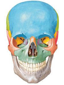
• Base of skull: occipital, sphenoid, temporal, palatine, maxilla bones
• Most of the bones of the skull are flat (type), with inner and outer “tables” (layers) of compact (cortical) bone surrounding trabecular bone and marrow space (diploë).
• Emissary veins connect diploic spaces with cerebral veins/sinuses (intracranial) and scalp and superficial veins: potential route for intracranial spread of infection.
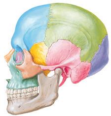
• Sutures
n Thin fibrous joints found only between skull and facial bones
n Produced by intramembranous ossification
n May be indented (e.g., coronal suture), planar, or squamous
• Most cranial and facial bones are pharyngeal arch derivatives.
• Occipital, sphenoid, and ethmoid bones develop from paraxial mesoderm, comparable to vertebrae.
Frontal bone
Nasal bone
Maxilla
Parietal bone
Pterion
Sphenoid bone
Zygomatic bone Greater wing
Temporal bone
Squamous part Zygomatic process
Mandible
Head of condylar process
Ramus
Body
Anterior and Lateral Aspects
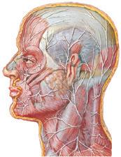
Scalp Layers
• Skin: thin (thicker in occipital region); well supplied with arteries, veins, lymphatic drainage
• Connective tissue: dense subcutaneous layer with rich neurovascular supply
• Aponeurosis of occipitofrontalis muscle, with lateral attachments of temporoparietalis and posterior auricular muscles (collectively the epicranius)
• Loose areolar tissue: allows aponeurosis movement; danger space for infections owing to emissary vein drainage into diploic spaces of cranium
• Pericranium: external periosteum, fibrously fused to sutures
NEUROVASCULAR SUPPLY
Arteries of Face and Cranium
External Carotid (Proximal to Distal)
• Lingual: to tongue and floor of mouth, may have common origin with facial
• Facial: superior, inferior labial, lateral nasal, angular branches; to anteromedial face
• Posterior auricular: posterior to ear and mastoid regions
• Occipital: lateral aspect of head behind ear
• Maxillary: deep auricular, anterior tympanic, deep temporal, middle meningeal, inferior alveolar, posterior alveolar, infraorbital branches; to deep face
• Transverse facial: lateral face, parallel to parotid duct
• Superficial temporal: anterior, lateral aspect of crania
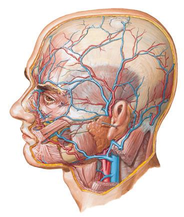
Transverse facial artery and vein
Supraorbital artery and vein
Supratrochlear artery and vein
Angular artery and vein
Facial artery and vein
External carotid artery
Parietal Frontal
Branches of superficial temporal artery and vein
Parietal emissary vein
Common facial vein
Internal jugular vein
Posterior auricular artery and vein
Retromandibular vein
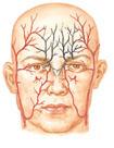
Sources of arterial supply of face
Black: from internal carotid artery (via ophthalmic artery)
Red: from external carotid artery
Superficial Arteries and Veins of Face and Scalp
Internal Carotid
• Anterior cerebral
n Ophthalmic artery: supraorbital, supratrochlear, anterior and posterior ethmoid branches
• Facial: face richly perfused, with anastomoses across midline, anterior to posterior, and between intra- and extracranial branches
• Kiesselbach’s area/plexus: anterior inferior nasal septal region, anastomoses between superior labial (facial), sphenopalatine, palatine (maxillary), and anterior ethmoid (anterior cerebral via ophthalmic) branches; frequent site of epistaxis
Venous Drainage
Internal Jugular Vein
Common Facial Vein
• Tributaries
n Facial: superior, inferior labial, deep facial, external nasal, angular ← orbital, inferior and superior palpebral n Submental
n Retromandibular: superficial temporal, middle temporal, maxillary
• Pterygoid venous plexus of deep face connects with deep facial and maxillary veins and with cavernous sinus via connections through foramen ovale.
• Facial veins have no valves: potential route for spread of infection from face and deep venous
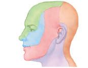
From ophthalmic division of trigeminal nerve (V1)
Supraorbital nerve
Supratrochlear nerve
Palpebral branch of lacrimal nerve
Infratrochlear nerve
External nasal branch of anterior ethmoidal nerve
From mandibular division of trigeminal nerve (V3)
Mental nerve
Buccal nerve
From maxillary division of trigeminal nerve (V2)
Zygomaticofacial nerve
Zygomaticotemporal nerve
Infraorbital nerve
Medial branches of dorsal rami of cervical spinal nerves
Greater occipital nerve (C2)
Auriculotemporal nerve
Trigeminal nerve (V)
Ophthalmic nerve (V1)
Maxillary nerve (V2)
Mandibular nerve (V3)
Branches from cervical plexus
Lesser occipital nerve (C2)
Great auricular nerve (C2, 3)
Dorsal rami cervical spinal nerves
Note: Auricular branch of vagus nerve to external acoustic meatus and small area on posteromedial surface of auricle
Branches from cervical plexus
Cutaneous Nerves of Head and Neck
sinuses to intracranial sinuses (e.g., cavernous sinus via angular and orbital veins)
• Common facial connects to external jugular vein
External Jugular Vein
• Drains posterior auricular
Innervation of the Head and Neck
• Cranial nerve deficits may be associated with specific regional fractures, trauma
• Olfactory (I): special somatic sensory to superior nasal cavity; foramina: cribriform plate of ethmoid; intranasal CSF leakage, anosmia with ethmoid fracture
• Optic (II): foramen–optic canal (sphenoid)
• Oculomotor (III), trochlear (IV): motor to extraocular muscles, travel through cavernous sinus, superior orbital fissure (sphenoid bone), and orbit
• Trigeminal nerve (V): sensory to most of face and head, superficial and deep, including sinuses and supratentorial dura; motor to muscles of mastication, tensor palati, and tensor tympani
n Ophthalmic division: foramen—superior orbital fissure (sphenoid bone)
n Maxillary division: foramen rotundum (sphenoid bone)
n Mandibular division: foramen ovale (sphenoid bone)
• Abducens (VI): runs along clivus and through cavernous sinus and superior orbital fissure to lateral rectus; clival fracture can cause lateral gaze paralysis
• Facial (VII)
n Supplies muscles of facial expression and stapedius
n Carries visceromotor fibers to lacrimal and submandibular and sublingual salivary glands
n Taste afferents for anterior 2/3 of tongue
n Exits stylomastoid foramen (temporal bone)
• Acousticovestibular (vestibuloacoustic, auditory) (VIII): from cochlea and vestibular apparatus (labyrinth) in temporal bone; nerve enters internal acoustic meatus (temporal bone)
• Glossopharyngeal (IX): taste and common sensation from posterior third of tongue and tonsillar fossa; exits jugular foramen (between temporal and occipital bones)
• Vagus (X): motor to palate, pharynx and larynx, thoracoabdominal viscera; exits jugular foramen (between temporal and occipital bones)
• (Spinal) accessory (XI): motor to sternomastoid and trapezius muscles; exits jugular foramen (between temporal and occipital bones)
• Hypoglossal (XII): motor to tongue muscles except for palatoglossus (X); exits hypoglossal canal (anterior supracondylar occipital bone)
• Cervical nerves
n No C1 dermatome exists.
n C2 spinal nerve: sensory to skull, skin from vertex down, infratentorial dura, parotid (auriculotemporal nerve), and infratemporal skin
n C3 spinal nerve: sensory to suboccipital region
CLINICAL CORRELATES
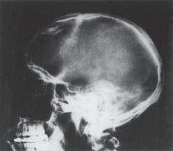
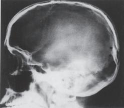
Skull Fractures
Classification
• Linear: fracture line is distinct
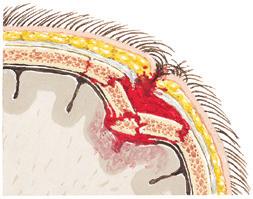
• Comminuted: multiple fragments, may be depressed with compression of dura and brain (image)
Compound depressed skull fracture. Note hair impacted into wound
Compound Depressed Skull Fractures
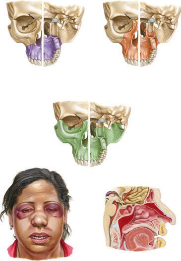
12 Skull and Face Fractures
Le Fort I fracture: horizontal detachment of maxilla at level of nasal floor
Anterior view Posterior view
Fracture line
Le Fort II fracture: fracture through maxillae, antra, nasal bones, and infraorbital rims
Anterior view Posterior view
Fracture line
Free-floating maxillary segment
Anterior view Posterior view
Fracture line
Free-floating maxilla
Edema
Facial asymmetry, especially elongation
Ecchymosis over midface
Malocclusion
Craniofacial dysjunction in Le Fort III fracture distorts facial symmetry
Free-floating maxillary segment
Le Fort III fracture: fracture through zygomatic bones and orbits, separating facial bones from cranial vault
Fracture in cranial vault
CSF leakage
Hematoma and massive edema may occlude nasal airway, necessitating tracheostomy