No part of this publication may be reproduced or transmitted in any form or by any means, electronic or mechanical, including photocopying, recording, or any information storage and retrieval system, without permission in writing from the publisher. Details on how to seek permission, further information about the Publisher’s permissions policies and our arrangements with organizations such as the Copyright Clearance Center and the Copyright Licensing Agency, can be found at our website: www.elsevier.com/permissions.
This book and the individual contributions contained in it are protected under copyright by the Publisher (other than as may be noted herein).
Notices
Knowledge and best practice in this field are constantly changing. As new research and experience broaden our understanding, changes in research methods, professional practices, or medical treatment may become necessary.
Practitioners and researchers must always rely on their own experience and knowledge in evaluating and using any information, methods, compounds, or experiments described herein. In using such information or methods they should be mindful of their own safety and the safety of others, including parties for whom they have a professional responsibility.
With respect to any drug or pharmaceutical products identified, readers are advised to check the most current information provided (i) on procedures featured or (ii) by the manufacturer of each product to be administered, to verify the recommended dose or formula, the method and duration of administration, and contraindications. It is the responsibility of practitioners, relying on their own experience and knowledge of their patients, to make diagnoses, to determine dosages and the best treatment for each individual patient, and to take all appropriate safety precautions.
To the fullest extent of the law, neither the Publisher nor the authors, contributors, or editors, assume any liability for any injury and/or damage to persons or property as a matter of products liability, negligence or otherwise, or from any use or operation of any methods, products, instructions, or ideas contained in the material herein.
Previous editions copyrighted 2010 and 2004.
Library of Congress Cataloging-in-Publication Data
Names: Stouffer, George A., editor. | Runge, Marschall Stevens, 1954- editor. | Patterson, Cam, editor. | Rossi, Joseph S., editor. | Netter, Frank H. (Frank Henry), 1906-1991, illustrator.
Title: Netter’s cardiology / edited by George A. Stouffer, Marschall S. Runge, Cam Patterson, Joseph S. Rossi ; illustrations by Frank H. Netter ; contributing illustrations, Carlos A. G. Machado [and 6 others].
Other titles: Cardiology
Description: 3rd edition. | Philadelphia, PA : Elsevier, [2019] | Includes bibliographical references and index.
Identifiers: LCCN 2018013552 | ISBN 9780323547260 (hardcover : alk. paper)
Classification: LCC RC667 | NLM WG 120 | DDC 616.1/2—dc23 LC record available at https://lccn.loc. gov/2018013552
Content Strategist: Marybeth Thiel
Publishing Services Manager: Catherine Albright Jackson
Senior Project Manager: Claire Kramer
Design Direction: Patrick Ferguson
George A. Stouffer, MD, was born in Indiana, Pennsylvania, and was graduated from Bucknell University and the University of Maryland, School of Medicine. He completed his internal medicine residency, cardiology fellowship, and interventional cardiology fellowship at the University of Virginia. During his cardiology fellowship, he completed a 2-year National Institutes of Health research fellowship in the laboratory of Gary Owens at the University of Virginia. He was on the faculty at the University of Texas Medical Branch from 1995 to 2000, where he became an associate professor and served as Co-Director of Clinical Trials in the Cardiology Division and as Associate Director of the Cardiac Catheterization Laboratory. He joined the faculty at the University of North Carolina in 2000 and currently serves as the Henry A. Foscue Distinguished Professor of Medicine and Chief of Cardiology. Dr. Stouffer’s main focus is clinical cardiology with an emphasis on interventional cardiology, but he is also involved in clinical and basic science research. His basic science research is in the areas of regulation of smooth muscle cell growth, the role of the smooth muscle cytoskeleton in regulating signaling pathways, thrombin generation, and renal artery stenosis.
Marschall S. Runge, MD, PhD, was born in Austin, Texas, and was graduated from Vanderbilt University with a BA in general biology and a PhD in molecular biology. He received his medical degree from the Johns Hopkins School of Medicine and trained in internal medicine at Johns Hopkins Hospital. He was a cardiology fellow and junior faculty member at Massachusetts General Hospital. Dr. Runge’s next position was at Emory University, where he directed the Cardiology Fellowship Training Program. He then moved to the University of Texas Medical Branch in Galveston, where he was Chief of Cardiology and Director of the Sealy Center for Molecular Cardiology. He was at the University of North Carolina from 2000 to 2015, where he served as Charles Addison and Elizabeth Ann Sanders Distinguished Professor of Medicine, Chair of the Department of Medicine, President of UNC Physicians, and Vice Dean for Clinical Affairs. He is currently Dean of the Medical School at the University of Michigan, Executive Vice President for Medical Affairs, and Chief Executive Officer of Michigan Medicine. Dr. Runge is board-certified in internal medicine and cardiovascular diseases and has spoken and published widely on topics in clinical cardiology and vascular medicine.
Cam Patterson, MD, MBA, was born in Mobile, Alabama. He was a Harold Sterling Vanderbilt Scholar and studied psychology and English at Vanderbilt University, graduating summa cum laude. Dr. Patterson attended Emory University School of Medicine, graduating with induction in the Alpha Omega Alpha Honor Society, and completed a residency in Internal Medicine at Emory University Hospitals and Chief Residency at Grady Memorial Hospital. He completed 3 years of research fellowship under the guidance of Edgar Haber at the Harvard School of Public Health, developing an independent research program in vascular biology and angiogenesis that was supported by a National Institutes of Health fellowship. He did a cardiology fellowship and was on the faculty at the University of Texas Medical Branch from 1996 to 2000. Dr. Patterson was at the University of North Carolina at Chapel Hill from 2000 to 2014 where he served as founding director of the UNC McAllister Heart Institute, Chief of Cardiology, and the Ernest and Hazel Craige Distinguished Professor of Cardiovascular Medicine. He received his MBA from the UNC Kenan-Flagler School of Business in 2008. He is an elected member of the American Society of Clinical Investigation and the Association of University Cardiologists. Until recently he was Senior Vice President and Chief Operating Officer at New York Presbyterian–Weill Cornell Medical Center and currently serves as Chancellor of the University of Arkansas for Medical Sciences.
Joseph S. Rossi, MD, was born in Hopedale, Illinois. He completed his undergraduate studies at the University of Illinois and then completed his medical education at the University of Illinois–Chicago, graduating with induction into the Alpha Omega Alpha Honor Society. He completed residency and fellowships in internal medicine, cardiovascular disease, and interventional cardiology at Northwestern University, where he also obtained a master’s degree in clinical investigation. Dr. Rossi is currently the Director of the Cardiac Catheterization Lab at the University of North Carolina. He is actively involved in multiple clinical trials and has received research grants to support his interest in the pharmacogenomics of dual antiplatelet therapy and complex coronary artery revascularization among Medicare beneficiaries. Dr. Rossi is particularly interested in pairing clinical and administrative data to enhance our knowledge of trends and resource utilization for patients with advanced vascular disease.
PREFACE
Our goal for the third edition of Netter’s Cardiology was to provide a concise and practical overview of cardiovascular medicine that has been updated to include new information and important clinical areas that were not well covered in the previous editions or in other cardiology texts. To accomplish this expansion while maintaining a focused text that could be used as a ready reference, we again avoided exhaustive treatment of topics. We also have made every effort to present the essential information in a reader-friendly format that increases the reader’s ability to learn the key facts without getting lost in details that can obfuscate the learning process.
The first two editions of Netter’s Cardiology were an effort to present in a concise and highly visual format the ever-increasing amount of medical information on cardiovascular disease. The challenge that clinicians face in “keeping up” with the medical literature has continued to grow in the 14 years since the first edition of Netter’s Cardiology. This need to process the ever-expanding medical information base and apply new findings to the optimal care of patients is acute in all areas of medicine, but perhaps it is most challenging in disciplines that require practitioners to understand a broad spectrum of evidence-based medicine, such as the field of cardiovascular diseases. The explosion of medical knowledge is also a real educational challenge for learners at all levels—students, residents, and practicing physicians—who must rapidly determine what is and is not important, organize the key information, and then apply these principles effectively in clinical settings.
The third edition includes substantial changes. All the chapters have been updated, there is a new section on Structural Heart Disease, and new chapters have been added on Basic Anatomy and Embryology of the Heart, Stem Cell Therapies for Cardiovascular Disease, Diabetes and Cardiovascular Events, Coronary Hemodynamics and Fractional Flow Reserve, Epidemiology of Heart Failure: Heart Failure with Preserved Ejection Fraction and Heart Failure with Reduced Ejection Fraction, Management of Acute Heart Failure, Cardiac Transplantation and Mechanical Circulatory Support Devices, Cardiovascular Manifestations of Rheumatic Fever, Clinical Presentation of Adults with Congenital Heart Disease, Transcatheter Aortic Valve Replacement, Transcatheter Mitral Valve Repair, Tricuspid and Pulmonic Valve Disease, Deep Vein Thrombosis and Pulmonary Embolism, Cardiac Tumors and Cardio-oncology, and Cardiovascular Disease in the Elderly.
As in the first two editions, the contributing authors have taken advantage of the genius of Frank Netter by carefully selecting the best of his artwork to illustrate the most important clinical concepts covered in each chapter. When Netter artwork was unavailable or difficult to apply to illustrate modern clinical concepts, we again used the great artistic talents of Carlos A. G. Machado, MD, to create new artwork or to skillfully edit and update some of Frank Netter’s drawings. The combination of Dr. Machado’s outstanding skills as a medical artist and his knowledge of the medical concepts being illustrated was an invaluable asset.
As in the first two editions, we chose to use authors from the University of North Carolina School of Medicine at Chapel Hill or those with close ties to the university. This allowed us to select authors who are clinical authorities, many of whom are also well known for their national and international contributions. All have active clinical practices that require daily use of the information covered in their chapters, and all are well aware of the approach to patient management used by their peers at other institutions and in other practice settings. Many of the contributing authors for this edition also contributed to prior editions of this textbook. Each author, whether a previous contributor or not, was given clearly defined guidelines that emphasized the need to distill the large amount of complex information in his or her field and to present it concisely in a carefully prescribed format maintained across all
chapters. The result is a text that is truly clinically useful and less of a compendium than is commonly the case in many medical texts. We believe that the changes we have made in the third edition substantially improve Netter’s Cardiology and ensure that it will continue to be a highly useful resource for all physicians, both generalists and subspecialists, who need to remain current in cardiology—from trainees to experienced practitioners. Whether we have succeeded will obviously be determined by our readers. We welcome the comments, suggestions, and criticisms of readers that will help us improve future editions of this work.
George A. Stouffer, MD
Ernest and Hazel Craige Distinguished Professor of Medicine
Chief, Division of Cardiology
Physician in Chief, UNC Heart and Vascular Service Line University of North Carolina at Chapel Hill Chapel Hill, North Carolina
Marschall S. Runge, MD, PhD
Professor of Internal Medicine
Dean, University of Michigan Medical School
Executive Vice President for Medical Affairs
Chief Executive Officer, Michigan Medicine Ann Arbor, Michigan
Cam Patterson, MD, MBA Chancellor University of Arkansas for Medical Sciences Little Rock, Arkansas
Joseph S. Rossi, MD
Associate Professor of Medicine
Director, Cardiac Catheterization Laboratory
University of North Carolina at Chapel Hill Chapel Hill, North Carolina
Algorithms have been color coded for quick reference.
Algorithm for Evaluating Patients in Whom Renal Artery Stenosis Is Suspected
Clinical findings associated with renal artery stenosis
Noninvasive evaluation
(duplex ultrasonography of renal arteries, magnetic resonance angiography, or computed tomographic angiography)
Nuclear imaging to estimate fractional flow to each kidney
Unilateral renal artery stenosis and asymmetric perfusion present
Follow clinically Treat risk factors
Unilateral renal artery stenosis and symmetric perfusion present
Follow clinically Treat risk factors
Consider revascularization
Bilateral renal artery stenosis present
ABOUT THE ARTISTS
Frank H. Netter, MD
Frank H. Netter was born in 1906 in New York City. He studied art at the Art Student’s League and the National Academy of Design before entering medical school at New York University, where he received his medical degree in 1931. During his student years, Dr. Netter’s notebook sketches attracted the attention of the medical faculty and other physicians, allowing him to augment his income by illustrating articles and textbooks. He continued illustrating as a sideline after establishing a surgical practice in 1933, but he ultimately opted to give up his practice in favor of a full-time commitment to art. After service in the United States Army during World War II, Dr. Netter began his long collaboration with the CIBA Pharmaceutical Company (now Novartis Pharmaceuticals). This 45-year partnership resulted in the production of the extraordinary collection of medical art so familiar to physicians and other medical professionals worldwide.
In 2005, Elsevier, Inc., purchased the Netter Collection and all publications from Icon Learning Systems. There are now more than 50 publications featuring the art of Dr. Netter available through Elsevier, Inc. (www.elsevierhealth.com).
Dr. Netter’s works are among the finest examples of the use of illustration in the teaching of medical concepts. The 13-book Netter Collection of Medical Illustrations, which includes the greater part of the more than 20,000 paintings created by Dr. Netter, became and remains one of the most famous medical works ever published. The Netter Atlas of Human Anatomy, first published in 1989, presents the anatomic paintings from the Netter Collection. Now translated into 16 languages, it is the anatomy atlas of choice among medical and health professions students the world over.
The Netter illustrations are appreciated not only for their aesthetic qualities, but also, more important, for their intellectual content. As Dr. Netter wrote in 1949, “. . . clarification of a subject is the aim and goal of illustration. No matter how beautifully painted, how delicately and subtly rendered a subject may be, it is of little value as a medical illustration if it does not serve to make clear some medical point.” Dr. Netter’s planning, conception, point of view, and approach are what inform his paintings and what makes them so intellectually valuable.
Frank H. Netter, MD, physician and artist, died in 1991.
Learn more about the physician-artist whose work has inspired the Netter Reference collection at https://www.netterimages.com/ artist-frank-h-netter.html.
Carlos A. G. Machado, MD
Carlos Machado was chosen by Novartis to be Dr. Netter’s successor. He continues to be the main artist who contributes to the Netter collection of medical illustrations.
Self-taught in medical illustration, cardiologist Carlos Machado has contributed meticulous updates to some of Dr. Netter’s original plates and has created many paintings of his own in the style of Netter as an extension of the Netter collection. Dr. Machado’s photorealistic expertise and his keen insight into the physician/ patient relationship inform his vivid and unforgettable visual style. His dedication to researching each topic and subject he paints places him among the premier medical illustrators at work today.
Learn more about his background and see more of his art at https://www.netterimages.com/artist-carlos-a-g-machado.html
ACKNOWLEDGMENTS
This third edition of Netter’s Cardiology benefited enormously from the hard work and talent of many dedicated individuals.
First, we thank the contributing authors. All are current or former faculty members at the University of North Carolina School of Medicine, Chapel Hill, or have close ties to the institution. Without their intellect, dedication, and drive for excellence, Netter’s Cardiology, third edition, could not have been published. We had a solid foundation on which to build the third edition, thanks to the hard work of the contributing authors of the second edition, many of whom we were fortunate to have continue on to this edition. We are also grateful for the invaluable editorial contribution that Dr. E. Magnus Ohman made to the first edition.
Special recognition goes to John A. Craig, MD, and Carlos A. G. Machado, MD. They are uniquely talented physician-artists who, through their work, brought to life important concepts in medicine in the new and updated figures included in this text. Marybeth Thiel at Elsevier was invaluable in bringing the third edition to fruition.
We would especially like to acknowledge our families: our wives— Susan Runge, Meg Stouffer, Emma Rossi, and Kristine Patterson— whose constant support, encouragement, and understanding made completion of this text possible; our children—Thomas, Elizabeth, William, John, and Mason Runge; Mark, Jeanie, Joy, and Anna Stouffer; Paul, Samuel, and James Rossi; and Celia, Anna Alyse, and Graham Patterson—who inspire us and remind us that there is life beyond the computer; and, finally, our parents—whose persistence, commitment, and work ethic got us started on this road many, many years ago.
CONTRIBUTORS
EDITORS
Cam Patterson, MD, MBA
Chancellor
University of Arkansas for Medical Sciences Little Rock, Arkansas
Joseph S. Rossi, MD
Associate Professor of Medicine
Director, Cardiac Catheterization Laboratory
University of North Carolina at Chapel Hill Chapel Hill, North Carolina
Marschall S. Runge, MD, PhD
Professor of Internal Medicine
Dean, University of Michigan Medical School
Executive Vice President for Medical Affairs
Chief Executive Officer, Michigan Medicine Ann Arbor, Michigan
George A. Stouffer, MD
Ernest and Hazel Craige Distinguished Professor of Medicine
Chief, Division of Cardiology
Physician in Chief, UNC Heart and Vascular Service Line
University of North Carolina at Chapel Hill
Chapel Hill, North Carolina
AUTHORS
Basil Abu-el-Haija, MD
Clinical Cardiac Electrophysiology
Staff Physician, Kaweah Delta Hospital Visalia, California
Tiffanie Aiken, BS MD Candidate 2019
University of South Carolina School of Medicine Greenville Greenville, South Carolina
Sameer Arora, MD
Cardiovascular Disease Fellow
University of North Carolina at Chapel Hill Chapel Hill, North Carolina
Matthew S. Baker, MD
Assistant Professor of Medicine
Division of Cardiology
University of North Carolina School of Medicine Chapel Hill, North Carolina
Charles Baggett, MD
Cardiologist
The Harbin Clinic Rome, Georgia
Thomas M. Bashore, MD
Professor of Medicine
Senior Vice Chief, Division of Cardiology
Duke University Medical Center Durham, North Carolina
Sharon Ben-Or, MD
Assistant Professor
Department of Surgery
University of South Carolina at Greenville Greenville, South Carolina
Hannah Bensimhon, MD
Cardiology Fellow
Department of Medicine
University of North Carolina at Chapel Hill School of Medicine
Chapel Hill, North Carolina
Christoph Bode, MD, PhD
Chairman of Internal Medicine
Medical Director, Department of Cardiology and Angiology Albert-Ludwigs-Universität Freiburg Freiburg, Germany
Michael Bode, MD
Cardiovascular Disease Fellow Department of Medicine
Division of Cardiology
University of North Carolina at Chapel Hill Chapel Hill, North Carolina
Weeranun D. Bode, MD Assistant Professor Department of Medicine Division of Cardiology University of North Carolina at Chapel Hill Chapel Hill, North Carolina
Mark E. Boulware, MD Interventional Cardiologist University of Colorado Health Heart and Vascular Center Colorado Springs, Colorado
Michael E. Bowdish, MD Director, Mechanical Circulatory Support Assistant Professor of Surgery
Keck School of Medicine of University of Southern California Los Angeles, California
Timothy Brand, MD Cardiothoraic Surgery Resident University of North Carolina Hospitals Chapel Hill, North Carolina
Bruce R. Brodie, MD, FACC Past President, LeBauer Cardiovascular Research Foundation Cone Health Heart and Vascular Center Greensboro, North Carolina
Adam W. Caldwell, MD
Cardiovascular Disease Fellow Division of Cardiology University of North Carolina School of Medicine Chapel Hill, North Carolina
Eric P. Cantey, MD Department of Medicine
Feinberg School of Medicine at Northwestern University Chicago, Illinois
Thomas G. Caranasos, MD
Assistant Professor Department of Surgery
University of North Carolina at Chapel Hill Chapel Hill, North Carolina
Wayne E. Cascio, MD
Director
Environmental Public Health Division
National Health and Environmental Effects Research Laboratory Office of Research and Development
U.S. Environmental Protection Agency Chapel Hill, North Carolina
Matthew A. Cavender, MD, MPH
Assistant Professor of Medicine
Department of Medicine
Division of Cardiology
University of North Carolina at Chapel Hill Chapel Hill, North Carolina
Patricia P. Chang, MD, MHS
Associate Professor of Medicine
Director of Heart Failure and Transplant Program Division of Cardiology
University of North Carolina at Chapel Hill Chapel Hill, North Carolina
Christopher Chien, MD FACC
Clinical Assistant Professor Division of Cardiology
University of North Carolina Chapel Hill, North Carolina
Medical Director, Heart Failure Clinic UNC-Rex Hospital Raleigh, North Carolina
Christopher D. Chiles, MD
Clinical Assistant Professor of Medicine
Texas A&M Health Science Center
Program Director, Cardiovascular Disease Fellowship
Baylor Scott & White Health/Texas A&M Temple, Texas
Eugene H. Chung, MD
Associate Professor
Cardiac Electrophysiology Service Department of Internal Medicine
Michigan Medicine
University of Michigan Ann Arbor, Michigan
David R. Clemmons, MD
Kenan Professor of Medicine Division of Internal Medicine
University of North Carolina School of Medicine
Chapel Hill, North Carolina
Romulo E. Colindres, MD, MSPH, FACP
Clinical Professor of Medicine
Division of Nephrology and Hypertension Department of Medicine
University of North Carolina at Chapel Hill Chapel Hill, North Carolina
Frank L. Conlon, PhD
Professor
Departments of Biology and Genetics
University of North Carolina at Chapel Hill Chapel Hill, North Carolina
Jason Crowner, MD
Assistant Professor of Surgery Division of Vascular Surgery
University of North Carolina School of Medicine
Chapel Hill, North Carolina
Xuming Dai, MD, PhD
Assistant Professor of Medicine
Division of Cardiology
University of North Carolina at Chapel Hill
Chapel Hill, North Carolina
Arjun Deb, MD
Associate Professor Department of Medicine (Cardiology) and Molecular, Cell, and Developmental Biology
Broad Stem Cell Research Center
University of California, Los Angeles Los Angeles, California
Cody S. Deen, MD
Assistant Professor of Medicine
Division of Internal Medicine/Cardiology
University of North Carolina at Chapel Hill Chapel Hill, North Carolina
Gregory J. Dehmer, MD, MACC, MSCAI
Vice President, Medical Director Cardiovascular Services
Baylor Scott & White Health Central Texas Division Temple, Texas
Professor of Medicine
Department of Internal Medicine
Division of Cardiology
Texas A&M College of Medicine Bryan, Texas
John S. Douglas, Jr., MD
Professor of Medicine
Director of Interventional Cardiology Fellowship Program Emory University School of Medicine Atlanta, Georgia
Allison G. Dupont, MD
Interventional Cardiologist
The Heart Center of Northeast George Medical Center Gainesville, Georgia
Fredy H. El Sakr, MD
Fellow in Cardiovascular Medicine
University of Michigan Hospital Ann Arbor, Michigan
Joseph J. Eron, MD
Professor of Medicine
Director, Clinical Core
University of North Carolina Center for AIDS Research Division of Infectious Disease
University of North Carolina School of Medicine
Chapel Hill, North Carolina
Mark A. Farber, MD, FACS
Professor of Radiology and Surgery
Division of Vascular Surgery
Director, Aortic Center
University of North Carolina School of Medicine Chapel Hill, North Carolina
Sunita Juliana Ferns, MD, MRCPCH(UK), FHRS
Assistant Professor of Pediatrics
Director, Pediatric Invasive Electrophysiology
University of North Carolina at Chapel Hill Chapel Hill, North Carolina
Michelle A. Floris-Moore, MD, MS
Associate Professor Department of Medicine
Division of Infectious Diseases
University of North Carolina School of Medicine Chapel Hill, North Carolina
H. James Ford, MD
University of North Carolina
Division of Pulmonary and Critical Care Chapel Hill, North Carolina
Elizabeth Boger Foreman, MD, FAASM
Sleep Medicine Specialist
Sentara Martha Jefferson Medical and Surgical Associates Charlottesville, Virginia
Elman G. Frantz, MD
Professor of Pediatrics Division of Cardiology
University of North Carolina School of Medicine
Director, Pediatric Cardiac Catheterization Laboratory North Carolina Children’s Hospital
Co-Director, Adult Congenital Heart Disease Program
University of North Carolina Heart and Vascular Center Chapel Hill, North Carolina
Anil K. Gehi, MD
Associate Professor of Medicine Director, Clinical Cardiac Electrophysiology Service Division of Cardiology
University of North Carolina School of Medicine Chapel Hill, North Carolina
Leonard S. Gettes, MD Professor Emeritus Department of Medicine
Division of Cardiology
University of North Carolina at Chapel Hill
Chapel Hill, North Carolina
Olivia N. Gilbert, MD
Advanced Heart Failure and Transplant Cardiologist
Novant Health Forsyth Heart and Wellness
Winston-Salem, North Carolina
Allie E. Goins, MD
Department of Medicine
Emory University
Atlanta, Georgia
Anna Griffith, MD
Clinical Fellow
Division of Hematology and Oncology
Department of Internal Medicine
University of North Carolina Hospitals
Chapel Hill, North Carolina
Thomas R. Griggs, MD
Professor Emeritus Medicine, Pathology, and Laboratory Medicine
University of North Carolina School of Medicine Chapel Hill, North Carolina
Benjamin Haithcock, MD
Associate Professor of Surgery and Anesthesiology
University of North Carolina Hospitals Chapel Hill, North Carolina
Eileen M. Handberg, PhD Professor of Medicine
Department of Medicine
University of Florida Gainesville, Florida
Alan L. Hinderliter, MD
Associate Professor of Medicine Division of Cardiology
University of North Carolina at Chapel Hill Chapel Hill, North Carolina
Lucius Howell, MD
Asheville Cardiology Associates/Mission Health Asheville, North Carolina
James P. Hummel, MD
Visiting Associate Professor of Medicine
Division of Cardiovascular Medicine
University of Wisconsin School of Medicine and Public Health Madison, Wisconsin
Thomas S. Ivester, MD, MPH
Professor of Maternal Fetal Medicine
Department of Obstetrics and Gynecology
University of North Carolina School of Medicine
Chief Medical Officer and Vice President for Medical Affairs
UNC Health Care Chapel Hill, North Carolina
Brian C. Jensen, MD
Associate Professor of Medicine and Pharmacology
Department of Medicine
Division of Cardiology
University of North Carolina School of Medicine
UNC McAllister Heart Institute Chapel Hill, North Carolina
Beth L. Jonas, MD
Reeves Foundation Distinguished Professor of Medicine Division of Rheumatology, Allergy, and Immunology
University of North Carolina at Chapel Hill Chapel Hill, North Carolina
Golsa Joodi, MD, MPH
Post-Doctoral Research Fellow Department of Medicine Division of Cardiology
University of North Carolina at Chapel Hill Chapel Hill, North Carolina
Jason N. Katz, MD, MHS Associate Professor of Medicine Department of Internal Medicine University of North Carolina Chapel Hill, North Carolina
Audrey Khoury, BS, AB Medical Student University of North Carolina School of Medicine Chapel Hill, North Carolina
J. Larry Klein, MD Professor of Medicine and Radiology Department of Cardiology and Radiology University of North Carolina at Chapel Hill Chapel Hill, North Carolina
Martyn Knowles, MD, FACS
Adjunct Assistant Professor of Surgery Division of Vascular Surgery
University of North Carolina at Chapel Hill Chapel Hill, North Carolina
David W. Lee, MD Chief Cardiology Fellow Division of Cardiology
University of North Carolina School of Medicine Chapel Hill, North Carolina
Daniel J. Lenihan, MD, FACC Professor of Medicine Director, Cardio-Oncology Center of Excellence Advanced Heart Failure Clinical Research Cardiovascular Division Washington University St. Louis, Missouri
Fong T. Leong, MBChB, PhD, FRCP, FHRS Consultant, Cardiac Electrophysiologist University Hospital of Wales Cardiff, United Kingdom
Gentian Lluri, MD, PhD Assistant Professor Department of Medicine Division of Cardiology University of California, Los Angeles Los Angeles, California
Robert Mendes, MD, FACS
Adjunct Assistant Professor Division of Vascular of Surgery
University of North Carolina School of Medicine Chapel Hill, North Carolina
Phil Mendys, PharmD Co-Director, Lipid and Prevention Clinic Department of Medicine Division of Cardiology University of North Carolina Healthcare Chapel Hill, North Carolina
Venu Menon, MD, FACC, FAHA Director, Cardiac Intensive Care Unit Director, Cardiovascular Fellowship Associate Director, C5 Research Professor of Medicine Cleveland Clinic Lerner College of Medicine Case Western Reserve University Cleveland, Ohio
Michael R. Mill, MD Professor of Surgery and Pediatrics University of North Carolina at Chapel Hill Chapel Hill, North Carolina
Paula Miller, MD
Clinical Associate Professor of Medicine and Cardiology Department of Medicine Division of Cardiology University of North Carolina School of Medicine Chapel Hill, North Carolina
Timothy A. Mixon, MD, FACC, FSCAI Interventional Cardiologist Baylor Scott & White Health Temple, Texas
Associate Professor of Medicine Department of Internal Medicine Division of Cardiology Texas A&M College of Medicine Bryan, Texas
J. Paul Mounsey, PhD, BSc, BM, BCh Chief of Electrophysiology, East Carolina Heart Institute Professor of Medicine Brody School of Medicine East Carolina University Greenville, North Carolina
E. Magnus Ohman, MD, FRCPI Professor of Medicine Associate Director, Duke Heart Center—Cardiology Clinics Director, Program for Advanced Coronary Disease Duke Clinical Research Institute
Duke University Medical Center Durham, North Carolina
Rikin Patel, DO Cardiovascular Disease Fellow
Baylor Scott & White Health/Texas A&M Temple, Texas
Kristine B. Patterson, MD Associate Professor of Medicine Division of Infectious Disease
Columbia University Medical Center New York, New York
Eric D. Pauley, MD
Cardiovascular Disease Fellow
University of North Carolina Hospitals Chapel Hill, North Carolina
Pamela S. Ro, MD
Associate Professor
Department of Pediatrics University of North Carolina at Chapel Hill Chapel Hill, North Carolina
Rachel D. Romero, MD Fellow
Division of Rheumatology, Allergy, and Immunology
University of North Carolina at Chapel Hill Chapel Hill, North Carolina
Lisa J. Rose-Jones, MD
Assistant Professor of Medicine
Division of Cardiology
University of North Carolina at Chapel Hill
UNC Center for Heart and Vascular Care Chapel Hill, North Carolina
Richard S. Schofield, MD Professor of Medicine
Division of Cardiovascular Medicine
University of Florida College of Medicine
Department of Veterans Affairs Medical Center Gainesville, Florida
Kristen A. Sell-Dottin, MD
Assistant Professor University of Louisville Louisville, Kentucky
Jay D. Sengupta, MD
Clinical Cardiac Electrophysiologist Minneapolis Heart Institute at Abbott Northwestern Hospital Minneapolis, Minnesota
Faiq Shaikh, MD
Molecular Imaging Physician Consultant Cellsight Technologies, Inc. San Francisco, California
Arif Sheikh, MD
Associate Professor Department of Radiology Columbia University New York, New York
David S. Sheps, MD, MSPH Professor Department of Epidemiology University of Florida Gainesville, Florida
Brett C. Sheridan, MD San Francisco Cardiology San Francisco, California
Ross J. Simpson, Jr., MD, PhD
Director of the Lipid and Prevention Clinic at University of North Carolina
Professor of Medicine and Adjuvant Professor of Epidemiology
University of North Carolina at Chapel Hill Chapel Hill, North Carolina
Christopher E. Slagle, PhD
Postdoctoral Fellow
Departments of Biology and Genetics
University of North Carolina at Chapel Hill Chapel Hill, North Carolina
Sidney C. Smith, Jr., MD, FAHA, FESC, FACP, MACC Professor of Medicine
Department of Medicine/Division of Cardiology
University of North Carolina at Chapel Hill Chapel Hill, North Carolina
Mark A. Socinski, MD Professor of Medicine
Division of Hematology and Oncology
Multidisciplinary Thoracic Oncology Program
Lineberger Comprehensive Cancer Center University of North Carolina School of Medicine Chapel Hill, North Carolina
Robert D. Stewart, MD, MPH
Staff, Pediatric and Congenital Heart Surgery
Heart and Vascular Institute Cleveland Clinic Cleveland, Ohio
Thomas D. Stuckey, MD, FACC
Medical Director, LeBauer Cardiovascular Research and Education Cone Health Heart and Vascular Center Greensboro, North Carolina
Carla A. Sueta, MD, PhD Professor of Medicine Emerita Division of Cardiology University of North Carolina School of Medicine Chapel Hill, North Carolina
Khola S. Tahir, MD
Cardiovascular Disease Fellow University of North Carolina at Chapel Hill Chapel Hill, North Carolina
Walter A. Tan, MD, MS
Associate Professor of Medicine Director, Cardiac Catheterization Laboratories Wake Forest—Baptist Health Winston-Salem, North Carolina
David A. Tate, MD
Associate Professor of Medicine Emeritus Division of Cardiology University of North Carolina School of Medicine Chapel Hill, North Carolina
Rebecca E. Traub, MD
Assistant Professor Department of Neurology University of North Carolina at Chapel Hill Chapel Hill, North Carolina
Bradley V. Vaughn, MD Professor of Neurology University of North Carolina School of Medicine Chapel Hill, North Carolina
John P. Vavalle, MD, MHS, FACC
Assistant Professor of Medicine Director of Structural Heart Disease
University of North Carolina at Chapel Hill Chapel Hill, North Carolina
Anirudh Vinnakota, MS
Case Western Reserve University School of Medicine Department of Thoracic and Cardiovascular Surgery Cleveland, Ohio
Raven A. Voora, MD
Assistant Professor of Medicine Division of Nephrology
University of North Carolina at Chapel Hill Chapel Hill, North Carolina
Thelsa Thomas Weickert, MD
Assistant Professor Department of Medicine Division of Cardiology
University of North Carolina at Chapel Hill Chapel Hill, North Carolina
Andy Wessels, PhD
Professor and Vice-Chair, Department of Regenerative Medicine and Cell Biology
Co-Director, Cardiovascular Developmental Biology Center
Medical University of South Carolina Charleston, South Carolina
John T. Wilkins, MD, MS
Assistant Professor of Medicine (Cardiology) and Preventive Medicine
Northwestern University Feinberg School of Medicine Chicago, Illinois
Park W. Willis IV, MD
Sarah Graham Kenan Distinguished Professor of Medicine and Pediatrics Emeritus
Director, Cardiac Ultrasound Laboratories
Division of Cardiology
University of North Carolina School of Medicine
Chapel Hill, North Carolina
Eric H. Yang, MD, MBA
Director of Interventional Cardiology and Cardiac Catheterization Laboratories
Department of Cardiovascular Disease
Mayo Clinic Arizona Phoenix, Arizona
Michael Yeung, MD
Assistant Professor of Medicine
University of North Carolina School of Medicine Chapel Hill, North Carolina
Andrew O. Zurick III, MD, MSEd, FACC, FASE
Director of Advanced Cardiovascular Imaging
Cardiovascular Medicine
St. Thomas Heart Nashville, Tennessee
Affiliated Assistant Professor Division of Internal Medicine in the Department of Clinical Medical Education
University of Tennessee Health Science Center, College of Medicine Knoxville, Tennessee
ONLINE CONTENTS
VIDEOS
SECTION VIII: STRUCTURAL HEART DISEASE
Chapter 52 Catheter-Based Therapies for Adult Congenital Heart Disease
Video 52-1 Bedside echo-guided balloon atrial septostomy
Video 52-2 Free pulmonary regurgitation after tetralogy of Fallot repair, stepwise Melody valve implantation after prestenting, and competent Melody valve postimplantation
Video 52-3 Stepwise transcatheter closure of a patent ductus arteriosus with the Amplatzer Duct Occluder
Video 52-4 Stepwise transcatheter closure of a secundum atrial septal defect with the Amplatzer Septal Occluder
Video 52-5 Stepwise transcatheter closure of a patent foramen ovale with an Amplatzer Occluder
3. MANAGING YOUR PERCUTANEOUS CORONARY INTERVENTION
4. MANAGING YOUR CORONARY ARTERY BYPASS SURGERY
5. MANAGING YOUR HYPERTROPHIC CARDIOMYOPATHY
6. MANAGING YOUR SUPRAVENTRICULAR TACHYCARDIA
7. MANAGING YOUR CARDIAC PACEMAKER AND DEFIBRILLATOR
8. MANAGING YOUR AORTIC VALVE DISEASE
9. MANAGING YOUR MITRAL VALVE DISEASE
10. MANAGING YOUR PULMONARY HYPERTENSION AND THROMBOEMBOLIC DISEASE
11. MANAGING YOUR SLEEP DISORDER AND HEART DISEASE
12. MANAGING YOUR EXERCISES AND CARDIOVASCULAR HEALTH
13. MANAGING YOUR ATRIAL FIBRILLATION
14. MANAGING YOUR ATRIAL SEPTAL DEFECT
15. MANAGING YOUR CONGESTIVE HEART FAILURE (CHF)
16. MANAGING YOUR CORONARY ARTERY DISEASE
17. MANAGING YOUR DEEP VEIN THROMBOSIS
18. MANAGING YOUR HEART ATTACK
19. MANAGING YOUR HIGH CHOLESTEROL
20. MANAGING YOUR HIGH BLOOD PRESSURE (HYPERTENSION)
21. MANAGING YOUR MYOCARDITIS
22. MANAGING YOUR RENAL ARTERY STENOSIS
23. MANAGING YOUR VENTRICULAR SEPTAL DEFECT
24. MANAGING YOUR HEART ATTACK: FOR WOMEN
Basic Anatomy and Embryology of the Heart
Frank L. Conlon, Christopher E. Slagle, Andy Wessels
ORIGINS OF CARDIAC PRECURSOR POPULATIONS
Heart development begins as the primary germ layers—ectoderm, mesoderm, and endoderm—are induced and progressively changed to various cell types during the morphogenetic process of gastrulation. Combinatorial networks of intercellular signaling events cooperate with massive tissue migrations and internalizations to lay out the basic body plan of the vertebrate embryo. Mesoderm-derived cardiac precursors are among the first cell populations to internalize, coalescing into 2 bilateral populations toward the anterior end of the embryo between 13 and 15 days of human development. The identity of these progenitor pools as cardiac precursors is defined and maintained by expression of a core cohort of developmental gene regulators or transcription factors. These cardiac transcription factors function cooperatively and hierarchically to induce expression of appropriate structural proteins, including components of the specialized cardiomyocyte contractile apparatus and ion channels. Many cardiac transcription factors function not only in the initial specification of cardiac precursors, but also in later aspects of heart morphogenesis, such as establishing chamber identity, chamber-vessel alignment, and conduction system development. Therefore proper spatial and temporal functions of cardiac transcription factors dictate development of a healthy and functional heart. This requirement of correct genetic regulation is exemplified by the numerous congenital heart defects associated with or caused by mutations in cardiac transcription factors.
Even at such early stages of embryonic development, the cardiac precursor pools have been subdivided into two distinct sources of progenitors according to expression of different subsets of cardiac transcription factors. The first, designated as the first heart field, will form the primitive linear heart tube, which will give rise to the left ventricle and most of the atrial tissues. The second heart field, incorporated into the primitive embryonic heart at various stages of development, contributes to the right ventricle and outflow tract. The developing heart receives further contributions from the cardiac neural crest and the mesothelium. The cardiac neural crest is made of ectodermal cells arising outside the heart fields at the lateral borders of the neural plate and because of neural induction from the midline ectoderm. The cardiac neural crest migrates to the heart-forming region, where it contributes to septation of the outflow tract into the arterial and pulmonary vessels. The mesothelium is the embryonic cell source that gives rise to the epicardium, an epithelium that covers the surface of the heart and that plays a role in a number of processes, such as the development of the coronary system and the formation of the annulus fibrosis.
FORMATION OF THE PRIMITIVE LINEAR HEART TUBE
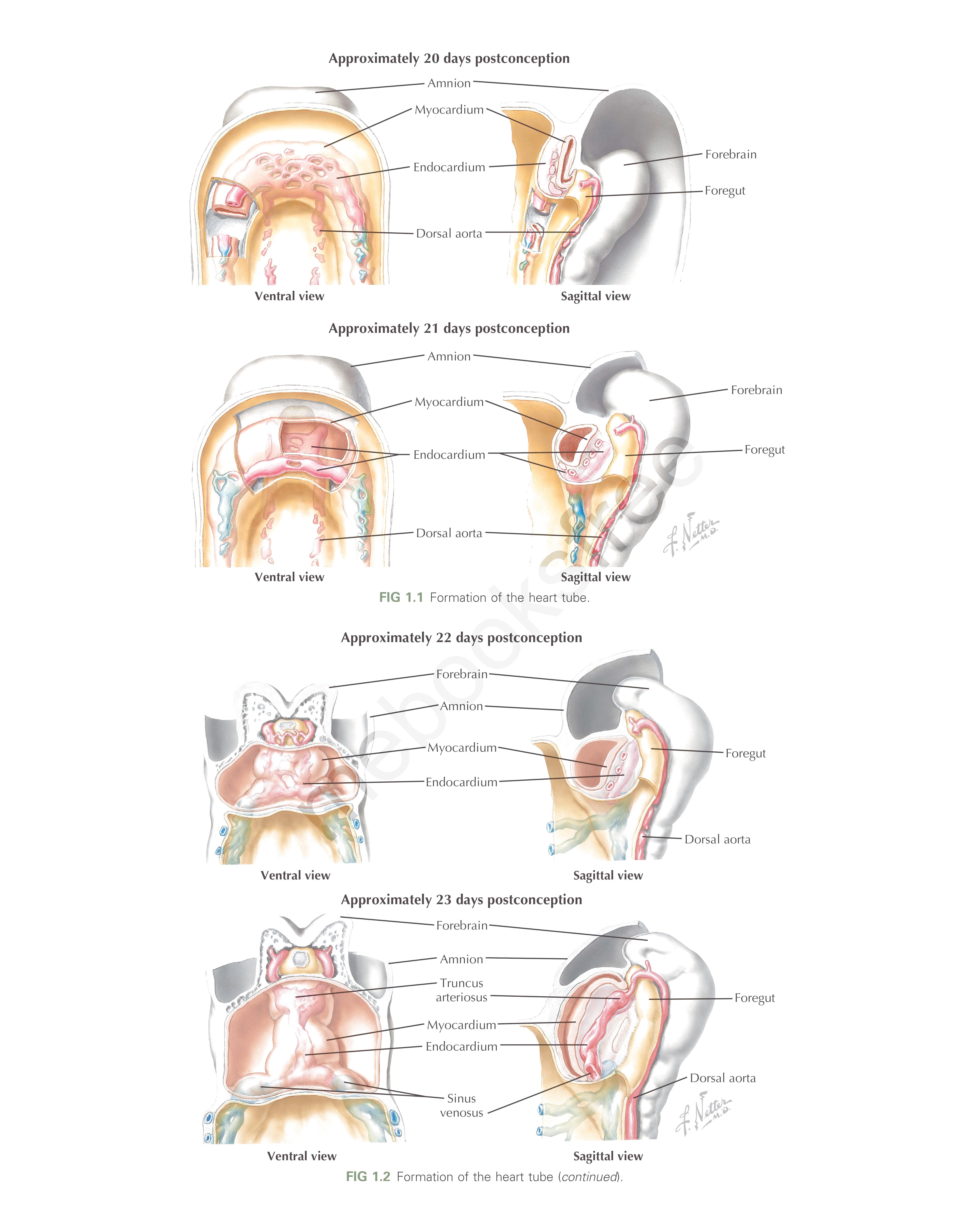
Even before gastrulation has completed, the internalized bilateral cardiac precursor pools continue to migrate in response to signaling cues from neighboring tissues. Remaining as cohesive epithelia, the heart fields
move anteriorly and ventrally between 15 and 20 days of development, fusing at the embryonic midline to form the transient cardiac crescent (Fig. 1.1). Proper midline fusion of the bilateral cardiac primordia is essential for development of the heart. Several cardiac transcription factors are required for this process, and loss of function of any one of them causes extensive defects in further morphogenesis, including cardia bifida in severe cases.
Newly united as the cardiac crescent, the multipotent cardiac progenitors coalesce further to form a linear tube by 3 weeks of development, segregating into the future endocardial lining and myocardial walls (Fig. 1.2). The linear heart tube consists exclusively of differentiated first heart field cells; the second heart field persists as a mesenchymal population, which is a loose association of rapidly dividing precursor cells adjacent to the heart tube. Although no specialized electrical conduction system has yet arisen, the myocardium of the linear heart tube already exhibits autonomous contractions. Compared with those of a mature heart, these contractions are slow and weak, driven only by the intrinsic depolarizing activity and conductivity of the still-maturing cardiomyocytes. Once the conduction system develops and connects to the mature working myocardium, it will serve as an extrinsic regulator of the electrical impulses within the myocardium. Sufficient contractile force will, in turn, allow the heart to beat at the strength required to circulate blood throughout the body.
LOOPING OF THE LINEAR HEART TUBE
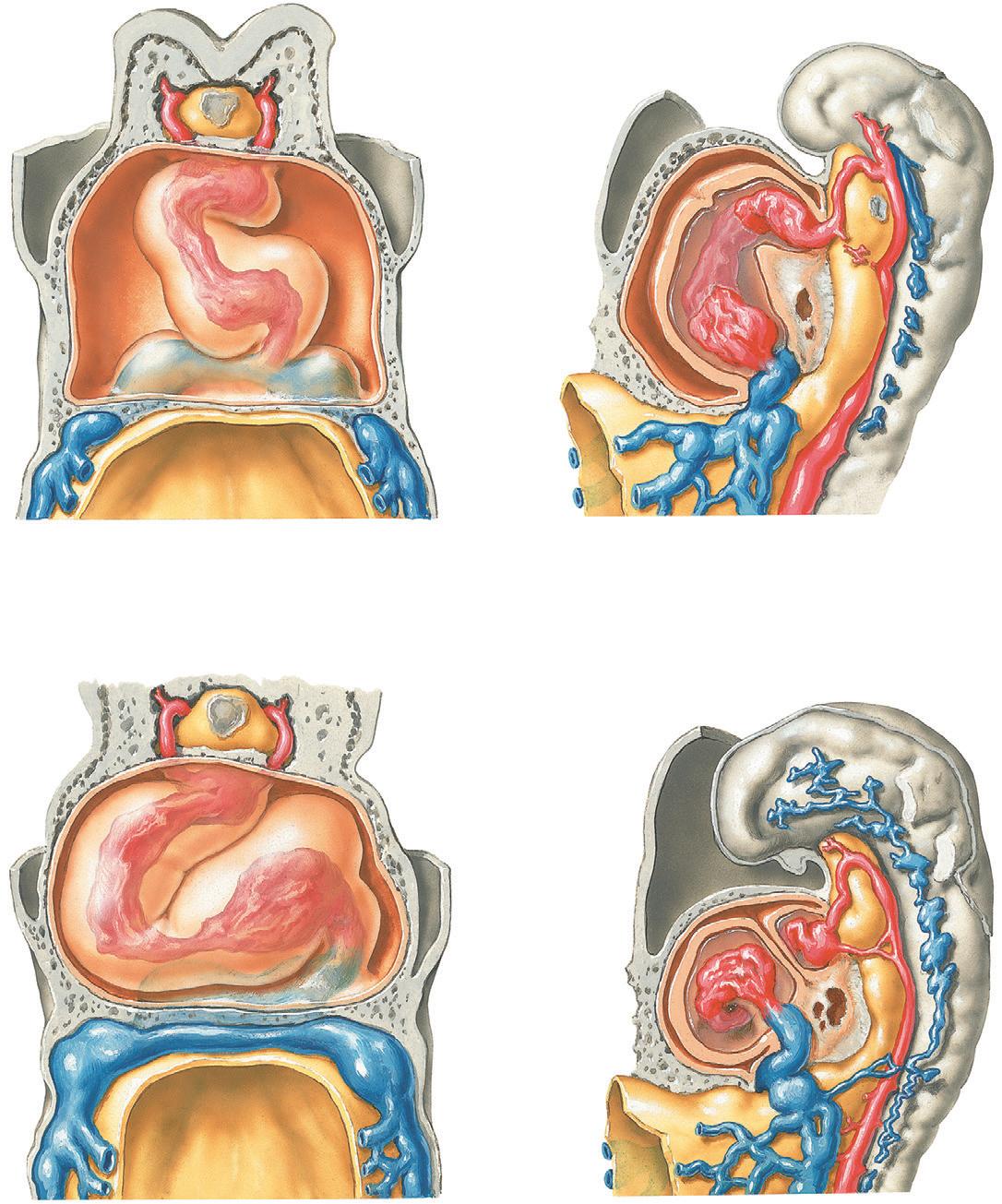
As a consequence of its formation, differentiation, and rudimentary functionality, the linear heart tube is mostly postmitotic. During the fourth week of human gestation, growth and elongation of the linear heart tube occur by means of contribution and division of second heart field cells at both the sinus venosus and truncus arteriosus (posterior and anterior poles, respectively). Concurrently, an embryo-wide genetic program breaks the final axis of symmetry—the left-right axis. Asymmetrical intercellular signaling on the left side of the embryo governs the migration and division of second heart field cells in the lengthening heart tube, leading to two major morphological cardiac asymmetries. First, the entire linear heart tube displaces to the right and rotates 90 degrees about its anterior-posterior axis, so that the original ventral surface of the linear tube is now the left side of a C-shaped tube (Fig. 1.3). Second, asymmetrical mitotic expansion of the second heart field contributions leads to localized “ballooning” of the primitive atrial and ventricular regions of the heart tube, transforming the C-shaped tube into an S-shaped heart (Fig. 1.3).
Further gross morphogenetic movements of the embryo bring the two poles in close apposition, anterior to the primitive chambers. This repositioning prepares the inflow and outflow tracts for appropriate connections to the developing vasculature, thereby contributing to proper segregation of oxygenated and deoxygenated blood flow among the heart, lungs, and body. By 30 days of gestation, the prospective atria
Ventral view
Approximately 20 days postconception
Sagittal view
Approximately 21 days postconception
Ventral view
Sagittal view
1.1 Formation of the heart<-tu?e.
Approximately 22 days postoonception
Ventral view
Sagittal view
Approximately 23 days postconception Myocardium---===------f/J
Amnion
FIG
Ventral view Sinus venosus
Sagittal view
FIG 1.2 Formation of the heart tube (continued).
Foregut
Approximately 23 days postconception
Forebrain
Truncus arteriosus
Myocardium
Endocardium
Sinus venosus
Approximately 24 days postconception
Truncus arteriosus
Primitive ventricle
Primitive atrium
Sinus venosus
are repositioned anterior to the ventricular region, marking the first resemblance of the embryonic heart to its future adult structure.
Formation of the S-looped heart overlaps with the beginnings of ventricular and outflow tract septation and valve development as endocardial cushions emerge within the atrioventricular junction and the outflow tract.
CHAMBER FORMATION
During the time of cardiac looping, at approximately 3 weeks of development, the arterial and venous poles of the heart decrease or cease cell division. At the same time, cardiomyocytes at two distinct locations within the intervening tissue reinitiate cell proliferation. This localized expansion of cardiomyocytes gives rise anteriorly to the atria and posteriorly to the left ventricle, with the area separating the two regions giving rise to the atrioventricular canal. Studies in chickens and mice demonstrated that the atria grow not only through proliferation but also by the recruitment of cells to the venous pole of the heart. The left ventricle and the atria are largely derived from a common pool of progenitors termed the first heart field (Fig. 1.4). In contrast, the second heart field gives rise to the dorsal mesenchymal protrusion and primary atrial septum, which are tissues that are critically important for atrioventricular septation, the outflow tract, and the right ventricle. A conserved role for the second heart field is supported by the observations that abnormalities that affect the expansion of the second heart field are associated with congenital heart disease in mouse models and humans, including atrial and atrioventricular defects, as well as outflow tract abnormalities.
Foregut
Dorsal aorta
Foregut
Dorsal aorta
Contribution of cells from the second heart field to the heart is complete by the fifth week of human development. At this stage, chamber identity can be established by inspecting anatomic features and/or by the expression of left or right ventricular chamber-specific genes. As the cardiovascular system develops to support postnatal systemic and pulmonary circulations, the heart goes through a series of complex remodeling events. Critical steps in this process are the formation of the septa between individual components of the heart, with the purpose of separating the respective blood flows within the heart, and the formation of valves facilitating unidirectional flow among the respective components. Together, these two events are commonly referred to as valvuloseptal morphogenesis.
SEPTATION
Atrial septation is initiated when the second heart field–derived dorsal mesenchymal protrusion and the myocardial primary atrial septum (or septum primum) extend ventrally into the, yet undivided, common atrium. In the mouse, this process takes place between embryonic day (ED) 9.5 to 10.5; in humans the process occurs around day 30. The space between the leading edge of the atrial septum and the fusing atrioventricular cushions in the atrioventricular canal is the primary atrial foramen. As the primary atrial septum grows toward the mesenchymal atrioventricular cushions, thereby closing the primary interatrial foramen, perforations appear in the upper part of the primary atrial septum. These perforations will eventually coalesce and form the secondary interatrial foramen. As this part of atrial septation process nears completion, the secondary atrial septum (or septum secundum) appears
Amnion
Ventral view
Sagittal view
Ventral view
Sagittal view
FIG 1.3 Formation of the heart loop.
Ascending aorta Pulmonary trunk
Aortic vestibule of left ventricle
Conus arteriosus of right ventricle
Trabecular walls of left and right ventricles
Auricles/pectinate muscle walls of left and right atria (smooth wall of left atrium from pulmonary veins)
Coronary
Smooth
in the space between the primary atrial septum and the left venous valve in the roof of the right atrium. Eventually, the upper part of the primary atrial septum will fuse with the secondary atrial septum, thereby closing off the secondary atrial foramen and completing the atrial septation process. Failure of fusion of the two atrial septa will lead to the congenital defect patent foramen ovale.
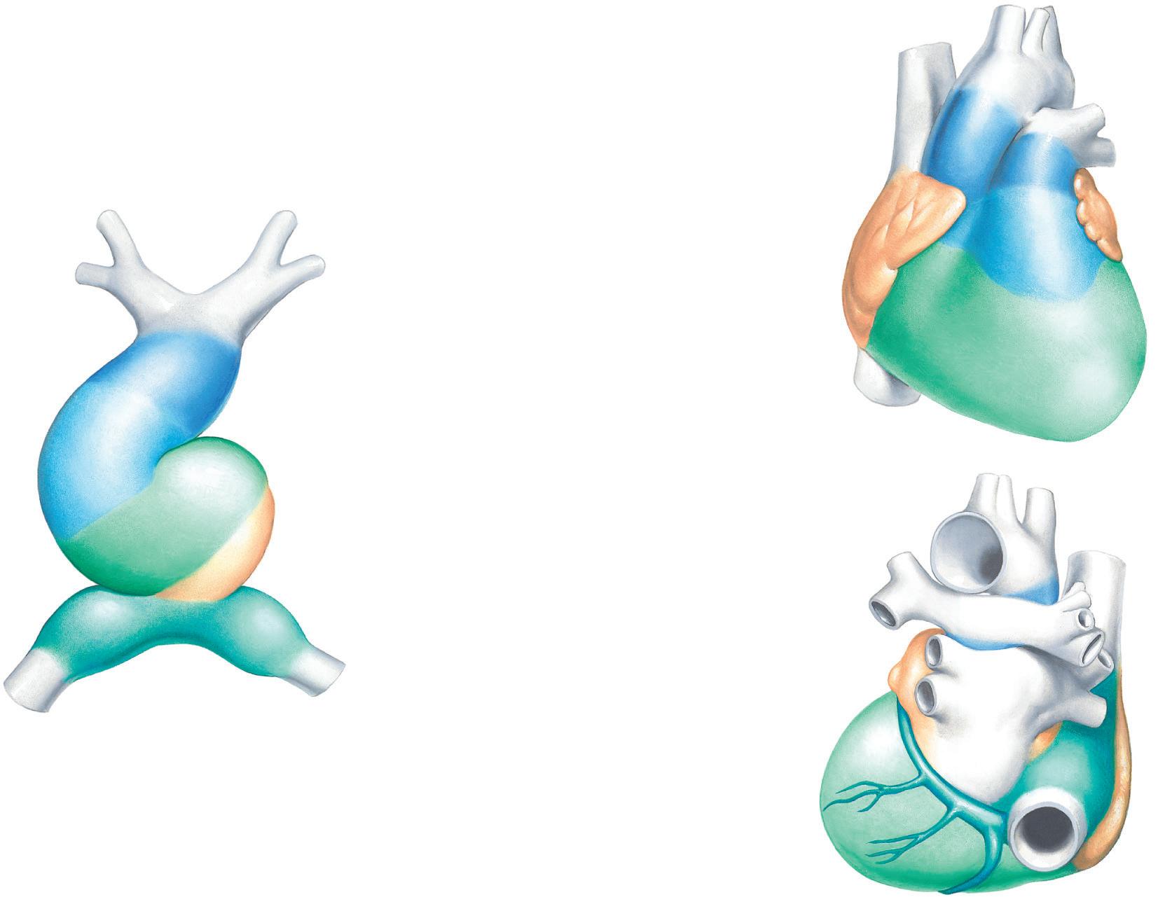
Compared with atrial septation, the creation of the ventricular septum is a rather straightforward process. As the tubular heart expands, undergoes looping, and remodels, distinctive left and right ventricular components appear. During this process, a myocardial ridge, the interventricular septum, emerges between the left and right ventricle. Subsequent outward expansion of the ventricles, a process sometimes referred to as “ballooning,” in combination with upward growth of the interventricular septum and eventual fusion of crest of the septum with the atrioventricular cushions, completes the process of ventricular septation. Cell lineage tracing experiments in the mouse demonstrated that, like the right ventricle, the interventricular septum is largely derived from the second heart field.
The third septal structure that is required for separating the respective blood flows in the heart is found in the outflow tract. After completion of cardiac looping, a single outflow tract can be found connected to the right ventricular component of the yet unseptated heart. Septation of this outflow tract is required for the formation of an aorta, which eventually connects to the left ventricle, and a pulmonary trunk that comes from the right ventricle. Two sets of endocardial ridges are located within the outflow tract, and as a result of their fusion, these will separate the common outflow tract into an aorta and a pulmonary trunk. Failure of fusion can lead to congenital defects, including a double outlet right
ventricle. The cardiac neural crest is also important in the septation process that separates aorta and pulmonary trunk. Abnormal development of the cardiac neural crest specifically affects the formation of the aorticopulmonary septum downstream of the semilunar valves ( Fig. 1.5). This can result in the congenital defect common arterial trunk (or truncus arteriosus) or in aorticopulmonary window.
FORMATION OF THE CARDIAC VALVES
The fully formed heart contains two sets of one-way valves. In the atrioventricular junction, the atrioventricular valves facilitate unidirectional flow through the left and right atrioventricular orifices, whereas at the ventriculoarterial junction, the semilunar valves serve the same function at the junction of the left ventricle and the aorta, and at the junction of the right ventricle and the pulmonary trunk.
Atrioventricular valve formation is initiated at the atrioventricular junction of the looping heart (see previous description); two atrioventricular cushions appear as a result of local accumulation of extracellular matrix between the atrioventricular endocardium and myocardium. A process of endothelial-to-mesenchymal transformation leads to the generation of a population of mesenchymal cells that colonize the cushions. As the heart grows, and these major atrioventricular cushions become bigger, they eventually fuse, thereby separating the common atrioventricular junction into the left and right atrioventricular orifices. As this process takes place, on the lateral walls of these respective orifices, two additional atrioventricular cushions form. These are known as the left and right lateral atrioventricular cushions. These lateral cushions also become populated with endocardially derived mesenchyme. Recent
Aortic arches (AA)
Heart tube derivatives
Heart tube primordia
Adult heart, anterior view
Adult heart, posterior view
Truncus arteriosus (TA)
Bulbus cordis (BC)
Ventricle (V)
Atrium (A)
Sinus venosus (SV)
sinus
wall of right atrium
FIG 1.4 Summary of heart tube derivatives.
Ectoderm
Neural crest
Fused neural folds
1st occipital somite
1st cervical somite
1st thoracic somite
Caudal neuropore
Ectoderm
Neural crest
Neural tube (spinal cord)
Notochord
Level of section
Neural tube
Sulcus limitans
Embryo at 24 days (dorsal view)
Embryo at 4 weeks (transverse view)
FIG 1.5 Nervous tissue of embryo at 24 days and 4 weeks.
cell fate studies in mice showed that epicardially derived cells migrate into these lateral cushions. Epicardially derived cells do not populate the major cushions. Further remodeling of the cushion-derived tissues eventually leads to the formation of the mitral valve leaflets in the left atrioventricular orifice and the tricuspid valve leaflets in the right atrioventricular orifice.
In many respects, the development of the semilunar valves is similar to that of the atrioventricular valves. It involves the fusion of two mesenchymal tissues, the parietal and septal endocardial ridges, which result in the separation of the left and right ventricular outflows. The emergence of a set of smaller endocardial ridges, the intercalated ridges at the opposite sides of the formed septum, resembles the process of formation of the lateral cushions in the atrioventricular junction. The remodeling of these two sets of mesenchymal ridges will eventually lead to the formation of the semilunar valves.
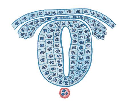
CARDIAC NEURAL CREST
The neural crest is a transient population of cells that form from the dorsal ectoderm at the time of neural tube closure (Fig. 1.5). The neural crest population arises through a series of inductive interactions with surrounding tissues around the fourth week of development. Once formed, the cells undergo an epithelial-to-mesenchymal transition, migrating ventrally and laterally to contribute to a wide array of tissue types, including the epinephrine-producing cells of the adrenal gland, the parasympathetic neurons, cartilage, bone, connective tissue, and pigment cells. The neural crests themselves are multipotential at the
time of their formation; their ultimate fate is a reflection of their relative position along the anterior-to-posterior axis of the embryo. In the cranial portions of the embryo, classic fate mapping studies showed that a subpopulation of neural crest cells enter the arterial pole or the venous pole of the heart to give rise to all of the parasympathetic innervation of the heart, the smooth muscle layer of the great vessels, and portions of the outflow tract. This population is termed the cardiac neural crest. Ablation studies in chicks and genetic studies in mammals demonstrated not only that the cardiac neural crest cells contribute to these regions of the heart but also that they are also essential for the proper formation of each of these structures.
EPICARDIUM AND EPICARDIALLY DERIVED CELLS
The walls of the developed heart essentially consist of three cell layers: the endocardium, the myocardium, and the epicardium. The endocardium and myocardium are generated early in development during the formation of the primitive linear heart tube (see previous description). However, the epicardium, a layer of epithelial cells covering the heart, is like the cardiac neural crest, a late addition to the developing heart. The source of the epicardium is the proepicardium, a local proliferation of the mesothelium found in association with the sinus venosus at the venous pole. In the mouse, the proepicardium can be seen around ED 9.5; in humans, this happens around day 30. Shortly after its generation, the proepicardium attaches to the myocardial surface in the atrioventricular junction. From there, the cells spread out as an epicardial sheet and eventually cover nearly the entire heart. Cell fate studies in animal
models indicated that the epicardial-like cells covering the distal part of the outflow tract are not derived from the proepicardium proper but instead come from the pericardial mesothelium associated with the aortic sac.
After formation of the epicardium, epithelial-to-mesenchymal transformation of a subpopulation of epicardial cells leads to the formation of epicardially derived cells that migrate into the space between the epicardium and the myocardium. This process is most pronounced at the junction between the atria and ventricles, where it leads to the formation of the atrioventricular sulcus. Furthermore, cell fate studies in animal model systems demonstrated that epicardially derived cells also migrate into the ventricular myocardial walls where they differentiate into interstitial fibroblasts and coronary smooth muscle cells. In addition, these animal studies also revealed that epicardially derived cells contribute significantly to the leaflets of the atrioventricular valves that are derived from the lateral atrioventricular cushions.
ADDITIONAL RESOURCES
Bruneau BG. Signaling and transcriptional networks in heart development and regeneration. Cold Spring Harb Perspect Biol. 2013;5:a008292.
A comprehensive review of primary literature on the genetic and molecular underpinnings of cardiac morphogenesis.
De la Cruz M, Markwald RR, eds. Living Morphogenesis of the Heart Birkhauser.
An excellent detailed summary of the original studies in cardiac development. Kirby ML. Cardiac Development. Oxford University Press; 2007.
An outstanding comprehensive text on vertebrate heart development.
Männer J. Cardiac looping in the chick embryo: a morphological review with special reference to terminological and biomechanical aspects of the looping process. Anat Record. 2000;259:248–262.
An in-depth review of primary literature documenting looping of the linear heart tube, illustrated with electron micrographs of the morphological process in chicken embryos.
Rana MS, Christoffels VM, Moorman AFM. A molecular and genetic outline of cardiac morphogenesis. Acta Physiol. 2013;207:588–615.
A synthesis of literature detailing contributions of various cardiac progenitor sources to development of the mature vertebrate heart.
The History and Physical Examination
Marschall S. Runge, Fredy H. El Sakr, E. Magnus Ohman, George A. Stouffer
The ability to determine whether disease is present or absent—and how that patient should be treated—is the ultimate goal for clinicians who evaluate patients with suspected heart disease. Despite the number of diagnostic tests available, the importance of a careful history and physical examination has never been greater. Opportunities for errors in judgment are abundant, and screening patients for coronary risk using a broad and unfocused panel of laboratory and noninvasive tests can lead to incorrect diagnoses and unnecessary testing. Selection of the most appropriate test and therapeutic approach for each patient is based on a skillfully performed history and physical examination. Furthermore, interpretation of any test results is based on the previous probability of disease, which again is based on the history and physical. Although entire texts have been written on cardiac history and physical examination, this chapter specifically focuses on features of the cardiac history and the cardiovascular physical examination that help discern the presence or absence of heart disease.
THE CONCEPT OF PREVIOUS PROBABILITY
The history and physical examination should allow the clinician to establish the previous probability of heart disease, that is, the likelihood that the symptoms reported by the patient result from heart disease. A reasonable goal is to establish the risk of heart disease in a patient as “low,” “intermediate,” or “high.” One demonstration of this principle in clinical medicine is the assessment of patients with chest pain, in which exercise stress testing to accurately diagnose coronary heart disease (CHD) depends on the previous probability of disease. In patients with a low risk of CHD based on clinical findings, exercise stress testing results in a large number of false-positive test results. Because up to 15% of exercise stress tests produce positive results in individuals without CHD, use of this test in a low-risk population can result in an adverse ratio of false-positive to true-positive test results and unnecessary cardiac catheterizations. Conversely, in patients with a high risk of CHD based on clinical findings, exercise stress testing can result in false-negative test results, which is an equally undesirable outcome, because patients with significant coronary artery disease (CAD) and their physicians may be falsely reassured that no further evaluation or treatment is necessary.
Emphasis is increasing on quantifying previous probability to an even greater degree with various mathematical models. This is a useful approach in teaching and may be clinically feasible in some diseases. However, for most patients with suspected heart disease, categorizing risk as low, intermediate, and high is appropriate, reproducible, and feasible in a busy clinical practice. Therefore obtaining the history and physical examination represents a key step before any testing, to minimize use of inappropriate diagnostic procedures.
THE HISTORY
A wealth of information is available to clinicians who carefully assess the history of the patient. Key components are assessment of the chief complaint; careful questioning for related, often subtle symptoms that may further define the chief complaint; and determination of other factors that help categorize the likelihood of disease. Major symptoms of heart patients include chest discomfort, dyspnea, palpitations, and syncope or presyncope.
Chest Discomfort
Determining whether chest discomfort results from a cardiac cause is often a challenge. The most common cause of chest discomfort is myocardial ischemia, which produces angina pectoris. Many causes of angina exist, and the differential diagnosis for chest discomfort is extensive (Box 2.1). Angina that is reproducible and constant in frequency and severity is often referred to as stable angina. For the purposes of this chapter, stable angina is a condition that occurs when CAD is present, and coronary blood flow cannot be increased to accommodate for increased myocardial demand. However, as discussed in Chapters 12 through 14, there are many causes of myocardial ischemia, including fixed coronary artery stenoses and endothelial dysfunction, which lead to reduced vasodilatory capacity.
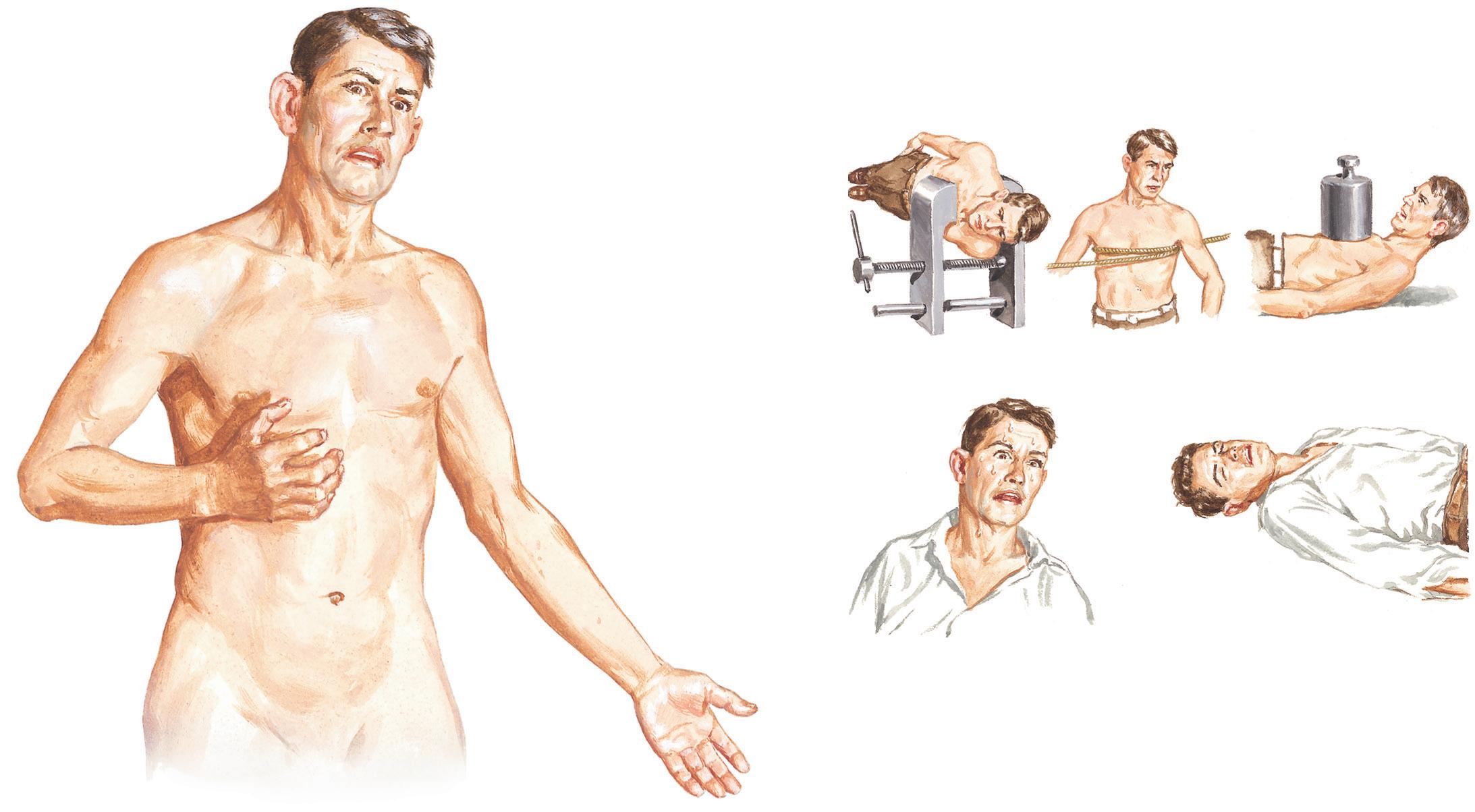
A description of chest discomfort can help establish whether the pain is angina or of another origin. First, characterization of the quality and location of the discomfort is essential (Fig. 2.1). Chest discomfort because of myocardial ischemia may be described as pain, a tightness, a heaviness, or simply an uncomfortable and difficult-to-describe feeling. The discomfort can be localized to the mid-chest or epigastric area, or may be characterized as pain in related areas, including the left arm, both arms, the jaw, or the back. The radiation of chest discomfort to any of these areas increases the likelihood of the discomfort being angina. Second, the duration of discomfort is important because chest discomfort due to cardiac causes generally lasts minutes. Therefore pain of short duration (“seconds” or “moments”), regardless of how typical it may be of angina, is less likely to be of cardiac origin. Likewise, pain that lasts for hours, on many occasions, in the absence of objective evidence of myocardial infarction (MI), is not likely to be of coronary origin. Third, the presence of accompanying symptoms should be considered. Chest discomfort may be accompanied by other symptoms (including dyspnea, diaphoresis, or nausea), any of which increase the likelihood that the pain is cardiac in origin. However, the presence of accompanying symptoms is not needed to define the discomfort as angina. Fourth, factors that precipitate or relieve the discomfort should be evaluated. Angina typically occurs during physical exertion, during emotional stress, or in other circumstances of increased myocardial
Abstract
This chapter reviews the role of the physical examination in cardiovascular medicine, as well as typical findings in common cardiovascular pathologies.
• Degenerative joint disease of the cervical or thoracic spine
• Costochondritis
• Herpes zoster
Psychogenic
• Anxiety
• Depression
• Cardiac psychosis
• Self-gain
oxygen demand. When exercise precipitates chest discomfort, relief after cessation of exercise substantiates the diagnosis of angina. Sublingual nitroglycerin also relieves angina, generally over a period of minutes. Instant relief or relief after longer periods lessens the likelihood that the chest discomfort was angina.
Although the presence of symptoms during exertion is important in assessing CHD risk, individuals, especially sedentary ones, may have angina-like symptoms that are not related to exertion. These include postprandial and nocturnal angina, or angina that occurs while the individual is at rest. As described herein, “rest-induced angina,” or the new onset of angina, connotes a pathophysiology different from effortinduced angina. Angina can also occur in persons with fixed CAD and
BOX 2.2 Conditions That Cause Increased Myocardial Oxygen Demand
• Hyperthyroidism
• Tachycardia of various etiologies
• Hypertension
• Pulmonary embolism
• Pregnancy
• Psychogenic
• Central nervous system stimulants
• Exercise
• Psychological stress
• Fever
increased myocardial oxygen demand due to anemia, hyperthyroidism, or similar conditions (Box 2.2). Angina occurring at rest, or with minimal exertion, may denote a different pathophysiology, one that involves platelet aggregation, which is clinically termed “unstable angina” or “acute coronary syndrome” (see Chapters 20 and 21).
Patients with heart disease need not present with chest pain at all. Anginal equivalents include dyspnea during exertion, abdominal discomfort, fatigue, or decreased exercise tolerance. Clinicians must be alert to and specifically ask about these symptoms. Often, a patient’s family member or spouse notices subtle changes in the endurance of the patient or that the individual no longer performs functions that require substantial physical effort. Sometimes, patients may be unable to exert themselves due to comorbidities. For instance, the symptoms of myocardial ischemia may be absent in patients with severe peripheral vascular disease who have limiting claudication. One should also be attuned to subtle or absent symptoms in individuals with diabetes mellitus (including type 1 and type 2 diabetes), which is a coronary risk equivalent as defined by the Framingham Risk Calculator.
When the likelihood that CHD accounts for a patient presenting with chest discomfort or any of the aforementioned variants is considered, assessment of the cardiac risk factor profile is important. The Framingham Study first codified the concept of cardiac risk factors, and over time, quantification of risk using these factors has become an increasingly useful tool in clinical medicine. Cardiac risk factors determined by the Framingham Study include a history of cigarette smoking, diabetes mellitus, hypertension, or hypercholesterolemia; a family history of CHD (including MI, sudden cardiac death, and first-degree relatives having undergone coronary revascularization); age; and sex (male). Although an attempt has been made to rank these risk factors, all are important, with a history of diabetes mellitus being perhaps the single most important factor. Subsequently, a much longer list of potential predictors of cardiac risk has been made (Box 2.3). Multiple risk calculators have since been created, such as the atherosclerotic cardiovascular disease algorithm used by the American College of Cardiology, the American Heart Association cholesterol guidelines, and the Multi-Ethnic Study of Atherosclerosis (MESA), which uses classic risk factors with the addition of a coronary artery calcium score to predict a 10-year risk of CHD.
Symptoms suggestive of vascular disease require special attention. Peripheral vascular disease may mask CHD because the individual may not be able to exercise sufficiently to provoke angina. A history of stroke, transient ischemic attack, or atheroembolism in any vascular distribution is usually evidence of significant vascular disease. Sexual dysfunction in men is not an uncommon presentation of peripheral vascular disease. The presence of Raynaud-type symptoms should also be elicited because such symptoms suggest abnormal vascular tone and function, and increase the risk that CHD is present.
Most commonly radiates to left shoulder and/or ulnar aspect of left arm and hand
May also radiate to neck, jaw, teeth, back, abdomen, or right arm
Common descriptions of pain
ViselikeConstricting
Other manifestations of myocardial ischemia
Chiefly retrosternal and intense
Cardiac Risk Factors
• Diabetes
• Smoking
• Hypertension
• High cholesterol
• Hyperlipidemia
• Sedentary lifestyle
• High-fat diet
• Stress
• Metabolic syndrome
• Family history of CHD (including history of MI, sudden cardiac death, and first-degree relatives who underwent coronary revascularization)
Determining whether the patient has stable or unstable angina is as important as making the diagnosis of angina. Stable angina is important to evaluate and treat but does not necessitate emergent intervention. However, unstable angina or acute coronary syndrome carries a significant risk of MI or death in the immediate future. The types of symptoms reported by patients with stable and unstable angina differ little, and the risk factors for both are identical. The severity of symptoms is not necessarily greater in patients with unstable angina, just as a lack of chest discomfort does not rule out significant CHD. The important distinction between stable and unstable coronary syndromes is whether the onset is new or recent, and/or progressive (e.g., occurring
more frequently or with less exertion). The initial presentation of angina is, by definition, unstable angina, although for a high percentage of individuals this may merely represent the first recognizable episode of angina. For those with unstable angina, the risk of MI in the near future is markedly increased. Likewise, when the patient experiences angina in response to decreased levels of exertion or when exertional angina has begun to occur at rest, these urgent circumstances require immediate therapy. The treatment of stable angina and acute coronary syndrome is discussed in Chapters 19 to 21.
The Canadian Cardiovascular Society Functional Classification of Angina Pectoris is a useful guide for everyday patient assessment (Box 2.4). Categorizing patients according to their class of symptoms is rapid and precise and can be used in follow-up. Class IV describes the typical patient with acute coronary syndrome.
Finally, it is important to distinguish those patients who have noncoronary causes of chest discomfort from those with CHD. Patients with gastroesophageal reflux disease (GERD) often present with symptoms that are impossible to distinguish from angina. In numerous studies, GERD was the most common diagnosis in patients who underwent diagnostic testing for angina and were found not to have CHD. The characteristics of the pain can be identical. Because exercise can increase intraabdominal pressure, GERD may be exacerbated with exercise, especially after meals. Symptoms from GERD can also be relieved with use of sublingual nitroglycerin. GERD can also result in early morning awakening (as can unstable angina) but tends to awaken individuals 2 to 4 hours after going to sleep, rather than 1 to 2 hours before arising, as is the case with unstable angina. Other causes (see Box 2.1) of anginalike pain can be benign or suggestive of other high-risk syndromes, such as aortic dissection or pulmonary embolus. Many of these “coronary mimics” can be ruled out by patient history, but others, such as valvular aortic stenosis, can be confirmed or excluded by physical examination.