For technical assistance: email studentconsult.help@elsevier.com call 1-800-401-9962 (inside the US) call +1-314-447-8300 (outside the US)
NOTE TO INSTRUCTORS
Contact your Elsevier Sales Representative for teaching resources—including image banks—for this title, or request these supporting materials at http://evolve.elsevier.com
NETTER’S ATLAS OF NEUROSCIENCE
4th Edition
David L. Felten, MD, PhD
Associate Dean of Clinical Sciences
University of Medicine and Health Sciences
New York, New York
M. Kerry O’Banion, MD, PhD
Professor and Vice Chair
Department of Neuroscience
Del Monte Neuroscience Institute
Director of the Medical Scientist Training Program
No part of this publication may be reproduced or transmitted in any form or by any means, electronic or mechanical, including photocopying, recording, or any information storage and retrieval system, without permission in writing from the publisher. Details on how to seek permission, further information about the Publisher’s permissions policies and our arrangements with organizations such as the Copyright Clearance Center and the Copyright Licensing Agency, can be found at our website: www.elsevier.com/permissions. This book and the individual contributions contained in it are protected under copyright by the Publisher (other than as may be noted herein.
Permission for Netter Art figures may be sought directly from Elsevier’s Health Science Licensing Department in Philadelphia, PA: phone 1-800-523-1649, ext. 3276, or (215) 239-3276; or email H.Licensing@elsevier.com.
Notices
Knowledge and best practice in this field are constantly changing. As new research and experience broaden our understanding, changes in research methods, professional practices, or medical treatment may become necessary.
Practitioners and researchers must always rely on their own experience and knowledge in evaluating and using any information, methods, compounds, or experiments described herein. In using such information or methods they should be mindful of their own safety and the safety of others, including parties for whom they have a professional responsibility.
With respect to any drug or pharmaceutical products identified, readers are advised to check the most current information provided (i) on procedures featured or (ii) by the manufacturer of each product to be administered, to verify the recommended dose or formula, the method and duration of administration, and contraindications. It is the responsibility of practitioners, relying on their own experience and knowledge of their patients, to make diagnoses, to determine dosages and the best treatment for each individual patient, and to take all appropriate safety precautions.
To the fullest extent of the law, neither the Publisher nor the authors, contributors, or editors, assume any liability for any injury and/or damage to persons or property as a matter of products liability, negligence or otherwise, or from any use or operation of any methods, products, instructions, or ideas contained in the material herein.
Senior Content Development Specialist: Marybeth Thiel
Publishing Services Manager: Catherine Jackson
Senior Project Manager/Specialist: Carrie Stetz
Design Direction: Patrick Ferguson
ABOUT THE AUTHORS
DAVID L. FELTEN, MD, PhD, is currently Associate Dean of Clinical Sciences at the University of Medicine and Health Sciences. He counsels and advised MD candidates to assist them in passing the USMLE clinical board exams (Step 2 CK and CS) and the basic sciences board exam (Step 1). He was formerly Vice President for Research and Medical Director of the Research Institute at William Beaumont Health System in Royal Oak, Michigan and the Founding Associate Dean for Research at Oakland University William Beaumont School of Medicine. He previously served as Dean of the School of Graduate Medical Education at Seton Hall University in South Orange, New Jersey; the Founding Executive Director of the Susan Samueli Center for Integrative Medicine and Professor of Anatomy and Neurobiology at the UC Irvine School of Medicine; the Founding Director of the Center for Neuroimmunology at Loma Linda School of Medicine; and the Kilian J. and Caroline F. Schmitt Professor and Chair of the Department of Neurobiology and Anatomy, and Director of the Markey Charitable Trust Institute for Neurobiology and Neurodegenerative Diseases and Aging at the University of Rochester School of Medicine in Rochester, New York. He received a bachelor of science degree from Massachusetts Institute of Technology and MD and PhD degrees (Anatomy, Institute of Neurological Sciences) from the University of Pennsylvania School of Medicine. Dr. Felten carried out pioneering studies of autonomic innervation of lymphoid organs and neural-immune signaling that underlie the mechanistic foundations for psychoneuroimmunology and many aspects of integrative medicine. Dr. Felten is the recipient of numerous honors and awards, including the prestigious John D. and Catherine T. MacArthur Foundation Prize Fellowship, two simultaneous NIH MERIT awards from the National Institutes of Mental Health and the National Institute on Aging, an Alfred P. Sloan Foundation Fellowship, an Andrew W. Mellon Foundation Fellowship, a Robert Wood Johnson Dean’s Senior Teaching Scholar Award, the Norman Cousins Award in Mind-Body Medicine, the Building Bridges of Integration Award from the Traditional Chinese Medicine Would Foundation, and numerous teaching awards.
Dr. Felten co-authored the definitive scholarly text in the field of neural-immune interactions, Psychoneuroimmunology (Academic Press, 3rd edition, 2001) and was a founding coeditor of the major journal in the field, Brain, Behavior and Immunity, with Drs. Robert Ader and Nicholas Cohen of the University of Rochester School of Medicine. He also is the author of three editions of Netter’s Neuroscience Flash Cards (fourth edition now in process) and, with Dr. Mary Maida, the first edition of Netter’s Neuroscience Coloring Book. Dr. Felten is the author of more than 210 peer-reviewed journal articles and reviews, many on links between the nervous system and immune system. His work has been featured on Bill Moyer’s PBS series and book, “Healing and the Mind,” “20/20,” and many other media venues. He served for over a decade on the National Board of Medical Examiners, including Chair of the Neurosciences Committee for the US Medical Licensure Examination. He has served on many Study Sections for the National Institutes of Health. Dr. Felten was named one of the “30 Most Influential Neuroscientists Alive Today” by Online Psychology Degree Guide, 2017.
M. KERRY O’BANION, MD, PhD, is Professor and Vice Chair of the Department of Neuroscience, member of the Del Monte Neuroscience Institute, and Director of the Medical Scientist Training Program at the University of Rochester School of Medicine in Rochester, New York. He received a bachelor of science degree and medical and doctoral degrees from the University of Illinois at Champaign-Urbana. As a postdoctoral fellow at the University of Rochester, Dr. O’Banion cloned cyclooxygenase-2 and discovered its critical role in mediating inflammation.
Dr. O’Banion has worked for more than 25 years in the field of neuroinflammation, with particular interests in how cytokines mediate disease pathology. His current work, funded by NIH and NASA, focuses on possible beneficial effects of modulating inflammation in Alzheimer disease, the persistent effects elicited by brain irradiation, and the potential risk of neurodegenerative disease in individuals exposed to cosmic radiation.
Dr. O’Banion has authored over 120 peer-reviewed journal articles and reviews on these and other topics.
Since 1997, Dr. O ‘Banion has co-directed the Medical Neural Science course (now called Mind, Brain, and Behavior I) at the University of Rochester School of Medicine, a role he assumed from Dr. Felten. Dr. O’Banion also helped design and direct Mind, Brain, and Behavior II, a basic science course that accompanies medical clerkships in neurology and psychiatry for third-year medical students. He has been program director of the University of Rochester MSTP since 2000 and has served on multiple national committees related to medical and doctoral training.
MARY E.S. MAIDA, PhD, divides her time among research, teaching, mentoring future medical scientists, mentoring future entrepreneurs, and leading a company she founded focused on translational research. She is an adjunct faculty member of the Department of Neuroscience at the University of Rochester School of Medicine, as well as an annually invited MBA Mentor for Entrepreneurship at the University of Rochester Simon School of Business. During her academic training she received bachelor of science degrees in microbiology/immunology, as well as finance and operations management. She returned to academic medicine as a nontraditional student after having raised her children, commencing at the University of Miami School of Medicine and subsequently at the University of Rochester School of Medicine, where she completed a master of science degree in neurobiology and anatomy, and a doctoral degree in molecular neuroscience under the mentorship of Drs. M. Kerry O ‘Banion, John Olschowka, Richard Phipps, and Denise Figlewicz.
Because her return to medical and basic sciences training resumed after she raised her children, her interest turned from microbiology/immunology to the broader field of neuroimmunology, which seeks to pinpoint how the CNS and immune systems are intricately involved in a delicate and elaborate dance of connectivity, everyday cross-talk, more elaborate communication when pathogens or damage is involved, give-and-take vs. give- and-go between the two systems (and among other systems), and many more descriptions than words can adequately capture.
Dr. Maida has received several honors and awards across many disciplines, including Outstanding Alumni of Distinction Award from Excelsior College, New York State Hall of Distinction Award, Partners in Lifelong Learning Award, Greater Rochester Excellence in Achievement Technology Award, Winning Mentor for Mark Ain Business Competition, 43North Semifinalist distinction, and winning finalist in several open invitation awards.
A firm proponent of fostering and living the spirit-mind-body relationship that clearly underlies optimal neural-immune health, Dr. Maida is devoted to her family, her Catholic faith, and the privilege of being a Eucharistic Minister. She is honored to be a community volunteer and board member for The EquiCenter, which provides equine therapy and other programs for US military veterans, developmentally and physically disabled children, and their families. She is a member of the board of trustees for Daystar Kids, which provides early education and respite care for medically fragile children and their families. Dr. Maida also serves as a board member for Boots on the Court, which brings weekend tennis fun to military bases across the United States. She has founded a scholarship fund at Excelsior College, named in honor of her parents. Dr. Maida is a fun-loving and enthusiastic competitor in tennis, pickleball, golf, cross-fitness training, and equestrianism and a lover of the arts as a patron, musician, and active performer.
DEDICATION
In memory of Walle J.H. Nauta, MD, PhD, Institute Professor of Neuroscience at the Massachusetts Institute of Technology
A distinguished, brilliant, and pioneering neuroscientist
An outstanding and inspirational teacher
A kind, supportive, insightful, and gracious mentor
An incredible role model and human being and
To my wife, Mary E.S. Maida, PhD
A wonderful wife, partner, and friend
My inspiration and motivation
A superb researcher, teacher, scientific innovator, and business leader
A woman who has it all—brains, beauty, kindness, and accomplishment
David L. Felten
In memory of Teresa Bellofatto, Joe Summo, Robert Summo, and Nicholas Summo
Beloved family and friends who faced overwhelming health challenges with determination and an unwaivering joy for life.
Daily, they demonstrated the strength of the human spirit and unshakable human kindness, even in the face of daunting physiological challenges.
They taught us that it is possible to live life fully, to laugh, and to be the best version of humanity in spite of the absence of a cure.
May their memory forever inspire us to strive for a better understanding of the molecular, physiological, and systemic mechanisms that underlie health and disease.
David L. Felten
Mary E.S. Maida
In memory of Fred Coyner and Nellie Rogers, sweet souls changed in old age, who turned my attention to brain dysfunction and neuroscience research, and
To my parents, Terry O’Banion and Mary Rogers, who both served as educators, teaching me the values of service in the name of learning and inspiring me to pursue my love of nature despite the piles of fossils, the stench of chemistry experiments, and some small fires they may still not know about, and
To my spouse, Dorothy Petrie, also an educator, for her love, her unconditional support through late nights and weekends of writing and looming deadlines, and her consistent reminder that the opportunity to do science is a gift to be shared with all.
M.
Kerry O’Banion
In honor of my mother, Mary D. Summo, MS, who endlessly gave her love, time, talent, intellect, and wise advice to the 6 of us, her children, and her 10 grandchildren, and still does to this very day. Thank you, Mom. and
In memory of my father, Dr. Anthony J. Summo, a true Renaissance man who embraced and promoted the reality of psychobiology, biopsychology, and PTSD well before they became accepted into mainstream medicine. And whose Ciba-Geigy Netter “green books” with the flip-over acetate pages sitting on our living room coffee table fascinated me and formed the basis of my love for science and medicine, and
To my husband, David L. Felten, MD, PhD, and my sons Michael and Matthew Maida, without whose love, encouragement, and support I would never be the woman I am today. In the spirit and words of our ancestors’ family motto: Avanti! Sempre Avanti!
Mary E.S. Maida
ACKNOWLEDGMENTS
For decades, Dr. Frank Netter’s beautiful and informative artwork has provided the visual basis for understanding anatomy, physiology, and relationships of great importance in medicine. Generations of physicians and healthcare professionals have “learned from the master” and have carried Dr. Netter’s legacy forward through their own knowledge and contributions to patient care. There is no way to compare Dr. Netter’s artwork to anything else because it stands in a class of its own. For many decades, the Netter Collection volume on the nervous system has been a flagship for the medical profession and for students of neuroscience. It was a great honor to provide the framework, organization, and new information for the updated first, second, and third editions, and now the fourth edition, of Netter’s Alas of Neuroscience. The opportunity to make a lasting contribution to the next generation of physicians and healthcare professionals is perhaps the greatest honor anyone could receive.
I also gratefully acknowledge Walle J.H. Nauta, MD, PhD, whose inspirational teaching of the nervous system at MIT contributed to the organizational framework for this atlas. Professor Nauta always emphasized the value of an overview; the plates in the beginning of Section II, Regional Neurosciences, on the conceptual organization of sensory, motor, and autonomic systems, especially reflect his approach. I am particularly honored to contribute to these updated editions of Netter’s Atlas of Neuroscience because I first learned neurosciences as an undergraduate in Professor Nauta’s laboratory at MIT through his personal mentorship, masterful insights, and explanations—using the first Nervous System “green book” volume by Dr. Frank Netter. It is my hope that continuing generations of students can benefit from the legacy of this wonderful teacher and great scientist.
I thank our outstanding artist and medical illustrator, James Perkins, MS, MFA, for his clear, creative, and beautiful contributions to this revised atlas. Jim is an excellent anatomist, with great insights for bringing otherwise complex systems and mechanisms into understandable illustrations. His accomplishments have received wide acclaim and many awards.
Special thanks go to the outstanding editors at Elsevier Clinical Solutions: Marybeth Thiel, Senior Content Development Specialist, Elyse O’Grady, Senior Content Strategist, and Carrie Stetz, Senior Project Manager. They helped guide the process of the fourth edition and gave us the latitude to introduce new components, such as the new molecular plates (especially in Chapter 1 ), new additions to forebrain anatomy, a new chapter on Global Neurological Functions, and new clinical correlations.
I also acknowledge and thank my friend, colleague, and co-author of this atlas, Kerry O’Banion. His insights, spanning from the molecular details to the systemic interactions of neural systems, are amazing. For more than 30 years we have had the privilege of working together, both in teaching and research arenas. As one of the premier experts on brain inflammation and a highly knowledgeable molecular neurobiologist, his expertise has been invaluable.
Continuing thanks also go to Ralph Jozefowicz, MD, the consummate neurology educator. It was a delight to work with him in the University of Rochester medical neurosciences course and to learn from him through his amazing insights into clinical neurology, and his ability to make those insights come alive for the benefits of both his students and colleagues.
And finally, to my wife Mary (Mary E.S. Maida), I again thank you for your unwavering love and your support and encouragement to continue this challenging project, and for your patience with the long hours and seemingly endless clutter of papers and folders you tolerated along the way. Your expertise as a molecular neuroscientist and your outstanding ability to take complex plates and explanations and help to clarify and re-express them in understandable terms for the readers has been a valuable addition.
David
L. Felten
First, I thank David Felten not only for the opportunity to contribute to this fourth edition but also for his long-standing support, encouragement, and friendship. Second, I thank Ralph Jozefowicz, MD, Professor of Neurology at the University of Rochester, who together with David Felten served as outstanding mentors for how to teach neuroscience. Finally, I am indebted to my professional colleagues and students, past and current, for the opportunity to learn new things as we pursue science together.
M. Kerry O’Banion
To this very day, I remember my fascination with the original Netter “green books” that sat prominently displayed on the coffee table in the living room of my childhood home. I would sit for hours turning each page, which added another colorful layer to the beauty and intricacy of the human body’s anatomy and physiology—and day after day trying to recall what I saw, let along make sense of it all. These original tomes that contained the original illustrations of Dr. Frank Netter in part formed the basis of my interest in, and pursuit of, science and medicine.
I thank my parents, Dr. Anthony J. and Mary D. Summo, for having provided us with such an enriched environment at home and for encouraging and allowing us to pursue our dreams.
I thank the University of Rochester School of Medicine and Dentistry Graduate Program in Neuroscience for providing me the opportunity to pursue my dreams as a nontraditional student. I also extend my deepest gratitude to my mentors M. Kerry O’Banion, MD, PhD, John Olschowka, PhD, Richard Phipps, PhD, and Denise Figlewicz, PhD, whom I have the privilege to know as friends as well as research colleagues.
Finally, I express my deepest gratitude to my husband, David Felten, and to my sons Michael and Matthew Maida—my biggest cheerleaders in life—who help me achieve far more than I believe I am capable of achieving and who adeptly help to keep my immune system healthy with the daily dose of humor and laughter we share.
Mary E.S. Maida
PREFACE
As in the three prior editions, Netter’s Atlas of Neuroscience, 4th edition, combines the richness and beauty of Dr. Frank Netter’s illustrations with key information about the many regions and systems of the brain, spinal cord, and periphery. Jim Perkins and John Craig have contributed outstanding illustrations to complement the original Netter illustrations.
The first edition included cross-sectional illustrations through the spinal cord and brainstem, as well as coronal and axial (horizontal) sections. The second edition built on the first edition with several additional illustrations and extensive new imaging using computed tomography (CT), magnetic resonance imaging (MRI), both T1and T2-weighted, positron emission tomography (PET) scanning, functional MRI (fMRI), and diffusion tensor imaging (DTI), which provides pseudocolor images of central axonal commissural, association, and projection pathways. Full-plate MRIs were included for direct side-by-side comparisons with Dr. John Craig’s illustrations of the brainstem cross sections and axial and coronal sections. More than 200 “clinical boxes” were added to offer succinct clinical discussions of the functional importance of key topics. These clinical discussions were intended to assist the reader in bridging the anatomy and physiology depicted in each relevant plate to important related clinical issues.
The third edition added many new components. Chapter 1, in the Overview section, “Neurons and Their Properties,” was extensively revised and reorganized. Approximately 15 new plates on molecular and cellular topics such as astrocytes, microglia, oligodendrocytes, axonal transport, growth and trophic factors, nuclear transcription factors, neuronal stem cell biology, and others were added. Almost 50 new plates were added throughout the atlas. Many of these plates reflect Jim Perkins’ outstanding ability to represent molecular and cellular concepts in lucid and beautiful form. We added histological cross sections of the spinal cord and brainstem to match the previous illustrations. We also added brainstem sections illustrating the major vascular syndromes of the medulla, pons, and midbrain. Many photomicrographs were introduced to plates throughout the atlas to add clarity to the illustrations. The third edition received three book awards: (1) Highly Commended, British Medical Association (2017); (2) Award of Merit, Association of Medical Illustrators (2016); and (3) Top 10 Neuroscience Textbooks (#2), Wiki Award (2018).
This fourth edition of this Atlas adds significant components that were not present in earlier editions, and are often are minimally covered in other sources. The Systemic Neurosciences Section has a new chapter, Chapter 17, Global Brain Functions. This chapter includes several plates on dementias, neuropsychiatric disorders, traumatic brain injury (TBI) and chronic traumatic encephalopathy (CTE), aphasias, nondominant hemispheric functions and dysfunctions, brain substrates of addictive behaviors, consciousness and coma, and aging and the nervous system.
Chapter 16, on Autonomic-Hypothalamic-Limbic Systems, includes new plates on circumventricular organs and their functions, hypothalamic regulation of sleep, bed nucleus of the stria terminalis, insular cortex, prefrontal cortex, and the functional role of major limbic and cortical regions.
Several other new plates include molecular techniques for studying neurons, genetic models for studying neurons and their diseases, normal pressure hydrocephalus, postnatal and adult neurogenesis, fetal alcohol syndrome, endogenous opioid systems, endogenous cannabinoid systems, somatosensory nuclei neuronal organization (dorsal column and thalamic nuclei), mechanisms of migraine headaches, neural mechanisms of swallowing (central and peripheral), surgical approaches to movement disorders, and others.
The fourth edition retains the organization of the previous three editions: (I) Overview, (II) Regional Neurosciences, and (III) Systemic Neurosciences. Further subdivisions in these sections into component chapters aides in ease of use. We have provided succinct figure legends to point out some of the major functional aspects of each illustration, particularly as they relate to problems that a clinician may encounter in the assessment of a patient with neurological symptoms. We believe that it is important for an atlas of the depth and clarity of Netter’s Atlas of Neuroscience to let the illustrations provide the focal point for learning, not long and detailed written explanations that constitute a full textbook in itself. However, the figure legends, combined with the excellent illustrations and the clinical discussions, provide content for a thorough understanding of the basic components, organization, and functional aspects of the region or system under consideration.
Netter’s Atlas of Neuroscience, 4th edition provides a comprehensive view of the entire nervous system, including the peripheral nerves and their target tissues, central nervous system, ventricular system, meninges, cerebral vascular system, developmental neuroscience, and neuroendocrine regulation. We have provided substantial but not exhaustive details and labels to permit the reader to understand the basics of human neuroscience, including the nervous system information usually presented in medical neurosciences courses, the nervous system components of anatomy courses, and neural components of physiology courses in medical school.
We are confronted with an era of rapid changes in healthcare and exploding knowledge in all fields of medicine, particularly with the continuing revolution in molecular biology. Medical school curricula are under enormous pressure to add more and more non-basic sciences components. It has become dangerously tempting to emphasize high-technology tests, readouts, imaging, and automated EMR drop-down menus as a substitute for the real foundations of medical practice—the history and the physical examination. Many medical schools strive to “decompress” the intensity of teaching and to incorporate more problem-based and small group teaching exercises (which we applaud), with a goal of hastening students into clinical experiences.
In the long run, much of the additional information crammed into the medical curriculum has come at the expense of the basic sciences, particularly anatomy, physiology, histology, and embryology. We believe that there is a fundamental core of knowledge that every physician must know. It is not sufficient for a medical student to learn only 3 of the 12 cranial nerves, their functional importance, and their clinical applications, as “representative examples,” in order to further reduce the length of basic sciences courses. Although medical students are always anxious to get
into the clinics and see patients, they need a substantial fund of knowledge to be even marginally competent, particularly if they strive to apply evidence-based practice, instead of rote memory, to patient care.
ORGANIZATION OF NETTER’S ATLAS OF NEUROSCIENCE
The Overview section of the atlas is a presentation of the basic components and organization of the nervous system, a “view from 30,000 feet”; this view is an essential foundation for understanding the details of regional and systemic neurosciences. The Overview includes chapters on neurons and their properties, an introduction to the forebrain, brainstem and cerebellum, spinal cord, meninges, ventricular system, cerebral vasculature, and developmental neuroscience.
The Regional Neurosciences section provides the structural components of the peripheral nervous system, the spinal cord, the brainstem and cerebellum, and the forebrain (diencephalon and telencephalon). We begin in the periphery and move from caudal to rostral. The peripheral nervous system section includes details about the somatic and autonomic innervation of peripheral nerves; we do not leave the learner at the boundary of CNS and PNS, and hope that they can find out about peripheral and autonomic nerves from a gross anatomy course. This detailed regional understanding is necessary to diagnose and understand the consequences of a host of lesions whose localization depends on regional knowledge—this includes strokes, local effects of tumors, injuries, specific demyelinating lesions, inflammatory reactions, a host of neuropathies, and many other localized problems. In this section many of the clinical correlations assist the reader in integrating a knowledge of the vascular supply with the consequences of infarcts (e.g., brainstem syndromes), which requires a detailed understanding of brainstem anatomy and relationships.
The Systemic Neurosciences section evaluates the sensory systems, motor systems (including cerebellum and basal ganglia, acknowledging that they also are involved in many other spheres of activity besides motor), autonomic-hypothalamic- limbic systems (including neuroendocrine), and global neural functions and dysfunctions, now named as a fourth section. We have organized each sensory system, when appropriate, with a sequential presentation of reflex channels, cerebellar channels, and lemniscal channels, reflecting Professor Nauta’s conceptual organization of sensory systems. For the motor systems, we begin with lower motor neurons and then show the various systems of upper motor neurons followed by cerebellum and basal ganglia, whose major motor influences are ultimately exerted through regulation of upper motor neuronal systems. For the autonomic-hypothalamiclimbic system, we begin with the autonomic preganglionic and postganglionic organization and then show brainstem and hypothalamic regulation of autonomic outflow, and finally limbic and cortical regulation of the hypothalamus and autonomic outflow The newly added Chapter 17 addresses global functions and dysfunctions of the nervous system. The systemic neurosciences constitute the basis for carrying out and interpreting the neurological examination. We believe that it is necessary for a student of neuroscience to understand both regional organization and systemic organization. Without this dual understanding, clinical
evaluation of a patient with a neurological problem would be incomplete.
In a discipline as complex as the neurosciences, the acquisition of a solid organization and understanding of the major regions and hierarchies of the nervous system is not just a “nice idea” or a luxury—it is essential. The fact that this approach has been stunningly successful for our students in a course organized and taught for 35 years by both authors of the first edition (David L. Felten, MD, PhD, and Ralph F. Jozefowicz, MD), and by M. Kerry O’Banion, MD, PhD, and Ralph F. Jozefowicz, MD, for more than 15 years is an added benefit but is not why we organized this Atlas as we have. A working competence for students in basic and clinical
neuroscience, and its value for delivering outstanding patient care, are always the main focus of our efforts. We truly value success in this arena. Knowledgeable and highly competent students are the finest outcome of our teaching that we could ever achieve. We hope that our students will come to appreciate both the beauty and the complexity of the nervous system and be inspired to contribute to the knowledge and functional application to patients of this greatest biological and medical frontier, which constitutes the substrate for human behavior and our loftiest human aspirations and endeavors.
David L. Felten
ABOUT THE ARTISTS
FRANK H. NETTER, MD was born in 1906 in New York City. He studied art at the Art Students League and the National Academy of Design before entering medical school at New York University, where he received his medical degree in 1931. During his student years, Dr. Netter ‘s notebook sketches attracted the attention of the medical faculty and other physicians, allowing him to augment his income by illustrating articles and textbooks. He continued illustrating as a sideline after establishing a surgical practice in 1933, but he ultimately opted to give up his practice in favor of a full-time commitment to art. After service in the United States Army during World War II, Dr. Netter began his long collaboration with the CIBA Pharmaceutical Company (now Novartis Pharmaceuticals). This 45-year partnership resulted in the production of the extraordinary collection of medical art so familiar to physicians and other medical professionals worldwide.
In 2005, Elsevier, Inc. purchased the Netter Collection and all publications from Icon Learning Systems. There are now more than 50 publications featuring the art of Dr. Netter available through Elsevier, Inc. (in the US: www.us.elsevierhealth.com/Netter ; outside the US: www.elsevierhealth.com).
Dr. Netter’s works are among the finest examples of the use of illustration in the teaching of medical concepts. The 13-book Netter Collection of Medical Illustrations, which includes the greater part of the more than 20,000 paintings created by Dr. Netter, became and remain one of the most famous medical works ever published. Dr. Netter’s Atlas of Human Anatomy, first published in 1989, presents the anatomical paintings from the Netter Collection. Now translated into 16 languages, it is the anatomy atlas of choice among medical and health professions students the world over.
The Netter illustrations are appreciated not only for their aesthetic qualities, but, more important, for their intellectual content. As Dr. Netter wrote in 1949, “. . . clarification of a subject is the aim and goal of illustration. No matter how beautifully painted, how delicately and subtly rendered a subject may be, it is of little value as a medical illustration if it does not serve to make clear some medical point.” Dr. Netter’s planning, conception, point of view, and approach are what inform his paintings and what makes them so intellectually valuable.
Frank H. Netter, MD, physician and artist, died in 1991.
Learn more about the physician-artist whose work has inspired the Netter Reference collection: http://www.netterimages.com/artist/netter.htm
CARLOS MACHADO, MD was chosen by Novartis to be Dr. Netter’s successor. He continues to be the main artist who contributes to the Netter Collection of medical illustrations.
Self-taught in medical illustration, cardiologist Carlos Machado has contributed meticulous updates to some of Dr. Netter’s original plates and has created many paintings of his own in the style of Netter as an extension of the Netter collection. Dr. Machado’s photorealistic expertise and his keen insight into the physician/patient relationship inform his vivid and unforgettable visual style. His dedication to researching each topic and subject he paints places him among the premier medical illustrators at work today.
Learn more about his background and see more of his art at: http://www.netterimages. com/artist/machado.htm
JAMES A. PERKINS, CMI, FAMI is Professor of Medical Illustration at Rochester Institute of Technology (RIT) where he teaches courses in anatomy, digital illustration, and scientific visualization. He is a Board Certified Medical Illustrator and Fellow of the Association of Medical Illustrators.
An expert in visualizing biological processes, Professor Perkins has illustrated more than 40 medical textbooks, particularly in the areas of pathology, physiology, and molecular biology. For more than 20 years, he has been the sole illustrator of the “Robbins” series of pathology texts published by Elsevier, including the flagship of the series, Robbins and Cotran Pathologic Basis of Disease. He has been a contributor to the Netter Collection since 2001, creating most of the new art for Netter’s Atlas of Human Physiology, Netter’s Illustrated Pharmacology, and Netter’s Atlas of Neuroscience and contributing to many other titles.
Professor Perkins received a bachelor degree in biology and geology from Cornell University and studied vertebrate paleontology and anatomy at the University of Texas and University of Rochester. He received a Master of Fine Arts degree in medical illustration from RIT and spent several years working in medical publishing and the medical legal exhibit field before returning to RIT to join the faculty. Learn more about his background and see more of his art at: http://www.netterimages.com/artist/perkins.htm
VIDEO CONTENTS
3.1 Sagittal Brain
3.2 Axial DT Imaging with Color Depiction of Pathways
6.1 Axial Sections with Surface Anatomy
6.2 Brain: High-Resolution Axial T2
7.1 Circle of Willis: Left to Right
7.2 Circle of Willis: Head to Foot
7.3 Carotid Artery: Left to Right
7.4 Carotid Artery: Head to Foot
7.5 Brain: Magnetic Resonance Venogram, Left to Right
7.6 Brain: Magnetic Resonance Venogram, Head to Foot
13.1 Axial Brain
13.2 Coronal Brain
13.3 Brain: Axial from Axial, Rotation Left to Right
13.4 Brain: Sagittal from Sagittal, Rotation Left to Right
Section I OVERVIEW OF THE NERVOUS SYSTEM
1. Neurons and Their Properties
Anatomical and Molecular Properties
Electrical Properties
Neurotransmitter and Signaling Properties
2. Skull and Meninges
3. Brain
4. Brainstem and Cerebellum
5. Spinal Cord
6. Ventricles and the Cerebrospinal Fluid
7. Vasculature
Arterial System
Venous System
8. Developmental Neuroscience
NEURONS AND THEIR PROPERTIES 1
Anatomical and Molecular Properties
1.1 Neuronal Structure
1.2 3D Neuronal Structure and Neurohistology
1.3 Neuronal Ultrastructure
1.4 Types of Synapses
1.5 Neuronal Cell Types
1.6 Molecular Techniques for Studying Neurons
1.7 Genetic Models for Studying Neurons and Their Disorders
1.8 Glial Cell Types
1.9 Astrocyte Biology
1.10 Microglial Biology
1.11 Oligodendrocyte Biology
1.12 Neuronal Growth Factors and Trophic Factors
1.13 Stem Cells in the CNS: Intrinsic and Extrinsic Mechanisms
1.14 Stem Cell Therapy
1.15 Blood-Brain Barrier
1.16 Inflammation in the CNS
1.17 Axonal Transport in the CNS and PNS
1.18 Myelination of CNS and PNS Axons
1.19 Development of Myelination and Axon Ensheathment
Electrical Properties
1.20 Neuronal Resting Potential
1.21 Neuronal Membrane Potential and Sodium Channels
1.22 Graded Potentials in Neurons
1.23 Mechanisms of Excitatory Postsynaptic Potentials and Inhibitory Postsynaptic Potentials
1.24 Action Potentials
1.25 Propagation of the Action Potential
1.26 Node of Ranvier and Conduction Velocity
1.27 Classification of Peripheral Nerve Fibers by Size and Conduction Velocity
1.28 Electromyography and Conduction Velocity Studies
1.29 Presynaptic and Postsynaptic Inhibition
1.30 Spatial and Temporal Summation
1.31 Normal Electrical Firing Patterns of Cortical Neurons and the Origin and Spread of Seizures
1.32 Electroencephalography
1.33 Types of Electrical Discharges in Generalized Seizures and Sites of Action of Antiseizure Medications
1.34 Visual and Auditory Evoked Potentials
Neurotransmitter and Signaling Properties
1.35 Synaptic Morphology
1.36 Mechanisms of Molecular Signaling in Neurons
1.37 Neurotransmitter Release
1.38 Multiple Neurotransmitter Synthesis, Release, and Signaling from Individual Neurons
1.39 Neuronal Signal Transduction: Local Regulation of Synaptic Strength at an Excitatory Synapse
1.40 Neuronal Signal Transduction: Regulation of Nuclear Signaling
1.41 Glucocorticoid Regulation of Neurons and Apoptosis
1.42 Chemical Neurotransmission
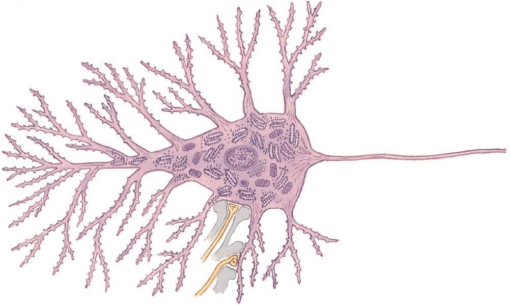
Dendrites
Dendritic spines (gemmules)
Rough endoplasmic reticulum (Nissl substance)
Ribosomes
Mitochondrion
Nucleus
Nucleolus
Axon hillock
Initial segment of axon
Neurotubules
Golgi apparatus
Lysosome
Cell body (soma)
Axosomatic synapse
Glial (astrocyte) process
Axodendritic synapse
Axon
ANATOMICAL AND MOLECULAR PROPERTIES
1.1
NEURONAL STRUCTURE
Neuronal structure reflects the functional characteristics of the individual neuron. Incoming information is projected to a neuron mainly through axonal terminations on the cell body and dendrites. These synapses are isolated and are protected by astrocytic processes. The dendrites usually make up the greatest surface area of the neuron. Some protrusions from dendritic branches (dendritic spines) are sites of specific axodendritic synapses. Each specific neuronal type has a characteristic dendritic branching pattern called the dendritic tree, or dendritic arborizations. The neuronal cell body varies from a few micrometers (μm) in diameter to more than 100 μm. The neuronal cytoplasm contains extensive rough endoplasmic reticulum (rough ER), reflecting the massive amount of protein synthesis necessary to maintain the neuron and its processes. The Golgi apparatus is involved in packaging potential signal molecules for transport and release. Large numbers of mitochondria are necessary to meet the huge energy demands of neurons, particularly those related to the maintenance of ion pumps and membrane potentials. Each neuron has a single (or occasionally no) axon, usually emerging from the cell body or occasionally from a dendrite (e.g., some hippocampal CA neurons). The cell body tapers to the axon at the axon hillock, followed by the initial segment of the axon, which contains the Na+ channels, the first site where action potentials are initiated. The axon extends for a variable distance from the cell body (up to 1 m or more). An axon larger than 1 to 2 μm in diameter is insulated by a sheath of myelin provided by oligodendroglia in the central nervous system (CNS) or Schwann cells in the peripheral nervous system (PNS). An axon may branch into more than 500,000 axon
terminals and may terminate in a highly localized and circumscribed zone (e.g., primary somatosensory axon projections used for fine discriminative touch) or may branch to many disparate regions of the brain (e.g., noradrenergic axonal projections of the locus coeruleus). A neuron whose axon terminates at a distance from its cell body and dendritic tree is called a macroneuron or a Golgi type I neuron; a neuron whose axon terminates locally, close to its cell body and dendritic tree, is called a microneuron, a Golgi type II neuron, a local circuit neuron, or an interneuron. There is no typical neuron because each type of neuron has its own specialization. However, pyramidal cells and lower motor neurons are commonly used to portray a so-called typical neuron.
CLINICAL POINT
Neurons require extraordinary metabolic resources to sustain their functional integrity, particularly that related to the maintenance of membrane potentials for the initiation and propagation of action potentials. Neurons require aerobic metabolism for the generation of adenosine triphosphate (ATP) and have virtually no ATP reserve, so they require continuous delivery of glucose and oxygen, generally in the range of 15% to 20% of the body’s resources, which is a disproportionate consumption of resources. During starvation, when glucose availability is limited, the brain can shift gradually to using beta-hydroxybutyrate and acetoacetate as energy sources for neuronal metabolism; however, this is not an instant process and is not available to buffer acute hypoglycemic episodes. An ischemic episode of even 5 minutes, resulting from a heart attack or an ischemic stroke, can lead to permanent damage in some neuronal populations such as pyramidal cells in the CA1 region of the hippocampus. In cases of longer ischemia, widespread neuronal death can occur. Because neurons are postmitotic cells, except for a small subset of interneurons, dead neurons are not replaced. One additional consequence of the postmitotic state of most neurons is that they are not sources of tumor formation. Brain tumors derive mainly from glial cells, ependymal cells, and meningeal cells.